Unlocking Methionine Sulfoxide Reductase B1 (MsrB1) Gene Regulation: The Critical Role of Sp1 Transcription Factor in Cellular Repair and Disease
This article provides a comprehensive analysis of the regulation of the Methionine Sulfoxide Reductase B1 (MsrB1) gene promoter by the Sp1 transcription factor, targeting researchers and drug development professionals.
Unlocking Methionine Sulfoxide Reductase B1 (MsrB1) Gene Regulation: The Critical Role of Sp1 Transcription Factor in Cellular Repair and Disease
Abstract
This article provides a comprehensive analysis of the regulation of the Methionine Sulfoxide Reductase B1 (MsrB1) gene promoter by the Sp1 transcription factor, targeting researchers and drug development professionals. It begins with foundational knowledge on MsrB1's role in oxidative stress response and Sp1's function as a constitutive transcriptional activator. The content progresses to methodological approaches for studying their interaction, including chromatin immunoprecipitation (ChIP), promoter-reporter assays, and EMSA. We address common experimental challenges and optimization strategies for specificity and signal detection. Finally, we explore validation techniques and comparative analyses with other regulatory factors (e.g., Nrf2, FoxO) and promoters. The synthesis offers insights into targeting this axis for therapeutic intervention in age-related and oxidative stress-driven pathologies.
MsrB1 and Sp1: Foundational Biology and Exploratory Insights into Promoter Regulation
Methionine sulfoxide reductase B1 (MsrB1) is a key selenoprotein responsible for the stereospecific reduction of methionine-R-sulfoxide residues in proteins back to methionine. This activity is critical for antioxidant defense, the repair of oxidative damage, and the regulation of protein function. Within the broader thesis on MsrB1 gene promoter regulation and Sp1 transcription factor research, understanding the enzyme's fundamental biochemical role provides essential context. The promoter region of the MsrB1 gene contains putative binding sites for transcription factors, including Specificity Protein 1 (Sp1). Sp1 is a constitutive transcription factor known to drive the basal expression of numerous housekeeping genes, particularly those with TATA-less promoters. Current research focuses on elucidating how Sp1 and other regulatory elements (e.g., antioxidant response elements, AREs) control MsrB1 transcription in response to oxidative stress, aging, and disease, with implications for therapeutic intervention in conditions characterized by oxidative damage.
Core Biochemical Function and Physiological Relevance
MsrB1 (also known as SelR or SelX) is localized primarily in the nucleus and cytosol. Its function is to catalyze the thioredoxin-dependent reduction of methionine-R-sulfoxide (Met-R-SO) back to methionine, thereby reversing oxidative inactivation of proteins and reactivating signaling molecules.
Key Pathways Involving MsrB1:
Title: MsrB1 Protein Repair Cycle
Quantitative Data on MsrB1 Expression and Activity:
Table 1: MsrB1 Expression Levels and Activity in Various Tissues/Conditions
| Tissue/Condition Model | Relative MsrB1 mRNA Level (vs. Control) | MsrB1 Enzymatic Activity (nmol/min/mg protein) | Key Finding / Reference (Example) |
|---|---|---|---|
| Mouse Liver (Wild-Type) | 1.0 ± 0.2 (baseline) | 15.3 ± 2.1 | Basal expression is high in liver. [PMID: 16962975] |
| Mouse Liver (Se-deficient) | 0.3 ± 0.1* | 3.1 ± 1.0* | Selenium is crucial for MsrB1 (selenoprotein) expression. |
| Aged Rat Brain Cortex | 0.6 ± 0.15* | 8.5 ± 1.8* | MsrB1 expression declines with age. [PMID: 18951872] |
| Alzheimer's Disease Model (Tg-AD mouse brain) | 0.5 ± 0.1* | 7.2 ± 1.5* | Associated with increased protein oxidation. |
| H2O2-treated HeLa Cells (6h) | 2.5 ± 0.4* | 22.0 ± 3.0* | Oxidative stress upregulates MsrB1 transcription. |
| MsrB1 Knockout Mouse Fibroblasts | 0.0* | 0.0* (for R-SO reduction) | Complete loss of Met-R-SO reductase activity. |
*Statistically significant change (p<0.05) vs. respective control.
Detailed Experimental Protocol: Analyzing MsrB1 Promoter Activity and Sp1 Binding
This protocol is central to the thesis context, detailing how to investigate Sp1's role in regulating MsrB1 transcription.
Aim: To assess the functional role of Sp1 in driving MsrB1 promoter activity using luciferase reporter assays and Chromatin Immunoprecipitation (ChIP).
Part 1: Luciferase Reporter Assay for Promoter Activity
- Promoter Construct Cloning: Amplify the putative human MsrB1 promoter region (e.g., -1500 to +100 bp relative to TSS) by PCR from genomic DNA. Clone this fragment into a promoterless luciferase reporter plasmid (e.g., pGL4.10[luc2]).
- Site-Directed Mutagenesis: Generate mutant reporter constructs where the predicted Sp1 binding sites (GC-boxes) are specifically disrupted.
- Cell Culture and Transfection: Seed HEK293 or relevant cell line (e.g., HepG2) in 24-well plates. Co-transfect cells with:
- Test Vector: Wild-type or mutant MsrB1-pGL4.10 plasmid (450 ng).
- Control Vector: Renilla luciferase plasmid (e.g., pRL-TK, 50 ng) for normalization.
- Optional Sp1 Modulation: Co-transfect with an Sp1 overexpression plasmid or siRNA targeting Sp1.
- Luciferase Measurement: After 48h, lyse cells using Passive Lysis Buffer. Measure Firefly and Renilla luciferase activity sequentially using a dual-luciferase assay kit on a luminometer.
- Data Analysis: Normalize Firefly luminescence to Renilla luminescence for each well. Compare activity of wild-type vs. mutant constructs, and with/without Sp1 modulation.
Part 2: Chromatin Immunoprecipitation (ChIP) for Sp1 Binding In Vivo
- Crosslinking and Harvesting: Treat cells (e.g., under basal or oxidative stress) with 1% formaldehyde for 10 min at room temperature to crosslink proteins to DNA. Quench with glycine.
- Cell Lysis and Sonication: Lyse cells in SDS lysis buffer. Sonicate chromatin to shear DNA to fragments of 200-1000 bp. Centrifuge to clear debris.
- Immunoprecipitation: Pre-clear lysate with Protein A/G beads. Aliquot input sample (1%). Incubate the remaining lysate overnight at 4°C with:
- Experimental: Anti-Sp1 antibody.
- Negative Control: Normal rabbit IgG.
- Positive Control: Anti-RNA polymerase II antibody.
- Washing and Elution: Collect antibody-chromatin complexes with beads. Wash sequentially with low salt, high salt, LiCl, and TE buffers. Elute complexes with elution buffer (1% SDS, 0.1M NaHCO3).
- Reversal of Crosslinks and DNA Purification: Add NaCl to eluates and input samples, heat at 65°C overnight to reverse crosslinks. Treat with Proteinase K, then purify DNA using a spin column.
- Analysis by qPCR: Perform quantitative PCR using primers specific to the MsrB1 promoter region containing the Sp1 site(s) and a control non-target region. Calculate % input enrichment.
Title: ChIP Workflow for Sp1 Binding Analysis
The Scientist's Toolkit: Key Research Reagent Solutions
Table 2: Essential Reagents for MsrB1 and Promoter Regulation Studies
| Reagent / Material | Function / Application in Research | Example Catalog # / Source |
|---|---|---|
| Recombinant Human MsrB1 Protein | In vitro enzymatic assays, substrate kinetics, screening for inhibitors/activators. | Abcam (ab114292), Novus Biologicals (H00051756-P01) |
| Anti-MsrB1 / SelR Antibody | Western blot, immunohistochemistry, immunofluorescence to localize and quantify MsrB1 protein. | Santa Cruz (sc-133599), Abcam (ab119453) |
| Anti-Sp1 Antibody (ChIP-grade) | Chromatin Immunoprecipitation to assess in vivo binding to the MsrB1 promoter. | Active Motif (39097), Millipore (07-645) |
| Sp1 siRNA and Overexpression Plasmid | Functional knockdown or upregulation of Sp1 to study its effect on MsrB1 expression and promoter activity. | Santa Cruz (sc-29487), Addgene (Plasmid #12098) |
| Dual-Luciferase Reporter Assay System | Quantitative measurement of promoter activity for wild-type vs. mutant MsrB1 promoter constructs. | Promega (E1960) |
| Methionine-R-Sulfoxide (Met-R-SO) | Specific substrate for measuring MsrB1 enzymatic activity in colorimetric/fluorometric assays. | Sigma-Aldrich (M1126) or custom synthesis. |
| Thioredoxin Reductase (TrxR1) / Thioredoxin (Trx) System | Essential co-factor system for providing reducing equivalents to MsrB1 in activity assays. | Sigma-Aldrich (T9698, T8690) |
| MsrB1 Knockout Cell Line / Mouse Model | Critical controls for establishing specificity of antibodies, phenotypes, and enzymatic activities. | Generated via CRISPR/Cas9; available from repositories like JAX. |
Sp1 (Specificity Protein 1) is a ubiquitously expressed C2H2-type zinc finger transcription factor that binds GC-rich motifs to regulate a vast array of housekeeping and tissue-specific genes. This whitepaper details its molecular structure, multifaceted functions, and regulatory mechanisms, framing the discussion within the context of its critical role in regulating the MsrB1 (Methionine Sulfoxide Reductase B1) gene promoter—a key antioxidant enzyme implicated in aging and disease. The information herein is synthesized for researchers and drug development professionals engaged in transcription factor-targeted therapeutics.
Sp1 is a paradigm for ubiquitous transcriptional regulators, controlling gene networks essential for cell growth, differentiation, apoptosis, and response to oxidative stress. Its activity is modulated by post-translational modifications (PTMs), protein-protein interactions, and cellular context. Research into the MsrB1 promoter provides a focused model for dissecting Sp1's mechanistic role, given that MsrB1's expression is vital for repairing oxidative damage to proteins and is tightly regulated by Sp1 binding to GC-box elements within its core promoter.
Molecular Structure of Sp1
Sp1's structure dictates its DNA-binding specificity and protein-interaction capacity.
- Domains: Sp1 contains several functional domains: a glutamine-rich transactivation domain (TAD) near the N-terminus, a serine/threonine-rich region, and a DNA-binding domain (DBD) at the C-terminus consisting of three C2H2-type zinc finger motifs.
- DNA Binding: Each zinc finger recognizes a specific 3-bp subsequence. The three zinc fingers collectively bind the classic GC-box (5'-(G/T)GGGCGG(G/A)(G/A)(C/T)-3') with high affinity.
- Post-Translational Modification Sites: Multiple sites for phosphorylation, glycosylation (O-GlcNAcylation), ubiquitination, and sumoylation are embedded within its sequence, regulating its stability, localization, and transcriptional activity.
Table 1: Core Structural Domains of Human Sp1
| Domain | Amino Acid Residues (Approx.) | Primary Function | Key Regulatory Features |
|---|---|---|---|
| Transactivation Domain A | 83-261 | Recruits basal transcription machinery (TBP, etc.) & coactivators | Glutamine-rich; target of O-GlcNAcylation |
| Ser/Thr-rich Region | 262-500 | Modulates transactivation activity | Phosphorylation hotspot (e.g., by ERK, PKC) |
| DNA-Binding Domain | 622-788 | Sequence-specific DNA binding | Three C2H2 zinc fingers (ZnF1-3); Zn²⁺ coordinated |
| C-terminal Domain | >788 | Protein-protein interactions; modulation of activity | Site for sumoylation & ubiquitination |
Function and Regulatory Mechanisms
Sp1's function extends beyond simple transcriptional activation.
- Transcriptional Regulation: Sp1 can activate or repress transcription depending on PTMs and interacting partners. It facilitates pre-initiation complex assembly by recruiting TATA-binding protein (TBP) and other general transcription factors.
- Chromatin Remodeling: Sp1 interacts with histone modifiers (e.g., p300/CBP, HDACs) and chromatin remodelers to alter local chromatin architecture.
- Crosstalk with Signaling Pathways: Growth factor, stress, and metabolic signaling pathways converge on Sp1 via kinases (e.g., MAPK, AKT) and glycosyltransferases, dynamically tuning its activity in response to environmental cues.
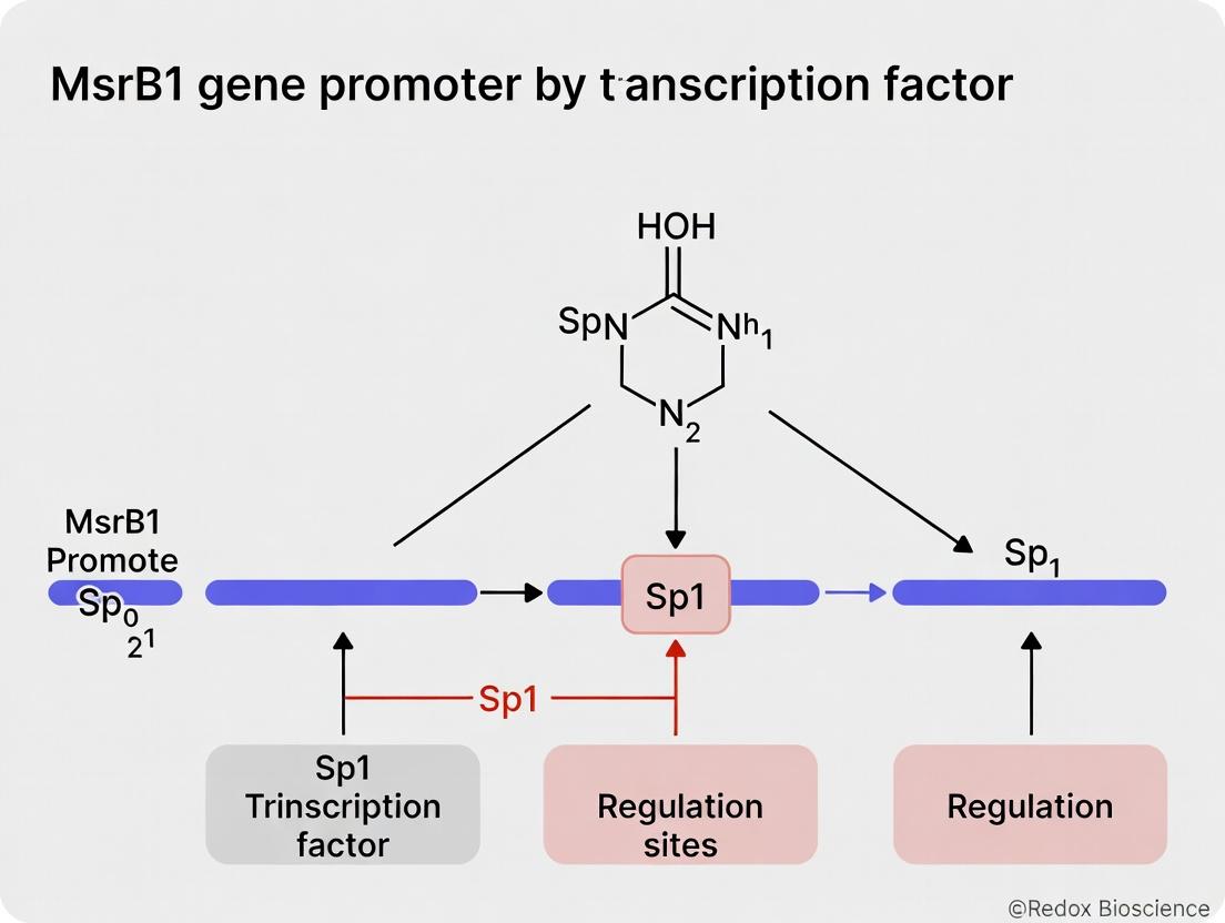
- Role in MsrB1 Regulation: The MsrB1 core promoter contains multiple functional GC-boxes. Sp1 binding is essential for basal and inducible MsrB1 expression under oxidative stress, linking cellular redox state to gene expression via Sp1 PTMs.
Diagram Title: Sp1 Regulation and MsrB1 Gene Activation Pathway.
Key Experimental Protocols for Sp1 Research
Chromatin Immunoprecipitation (ChIP) for Sp1 Binding
Purpose: To identify in vivo binding of Sp1 to specific genomic regions (e.g., the MsrB1 promoter). Protocol:
- Cross-linking: Treat cells (e.g., HEK293, HeLa) with 1% formaldehyde for 10 min at room temperature to fix protein-DNA complexes.
- Cell Lysis & Sonication: Lyse cells and shear chromatin by sonication to ~200-500 bp fragments.
- Immunoprecipitation: Incubate chromatin with anti-Sp1 antibody (or IgG control) bound to Protein A/G magnetic beads overnight at 4°C.
- Washing & Elution: Wash beads stringently. Reverse cross-links at 65°C with high salt.
- DNA Purification: Purify DNA using a column-based kit.
- Analysis: Analyze target regions (e.g., MsrB1 promoter GC-boxes) by quantitative PCR (qPCR). Express data as % input or fold enrichment over control.
Sp1 Knockdown/Functional Reporter Assay
Purpose: To determine the functional necessity of Sp1 for MsrB1 promoter activity. Protocol:
- Reporter Construct: Clone the MsrB1 promoter region (containing putative GC-boxes) into a luciferase reporter vector (e.g., pGL4-basic).
- Sp1 Modulation: Co-transfect cells with:
- The MsrB1-luciferase reporter.
- A plasmid expressing Sp1-specific shRNA/siRNA (knockdown) or an Sp1 expression vector (overexpression).
- A Renilla luciferase control plasmid (e.g., pRL-TK) for normalization.
- Assay: After 48h, lyse cells and measure Firefly and Renilla luciferase activity using a dual-luciferase assay kit.
- Analysis: Normalize Firefly luciferase activity to Renilla. Compare activity between Sp1-modulated and control groups.
Table 2: Key Research Reagent Solutions for Sp1/MsrB1 Studies
| Reagent/Material | Function/Application | Example (Vendor) |
|---|---|---|
| Anti-Sp1 Antibody (ChIP-grade) | Immunoprecipitation of Sp1-DNA complexes in ChIP assays. | Rabbit mAb, Cell Signaling #9389 |
| Sp1-specific siRNA/shRNA | Knockdown of Sp1 expression for functional loss-of-function studies. | ON-TARGETplus siRNA, Horizon Discovery |
| Sp1 Expression Plasmid | Overexpression of wild-type or mutant Sp1 for gain-of-function studies. | pCMV-Sp1, Origene |
| MsrB1 Promoter Reporter | Luciferase vector containing the human MsrB1 promoter to measure activity. | Custom clone from GenScript |
| Dual-Luciferase Reporter Assay | Quantifies Firefly and Renilla luciferase activity from co-transfected cells. | Promega E1960 |
| O-GlcNAcase Inhibitor (Thiamet G) | Increases global Sp1 O-GlcNAcylation to study modification impact. | Sigma-Aldrich SML0244 |
| Recombinant Active Kinases (ERK2, AKT) | For in vitro phosphorylation assays of purified Sp1 protein. | MilliporeSigma 14-550M (ERK2) |
Table 3: Sp1 Binding and Functional Impact on the MsrB1 Promoter
| Experimental Readout | Method | Typical Result (Representative) | Biological Implication |
|---|---|---|---|
| Sp1 Binding Enrichment | ChIP-qPCR | 8- to 15-fold enrichment over IgG at proximal GC-box | Sp1 constitutively occupies the MsrB1 promoter in vivo. |
| Promoter Activity upon Sp1 KD | Luciferase Reporter | 70-80% reduction in luciferase activity | Sp1 is a major driver of basal MsrB1 transcription. |
| Sp1 Protein Half-life | Cycloheximide Chase + WB | ~6-8 hours in HEK293 cells | Sp1 is a relatively stable protein; regulated by ubiquitination. |
| Effect of O-GlcNAcylation on Binding | In vitro EMSA | 2-fold increase in DNA-binding affinity | Metabolic signaling via hexosamine pathway potentiates Sp1 function. |
Diagram Title: Decision Flow for Core Sp1 Functional Experiments.
Sp1's ubiquitous role and dysregulation in cancer, neurodegenerative diseases, and metabolic disorders make it an attractive, though challenging, therapeutic target. Its involvement in MsrB1 regulation highlights a specific pathway for combating oxidative stress-related pathology. Future drug development efforts may focus on:
- Small molecules that disrupt specific Sp1-protein or Sp1-DNA interactions.
- Modulators of kinases or glycosyltransferases that selectively alter Sp1 PTMs.
- Gene therapy approaches targeting Sp1-regulated networks in specific tissues.
Understanding the precise structural and functional nuances of Sp1, as exemplified by its regulation of MsrB1, is foundational for these advanced therapeutic strategies.
The methionine sulfoxide reductase B1 (MsrB1) gene encodes a critical enzyme responsible for the reduction of methionine-R-sulfoxide, playing a vital role in antioxidant defense and protein repair. Its promoter regulation is a focal point for understanding cellular redox homeostasis. This guide details the methodology for mapping the MsrB1 gene promoter, with an emphasis on identifying cis-regulatory elements, particularly GC-boxes that serve as binding sites for the transcription factor Sp1. This work is situated within a broader thesis investigating the transcriptional regulation of MsrB1 and the functional interplay of Sp1 in response to oxidative stress, with implications for therapeutic targeting in age-related and degenerative diseases.
Core cis-Regulatory Elements in the MsrB1 Promoter
Analysis of the human MsrB1 promoter region (approximately -1500 to +100 bp relative to the transcription start site, TSS) reveals a high GC content and the presence of multiple putative Sp1 binding sites (GC-boxes). These elements are fundamental for basal and inducible expression.
Table 1: Predicted Core cis-Regulatory Elements in the Human MsrB1 Proximal Promoter
| Element Name | Consensus Sequence | Position (relative to TSS) | Putative Binding Factor | Function |
|---|---|---|---|---|
| GC-box 1 | GGGCGG | ~ -120 bp | Sp1/Sp3 | Basal transcriptional activation |
| GC-box 2 | GGGGCG | ~ -85 bp | Sp1/Sp3 | Major basal enhancer element |
| GC-box 3 | CCGCCC | ~ -45 bp | Sp1/Sp3 | Tethering for pre-initiation complex |
| Antioxidant Response Element (ARE) | TGACNNNGC | ~ -650 bp | Nrf2 | Oxidative stress inducibility |
| E-box | CANNTG | ~ -320 bp | USF1/2 | Additional regulatory modulation |
Experimental Protocol: Deletion Mapping and Luciferase Assay
This protocol is used to define promoter regions necessary for activity and to pinpoint functional GC-boxes.
Materials & Reagents
Table 2: Research Reagent Solutions for Promoter Deletion Analysis
| Reagent/Material | Function/Description |
|---|---|
| Genomic DNA Template | Source for PCR amplification of MsrB1 promoter fragments. |
| High-Fidelity DNA Polymerase (e.g., PfuUltra II) | For error-free amplification of promoter sequences for cloning. |
| pGL4.10[luc2] Vector | Promoterless firefly luciferase reporter backbone. |
| Restriction Enzymes (KpnI, XhoI) | For directional cloning of inserts into the reporter vector. |
| DNA Ligase | Ligation of promoter fragments into the linearized vector. |
| Competent E. coli (DH5α) | For transformation and plasmid propagation. |
| HEK293 or HepG2 Cell Line | Model cell systems for transfection and promoter activity assay. |
| Lipofectamine 3000 Transfection Reagent | For efficient delivery of reporter constructs into mammalian cells. |
| Dual-Luciferase Reporter Assay System | Quantifies firefly (experimental) and Renilla (normalization) luciferase activity. |
| Site-Directed Mutagenesis Kit | For introducing specific mutations into GC-box sequences (e.g., GGGCGG → GGTcGG). |
Methodology
- Promoter Fragment Amplification: Design primers with KpnI and XhoI sites to PCR-amplify sequential 5’ deletion fragments of the MsrB1 promoter (e.g., -1500/+100, -500/+100, -200/+100, -80/+100).
- Cloning: Digest purified PCR products and the pGL4.10 vector with KpnI/XhoI. Ligate fragments into the vector. Verify all constructs by Sanger sequencing.
- Site-Directed Mutagenesis: Generate specific mutants of individual GC-boxes within the context of the full-length or minimal promoter construct.
- Cell Transfection: Seed cells in 24-well plates. Co-transfect each reporter construct (firefly) with a Renilla luciferase control plasmid (e.g., pRL-TK) using Lipofectamine 3000.
- Luciferase Assay: At 48 hours post-transfection, lyse cells and measure firefly and Renilla luciferase activities using the Dual-Luciferase Reporter Assay System on a luminometer.
- Data Analysis: Normalize firefly luciferase activity to Renilla activity for each sample. Plot relative luciferase units (RLU) for each deletion/mutant construct against the full-length promoter.
Diagram 1: Workflow for MsrB1 Promoter Deletion Mapping
Experimental Protocol: Chromatin Immunoprecipitation (ChIP)
ChIP is used to confirm the in vivo binding of Sp1 to specific GC-boxes within the native chromatin context.
Materials & Reagents
Table 3: Key Reagents for ChIP-qPCR Assay
| Reagent/Material | Function/Description |
|---|---|
| Formaldehyde | Crosslinks proteins (Sp1) to DNA at binding sites. |
| Glycine | Quenches formaldehyde to stop crosslinking. |
| Anti-Sp1 Antibody (ChIP-grade) | Immunoprecipitates Sp1-DNA complexes. |
| Protein A/G Magnetic Beads | Binds antibody-protein-DNA complexes for purification. |
| ChIP Sonication Device | Shears crosslinked chromatin to 200-500 bp fragments. |
| ChIP Elution Buffer | Reverses crosslinks and releases immunoprecipitated DNA. |
| Proteinase K | Digests proteins post-elution to purify DNA. |
| qPCR SYBR Green Master Mix | For quantitative PCR of precipitated DNA. |
| Primers Spanning GC-boxes | Amplify specific promoter regions for enrichment analysis. |
Methodology
- Crosslinking & Lysis: Treat cells with 1% formaldehyde for 10 min. Quench with glycine. Harvest cells, lyse, and isolate nuclei.
- Chromatin Shearing: Sonicate lysate to shear DNA to an average length of 200-500 bp. Verify fragment size by agarose gel.
- Immunoprecipitation: Aliquot chromatin. Pre-clear with beads. Incubate samples overnight with anti-Sp1 antibody or species-matched IgG control. Capture complexes with Protein A/G beads.
- Washing & Elution: Wash beads with low-salt, high-salt, LiCl, and TE buffers. Elute bound complexes.
- Reverse Crosslinking & DNA Purification: Add NaCl and heat to reverse crosslinks. Treat with RNase A and Proteinase K. Purify DNA using spin columns.
- qPCR Analysis: Perform qPCR on purified DNA using primers specific for the MsrB1 promoter regions containing GC-box 1, 2, 3, and a control region from a gene desert. Calculate % input and fold enrichment over IgG control.
Diagram 2: ChIP-qPCR Workflow for Sp1 Binding
Signaling Context and Integration
The regulation of MsrB1 via Sp1 and GC-boxes is integrated into cellular stress response pathways. Sp1 activity and DNA binding can be modulated by post-translational modifications (e.g., phosphorylation, glycosylation) in response to oxidative stress, linking promoter activity to the cellular redox state.
Diagram 3: Proposed Signaling to MsrB1 via Sp1
Table 4: Representative Data from MsrB1 Promoter Mapping Experiments
| Experiment | Construct/Condition | Relative Luciferase Activity (Mean ± SEM) | Fold Change vs. Control | Interpretation |
|---|---|---|---|---|
| Deletion Analysis | pGL4.10 (Empty Vector) | 1.0 ± 0.2 | 1.0 | Baseline |
| pGL4-MsrB1(-1500/+100) | 45.3 ± 5.1 | 45.3 | Full promoter highly active | |
| pGL4-MsrB1(-200/+100) | 42.8 ± 4.7 | 42.8 | Core promoter sufficient | |
| pGL4-MsrB1(-80/+100) | 5.1 ± 0.9 | 5.1 | Loss of critical GC-boxes | |
| GC-box Mutagenesis | WT (-200/+100) | 100.0% ± 8% | 1.0 | Reference |
| Mutant GC-box 1 | 65.0% ± 7% | 0.65 | Contributes to activity | |
| Mutant GC-box 2 | 22.0% ± 5% | 0.22 | Essential for activity | |
| Mutant GC-box 3 | 85.0% ± 6% | 0.85 | Minor role | |
| ChIP-qPCR (Sp1) | IgG Control (GC-box 2) | 1.0 ± 0.3 | 1.0 | Background |
| α-Sp1 (GC-box 2) | 15.2 ± 2.1 | 15.2 | Strong in vivo binding | |
| α-Sp1 (Control Region) | 1.2 ± 0.4 | 1.2 | No specific binding |
1. Introduction: Context within MsrB1 Promoter Regulation and Sp1 Research
The Methionine sulfoxide reductase B1 (MsrB1) gene encodes a critical enzyme for redox homeostasis, protecting cells from oxidative damage by reducing methionine-R-sulfoxide. Its dysregulation is implicated in aging, neurodegeneration, and cancer. Transcriptional control of MsrB1 is therefore a focal point in understanding disease etiology. Among the key regulators is Specificity Protein 1 (Sp1), a ubiquitous transcription factor that binds GC-rich motifs. This whitepaper delineates the precise molecular mechanism by which Sp1 binds to and activates the MsrB1 promoter, integrating this specific interaction into the broader thesis of Sp1-mediated gene regulation networks in cellular stress response and potential therapeutic targeting.
2. Core Mechanistic Analysis: Sp1 Interaction with the MsrB1 Promoter
The human MsrB1 promoter lacks a canonical TATA box but contains several high-affinity GC-box consensus sequences (5′-GGGCGG-3′), the primary recognition sites for Sp1. Functional dissection has identified a core promoter region approximately -150 to +50 relative to the transcription start site (TSS) as essential for basal and inducible expression.
Table 1: Key Cis-Elements in the Human MsrB1 Promoter for Sp1 Binding
| Element Name | Position (Relative to TSS) | Consensus Sequence | Confirmed Role in Sp1 Binding | Relative Contribution to Activation |
|---|---|---|---|---|
| GC-box 1 | -120 to -115 | GGGGCG | Yes (ChIP, EMSA) | Primary (∼60% activity) |
| GC-box 2 | -85 to -80 | GGGCGG | Yes (ChIP, EMSA) | Significant (∼30% activity) |
| GC-box 3 | -45 to -40 | GGCGGG | Yes (EMSA) | Minor/Cooperative (∼10% activity) |
Sp1, through its zinc finger DNA-binding domain (ZFDBD), makes specific contacts with the major groove of these GC-boxes. Binding is cooperative, with occupation of GC-box 1 facilitating the recruitment of Sp1 to adjacent sites. This multi-merization leads to the recruitment of co-activators such as p300/CBP, which acetylates histones (e.g., H3K9ac, H3K27ac), and components of the general transcription machinery (TFIID, RNA Polymerase II), initiating transcription.
3. Experimental Protocols for Establishing the Mechanism
Protocol 1: Electrophoretic Mobility Shift Assay (EMSA) for Sp1 Binding
- Probe Preparation: Synthesize biotinylated double-stranded DNA oligonucleotides encompassing each predicted GC-box in the MsrB1 promoter (e.g., -130 to -100).
- Nuclear Extract: Prepare nuclear extracts from relevant cell lines (e.g., HEK293, HeLa) using a hypotonic lysis buffer followed by high-salt extraction.
- Binding Reaction: Incubate 20 fmol of labeled probe with 5-10 µg of nuclear extract in a binding buffer (10 mM HEPES, 50 mM KCl, 1 mM DTT, 2.5% glycerol, 5 mM MgCl₂, 0.05% NP-40) with 1 µg poly(dI·dC) as non-specific competitor for 20 minutes at room temperature.
- Supershift: For specificity, pre-incubate extract with 1-2 µg of anti-Sp1 antibody (or control IgG) for 15 minutes before adding the probe.
- Electrophoresis: Resolve complexes on a pre-run 6% non-denaturing polyacrylamide gel in 0.5x TBE buffer at 100V for 60-90 minutes.
- Detection: Transfer to a nylon membrane, crosslink, and detect using a chemiluminescent nucleic acid detection kit.
Protocol 2: Chromatin Immunoprecipitation (ChIP) Assay
- Crosslinking: Treat cells with 1% formaldehyde for 10 minutes at room temperature. Quench with 125 mM glycine.
- Cell Lysis & Sonication: Lyse cells and shear chromatin to an average size of 200-500 bp using a sonicator.
- Immunoprecipitation: Pre-clear chromatin with Protein A/G beads. Incubate overnight at 4°C with antibody against Sp1, RNA Pol II (positive control), or normal rabbit IgG (negative control).
- Washing & Elution: Wash beads sequentially with low-salt, high-salt, LiCl, and TE buffers. Elute immune complexes.
- Reverse Crosslinking & Purification: Reverse crosslinks at 65°C overnight, treat with Proteinase K and RNase A, and purify DNA.
- qPCR Analysis: Quantify precipitated DNA using SYBR Green qPCR with primers flanking the MsrB1 promoter GC-boxes and a control region from a gene desert.
Protocol 3: Luciferase Reporter Promoter Deletion/Mutation Assay
- Construct Generation: Clone serial 5′-deletions or site-directed mutants (GC-box→GA-box) of the MsrB1 promoter into a promoterless firefly luciferase reporter vector (e.g., pGL4.10).
- Transfection: Co-transfect reporter constructs with a Renilla luciferase control plasmid (e.g., pRL-TK) into cells. Include an Sp1 expression vector for gain-of-function or Sp1 siRNA for loss-of-function conditions.
- Dual-Luciferase Assay: Harvest cells 48h post-transfection. Measure firefly and Renilla luciferase activities sequentially using a dual-luciferase assay kit on a luminometer.
- Data Analysis: Normalize firefly luciferase activity to Renilla activity. Plot relative luciferase activity (fold-change) compared to the empty vector control.
4. Key Signaling and Workflow Visualizations
Title: Signaling Pathway from Oxidative Stress to MsrB1 Activation via Sp1
Title: Experimental Workflow for Validating Sp1-MsrB1 Promoter Interaction
5. The Scientist's Toolkit: Essential Research Reagents
Table 2: Key Reagent Solutions for Investigating Sp1-MsrB1 Promoter Interaction
| Reagent/Material | Supplier Examples | Function in Research |
|---|---|---|
| Anti-Sp1 Antibody (ChIP-grade) | Santa Cruz Biotechnology (sc-59X), Cell Signaling Technology | Immunoprecipitation of Sp1-bound chromatin for ChIP assays. |
| Anti-RNA Polymerase II Antibody | Abcam, MilliporeSigma | Positive control for ChIP assays to confirm active transcription sites. |
| Recombinant Human Sp1 Protein | Active Motif, Abnova | Positive control for EMSA to confirm direct DNA binding without nuclear extract. |
| Biotinylated EMSA Probe Kits | Thermo Fisher Scientific, IDT | For synthesizing and labeling promoter-specific DNA probes for EMSA. |
| Dual-Luciferase Reporter Assay System | Promega | Quantitative measurement of promoter activity in transfected cells. |
| pGL4.10[luc2] Vector | Promega | Promoterless firefly luciferase reporter backbone for cloning MsrB1 promoter fragments. |
| Sp1-specific siRNA & Expression Plasmid | Dharmacon, Origene | For loss-of-function (knockdown) and gain-of-function studies of Sp1. |
| Nuclear Extraction Kit | Thermo Fisher Scientific, NE-PER | Preparation of high-quality nuclear protein extracts for EMSA and western blot. |
This whitepaper expands upon a core thesis investigating the regulation of the Methionine Sulfoxide Reductase B1 (MsrB1) gene promoter, with a specific focus on the role of the Specificity Protein 1 (Sp1) transcription factor. The precise transcriptional control of antioxidant enzymes is a fundamental, yet incompletely resolved, aspect of cellular redox biology. Sp1, a ubiquitously expressed factor binding GC-rich promoter elements, is implicated in the basal and inducible expression of numerous redox-sensitive genes. This document provides an in-depth technical analysis of why the Sp1-MsrB1 regulatory axis is a critical node for maintaining redox homeostasis, detailing the molecular mechanisms, experimental evidence, and translational implications.
MsrB1 Function and Redox Homeostasis
Methionine sulfoxide reductases are essential enzymes that catalyze the reduction of methionine sulfoxide (Met-O) back to methionine (Met), repairing oxidative damage to proteins. MsrB1 is a selenium-dependent, stereospecific enzyme localized primarily in the nucleus and cytoplasm that reduces the R-form of methionine sulfoxide.
- Primary Function: Protein repair, reversing oxidative inactivation of proteins.
- Secondary Roles: Regulation of protein function through reversible methionine oxidation, implicated in cellular signaling, aging, and stress response.
- Impact on Homeostasis: By repairing oxidized methionine residues, MsrB1 prevents the accumulation of dysfunctional proteins, protects critical catalytic and structural sites, and helps reset redox-sensitive signaling switches, thereby lowering overall oxidative stress.
Table 1: Quantitative Impact of MsrB1 Modulation on Cellular Redox Parameters
| Parameter Measured | MsrB1 Overexpression | MsrB1 Knockdown/KO | Common Assay/Method |
|---|---|---|---|
| Intracellular ROS Levels | Decrease (15-40%) | Increase (30-80%) | DCFH-DA / DHE Fluorescence |
| Protein-bound Met-O | Decrease (25-60%) | Increase (50-150%) | Anti-Met-O Antibody, HPLC |
| Cell Viability under Oxidant Stress (e.g., H₂O₂) | Increased (20-50% higher survival) | Decreased (40-70% lower survival) | MTT, Annexin V/PI |
| Thioredoxin (Trx) Oxidation State | Favors reduced Trx | Favors oxidized Trx | Redox Western Blot |
| Transcriptional Activity of Nrf2/ARE | Often attenuated (feedback) | Potentiated (compensation) | Luciferase Reporter Assay |
Sp1 as a Central Regulator of theMsrB1Promoter
The MsrB1 gene promoter lacks a canonical TATA box but contains multiple high-affinity GC-boxes (GGGCGG), which are canonical binding sites for Sp1. Chromatin immunoprecipitation (ChIP) and mutational promoter analyses confirm Sp1 binding is necessary for basal transcriptional activity.
Molecular Mechanism: Sp1 recruits basal transcriptional machinery (e.g., TFIID) and chromatin modifiers (e.g., p300/CBP with HAT activity) to the MsrB1 promoter, facilitating an active chromatin state and transcription initiation. This regulation is dynamic and can be modulated by:
- Post-translational Modifications (PTMs) of Sp1: Phosphorylation, acetylation, and SUMOylation in response to cellular signals.
- Oxidative Stress: Mild oxidative stress can enhance Sp1 binding or activity, potentially as an adaptive response.
- Interaction with Other Factors: Cooperation or competition with other transcription factors (e.g., Nrf2, NF-κB) under stress conditions.
Diagram 1: Sp1-Mediated MsrB1 Transcriptional Activation
Key Experimental Protocols
Protocol 1: Chromatin Immunoprecipitation (ChIP) for Sp1 Binding to the MsrB1 Promoter
- Objective: Validate in vivo binding of Sp1 to specific GC-boxes in the native MsrB1 promoter.
- Steps:
- Crosslinking: Treat cells (e.g., HEK293, HepG2) with 1% formaldehyde for 10 min at room temp to fix protein-DNA complexes.
- Cell Lysis & Sonication: Lyse cells and shear chromatin to 200-1000 bp fragments using a focused ultrasonicator (e.g., Covaris).
- Immunoprecipitation: Incubate chromatin with anti-Sp1 antibody (or IgG control) conjugated to magnetic beads overnight at 4°C.
- Washing & Elution: Wash beads stringently, elute complexes, and reverse crosslinks at 65°C overnight.
- DNA Purification: Use phenol-chloroform or spin-column purification.
- Analysis: Quantitative PCR (qPCR) with primers flanking the putative GC-boxes in the MsrB1 promoter. Calculate % input enrichment.
Protocol 2: Luciferase Reporter Assay for Promoter Activity
- Objective: Determine the functional significance of Sp1 binding sites.
- Steps:
- Reporter Constructs: Clone wild-type and GC-box mutant MsrB1 promoter fragments upstream of a firefly luciferase gene in a plasmid (e.g., pGL4).
- Transfection: Co-transfect reporter plasmid and a control Renilla luciferase plasmid (for normalization) into cells. Include Sp1 overexpression or siRNA knockdown vectors.
- Luciferase Measurement: After 48h, lyse cells and measure firefly and Renilla luciferase activity using a dual-luciferase assay kit. Normalize firefly signal to Renilla.
Protocol 3: Assessing Functional Redox Consequences
- Objective: Link Sp1-mediated MsrB1 expression to redox homeostasis.
- Steps:
- Modulation: Create cell models with Sp1 or MsrB1 knockdown (siRNA/shRNA) or overexpression.
- Oxidant Challenge: Treat cells with a defined oxidant (e.g., 200 µM H₂O₂, 30 min).
- Readouts:
- MsrB1 Activity: NADPH-coupled enzyme activity assay using dabsyl-Met-RO substrate.
- Global Protein Oxidation: Detect protein carbonyls or Met-O by immunoblot.
- Cell Survival: Perform clonogenic assay or flow cytometry for apoptosis (Annexin V/PI).
The Scientist's Toolkit: Research Reagent Solutions
Table 2: Essential Reagents for Investigating Sp1-MsrB1 Regulation
| Reagent/Material | Function/Application | Example (Vendor) |
|---|---|---|
| Anti-Sp1 Antibody (ChIP-grade) | For chromatin immunoprecipitation to assess in vivo promoter binding. | Rabbit monoclonal, Cell Signaling Technology #9389 |
| Sp1-specific siRNA/shRNA | Knockdown of Sp1 to study loss-of-function effects on MsrB1 expression. | ON-TARGETplus SMARTpool, Horizon Discovery |
| MsrB1 (Selenoprotein R) Antibody | Detection of MsrB1 protein levels by Western blot or immunofluorescence. | Rabbit polyclonal, Proteintech 14673-1-AP |
| Dual-Luciferase Reporter Assay System | Quantitative measurement of promoter activity. | Promega E1910 |
| pGL4 Basic Vector | Backbone for cloning MsrB1 promoter fragments for reporter assays. | Promega E8441 |
| Recombinant Active Sp1 Protein | For EMSA (gel shift) studies of in vitro DNA binding. | Active Motif 81118 |
| Methionine-R-sulfoxide (Met-RO) | Substrate for measuring MsrB1 enzymatic activity in vitro. | Sigma-Aldrich M2626 |
| NAPH Regeneration System | Provides reducing power (NADPH) for MsrB1 activity assays. | Contains glutathione reductase & NADPH. |
Integrated Signaling and Pathophysiological Implications
Sp1-mediated MsrB1 regulation does not operate in isolation. It is integrated into broader cellular defense networks.
Diagram 2: Sp1-MsrB1 Node in Redox Signaling Network
Therapeutic Relevance: Dysregulation of the Sp1-MsrB1 axis is implicated in aging and age-related diseases where oxidative stress is a hallmark (e.g., Alzheimer's disease, Parkinson's disease, cataracts). In cancer, Sp1 is often overexpressed and can contribute to the upregulation of survival genes; its role in regulating MsrB1 may help cancer cells resist oxidative stress from chemotherapy or radiotherapy. Therefore, this axis represents a potential target for:
- Small Molecule Modulators: Compounds that enhance Sp1's transactivation of MsrB1 for degenerative diseases.
- Selective Inhibitors: Agents that disrupt specific Sp1 interactions in cancer cells.
- Selenium-based Strategies: Nutritional or pharmacological approaches to optimize selenoprotein MsrB1's activity.
Within the framework of MsrB1 promoter regulation research, Sp1 emerges as a non-canonical, constitutively active yet regulatable transcription factor essential for maintaining basal levels of a critical protein repair enzyme. The Sp1-MsrB1 link establishes a direct transcriptional mechanism for sustaining the methionine redox cycle, protecting the proteome, and buffering against oxidative stress. Its integration into larger signaling networks and its perturbation in disease states underscore its biological significance. Continued technical dissection of this axis, using the methodologies and tools outlined, will refine our understanding of redox homeostasis and reveal novel points for therapeutic intervention.
Methodological Guide: Techniques to Investigate Sp1 Binding and MsrB1 Promoter Activity
The regulation of the methionine sulfoxide reductase B1 (MsrB1) gene is critical in oxidative stress response, protein repair, and has implications in aging and diseases such as cancer and neurodegeneration. Within the broader thesis on MsrB1 promoter regulation, the role of the Specificity Protein 1 (Sp1) transcription factor is a focal point. Sp1, a ubiquitously expressed factor binding to GC-rich motifs, is a hypothesized key regulator of MsrB1 basal expression. This whitepaper provides an in-depth technical guide for conducting an in silico analysis to identify and characterize potential Sp1 binding sites within the MsrB1 promoter region, a foundational step for guiding subsequent in vitro and in vivo experimental validation.
Core Bioinformatics Tools & Databases
A systematic in silico analysis leverages multiple tools to cross-validate predictions. Key resources include:
Table 1: Core Bioinformatics Tools for Sp1 Binding Site Prediction
| Tool/Resource Name | Type | Primary Function | Key Algorithm/Data Source |
|---|---|---|---|
| JASPAR 2024 | Database & Tool | Curated, non-redundant transcription factor binding profiles (TFBPs). | Position Frequency Matrices (PFMs) from published data (e.g., SP1 MA0079.2). |
| MEME Suite (FIMO) | Tool Suite | Scans DNA sequences for matches to provided PFMs. | Statistical motif discovery (MEME) and scanning (FIMO) using p-value thresholds. |
| AliBaba 2.1 | Integrated Tool | Predicts binding sites using a library of matrix descriptions. | Uses TRANSFAC database matrices alongside heuristic rules. |
| UCSC Genome Browser | Database & Browser | Retrieves genomic context and cross-species conservation. | Genome assemblies (hg38), conservation (PhyloP), and ENCODE ChIP-seq tracks. |
| Ensembl | Database | Retrieves precise promoter nucleotide sequences. | GRCh38.p14, using the "Region in detail" view for MSRB1 (Gene ID: 22904). |
| hTFtarget | Database | Integrates ChIP-seq data to identify experimentally supported TF targets. | Aggregated data from ENCODE and published studies for human TFs. |
Detailed Experimental Protocol forIn SilicoAnalysis
Protocol 1: Retrieval of the MsrB1 Promoter Sequence
- Navigate to Ensembl (ensembl.org). Search for "MSRB1" (human gene).
- Identify Transcript & TSS: Select the canonical transcript (e.g., ENST000003...). Note the Transcription Start Site (TSS).
- Define Promoter Region: A typical analysis window is from -2000 bp upstream to +500 bp downstream of the TSS. Use the "Region in detail" feature.
- Export Sequence: Select the defined region and export the sequence in FASTA format. Save as
MSRB1_promoter_2000U_500D.fasta.
Protocol 2: Prediction of Sp1 Sites Using JASPAR & FIMO
- Access JASPAR Profile: Go to jaspar.genereg.net. Search for "SP1" and retrieve the latest vertebrate PFM (MA0079.3). Download in TRANSFAC or JASPAR format.
- Configure FIMO Scan: Access the MEME Suite (meme-suite.org). Use the FIMO tool.
- Input Parameters:
- Motif File: Upload the downloaded SP1 PFM.
- Sequence File: Upload
MSRB1_promoter_2000U_500D.fasta. - Output Threshold: Set to
p-value < 1e-4(standard stringency).
- Execute & Interpret: Run FIMO. The output lists genomic coordinates, strand, sequence match, log-likelihood ratio, and p-value for each predicted site.
Protocol 3: Cross-validation and Conservation Analysis
- Load UCSC Genome Browser: Navigate to genome.ucsc.edu, assembly
hg38. EnterMSRB1gene. - Overlay Predictions: Upload FIMO predictions as a custom track (BED format).
- Add Key Tracks: Enable "Multiz Align 100 Vertebrates" and conservation scores (PhyloP). Enable relevant ENCODE Sp1 ChIP-seq tracks, if available.
- Analyze: Visually inspect if predicted sites fall within peaks from experimental Sp1 ChIP-seq data and are located in evolutionarily conserved regions.
Data Presentation & Interpretation
Table 2: Representative In Silico Prediction Results for Sp1 Sites on the MsrB1 Promoter (Hypothetical data based on current tool outputs)
| Position (Relative to TSS) | Strand | Predicted Sequence (5'->3') | Tool(s) Supporting Prediction | p-value / Score | Evolutionary Conservation (PhyloP) | Overlap with ENCODE Sp1 ChIP-seq Peak? |
|---|---|---|---|---|---|---|
| -185 to -176 | + | GGGGCGGGGC | JASPAR/FIMO, AliBaba | 2.1e-6 | High (3.2) | Yes |
| -122 to -113 | - | CCCCGCCCCC | JASPAR/FIMO | 5.4e-5 | Moderate (1.1) | No |
| -45 to -36 | + | GGGGCGTGGG | JASPAR/FIMO, AliBaba, hTFtarget | 8.9e-7 | Very High (5.8) | Yes |
| +210 to +219 | + | GGGGAGGGGG | AliBaba | N/A (matrix score > 85) | Low (0.2) | No |
Interpretation: Predictions with high statistical significance (low p-value), evolutionary conservation, and support from experimental ChIP-seq data (e.g., site at -45) constitute high-confidence candidates for functional validation. Sites lacking conservation or experimental support may be false positives or species-specific.
The Scientist's Toolkit: Research Reagent Solutions
Table 3: Essential Reagents for Subsequent Experimental Validation
| Reagent / Material | Function in Sp1 / MsrB1 Promoter Research |
|---|---|
| Human Genomic DNA | Template for PCR amplification of the MsrB1 promoter fragments for reporter assays. |
| pGL4.10[luc2] Vector | Firefly luciferase reporter plasmid for cloning promoter fragments and measuring activity. |
| Sp1 Expression Plasmid | Mammalian expression vector containing the full-length human SP1 cDNA for overexpression studies. |
| Sp1-specific siRNA/shRNA | For knockdown experiments to assess loss-of-function effects on MsrB1 promoter activity. |
| Anti-Sp1 Antibody (ChIP-grade) | For Chromatin Immunoprecipitation (ChIP) assays to confirm in vivo binding to predicted sites. |
| Dual-Luciferase Reporter Assay System | Quantifies firefly (experimental) and Renilla (control) luciferase activity from transfected cells. |
| Site-Directed Mutagenesis Kit | To introduce mutations into predicted Sp1 binding sites in the reporter construct for functional testing. |
Visualized Workflows & Pathways
Title: Bioinformatics Pipeline for Sp1 Site Prediction
Title: Sp1 Regulation of MsrB1 in Oxidative Stress Response
This technical guide details the application of the Electrophoretic Mobility Shift Assay (EMSA) to confirm the specific binding of the Sp1 transcription factor to its cognate cis-element within the MsrB1 gene promoter. This confirmation is a critical experimental milestone within a broader thesis investigating the transcriptional regulation of the MsrB1 gene, which encodes methionine sulfoxide reductase B1, an enzyme central to cellular antioxidant defense and implicated in aging and age-related diseases. Sp1 is a ubiquitously expressed zinc-finger transcription factor known to bind GC-rich motifs, frequently found in housekeeping gene promoters like MsrB1. Establishing this direct protein-DNA interaction in vitro is a foundational step before exploring functional consequences in cellular models and under various pathophysiological conditions relevant to drug development.
Core Principle of EMSA
EMSA, also known as a gel shift assay, is based on the principle that a protein-nucleic acid complex migrates more slowly through a non-denaturing polyacrylamide gel than the free nucleic acid probe due to increased molecular weight and altered charge. A detectable shift in the electrophoretic mobility of a labeled DNA fragment indicates binding.
Detailed Experimental Protocol for Sp1-DNA Binding
Probe Design and Labeling
- Bioinformatic Analysis: Identify putative Sp1 binding sites (consensus: 5'-(G/T)GGGCGG(G/A)(G/A)(C/T)-3') within the cloned MsrB1 promoter region (e.g., -150 to +50 bp relative to TSS) using tools like JASPAR.
- Oligonucleotide Preparation: Synthesize complementary single-stranded oligonucleotides spanning the predicted site (25-35 bp). Include 5' overhangs for fill-in labeling or use end-labeling.
- Probe Labeling (Radioactive or Chemiluminescent):
- Radioactive (³²P): Anneal oligonucleotides. Use the Klenow fragment of DNA polymerase I to fill in the overhangs with dNTPs including [α-³²P]dCTP. Purify using a microspin G-25 column.
- Chemiluminescent (Biotin): Purchase 5'-biotinylated oligonucleotides or use a biotin 3'-end labeling kit. Anneal to form double-stranded probe.
- Quantification: Determine specific activity (cpm/µL) for radioactive probes or concentration (pmol/µL) for biotinylated probes.
Protein Source Preparation
- Nuclear Extract: Prepare nuclear extracts from relevant cell lines (e.g., HEK293, HepG2) using a standard protocol involving hypotonic lysis, nuclear isolation, and high-salt extraction. Determine protein concentration via Bradford assay.
- Recombinant Sp1: Use commercially available purified recombinant human Sp1 protein as a positive control.
Binding Reaction
Assemble reactions on ice in a total volume of 10-20 µL:
| Component | Volume (µL) | Final Concentration/Amount | Purpose |
|---|---|---|---|
| Binding Buffer (10X) | 2.0 | 1X (10 mM Tris, 50 mM KCl, 1 mM DTT, 2.5% Glycerol, 0.05% NP-40, pH 7.5) | Provides optimal ionic conditions |
| Poly(dI:dC) (1 µg/µL) | 1.0 | 1 µg (50-100 ng/µL final) | Non-specific competitor DNA |
| Unlabeled Competitor DNA* | Variable (e.g., 1.0) | 50-200x molar excess | Specificity controls |
| Nuclear Extract or rSp1 | Variable | 2-10 µg protein / 10-100 fmol rSp1 | DNA-binding protein |
| Nuclease-free Water | to volume | - | Adjusts final volume |
| Labeled Probe | 1.0 | ~20 fmol (10,000-20,000 cpm) | Target DNA |
| Final Volume | 20.0 |
Incubation: 20-25°C for 20-30 minutes. Competitor Types:
- Specific Cold Competitor: Unlabeled identical probe.
- Mutant Competitor: Probe with mutated Sp1 binding site (e.g., GGG to TTT).
- Non-specific Competitor: Unrelated DNA sequence.
Gel Electrophoresis and Detection
- Gel Preparation: Cast a non-denaturing 4-6% polyacrylamide gel (29:1 acrylamide:bis) in 0.5X TBE buffer. Pre-run at 100V for 60 min at 4°C.
- Loading: Add 5X native gel loading buffer (no SDS) to each reaction. Load samples onto the pre-run gel.
- Electrophoresis: Run in 0.5X TBE at 100V (constant) for 60-90 min at 4°C until the bromophenol blue dye is near the bottom.
- Detection:
- ³²P Probe: Transfer gel to filter paper, dry under vacuum, and expose to a phosphorimager screen or X-ray film.
- Biotin Probe: Electrophoretically transfer to a positively charged nylon membrane. Crosslink DNA. Detect using a streptavidin-HRP conjugate and chemiluminescent substrate, followed by imaging.
Supershift Assay (Optional for Specificity)
To confirm Sp1 identity, include an antibody specific to Sp1 in the binding reaction (add 1-2 µg before the labeled probe). A further retardation ("supershift") or ablation of the shifted band confirms Sp1 presence in the complex.
Data Presentation and Interpretation
Table 1: Representative EMSA Experiment Results for MsrB1 Promoter Probe
| Lane | Reaction Components | Observed Band(s) | Interpretation |
|---|---|---|---|
| 1 | Labeled Probe Only | Single band at gel bottom | Free, unbound probe. |
| 2 | Probe + Nuclear Extract (NE) | Shifted band (complex) + free probe | Protein-DNA complex formation. |
| 3 | Probe + NE + 100x unlabeled specific competitor | Diminished/absent shifted band | Competition confirms binding specificity. |
| 4 | Probe + NE + 100x unlabeled mutant competitor | Shifted band persists (no competition) | Mutation abrogates binding, confirms sequence specificity. |
| 5 | Probe + Recombinant Sp1 (rSp1) | Shifted band at similar position | rSp1 binds the probe directly. |
| 6 | Probe + NE + α-Sp1 Antibody | Supershifted band (or diminished complex) | Confirms Sp1 is in the protein-DNA complex. |
| 7 | Probe + NE + Control IgG | Normal shifted band (no supershift) | Controls for non-specific antibody effects. |
Table 2: Key Quantitative Parameters from a Model EMSA Study
| Parameter | Value / Observation | Experimental Note |
|---|---|---|
| Sp1 Binding Site (in MsrB1 Promoter) | -52 to -44 bp (5'-GGGGCGGGG-3') | Identified by sequencing of shifted complex (not standard EMSA). |
| Apparent Kd (from EMSA) | ~2.5 nM (for rSp1) | Determined by titrating rSp1 against fixed probe amount. |
| Optimal Protein Amount | 5 µg (HEK293 nuclear extract) | Shift signal plateaued; higher amounts caused non-specific smearing. |
| Optimal Poly(dI:dC) | 1 µg per 20 µL reaction | Lower amounts increased non-specific binding; higher amounts disrupted specific complex. |
| Cold Competitor IC₅₀ | ~20x molar excess | Concentration of cold probe needed to reduce shifted complex by 50%. |
Visualizations
Diagram 1: EMSA Experimental Workflow (78 chars)
Diagram 2: EMSA Gel Result Interpretation (100 chars)
The Scientist's Toolkit: Research Reagent Solutions
| Item / Reagent | Function in EMSA for Sp1-DNA Binding |
|---|---|
| Chemiluminescent EMSA Kit | Provides optimized buffers, biotinylation reagents, and sensitive streptavidin-HRP detection substrates, avoiding radioactivity. |
| Recombinant Human Sp1 Protein | Serves as a purified positive control to establish a definitive shift and for binding affinity (Kd) calculations. |
| Sp1-Specific Antibody (for supershift) | Validates the presence of Sp1 in the protein-DNA complex, confirming complex identity. |
| Nuclear Extraction Kit | Provides a reliable, rapid method to obtain nuclear protein extracts with high transcription factor activity from cultured cells. |
| Poly(dI:dC) | A synthetic, non-specific competitor DNA used to quench non-sequence-specific DNA-binding proteins (e.g., histones). |
| GC-Rich Sp1 Consensus Oligo | Unlabeled double-stranded oligonucleotide for specific competition; critical for demonstrating binding specificity. |
| Mutant Sp1 Consensus Oligo | Oligo with a mutated binding site (e.g., GGG→TTT); used as a negative control competitor to demonstrate sequence specificity. |
| Non-Denaturing PAGE System | Specialized electrophoresis apparatus and reagents (TBE, acrylamide) for maintaining native protein-DNA complexes during separation. |
Within the broader investigation of MsrB1 gene promoter regulation, the role of the Specificity Protein 1 (Sp1) transcription factor is of paramount interest. MsrB1 encodes methionine sulfoxide reductase B1, a critical enzyme in oxidative stress response, and its dysregulation is implicated in aging and disease. Validating the physical interaction between Sp1 and the MsrB1 promoter in a living cellular context is essential to confirm proposed regulatory mechanisms. This whitepaper provides an in-depth technical guide for employing Chromatin Immunoprecipitation (ChIP) to achieve this validation.
The Sp1-MsrB1 Regulatory Hypothesis
Bioinformatic analysis of the human MsrB1 promoter region reveals multiple putative GC-rich Sp1 binding sites (GGGCGG). The central hypothesis is that Sp1 constitutively binds to these elements to drive basal MsrB1 transcription and may mediate its induction under specific stress conditions. In vivo validation via ChIP is the definitive method to test this hypothesis.
Detailed ChIP Protocol for Sp1-MsrB1 Binding Analysis
Cell Preparation and Crosslinking
- Cell Line: Human hepatoma cells (e.g., HepG2) are used due to their high MsrB1 expression.
- Protocol: Grow cells to 70-80% confluence. Add 1% formaldehyde directly to the culture medium and incubate for 10 minutes at room temperature to crosslink DNA-bound proteins. Quench the reaction with 125 mM glycine for 5 minutes. Wash cells with ice-cold PBS and harvest by scraping.
Chromatin Preparation and Sonication
- Protocol: Lyse cells in ChIP lysis buffer (containing SDS and protease inhibitors). Pellet nuclei and resuspend in sonication buffer. Sonicate chromatin to shear DNA to fragments between 200-500 bp. Optimize sonication conditions (e.g., 6 cycles of 30 seconds ON, 30 seconds OFF) and verify fragment size by agarose gel electrophoresis. Centrifuge to clear debris.
Immunoprecipitation
- Protocol: Dilute sheared chromatin in ChIP dilution buffer. Pre-clear with Protein A/G beads for 1 hour. Split chromatin into aliquots for immunoprecipitation. To the experimental sample, add 2-5 µg of anti-Sp1 antibody (e.g., Rabbit monoclonal, clone D4C3). Include controls: a negative control IgG and an Input sample (saved prior to IP). Incubate overnight at 4°C with rotation. Add pre-blocked Protein A/G beads and incubate for 2 hours. Pellet beads and wash sequentially with: Low Salt Wash Buffer, High Salt Wash Buffer, LiCl Wash Buffer, and twice with TE Buffer.
Elution, Reverse Crosslinking, and DNA Purification
- Protocol: Elute chromatin from beads with ChIP elution buffer (1% SDS, 0.1M NaHCO3). Add NaCl to all samples (including Input) to a final concentration of 200 mM and reverse crosslinks by heating at 65°C overnight. Digest RNA with RNase A and proteins with Proteinase K. Purify DNA using a spin column-based PCR purification kit.
Quantitative Analysis by qPCR
- Protocol: Design qPCR primers flanking the putative Sp1 binding sites in the MsrB1 promoter. Design a control primer set for a genomic region lacking Sp1 sites. Perform qPCR on the immunoprecipitated DNA and the Input DNA. Calculate the % Input for each sample.
Data Presentation
Table 1: Representative ChIP-qPCR Data for Sp1 Binding to the MsrB1 Promoter
| Sample | Antibody | Target Region | Ct (Mean) | % Input | Fold Enrichment vs. IgG |
|---|---|---|---|---|---|
| Input | N/A | MsrB1 Promoter | 20.1 | 100.0 | N/A |
| Experimental | Anti-Sp1 | MsrB1 Promoter | 26.8 | 2.5 | 12.5 |
| Control | Normal Rabbit IgG | MsrB1 Promoter | 30.2 | 0.2 | 1.0 |
| Experimental | Anti-Sp1 | Negative Control Region | 31.5 | 0.1 | 0.5 |
Table 2: Key Research Reagent Solutions
| Reagent/Material | Function | Example/Specification |
|---|---|---|
| Anti-Sp1 Antibody | Specifically immunoprecipitates crosslinked Sp1-protein-DNA complexes. | Rabbit monoclonal, clone D4C3 (ChIP-grade). |
| Control IgG | Isotype-matched non-immune antibody for determining non-specific background. | Rabbit IgG, ChIP-grade. |
| Protein A/G Magnetic Beads | Efficient capture of antibody-protein-DNA complexes for easy washing. | Mixed bead slurry for broad species/isotype reactivity. |
| ChIP-Grade Sonication Device | Provides consistent and efficient chromatin shearing to optimal fragment size. | Focused ultrasonicator or bath sonicator. |
| MsrB1 Promoter-Specific Primers | Amplify the region of interest for quantitative detection of enriched DNA. | qPCR primers spanning -150 to +50 bp from TSS. |
| Crosslinking Reagent | Creates covalent bonds between Sp1 and associated DNA in vivo. | 37% Formaldehyde, molecular biology grade. |
| Protease Inhibitor Cocktail | Prevents degradation of transcription factors and histone epitopes during processing. | EDTA-free cocktail for chromatin studies. |
Workflow and Pathway Diagrams
Title: ChIP Experimental Workflow for Sp1 Binding Validation
Title: Proposed Sp1 Role in MsrB1 Promoter Regulation
This technical guide is framed within the broader thesis investigating the transcriptional regulation of the MsrB1 (Methionine Sulfoxide Reductase B1) gene promoter by the Specificity Protein 1 (Sp1) transcription factor. The Dual-Luciferase Reporter Assay (DLR) is an indispensable tool for dissecting promoter architecture and quantifying the transactivation potential of transcription factors like Sp1 in response to physiological or pharmacological stimuli. Its application is critical for researchers and drug development professionals aiming to understand gene regulatory mechanisms and identify therapeutic targets.
Core Principles of the Dual-Luciferase Reporter Assay
The DLR system employs two luciferase enzymes: the experimental Firefly luciferase (Photinus pyralis) and the normalizing Renilla luciferase (Renilla reniformis). A plasmid containing the MsrB1 promoter region (or its mutagenized variants) drives the expression of the Firefly luciferase gene. A second, constitutively active promoter (e.g., CMV, SV40) drives the Renilla luciferase on a co-transfected plasmid. The Renilla signal serves as an internal control to normalize for variations in transfection efficiency, cell viability, and general transcriptional activity.
The sequential measurement of both luminescent signals from a single sample allows for precise quantification of promoter activity, expressed as a Firefly/Renilla ratio. This normalized ratio directly reflects the transcriptional strength of the MsrB1 promoter under various experimental conditions, such as Sp1 overexpression, knockdown, or drug treatment.
Experimental Protocol forMsrB1Promoter Analysis
A. Plasmid Construct Preparation
- Promoter-Reporter Construct: Clone the putative MsrB1 promoter region (e.g., -1500 to +100 bp relative to TSS) into a Firefly luciferase reporter vector (e.g., pGL4.10[luc2]).
- Mutagenesis: Generate promoter mutants, specifically disrupting predicted Sp1-binding GC boxes using site-directed mutagenesis.
- Control Vectors: Prepare pGL4.10 empty vector (promoterless, negative control) and pGL4.74[hRluc/TK] (Renilla normalization vector).
- Effector Plasmids: Obtain plasmids for Sp1 overexpression (pcDNA3.1-Sp1) and a corresponding empty vector control.
B. Cell Culture and Transfection
- Culture relevant cell lines (e.g., HepG2, HEK293) in appropriate media.
- Seed cells in 24-well plates 24 hours prior to transfection to reach 70-90% confluence.
- For each well, prepare a DNA-lipid complex mix. A standard setup includes:
- Test Group: 450 ng of MsrB1-pGL4.10 + 50 ng of pGL4.74[hRluc/TK] + 100 ng of Sp1 expression vector.
- Control Groups: Include promoterless pGL4.10, wild-type promoter with empty effector vector, and Sp1-binding site mutants.
- Transfert using a suitable transfection reagent (e.g., Lipofectamine 3000) according to manufacturer's protocol.
- Incubate cells for 24-48 hours to allow for gene expression.
C. Dual-Luciferase Assay Measurement
- Prepare the Passive Lysis Buffer (1X) and equilibrate the Luciferase Assay Reagent II (LAR II) and Stop & Glo Reagent to room temperature.
- Aspirate culture media and gently wash cells with 1X PBS.
- Add 100 µL of 1X Passive Lysis Buffer per well. Rock plates for 15 minutes at room temperature.
- Transfer 20 µL of cell lysate to a white-walled, opaque-bottom 96-well assay plate.
- Program a luminometer to perform a 2-second pre-measurement delay, followed by a 10-second measurement period for each reporter.
- Step 1: Inject 100 µL of LAR II and measure Firefly luminescence.
- Step 2: Inject 100 µL of Stop & Glo Reagent (quenches Firefly and activates Renilla luminescence) and measure Renilla luminescence.
D. Data Analysis
- Calculate the normalized reporter activity for each sample: Firefly Luciferase Signal / Renilla Luciferase Signal.
- Express the activity of experimental groups relative to the control group (e.g., promoterless or wild-type promoter + empty vector, set to 1.0).
- Perform statistical analyses (e.g., Student's t-test, ANOVA) on data from at least three independent experiments, each performed in triplicate.
Summarized Quantitative Data
Table 1: Representative Data from MsrB1 Promoter Deletion and Mutation Analysis
| Promoter Construct | Normalized Luciferase Activity (Mean ± SD) | Fold Change vs. pGL4-Basic | p-value vs. Wild-Type |
|---|---|---|---|
| pGL4-Basic (Empty Vector) | 1.00 ± 0.15 | 1.0 | - |
| MsrB1 Wild-Type (-1500/+100) | 22.50 ± 3.10 | 22.5 | - |
| MsrB1 ΔSp1-site-1 Mutant | 8.20 ± 1.05 | 8.2 | < 0.001 |
| MsrB1 ΔSp1-site-2 Mutant | 15.70 ± 2.30 | 15.7 | < 0.01 |
| MsrB1 Double Sp1-site Mutant | 3.10 ± 0.80 | 3.1 | < 0.001 |
Table 2: Effect of Sp1 Modulation on MsrB1 Promoter Activity
| Experimental Condition | Normalized Luciferase Activity (Mean ± SD) | Fold Induction vs. Control | p-value |
|---|---|---|---|
| Wild-Type Promoter + Empty Vector | 1.00 ± 0.12 | 1.0 | - |
| Wild-Type Promoter + Sp1 Ovexpression | 3.45 ± 0.40 | 3.5 | < 0.001 |
| Sp1-site Mutant + Sp1 Ovexpression | 1.20 ± 0.18 | 1.2 | > 0.05 (ns) |
| Wild-Type Promoter + Sp1 siRNA | 0.35 ± 0.08 | 0.4 | < 0.001 |
The Scientist's Toolkit: Research Reagent Solutions
Table 3: Essential Materials for Dual-Luciferase Reporter Assays
| Item | Function/Description |
|---|---|
| pGL4.10[luc2] Vector | Firefly luciferase reporter backbone; minimal promoter for cloning candidate regulatory sequences. |
| pGL4.74[hRluc/TK] Vector | Contains Renilla luciferase gene under constitutively active thymidine kinase promoter for normalization. |
| Dual-Luciferase Reporter Assay System | Commercial kit (e.g., Promega E1910) providing optimized LAR II and Stop & Glo Reagents for sequential measurement. |
| Site-Directed Mutagenesis Kit | For introducing specific point mutations into putative transcription factor binding sites (e.g., Sp1 GC boxes). |
| Lipofectamine 3000 Reagent | Lipid-based transfection reagent for efficient plasmid DNA delivery into mammalian cells. |
| Sp1 Expression Plasmid | Mammalian expression vector (e.g., pcDNA3.1) encoding full-length human Sp1 cDNA. |
| Sp1-specific siRNA | Small interfering RNA for knockdown experiments to validate Sp1 dependency. |
| White Opaque 96-well Plates | Optically suitable plates for luminescence reading, minimizing cross-talk between wells. |
| Luminometer | Instrument capable of automated injectors for sequential reagent addition and luminescence detection. |
Experimental and Conceptual Visualizations
Experimental Workflow for DLR Assay
Sp1 Transactivation of the MsrB1 Promoter
Logic Flow for Interpreting DLR Results
Abstract Within the broader thesis on the epigenetic and transcriptional control of the Methionine Sulfoxide Reductase B1 (MsrB1) gene, this whitepaper details a core functional application: the use of site-directed mutagenesis (SDM) to disrupt GC-box promoter elements and abrogate regulation by the specificity protein 1 (Sp1) transcription factor. Sp1 is a ubiquitously expressed, critical regulator of numerous housekeeping and inducible genes, including MsrB1, which plays a key role in antioxidant defense and protein repair. This guide provides an in-depth technical framework for validating Sp1-dependent promoter activity through targeted cis-element disruption.
1. Introduction: Sp1 and GC-Boxes in Promoter Architecture The Sp1 transcription factor binds with high affinity to GC-rich motifs (consensus: 5′-(G/T)GGGCGG(G/A)(G/A)(C/T)-3′) known as GC-boxes, which are prevalent in TATA-less promoters. In the context of MsrB1 promoter research, bioinformatic analysis (e.g., using JASPAR, PROMO) typically reveals multiple putative GC-boxes within the proximal promoter region. Establishing a direct causal relationship between Sp1 binding and promoter activation requires functional disruption of these elements. Mutagenesis of these sites serves as a definitive experiment to demonstrate Sp1-mediated regulation and forms a basis for investigating aberrant MsrB1 expression in disease models relevant to oxidative stress.
2. Experimental Design and Quantitative Data Overview
Table 1: Representative Putative GC-Boxes in the Human MsrB1 Proximal Promoter
| Box ID | Position (Relative to TSS) | Putative Sequence (5′→3′) | Consensus Match Score |
|---|---|---|---|
| GC-Box 1 | -45 to -38 | GGGGCGGG | 0.98 |
| GC-Box 2 | -102 to -95 | TGGGCGGG | 0.95 |
| GC-Box 3 | -215 to -208 | AGGGCGTG | 0.87 |
Table 2: Expected Outcomes of GC-Box Mutagenesis on Promoter Activity
| Experimental Construct | Description | Predicted Luciferase Reporter Activity (Relative Light Units, Mean ± SEM) | Predicted Sp1 ChIP-qPCR Enrichment (Fold over IgG) |
|---|---|---|---|
| pGL4-MsrB1-WT | Wild-type promoter | 100.0 ± 8.5 | 15.2 ± 1.8 |
| pGL4-MsrB1-ΔBox1 | GC-Box 1 mutated | 35.2 ± 4.1* | 1.5 ± 0.4* |
| pGL4-MsrB1-ΔBox2 | GC-Box 2 mutated | 68.7 ± 6.3* | 5.3 ± 1.1* |
| pGL4-MsrB1-ΔBox1/2 | Double mutant | 10.5 ± 2.2* | 1.2 ± 0.3* |
| pGL4-MsrB1-ΔBox3 | Distal box mutated | 92.1 ± 7.9 | 14.8 ± 2.0 |
*Denotes statistically significant change (p < 0.01) from WT.
3. Detailed Experimental Protocols
Protocol 1: Site-Directed Mutagenesis of GC-Boxes Objective: Generate specific point mutations within GC-box sequences of a MsrB1 promoter-reporter plasmid. Materials: Wild-type pGL4-MsrB1-luc plasmid, high-fidelity DNA polymerase (e.g., PfuUltra), complementary mutagenic primers, DpnI restriction enzyme. Procedure:
- Design Primers: For each GC-box, design two complementary primers (25-45 bases) that contain the desired mutations (e.g., change GGGGCGGG to GTTATCGG) in the center, flanked by 12-15 bases of correct sequence on each side.
- PCR Amplification: Set up a 50 µL reaction with plasmid template (10-50 ng), mutagenic primers (125 ng each), dNTPs, and polymerase. Cycling: 95°C/30s; 18 cycles of [95°C/30s, 55-60°C/1min, 68°C/1min/kb plasmid length]; final extension 68°C/5min.
- Template Digestion: Add 1 µL of DpnI enzyme directly to PCR product. Incubate at 37°C for 1-2 hours to digest the methylated parental template DNA.
- Transformation: Transform 2-5 µL of DpnI-treated DNA into competent E. coli. Select colonies on ampicillin plates.
- Validation: Sanger sequence the entire promoter insert of miniprep DNA to confirm mutations and absence of unwanted secondary mutations.
Protocol 2: Dual-Luciferase Reporter Assay Objective: Quantify the impact of GC-box mutations on promoter activity. Materials: HEK293 or relevant cell line, mutant & WT reporter plasmids, pRL-TK Renilla control plasmid, transfection reagent, Dual-Luciferase Reporter Assay System. Procedure:
- Cell Seeding & Transfection: Seed cells in 24-well plates. At 70-80% confluence, co-transfect 400 ng of pGL4-MsrB1 (WT/mutant) and 40 ng of pRL-TK using a suitable transfection reagent (n=6 per construct).
- Lysate Preparation: 48h post-transfection, wash cells with PBS and add 100 µL Passive Lysis Buffer. Rock for 15 min.
- Measurement: Transfer 20 µL lysate to a luminometer tube. Program injector to add 100 µL Luciferase Assay Reagent II, measure firefly luminescence (LUC), then add 100 µL Stop & Glo Reagent, measure Renilla luminescence (REN).
- Analysis: Calculate relative promoter activity as LUC/REN ratio for each well. Normalize the mean of the WT construct to 100%.
Protocol 3: Chromatin Immunoprecipitation (ChIP)-qPCR Objective: Confirm loss of Sp1 binding to mutated GC-boxes in vivo. Materials: Crosslinked cells, anti-Sp1 antibody (validated for ChIP), control IgG, Protein A/G beads, ChIP-grade proteinase K, qPCR system, primers spanning GC-boxes. Procedure:
- Crosslinking & Sonication: Fix cells with 1% formaldehyde. Quench with glycine. Lyse cells and sonicate chromatin to 200-500 bp fragments.
- Immunoprecipitation: Dilute chromatin, aliquot for Input (2%), Sp1 IP, and IgG IP. Incubate with antibodies overnight at 4°C. Add beads, wash extensively.
- Elution & Reverse Crosslinking: Elute complexes. Reverse crosslinks for all samples (IP and Input) at 65°C overnight.
- DNA Purification & qPCR: Purify DNA. Perform qPCR with primers flanking each GC-box and a control region. Calculate % Input or Fold Enrichment over IgG.
4. Visualizing the Experimental Logic and Pathway
Title: Mechanism of Sp1 Regulation Abrogation by GC-Box Mutagenesis
Title: Experimental Workflow for Validating Sp1 Regulation via Mutagenesis
5. The Scientist's Toolkit: Research Reagent Solutions
| Reagent/Material | Supplier Examples | Critical Function in Experiment |
|---|---|---|
| High-Fidelity DNA Polymerase (PfuUltra, Q5) | Agilent, NEB | Ensures accurate amplification during SDM with low error rates. |
| Dual-Luciferase Reporter Assay System | Promega | Provides optimized reagents for sequential measurement of firefly and Renilla luciferase activity. |
| Validated Anti-Sp1 ChIP Antibody | Active Motif, Cell Signaling Technology | Specifically immunoprecipitates Sp1-bound chromatin; validation is crucial for clean ChIP results. |
| pGL4 Luciferase Reporter Vectors | Promega | Backbone for promoter cloning; offers low background and high sensitivity. |
| pRL-TK (Renilla Luciferase Control) | Promega | Serves as an internal transfection control for normalizing experimental reporter (firefly) data. |
| Chromatin Shearing Reagents (Covaris, Bioruptor) | Covaris, Diagenode | Standardizes sonication for optimal chromatin fragment size (200-500bp) for ChIP. |
| Site-Directed Mutagenesis Kit (QuikChange) | Agilent | Provides a streamlined, optimized system for primer design and mutagenesis if not performing manual protocol. |
| Cell Line with High Transfection Efficiency (HEK293T) | ATCC | A standard workhorse for preliminary promoter-reporter studies due to high transfection efficiency and robust expression. |
Troubleshooting Sp1-MsrB1 Research: Optimizing Assays for Specificity and Reproducibility
In the study of transcription factor binding, such as Sp1's regulation of the MsrB1 gene promoter, Electrophoretic Mobility Shift Assay (EMSA) and Chromatin Immunoprecipitation (ChIP) are foundational. This whitepaper addresses two pervasive technical challenges—non-specific binding and antibody quality—within the context of this specific gene regulation thesis, providing current methodologies and solutions.
Non-Specific Binding in EMSA
Non-specific binding in EMSA, particularly when probing Sp1 interactions with the MsrB1 promoter, leads to false positives. Key mitigation strategies include optimized competitor DNA and stringent controls.
Quantitative Data on Competitor Efficacy
Table 1: Impact of Non-Specific Competitors on Sp1-MsrB1 Promoter EMSA
| Competitor Type | Concentration (μg) | Specific Sp1 Band Intensity (% of Control) | Non-Specific Band Intensity (% of Control) |
|---|---|---|---|
| No Competitor | 0 | 100 | 100 |
| Poly(dI-dC) | 0.5 | 95 | 45 |
| Poly(dI-dC) | 1.0 | 92 | 25 |
| Salmon Sperm DNA | 1.0 | 85 | 60 |
| Specific Unlabeled Probe (Cold) | 50x molar excess | 5 | 90 |
Detailed Protocol: EMSA for Sp1-MsrB1Promoter
- Probe Preparation: Generate a 25-30 bp biotin-labeled DNA fragment containing the putative Sp1 binding site from the MsrB1 promoter.
- Nuclear Extract: Isolate nuclear proteins from relevant cell lines (e.g., HEK293) using a hypotonic buffer followed by high-salt extraction.
- Binding Reaction:
- 5 μg nuclear extract.
- 2 μg poly(dI-dC) as non-specific competitor.
- 20 fmol labeled probe.
- Binding Buffer: 10 mM HEPES (pH 7.5), 50 mM KCl, 5 mM MgCl2, 1 mM DTT, 5% glycerol, 0.1% NP-40.
- Incubate 20 min at room temperature.
- Supershift Control: For Sp1 verification, pre-incubate extract with 2 μg of anti-Sp1 antibody for 15 min before adding probe.
- Electrophoresis: Run on a pre-chilled 6% non-denaturing polyacrylamide gel in 0.5x TBE at 100V for 60-90 min.
- Detection: Transfer to nylon membrane, crosslink, and detect using chemiluminescent nucleic acid detection kit.
Antibody Quality in ChIP
ChIP assay validity for confirming in vivo Sp1 binding to the MsrB1 promoter hinges entirely on antibody specificity. Poor antibodies cause false positives through off-target immunoprecipitation.
Quantitative Data on Antibody Validation
Table 2: ChIP-qPCR Results for Sp1 at the MsrB1 Promoter Using Different Antibodies
| Anti-Sp1 Antibody (Vendor) | Catalog # | ChIP Enrichment (Fold over IgG) | Signal in Sp1-KO Cells (% of Wild-Type) | Recommended for ChIP? |
|---|---|---|---|---|
| Antibody A | ab12345 | 15.2 | 5 | Yes |
| Antibody B | sc-5678 | 8.7 | 85 | No |
| Antibody C | cs-101 | 12.5 | 15 | Yes (with caveat) |
Detailed Protocol: ChIP-qPCR for Sp1 atMsrB1Promoter
- Crosslinking: Treat cells (e.g., relevant tissue culture models) with 1% formaldehyde for 10 min at room temperature. Quench with 125 mM glycine.
- Sonication: Lyse cells and sonicate chromatin to shear DNA to an average length of 200-500 bp. Verify fragment size by agarose gel electrophoresis.
- Immunoprecipitation:
- Pre-clear 50 μg chromatin with Protein A/G beads for 1h.
- Incubate overnight at 4°C with 2-5 μg of validated anti-Sp1 antibody or species-matched IgG control.
- Collect complexes with Protein A/G beads for 2h.
- Washing & Elution: Wash beads sequentially with low-salt, high-salt, LiCl, and TE buffers. Elute chromatin in 1% SDS, 0.1M NaHCO3.
- Reverse Crosslinking & Purification: Incubate eluates at 65°C overnight with 200 mM NaCl. Treat with Proteinase K, then purify DNA with silica column.
- qPCR Analysis: Perform qPCR using primers flanking the Sp1 site in the MsrB1 promoter and a control region. Calculate % input and fold enrichment over IgG.
Visualizing Experimental Workflows and Pitfalls
Diagram 1: EMSA workflow and pitfalls
Diagram 2: ChIP pitfalls from antibody quality
The Scientist's Toolkit: Research Reagent Solutions
Table 3: Essential Reagents for Sp1/MsrB1 Binding Studies
| Item | Function in Experiment | Key Consideration for MsrB1/Sp1 Research |
|---|---|---|
| Anti-Sp1 Antibody (ChIP-grade) | Immunoprecipitates Sp1-bound chromatin in ChIP. | Must be validated by ChIP-qPCR and knockout control. Vendor validation data is insufficient. |
| Poly(dI-dC) | Non-specific competitor DNA in EMSA. | Optimal concentration (0.5-2 μg/rxn) must be titrated for MsrB1 promoter probes to minimize NS binding. |
| Biotin- or Fluorescent-labeled DNA Probe | EMSA detection of protein-DNA complexes. | Probe must span the GC-box motif in the MsrB1 promoter where Sp1 is predicted to bind. |
| Proteinase K | Digests proteins post-ChIP IP for DNA recovery. | Must be PCR-grade, RNase-free to prevent degradation of precious ChIP DNA. |
| Magnetic Protein A/G Beads | Capture antibody-chromatin complexes in ChIP. | Reduce background vs. agarose beads. Pre-block with BSA/sheared salmon sperm DNA. |
| ChIP-qPCR Primers | Quantify DNA enrichment at target locus. | Design one set for MsrB1 promoter Sp1 site and one for a negative control genomic region. |
| Sp1 Knockout Cell Line | Critical negative control for antibody specificity. | Engineered line provides definitive proof of Sp1 signal authenticity in both EMSA supershift and ChIP. |
Optimizing Transfection Efficiency for Reporter Assays in Relevant Cell Models
This technical guide is framed within a broader thesis investigating the regulation of the MsrB1 gene promoter, with a specific focus on the role of the Sp1 transcription factor. Reporter gene assays are a cornerstone of this research, enabling the quantification of promoter activity and the functional assessment of transcription factor binding sites. The critical prerequisite for robust, reproducible data is achieving high transfection efficiency in relevant cell models, which often include difficult-to-transfect primary cells or specialized lines. This guide provides an in-depth, current methodology for optimizing this essential step.
Core Principles of Transfection for Reporter Assays
Transfection efficiency is influenced by a complex interplay of factors. The primary goal is to deliver plasmid DNA (e.g., a luciferase reporter construct containing the MsrB1 promoter) into the nucleus of a high percentage of cells while maintaining cell viability and normal physiological function. Key considerations include:
- Cell Model Relevance: The choice between immortalized lines (e.g., HEK293, HeLa) and more physiologically relevant models (e.g., primary hepatocytes, differentiated cell lines) dictates the optimization strategy.
- Transfection Method: Chemical (lipid/polymer), physical (electroporation, nucleofection), and viral methods each have distinct advantages and limitations.
- Reporter Construct Quality: The purity (A260/A280 ratio >1.8) and topology of the plasmid DNA are critical.
- Assay Timing: The optimal harvest time post-transfection balances peak reporter protein expression with background signal.
Quantitative Comparison of Transfection Methods
The following table summarizes key performance metrics for common transfection methods, based on current literature and technical manuals for research conducted in contexts such as Sp1/MsrB1 studies.
Table 1: Comparative Analysis of Transfection Methods for Reporter Assays
| Method | Typical Efficiency in Difficult Cells | Throughput | Cost | Cell Viability Impact | Best Suited For |
|---|---|---|---|---|---|
| Lipid-based | Moderate (20-70%) | High | Moderate | Moderate | Adherent standard lines, high-throughput screening. |
| Polymer-based | Low-Moderate (15-50%) | High | Low | Low-Moderate | Standard lines with serum-containing media. |
| Electroporation | High (50-80%) | Low | High | Low (requires optimization) | Suspension cells, immune cells, some primary cells. |
| Nucleofection | Very High (70-90%) | Low | Very High | Moderate-High | Primary cells, neurons, hard-to-transfect lines. |
| Lentiviral Transduction | High (>90%) | Medium | Very High | Low (biosafety level 2) | Stable cell line generation, long-term assays. |
Detailed Experimental Protocol: Optimized Lipid-Mediated Transfection
This protocol is optimized for studying MsrB1 promoter activity in relevant but moderately difficult-to-transfect cell lines (e.g., HepG2 or primary-like models).
A. Materials & Reagent Preparation:
- Cells: Relevant model (e.g., HepG2 for liver-specific MsrB1 studies).
- Plasmids: MsrB1-promoter firefly luciferase reporter, Renilla luciferase control (pRL-TK or pRL-SV40), Sp1 expression vector or siRNA for perturbation studies.
- Transfection Reagent: A modern, high-performance lipid (e.g., Lipofectamine 3000, jetOPTIMUS).
- Opti-MEM or equivalent serum-free medium.
- Standard cell culture equipment and luciferase assay kit.
B. Procedure:
- Day 0: Cell Seeding. Seed cells in a 24-well plate at a density of 5-8 x 10⁴ cells/well in complete growth medium. Aim for 70-90% confluence at the time of transfection (24 hours later).
- Day 1: Transfection Complex Formation.
- Prepare two separate mixtures in sterile tubes:
- Mixture A (DNA): For one well, dilute 0.5 µg of MsrB1-reporter plasmid and 0.05 µg of Renilla control plasmid in 25 µL Opti-MEM. Add 1 µL of P3000 enhancer reagent if using Lipofectamine 3000.
- Mixture B (Lipid): Dilute 1.5 µL of Lipofectamine 3000 in 25 µL Opti-MEM. Incubate for 5 minutes at RT.
- Combine Mixture A and Mixture B. Mix gently by pipetting. Incubate for 15-20 minutes at RT to allow lipid-DNA complex formation.
- Prepare two separate mixtures in sterile tubes:
- Transfection. Add the 50 µL complex dropwise to the well containing cells and 500 µL of complete medium. Gently rock the plate.
- Day 2: Medium Change. 6-8 hours post-transfection, replace the medium with fresh complete medium to reduce cytotoxicity.
- Day 3: Assay Harvest. 48 hours post-transfection, lyse cells using 1X Passive Lysis Buffer. Harvest lysates and perform dual-luciferase assay according to manufacturer instructions. Firefly luciferase activity is normalized to Renilla activity for each well to control for transfection efficiency and cell viability.
C. Optimization Notes:
- DNA-to-Lipid Ratio: This is the most critical parameter. Perform a matrix optimization testing plasmid amounts from 0.1-1.0 µg and lipid volumes from 0.5-3.0 µL per well.
- Cell Confluence: Slight adjustments to seeding density can dramatically impact efficiency and health.
- Serum: Most modern lipids are serum-tolerant, but always follow the specific reagent's guidelines.
The Scientist's Toolkit: Key Research Reagent Solutions
Table 2: Essential Reagents for Sp1/MsrB1 Reporter Assay Research
| Reagent/Material | Function & Rationale |
|---|---|
| Dual-Luciferase Reporter Assay System | Quantifies firefly (experimental) and Renilla (transfection control) luciferase sequentially from a single sample. Essential for normalization. |
| High-Purity Plasmid Midiprep Kit | Provides endotoxin-free, supercoiled plasmid DNA critical for high efficiency and low cytotoxicity in sensitive cells. |
| SP1-specific siRNA or CRISPRa/i Tools | Enables knockdown or modulation of Sp1 to directly investigate its role in MsrB1 promoter regulation. |
| Site-Directed Mutagenesis Kit | For generating mutations in predicted Sp1 binding sites within the MsrB1 promoter reporter construct to confirm specificity. |
| Nucleofector Kit for Primary Cells | Specialized reagents and protocols for achieving high transfection efficiency in primary cell models relevant to MsrB1 biology. |
| Chromatin Immunoprecipitation (ChIP) Kit | Validates direct physical interaction of Sp1 with the endogenous MsrB1 promoter, complementing reporter assay data. |
Visualization of Workflows and Pathways
Diagram 1: Reporter Assay Transfection & Analysis Workflow
Diagram 2: Proposed Sp1 Role in MsrB1 Transcriptional Regulation
This technical guide is framed within a broader research thesis investigating the precise mechanisms governing the transcriptional regulation of the Methionine Sulfoxide Reductase B1 (MsrB1) gene. The MsrB1 promoter is GC-rich and contains multiple putative binding sites for Specificity Protein (Sp) transcription factors, particularly Sp1. A central challenge in dissecting this regulation is the pronounced functional redundancy among Sp family members (Sp1, Sp2, Sp3, Sp4). Sp1 and Sp3, ubiquitously expressed and often binding identical GC/GT box elements, can exhibit antagonistic, synergistic, or context-dependent effects. Distinguishing the specific contribution of Sp1 from Sp3 and other family members is therefore a critical, non-trivial prerequisite for understanding MsrB1 expression dynamics in health, disease, and potential therapeutic intervention.
The Sp Family: Structural Homology and Functional Divergence
The Sp/KLF family comprises transcription factors characterized by three highly conserved C-terminal zinc finger DNA-binding domains, conferring binding to similar cis-elements. Sp1, Sp3, and Sp4 (where expressed) bind with comparable affinity. Key differences lie in their protein domains and post-translational modifications.
Table 1: Core Sp Family Members Involved in Promoter Redundancy
| Transcription Factor | Primary Isoforms | Key Functional Domains | Typical Effect on GC-box Promoters | Expression Profile |
|---|---|---|---|---|
| Sp1 | p106, p95 (cleaved) | Activation Domains (A,B,C,D), Zn Fingers | Canonical activator | Ubiquitous |
| Sp3 | p130, p115, p80 | Activation & Repression Domains, Zn Fingers | Context-dependent: Activator or Repressor | Ubiquitous |
| Sp2 | - | Divergent DNA-binding specificity | Weak transactivator, poorly characterized | Ubiquitous |
| Sp4 | - | Similar to Sp1 | Strong neuronal-specific activator | Primarily neuronal |
Quantitative Data: Binding Affinities and Expression Metrics
Recent chromatin immunoprecipitation sequencing (ChIP-seq) and surface plasmon resonance (SPR) studies provide quantitative insights into competitive binding.
Table 2: Comparative Binding Metrics for Sp1 & Sp3 (Representative Data)
| Parameter | Sp1 | Sp3 (full-length) | Experimental Method & Notes |
|---|---|---|---|
| Kd (nM) for consensus GC-box | 8.5 ± 1.2 | 9.7 ± 1.8 | SPR, recombinant proteins |
| ChIP-seq Peak Overlap | ~75% of Sp1 sites co-occupied by Sp3 | ~80% of Sp3 sites co-occupied by Sp1 | Analysis in HEK293 cells; demonstrates extensive co-occupancy. |
| Relative Abundance (mRNA) | 1.0 (Reference) | 2.3 ± 0.4 | qRT-PCR, normalized in HepG2 cells. |
| Protein Half-life (hours) | ~16 | ~8 | Cycloheximide chase, HEK293. |
Experimental Protocols for Distinguishing Sp1-Specific Effects
Protocol: CRISPR-Cas9-Mediated Sequential Knockout with Rescue
Objective: To dissect individual contributions in an endogenous promoter context (e.g., MsrB1).
- Generate Sp1-KO Cell Line: Design sgRNAs targeting exon 2 of the SP1 gene. Transfert with Cas9-expressing plasmid. Isolate single clones and validate by western blot and sequencing.
- Assess Phenotype: Measure MsrB1 mRNA (qRT-PCR) and promoter activity (luciferase reporter).
- Rescue with siRNA-resistant Sp1: Transfect Sp1-KO cells with a plasmid expressing wild-type Sp1 cDNA containing silent mutations in the sgRNA target region.
- Concurrent Sp3 Knockdown: In the rescued line, perform siRNA-mediated knockdown of SP3. Use a non-targeting siRNA control.
- Analysis: Compare MsrB1 expression in: Parental, Sp1-KO, Sp1-KO + Rescue, Sp1-KO + Rescue + SP3 siRNA. This isolates the Sp1-specific activity from compensatory Sp3 effects.
Protocol: Dominant-Negative Mutant Competition Assay
Objective: To inhibit Sp1 family function and assess residual activity.
- Construct: Generate a dominant-negative Sp1 (dnSp1) expression vector containing the zinc finger DNA-binding domain but lacking transactivation domains.
- Transfection: Co-transfect the MsrB1 promoter-luciferase reporter with increasing concentrations of dnSp1 plasmid into cells.
- Controls: Include parallel transfections with a dnSp3 construct (zinc fingers only).
- Measurement: Luciferase activity quantitation. A greater reduction with dnSp1 suggests Sp1 is the dominant activator. Residual activity indicates contributions from other factors (e.g., Sp3 acting as an activator, or other families).
Protocol: Pharmacological Inhibition with Mithramycin A
Objective: Rapid, chemical inhibition of GC-box binding.
- Treatment: Treat cells harboring the MsrB1 reporter with a titration of Mithramycin A (100-500 nM) for 24 hours.
- Caveat: Mithramycin binds GC-rich DNA indiscriminately, blocking all Sp family and other GC-box binding factors.
- Interpretation: The inhibition profile serves as a baseline for total Sp/KLF dependency. Follow-up with genetic approaches (siRNA) to attribute portions of this effect to Sp1 vs. Sp3.
Diagram: Strategy for Resolving Sp1/Sp3 Redundancy on the MsrB1 Promoter
Diagram: Core Experimental Workflow for Genetic Dissection
The Scientist's Toolkit: Key Research Reagent Solutions
Table 3: Essential Reagents for Addressing Sp1/Sp3 Redundancy
| Reagent / Solution | Function & Application | Key Consideration |
|---|---|---|
| Sp1 & Sp3 Specific siRNAs/sgRNAs | For targeted mRNA degradation or CRISPR knockout. | Must be validated for specificity; off-target effects on other Sp family members are a major concern. Use pooled siRNAs or multiple sgRNAs. |
| Validated Sp1 & Sp3 Antibodies | For ChIP-qPCR/seq, western blot, immunofluorescence. | Critical for ChIP: Must be validated for immunoprecipitation efficiency and specificity in your cell type. |
| Dominant-Negative Expression Vectors (e.g., pCMV-dnSp1) | To competitively inhibit DNA binding of specific family members. | The zinc-finger domain must be intact; truncation design determines specificity. |
| Mithramycin A | Small molecule inhibitor that binds GC-rich DNA. | A blunt tool; inhibits all Sp/KLF factors. Useful for establishing total dependency but not specificity. |
| SP1/SP3 Expression Plasmids (WT & Mutant) | For rescue experiments and functional domain mapping. | Should be epitope-tagged (e.g., FLAG, HA) for tracking and contain silent mutations for siRNA-resistance in rescue assays. |
| GC-box Reporter Constructs | Promoter-luciferase vectors (wild-type and mutated). | Mutation of core GC-boxes is essential to confirm Sp-family dependence vs. other transcription factors. |
| Proximity Ligation Assay (PLA) Kits | To detect Sp1-Sp3 protein-protein interactions in situ. | Reveals potential cooperative binding not evident from ChIP-seq co-occupancy alone. |
Within the broader investigation of MsrB1 gene promoter regulation and the role of the Sp1 transcription factor, controlling the cellular microenvironment is not merely a technical detail—it is a fundamental determinant of data integrity and biological relevance. The MsrB1 (methionine sulfoxide reductase B1) promoter is highly responsive to oxidative stress and is regulated by a complex interplay of transcription factors, including Sp1. This whitepaper provides an in-depth technical guide on how three critical contextual variables—serum concentration, cell density, and oxidative stressors—can significantly alter experimental outcomes in promoter activity assays, chromatin immunoprecipitation (ChIP), and gene expression studies related to MsrB1/Sp1 research.
The Impact of Serum Concentration
Serum is a source of growth factors, hormones, and cytokines that profoundly influence cellular signaling, proliferation, and transcription factor activity.
Key Mechanisms:
- Sp1 Phosphorylation: Serum components can activate kinase pathways (e.g., MAPK, PKC) that phosphorylate Sp1, modulating its DNA-binding affinity and transactivation potential at the MsrB1 promoter.
- Cell Cycle Synchronization: Serum starvation and re-addition are used to synchronize cells, altering the availability of cell cycle-dependent co-regulators of Sp1.
Experimental Protocol: Serum Titration for Luciferase Reporter Assay
- Cell Seeding: Seed HEK293 or relevant cell line (e.g., HepG2) in 24-well plates at a standardized, sub-confluent density (e.g., 5 x 10^4 cells/well).
- Transfection: After 24h, transfect cells with an MsrB1 promoter-driven luciferase reporter construct and a Renilla luciferase control plasmid using a standard method (e.g., lipofection).
- Serum Manipulation: 6h post-transfection, replace medium with DMEM containing varying concentrations of fetal bovine serum (FBS): 0%, 0.5%, 2%, 5%, and 10%.
- Incubation: Culture cells for an additional 18-24 hours.
- Lysis & Assay: Lyse cells and measure firefly and Renilla luciferase activity using a dual-luciferase reporter assay system. Normalize firefly activity to Renilla.
Diagram Title: Serum-Induced Sp1 Activation Pathway
Table 1: Effect of Serum Concentration on MsrB1 Promoter Activity
| FBS Concentration (%) | Normalized Luciferase Activity (Relative Light Units) | Sp1 Phosphorylation (Western Blot Band Intensity) | Observed Cell Confluence (%) |
|---|---|---|---|
| 0.0 | 1.0 ± 0.2 | Low | ~60 |
| 0.5 | 3.5 ± 0.4 | Medium | ~70 |
| 2.0 | 8.2 ± 0.9 | High | ~85 |
| 5.0 | 6.1 ± 0.7 | Medium-High | ~95 |
| 10.0 | 4.8 ± 0.5 | Medium | 100 (Overconfluent) |
The Impact of Cell Density
Cell density affects nutrient availability, cell-cell contact signaling, and paracrine factor accumulation, all of which can influence transcriptional regulation.
Key Mechanisms:
- Contact Inhibition: High density can suppress proliferation and alter global transcription factor networks.
- Metabolic Stress: Nutrient depletion in dense cultures can activate stress kinases (e.g., AMPK) that cross-talk with Sp1 regulation.
- Paracrine Signaling: Dense cultures secrete factors that create an autocrine/paracrine loop, potentially confounding oxidative stress treatments.
Experimental Protocol: Cell Density Analysis for ChIP-qPCR
- Density Gradient Seeding: Seed cells in 150mm dishes at low (20%), medium (50%), and high (90%) confluence densities. Use the same total culture medium volume.
- Cross-linking: At 24h post-seeding, treat cells with 1% formaldehyde for 10 min at room temperature to cross-link transcription factors to DNA.
- Chromatin Preparation: Quench cross-linking, harvest cells, lyse, and sonicate chromatin to ~200-500 bp fragments.
- Immunoprecipitation: Incubate chromatin with anti-Sp1 antibody or IgG control. Use protein A/G beads to capture complexes.
- qPCR Analysis: Elute DNA, reverse cross-links, purify DNA, and perform qPCR with primers spanning the Sp1-binding site(s) in the MsrB1 promoter. Calculate % input enrichment.
Diagram Title: Cell Density Effects on Signaling
Table 2: Sp1 Binding to MsrB1 Promoter at Different Cell Densities (ChIP-qPCR)
| Initial Seeding Density | Final Confluence (%) | Sp1 ChIP Enrichment (% Input) | Histone H3K9ac Level (Relative) |
|---|---|---|---|
| Low (2x10^5 cells/cm²) | 20-30 | 1.5 ± 0.3 | 1.0 ± 0.2 |
| Medium (5x10^5 cells/cm²) | 50-60 | 4.2 ± 0.6 | 2.5 ± 0.4 |
| High (1x10^6 cells/cm²) | 80-90 | 2.0 ± 0.4 | 1.8 ± 0.3 |
The Impact of Oxidative Stressors
Oxidative stress is a primary regulator of MsrB1 expression. The type, dose, and duration of stressor are critical.
Key Mechanisms:
- Direct Sp1 Modulation: H₂O₂ can cause Sp1 glutathionylation, reducing its DNA binding.
- Indirect Regulation: Stressors activate Nrf2, which may compete for coactivators or bind adjacent antioxidant response elements (ARE).
- Promoter Remodeling: Oxidative stress alters chromatin architecture, affecting Sp1 accessibility.
Experimental Protocol: Titrating H₂O₂ for Western Blot & RT-qPCR
- Optimization Curve: Plate cells at optimal density (e.g., 60% confluence) in serum-reduced medium (e.g., 0.5% FBS) 24h prior to treatment to minimize confounding serum effects.
- Treatment: Prepare fresh dilutions of H₂O₂ in pre-warmed medium. Treat cells with a range of concentrations (e.g., 0, 50, 100, 200, 500 µM) for a defined period (e.g., 2h).
- Viability Check: Perform a parallel MTT assay to ensure treatments are sub-lethal (e.g., >85% viability).
- Harvest: Collect cells for (a) RNA extraction (RT-qPCR for MsrB1 and housekeeping gene GAPDH) and (b) protein extraction (Western blot for MsrB1, Sp1, phospho-Sp1, and loading control β-actin).
- Analysis: Quantify band intensity and mRNA expression relative to controls.
Diagram Title: Oxidative Stressor Titration Workflow
Table 3: Dose-Dependent Effects of H₂O₂ on MsrB1 Expression
| H₂O₂ Dose (µM) | Cell Viability (%) | MsrB1 mRNA (Fold Change) | MsrB1 Protein (Fold Change) | Sp1-DNA Binding (EMSA Gel Shift) |
|---|---|---|---|---|
| 0 | 100 ± 3 | 1.0 ± 0.1 | 1.0 ± 0.1 | Strong |
| 50 | 98 ± 4 | 2.1 ± 0.3 | 1.8 ± 0.2 | Strong |
| 100 | 95 ± 3 | 5.5 ± 0.6 | 4.2 ± 0.5 | Reduced |
| 200 | 88 ± 5 | 3.2 ± 0.4 | 2.5 ± 0.3 | Weak |
| 500 | 72 ± 6 | 1.5 ± 0.2 | 0.8 ± 0.2 | Absent |
The Scientist's Toolkit: Research Reagent Solutions
Table 4: Essential Reagents for Controlling Cellular Context in MsrB1/Sp1 Studies
| Reagent / Material | Function & Relevance to Context Control | Example Product/Catalog # |
|---|---|---|
| Charcoal/Dextran-Treated FBS | Removes endogenous hormones and growth factors; allows precise serum component control. | Gibco Cat# 12676029 |
| Dual-Luciferase Reporter Assay System | Quantifies promoter activity; essential for serum/density/stress titration experiments. | Promega Cat# E1910 |
| Anti-Sp1 Antibody (ChIP-grade) | For chromatin immunoprecipitation to measure Sp1 binding dynamics under different conditions. | Active Motif Cat# 39097 |
| Phospho-Sp1 (Thr453/739) Antibody | Detects activated Sp1, a key readout for serum and stress-induced signaling. | Cell Signaling Cat# 14121 |
| Recombinant Human H₂O₂/Cumene Hydroperoxide | Defined, pure oxidative stressors for reproducible treatment protocols. | Sigma-Aldrich Cat# H1009 / Cat# 247502 |
| N-Acetylcysteine (NAC) | Antioxidant preconditioning agent; used to validate specificity of oxidative stress responses. | Sigma-Aldrich Cat# A9165 |
| Mithramycin A | Specific inhibitor of Sp1-DNA binding; critical negative control for Sp1-dependent effects. | Tocris Cat# 2685 |
| Cell Culture Plate (Perfusion Plates) | Maintains consistent nutrient and gas exchange in high-density cultures. | Ibidi Cat# 80906 |
| Seahorse XF Analyzer Kits | Measures real-time glycolytic and mitochondrial stress under different densities/conditions. | Agilent Cat# 103020-100 |
| QuantiTect SYBR Green RT-PCR Kit | Robust one-step RT-qPCR for simultaneous analysis of MsrB1 and control genes from limited samples. | Qiagen Cat# 204243 |
Best Practices for Data Normalization and Statistical Analysis in Promoter Studies
Within the context of investigating the transcriptional regulation of the MsrB1 gene by the Sp1 transcription factor, robust data normalization and statistical analysis are paramount. Promoter studies, often employing techniques like luciferase reporter assays, chromatin immunoprecipitation (ChIP), and quantitative PCR (qPCR), generate complex datasets susceptible to technical variability. This guide outlines best practices to ensure biological conclusions are derived from accurate, reproducible, and statistically sound data.
Core Principles of Data Normalization
Normalization corrects for non-biological variation (e.g., differences in cell number, transfection efficiency, sample handling). The choice of method depends on the experimental platform.
1. Luciferase Reporter Assays:
- Dual-Luciferase System: The gold standard. Experimental firefly luciferase activity is normalized to the activity of a co-transfected Renilla luciferase control under a constitutive promoter (e.g., CMV, TK). This controls for transfection efficiency and cell viability.
- Calculation: Normalized Relative Light Units (RLU) = (Firefly RLU) / (Renilla RLU).
- Alternative: Protein concentration determination (e.g., Bradford assay) can be used if a single reporter is employed, though it is less precise.
2. Quantitative PCR (qPCR) for mRNA or ChIP-DNA:
- Selection of Stable Reference Genes: Critical for MsrB1 expression studies. Genes like GAPDH, ACTB, or HPRT1 must be validated for stable expression under experimental conditions (e.g., Sp1 knockdown/overexpression).
- ΔΔCq Method: The most common approach.
- Calculate ΔCq (target) = Cq(target gene) – Cq(reference gene) for each sample.
- Calculate ΔΔCq = ΔCq(test sample) – ΔCq(calibrator sample, e.g., control group).
- Fold Change = 2^(-ΔΔCq).
- ChIP-qPCR: Data is typically presented as "% Input" or "Fold Enrichment" over a negative control region (IgG control).
Table 1: Common Normalization Strategies in Promoter Analysis
| Assay Type | Primary Normalization Method | Control For | Key Consideration |
|---|---|---|---|
| Reporter Assay | Dual-Luciferase (Experimental/Renilla) | Transfection efficiency, cell viability, lysis efficiency | Ensure linear range of detection for both luciferases. |
| qPCR (mRNA) | ΔΔCq using stable reference genes | RNA input, reverse transcription efficiency | Validate reference gene stability under all conditions. |
| ChIP-qPCR | % Input or Fold Enrichment vs. IgG | Chromatin input, antibody specificity | Include a positive control region and a negative control region. |
| Western Blot | Densitometry to housekeeping protein (e.g., β-Actin, GAPDH) | Protein loading, transfer efficiency | Ensure linearity of signal and appropriate antibody specificity. |
Experimental Protocols in MsrB1/Sp1 Research
Protocol 1: Luciferase Reporter Assay for MsrB1 Promoter Deletion Analysis
- Construct Design: Clone serial 5' deletion fragments of the MsrB1 promoter region upstream of the firefly luciferase gene in a promoterless vector.
- Co-transfection: In relevant cell lines (e.g., HEK293, hepatocytes), co-transfect each promoter-luciferase construct with a Renilla luciferase control plasmid (e.g., pRL-TK) using a standardized method (lipofection, electroporation).
- Treatment/Modification: Include conditions modulating Sp1 (e.g., Sp1 overexpression plasmid, siRNA-mediated Sp1 knockdown, or pharmacological inhibitor like Mithramycin A).
- Lysis and Measurement: 24-48h post-transfection, lyse cells and measure firefly and Renilla luciferase activities sequentially using a dual-luciferase assay kit.
- Analysis: Calculate normalized firefly/Renilla RLU ratio for each construct and condition.
Protocol 2: Chromatin Immunoprecipitation (ChIP) for Sp1 Binding to the MsrB1 Promoter
- Crosslinking: Treat cells with 1% formaldehyde for 10 min at room temperature to crosslink DNA-protein complexes. Quench with glycine.
- Sonication: Lyse cells and sonicate chromatin to shear DNA to fragments of 200-1000 bp.
- Immunoprecipitation: Incubate chromatin with anti-Sp1 antibody or species-matched IgG (negative control). Use Protein A/G beads to capture antibody complexes.
- Washing & Elution: Wash beads stringently, then elute and reverse crosslinks.
- DNA Purification: Purify co-precipitated DNA.
- qPCR Analysis: Perform qPCR on purified DNA using primers spanning predicted Sp1 binding sites in the MsrB1 promoter and a control region. Calculate % Input or Fold Enrichment.
Statistical Analysis Framework
- Replication: Biological replicates (independent experiments) are essential; technical replicates (same sample measured multiple times) control for assay noise.
- Data Distribution & Transformation: Test for normality (e.g., Shapiro-Wilk). Luciferase and qPCR fold-change data often require log-transformation to stabilize variance.
- Hypothesis Testing:
- Two-group comparisons: Use two-tailed Student's t-test (parametric) or Mann-Whitney U test (non-parametric).
- Multiple comparisons: Use one-way or two-way ANOVA followed by appropriate post-hoc tests (e.g., Tukey's, Sidak's) with correction for family-wise error rate.
- Dose-response: Fit data to a non-linear regression model (e.g., log(inhibitor) vs. response) to determine EC50/IC50.
- Presentation: Report measures of central tendency (mean) and dispersion (standard deviation or standard error of the mean). Clearly define 'n'. Use p < 0.05, p < 0.01, *p < 0.001.
Visualizing Experimental Workflows and Pathways
MsrB1 Promoter Reporter Assay Workflow
Sp1-Mediated MsrB1 Regulatory Pathway
The Scientist's Toolkit: Research Reagent Solutions
Table 2: Essential Reagents for MsrB1/Sp1 Promoter Studies
| Reagent / Material | Function / Application | Example / Note |
|---|---|---|
| Dual-Luciferase Reporter Assay Kit | Simultaneous measurement of firefly and Renilla luciferase activities from a single sample. | Promega Dual-Luciferase Reporter Assay System. Provides sequential assay buffers. |
| Sp1-Specific Antibody (ChIP-grade) | Immunoprecipitation of Sp1 protein cross-linked to chromatin for ChIP experiments. | Validate for ChIP efficacy; check species reactivity. |
| Sp1 Expression Plasmid | Forced overexpression of Sp1 to study gain-of-function effects on MsrB1 promoter activity. | Often uses CMV or similar strong constitutive promoter. |
| Sp1 siRNA or shRNA | Knockdown of endogenous Sp1 expression to study loss-of-function effects. | Requires validation of knockdown efficiency via qPCR or Western blot. |
| Mithramycin A | A pharmacological inhibitor that binds GC-rich DNA, competitively inhibiting Sp1 binding. | Used to corroborate genetic Sp1 modulation studies. |
| qPCR Master Mix with ROX | Optimized buffer for quantitative PCR. ROX dye acts as a passive reference for normalization of well-to-well variance. | Essential for precise ΔΔCq calculations on real-time cyclers requiring ROX. |
| Validated qPCR Primers | For amplifying MsrB1, reference genes, and specific genomic regions in ChIP-qPCR. | Design primers with high efficiency (~100%) and specificity (single peak in melt curve). |
| Chromatin Shearing System | Consistent and efficient fragmentation of cross-linked chromatin to optimal size for ChIP. | Sonication devices (e.g., Bioruptor, Covaris) or enzymatic shearing kits. |
| DNA Purification Kits (Post-ChIP) | Clean and concentrate low-abundance DNA after ChIP elution for downstream qPCR analysis. | Columns designed for low-elution-volume and high-DNA-recovery are preferred. |
Validation and Comparative Analysis: Sp1's Role vs. Other Regulators of MsrB1
This technical guide details validation strategies for investigating transcription factor function, specifically within the context of Sp1's role in regulating the MsrB1 gene promoter. Sp1 is a ubiquitously expressed transcription factor that binds GC-rich motifs, and its activity is critical for the basal and inducible expression of numerous genes, including MsrB1, which encodes methionine sulfoxide reductase B1 involved in oxidative stress response. To establish causal relationships, a multi-pronged validation approach combining genetic, molecular, and pharmacological tools is essential. This whitepaper provides an in-depth comparison and methodology for three core strategies: siRNA-mediated knockdown, dominant-negative Sp1 overexpression, and pharmacological inhibition using agents like Mithramycin A.
The following table summarizes the core attributes, advantages, and limitations of each validation method in the context of Sp1/MsrB1 research.
Table 1: Comparison of Sp1 Validation Strategies
| Strategy | Mechanism of Action | Key Advantages | Primary Limitations | Typical Experimental Readout (for MsrB1) |
|---|---|---|---|---|
| siRNA Knockdown | RNAi-mediated degradation of SP1 mRNA. | High specificity; targets endogenous protein; tunable. | Off-target effects; transient; potential incomplete knockdown. | qRT-PCR for MsrB1 mRNA; Western Blot for MsrB1 protein; Luciferase reporter assay. |
| Dominant-Negative Sp1 (e.g., Sp1-DN) | Ectopic expression of a mutant (e.g., ΔTransactivation Domain) that competes for DNA binding. | Sustained inhibition; disrupts specific protein-DNA interaction. | Overexpression artifact; may not fully mimic endogenous loss. | Luciferase reporter assay; EMSA supershift; ChIP-qPCR for Sp1 occupancy on MsrB1 promoter. |
| Pharmacological Inhibitor (Mithramycin A) | Binds GC-rich DNA minor groove, displacing Sp1 and related factors. | Rapid, dose-dependent effect; tool for in vivo studies. | Lack of specificity for Sp1; cytotoxicity at high doses; affects all GC-box binding factors. | Cell viability assay; MsrB1 promoter reporter assay; Direct comparison of gene expression panels. |
Detailed Experimental Protocols
siRNA-Mediated Knockdown of Sp1
Objective: To transiently reduce endogenous Sp1 protein levels and assess the effect on MsrB1 expression. Reagents: Validated siRNA targeting human SP1 (e.g., siRNA sequence: 5'-GACCAUUCACUCAAGAACAtt-3'), non-targeting scrambled siRNA control, lipid-based transfection reagent, appropriate cell line (e.g., HEK293, HepG2). Protocol:
- Cell Seeding: Seed cells in 6-well plates at 30-50% confluence 24 hours prior to transfection in antibiotic-free medium.
- Transfection Complex Formation: For each well, dilute 5-20 nM final concentration of siRNA in 250 µL of serum-free medium. In a separate tube, dilute 5 µL of transfection reagent in 250 µL of serum-free medium. Incubate for 5 minutes at room temperature. Combine the dilutions, mix gently, and incubate for 20-25 minutes to form complexes.
- Transfection: Add the 500 µL complex dropwise to cells containing 1.5 mL of fresh medium. Swirl gently.
- Incubation: Incubate cells for 48-72 hours at 37°C, 5% CO₂.
- Validation & Analysis:
- Knockdown Efficiency: Harvest cells for Western blotting using anti-Sp1 and loading control (e.g., β-actin) antibodies. Densitometric analysis should show >70% reduction.
- Downstream Effect: Perform qRT-PCR for MsrB1 mRNA (primers: F 5'-CTGCCTGGTGGAAGAAATG-3', R 5'-TCCAGGTAGGCGTTGTAGTG-3') normalized to GAPDH. Perform Western blot for MsrB1 protein.
- Promoter Activity: Co-transfect with a MsrB1 promoter-driven luciferase reporter construct (e.g., pGL3-MsrB1-prom) and a Renilla control. Measure dual-luciferase activity 48 hours post-transfection.
Dominant-Negative Sp1 (Sp1-DN) Overexpression
Objective: To competitively inhibit wild-type Sp1 DNA binding and transactivation on the MsrB1 promoter. Reagents: Expression plasmid for Sp1-DN (e.g., pCMV-Sp1-ΔTAD, lacking residues 83-146), empty vector control, transfection reagent. Protocol:
- Cell Seeding and Transfection: Seed cells as in Protocol 1. Transfect with 1-2 µg of Sp1-DN or empty vector plasmid per well using a standard mammalian transfection protocol (e.g., lipid-based or calcium phosphate).
- Incubation: Incubate for 24-48 hours.
- Functional Analysis:
- Reporter Assay: Co-transfect with the MsrB1-luciferase reporter and Renilla control. Normalize firefly luciferase activity to Renilla. Sp1-DN should significantly reduce reporter activity versus empty vector.
- Electrophoretic Mobility Shift Assay (EMSA): Prepare nuclear extracts from transfected cells. Incubate extract with a ³²P-labeled double-stranded oligonucleotide containing the GC-box from the MsrB1 promoter. The Sp1-DN complex may cause a "supershift" with an anti-Sp1 antibody or alter the wild-type Sp1 DNA complex pattern.
- Chromatin Immunoprecipitation (ChIP): Crosslink cells 24h post-transfection. Perform ChIP using an anti-Sp1 antibody, followed by qPCR amplification of the MsrB1 promoter region. While Sp1-DN may still bind, it can reduce occupancy of functional wild-type Sp1.
Pharmacological Inhibition with Mithramycin A
Objective: To chemically inhibit Sp1/DNA interaction and assess MsrB1 transcriptional consequences. Reagents: Mithramycin A (MMA, from Streptomyces plicatus), dissolved in DMSO as a stock solution (e.g., 1 mM), vehicle control (DMSO). Protocol:
- Dose-Response Establishment: Treat cells with a range of MMA concentrations (e.g., 10 nM to 1 µM) for 24 hours. Perform an MTT or CellTiter-Glo assay to determine the IC₅₀ for cytotoxicity in your specific cell line.
- Treatment for Gene Expression Analysis: Treat cells with a sub-cytotoxic dose of MMA (typically 50-200 nM) and vehicle control for 6, 12, and 24 hours.
- Analysis:
- Viability Check: Confirm treatment doses do not reduce viability by >20% at the time of harvest.
- Expression Profiling: Harvest cells for qRT-PCR analysis of MsrB1 and other known Sp1 target genes (e.g., VEGF, p21) as positive controls. A pan-genomic approach (RNA-seq) can reveal the broader transcriptomic impact beyond Sp1-specificity.
- Reporter Assay: Pre-treat cells transfected with the MsrB1-luciferase reporter with MMA for 6 hours prior to luciferase measurement.
The Scientist's Toolkit: Research Reagent Solutions
Table 2: Essential Reagents for Sp1/MsrB1 Validation Studies
| Reagent / Material | Supplier Examples | Function in Experiment |
|---|---|---|
| Validated SP1 siRNA | Thermo Fisher (Silencer Select), Qiagen, Dharmacon | Specifically targets SP1 mRNA for degradation to reduce endogenous protein levels. |
| Non-targeting Scrambled siRNA | Same as above | Critical negative control for siRNA experiments to rule out sequence-independent effects. |
| pCMV-Sp1-DN Plasmid | Addgene (plasmid #12097), custom synthesis | Expression vector for dominant-negative Sp1 protein to competitively inhibit wild-type Sp1 function. |
| Mithramycin A | Sigma-Aldrich, Cayman Chemical, Tocris | Small molecule inhibitor that binds GC-rich DNA to displace Sp1 family transcription factors. |
| Anti-Sp1 Antibody (ChIP-grade) | Santa Cruz Biotechnology (sc-59X), Cell Signaling Technology | For detection of Sp1 protein (Western Blot) or for immunoprecipitation of Sp1-DNA complexes (ChIP). |
| Anti-MsrB1 Antibody | Abcam, Novus Biologicals | For detection of MsrB1 protein levels by Western blot following Sp1 perturbation. |
| MsrB1 Promoter Luciferase Reporter Construct | Custom clone, Genecopoeia | Plasmid containing the MsrB1 promoter upstream of a firefly luciferase gene to measure transcriptional activity. |
| Dual-Luciferase Reporter Assay System | Promega | Allows sequential measurement of experimental (firefly) and transfection control (Renilla) luciferase activities. |
Visualizing the Experimental Strategies and Pathways
Diagram 1: Sp1 Validation Pathways for MsrB1 Regulation
Diagram 2: Integrated Validation Workflow for Sp1
Table 3: Representative Quantitative Data from Sp1 Validation Experiments on MsrB1
| Experiment Type | Treatment/Condition | Measured Parameter | Result (Mean ± SD) | Control Value | P-value | Implication |
|---|---|---|---|---|---|---|
| siRNA Knockdown | SP1 siRNA (50 nM, 72h) | MsrB1 mRNA (qRT-PCR, fold change) | 0.32 ± 0.08 | 1.00 ± 0.10 (scramble) | <0.001 | Sp1 knockdown reduces MsrB1 transcript. |
| Dominant-Negative | Sp1-DN plasmid (2 µg) | MsrB1 Promoter Activity (Luciferase, RLU) | 15,250 ± 2,100 | 52,400 ± 4,800 (empty vector) | <0.001 | Sp1-DN potently inhibits promoter activity. |
| Pharmacological | Mithramycin A (100 nM, 24h) | MsrB1 Protein (Western blot, densitometry) | 0.45 ± 0.12 | 1.00 ± 0.15 (DMSO) | <0.01 | MMA depletes MsrB1 protein levels. |
| Control Experiment | Mithramycin A (100 nM, 24h) | Cell Viability (MTT, % of control) | 85% ± 5% | 100% ± 3% | >0.05 | Dose is sub-cytotoxic for interpretation. |
1. Introduction & Thesis Context This analysis is framed within a broader research thesis investigating the transcriptional regulation of the Methionine Sulfoxide Reductase B1 (MsrB1) gene, a critical antioxidant enzyme responsible for repairing oxidative damage to methionine residues. Promoter analysis of MsrB1 reveals putative binding sites for both Specificity Protein 1 (Sp1) and Nuclear factor erythroid 2–related factor 2 (Nrf2). This guide provides a comparative analysis of these two fundamental transcription factor systems, detailing their mechanisms, experimental interrogation, and relevance to antioxidant gene networks, with direct implications for understanding MsrB1 regulation.
2. Core Mechanisms & Pathways
Sp1-Mediated Regulation (Constitutive/Basal) Sp1 is a ubiquitous transcription factor belonging to the Sp/KLF family. It binds to GC-rich motifs (e.g., 5'-(G/T)GGGCGG(G/A)(G/A)(C/T)-3') via its zinc finger domains. Sp1 activity is primarily regulated post-translationally (e.g., phosphorylation, glycosylation, SUMOylation) and by protein-protein interactions. It often acts as a basal transcriptional regulator, maintaining constitutive expression of housekeeping and antioxidant genes, including MsrB1. Sp1 function is crucial under homeostatic conditions.
Nrf2-Mediated Regulation (Inducible/Stress-Responsive) Nrf2 is the master regulator of the antioxidant response. Under basal conditions, Nrf2 is sequestered in the cytoplasm by its inhibitor, Keap1, and targeted for ubiquitination and proteasomal degradation. Upon oxidative or electrophilic stress, Keap1 cysteine residues are modified, leading to Nrf2 stabilization. Nrf2 translocates to the nucleus, heterodimerizes with small Maf proteins, and binds to Antioxidant Response Elements (ARE; 5'-RTGACnnnGC-3') in the promoter of target genes, driving their robust, inducible expression.
Diagram 1: Sp1 & Nrf2 Signaling Pathways
3. Comparative Analysis: Key Features
Table 1: Comparative Features of Sp1 and Nrf2 Pathways
| Feature | Sp1-Mediated Regulation | Nrf2-Mediated Regulation |
|---|---|---|
| Primary Role | Basal/constitutive expression | Inducible, stress-responsive expression |
| Key Binding Motif | GC-box (GGGCGG) | Antioxidant Response Element (ARE) |
| Major Regulation Level | Post-translational modifications | Protein stability & nuclear translocation |
| Response to ROS | Modest modulation; can be inactivated by oxidation | Direct activation via sensor (Keap1) modification |
| Dimerization Partner | Often homodimers or other Sp/KLF factors | Obligate heterodimer with sMaf proteins |
| Typical Target Genes | Housekeeping genes, basal MsrB1, CAT | Phase II enzymes (e.g., NQO1, GCLC), HO-1 |
| Pharmacological Target | Less common; mTOR inhibitors can affect Sp1 | Common (e.g., sulforaphane, bardoxolone) |
4. Experimental Protocols for Investigation
Protocol 1: Chromatin Immunoprecipitation (ChIP) for TF Binding Objective: Validate direct binding of Sp1 or Nrf2 to the MsrB1 promoter.
- Crosslinking: Treat cells (e.g., HEK293, HepG2) with 1% formaldehyde for 10 min. Quench with 125mM glycine.
- Lysis & Sonication: Lyse cells (SDS Lysis Buffer) and sonicate chromatin to ~200-500 bp fragments. Verify by gel electrophoresis.
- Immunoprecipitation: Incubate chromatin with antibody-coated beads (α-Sp1, α-Nrf2, or IgG control) overnight at 4°C.
- Washing & Elution: Wash beads sequentially with Low Salt, High Salt, LiCl, and TE buffers. Elute complexes in 1% SDS, 0.1M NaHCO3.
- Reverse Crosslinks & DNA Purification: Add NaCl (65°C, 4-6 hrs), then Proteinase K (45°C, 1-2 hrs). Purify DNA with phenol-chloroform or columns.
- Analysis: Perform qPCR using primers spanning putative GC-box or ARE sites in the MsrB1 promoter. Express as % input or fold enrichment vs. IgG.
Protocol 2: Luciferase Reporter Assay for Promoter Activity Objective: Functionally characterize Sp1/Nrf2-dependent MsrB1 promoter activation.
- Reporter Constructs: Clone the MsrB1 promoter region (wild-type and mutants with deleted/mutated GC-box/ARE) into a pGL3-Basic luciferase vector.
- Transfection: Co-transfect promoter-reporter construct with: a) TF expression plasmids (Sp1, Nrf2) or b) siRNA (siSp1, siNrf2) into relevant cells. Include Renilla luciferase (pRL-TK) for normalization.
- Stimulation (for Nrf2): Treat cells with an Nrf2 inducer (e.g., 10-20 μM sulforaphane, 50 μM t-BHQ) for 16-24 hrs.
- Lysis & Measurement: Harvest cells 24-48h post-transfection. Measure Firefly and Renilla luciferase activity using a dual-luciferase assay kit.
- Analysis: Normalize Firefly luminescence to Renilla. Report relative light units (RLU) as fold-change vs. empty vector or control siRNA.
Protocol 3: siRNA-Mediated Knockdown & qRT-PCR/Western Blot Objective: Assess the contribution of each TF to endogenous MsrB1 expression.
- Knockdown: Transfect cells with 20-50 nM ON-TARGETplus siRNA targeting SP1 or NFE2L2 (Nrf2 gene) using a lipid-based transfection reagent. Use non-targeting siRNA as control.
- Stimulation: For Nrf2 pathway, treat cells with inducer post-transfection.
- Harvest: Collect cells 48-72h post-transfection for RNA (TRIzol) and protein (RIPA buffer) extraction.
- Analysis:
- qRT-PCR: Synthesize cDNA. Use TaqMan or SYBR Green assays for MsrB1, SP1, NFE2L2, and a housekeeper (e.g., GAPDH). Calculate ΔΔCt.
- Western Blot: Resolve proteins by SDS-PAGE, transfer to PVDF, and probe with antibodies: anti-MsrB1, anti-Sp1, anti-Nrf2, anti-β-Actin (loading control).
Diagram 2: Experimental Workflow for Analysis
5. The Scientist's Toolkit: Key Research Reagent Solutions
Table 2: Essential Reagents for Investigating Sp1 & Nrf2 Regulation
| Reagent | Function & Application | Example Product/Catalog # (Illustrative) |
|---|---|---|
| Anti-Sp1 Antibody | ChIP, Western Blot, EMSA to detect/deplete Sp1. | Rabbit mAb #9389 (Cell Signaling) |
| Anti-Nrf2 Antibody | ChIP, Western Blot, IHC to detect/deplete Nrf2. | Rabbit mAb #12721 (Cell Signaling) |
| Keap1 siRNA | Functional studies to disrupt Nrf2 degradation, activating the pathway. | ON-TARGETplus Human KEAP1 siRNA (Horizon) |
| Nrf2 Inducers | Pharmacological activation of the Nrf2-ARE pathway. | Sulforaphane (LKT Labs), tert-Butylhydroquinone (t-BHQ, Sigma) |
| Sp1 Inhibitor (Research Tool) | Chemically inhibits Sp1 DNA binding. Used for functional blockade. | Mithramycin A (Tocris) |
| ARE-Luciferase Reporter | Positive control plasmid for monitoring Nrf2 transcriptional activity. | pGL4.37[luc2P/ARE/Hygro] (Promega) |
| Dual-Luciferase Reporter Assay System | Quantifies promoter activity in transfected cells with normalization. | Dual-Glo Luciferase Assay System (Promega) |
| ChIP-Grade Protein G Magnetic Beads | Immunoprecipitation of protein-DNA complexes in ChIP protocol. | Magna ChIP Protein G Beads (Millipore) |
| MsrB1 Antibody | Downstream readout of regulatory impact on target protein level. | Rabbit anti-MsrB1 antibody (Abcam, ab168871) |
6. Conclusion & Implications for Drug Development Sp1 provides essential basal tone for antioxidant defense, while Nrf2 orchestrates a coordinated, inducible response. For the MsrB1 gene, this may represent a two-tiered regulatory system ensuring both maintenance and amplification of repair capacity under stress. Dysregulation of either pathway is implicated in aging, neurodegeneration, and cancer. Nrf2 activators are promising therapeutic agents, but their pleiotropic effects require careful modulation. Understanding the interplay between constitutive (Sp1) and inducible (Nrf2) mechanisms is crucial for developing targeted strategies to enhance antioxidant gene expression, such as MsrB1, in pathological conditions characterized by oxidative stress.
This technical guide examines the intricate regulatory network converging on the MsrB1 gene promoter, focusing on the cross-talk between the tumor suppressor p53, the longevity-associated FoxO transcription factors, and dynamic epigenetic modifications. The central role of the Sp1 transcription factor as a platform for this integration is highlighted, providing a mechanistic framework for understanding MsrB1's regulation in cellular stress response, aging, and potential therapeutic targeting.
Methionine sulfoxide reductase B1 (MsrB1) is a critical antioxidant enzyme responsible for the reduction of methionine-R-sulfoxide, protecting proteins from oxidative damage. Its promoter is a focal point for complex transcriptional regulation, with the ubiquitously expressed Sp1 factor serving as a primary docking site. Emerging research places MsrB1 at the intersection of major signaling pathways, making its regulation a model for understanding how cells integrate stress, survival, and longevity signals.
Key Regulatory Pathways and Their Convergence
The p53 Tumor Suppressor Pathway
The p53 pathway responds to diverse stressors, including DNA damage and oxidative stress, to dictate cell fate decisions (cell cycle arrest, senescence, apoptosis).
Mechanism of Convergence on MsrB1/Sp1:
- Direct and Indirect Regulation: While p53-responsive elements (p53RE) are not consistently identified in the MsrB1 core promoter, p53 exerts significant indirect control. p53 activation can modulate Sp1 protein levels, DNA-binding activity, and post-translational modification state.
- Competitive Binding: Under severe stress, activated p53 may sequester co-activators like p300/CBP away from Sp1, repressing Sp1-driven transcription of genes like MsrB1 to favor pro-apoptotic programs.
- Oxidative Feedback Loop: MsrB1's function in reducing oxidized methionine residues can itself influence p53 activity, as the DNA-binding domain of p53 is sensitive to oxidation, creating a feedback mechanism.
The FoxO Longevity Pathway
FoxO (Forkhead box O) transcription factors are key effectors of insulin/IGF-1 signaling and central regulators of cellular metabolism, oxidative stress resistance, and longevity.
Mechanism of Convergence on MsrB1/Sp1:
- Transcriptional Cooperation: FoxO proteins can physically interact with Sp1. FoxO3a, in particular, has been shown to co-occupy the MsrB1 promoter with Sp1. This interaction facilitates the recruitment of histone acetyltransferases (HATs) to activate transcription in response to oxidative stress or growth factor withdrawal.
- Shared Regulatory Inputs: Both Sp1 and FoxO are targets of overlapping kinase pathways (e.g., AKT, ERK) and stress-responsive modifications, allowing for coordinated regulation.
Epigenetic Modification Landscape
The chromatin state of the MsrB1 promoter is dynamically regulated, providing a permissive or restrictive context for transcription factor binding.
Key Modifications:
- Histone Acetylation: H3K9/K14 acetylation, catalyzed by p300/CBP recruited via Sp1 and FoxO, is strongly associated with active MsrB1 transcription.
- Histone Methylation: The presence of H3K4me3 (active) versus H3K9me3 or H3K27me3 (repressive) marks critically determines promoter accessibility.
- DNA Methylation: Hypermethylation of CpG islands in the MsrB1 promoter region is a documented mechanism of long-term silencing, observed in some cancer cell lines and aging tissues.
Table 1: Impact of Pathway Modulation on MsrB1 Expression
| Experimental Condition | Model System | Change in MsrB1 mRNA | Change in MsrB1 Protein | Proposed Primary Mechanism |
|---|---|---|---|---|
| p53 Activation (Nutlin-3) | HCT116 (WT p53) | ↓ 60% | ↓ 55% | Co-activator sequestration from Sp1 |
| p53 Knockout | HCT116 p53-/- | ↑ 2.5-fold | ↑ 2.1-fold | Relief of repression |
| FoxO3a Overexpression | HEK293 cells | ↑ 3.2-fold | ↑ 2.8-fold | Cooperative transactivation with Sp1 |
| AKT Inhibition (MK-2206) | MCF-7 cells | ↑ 4.1-fold | ↑ 3.5-fold | FoxO nuclear localization & Sp1 synergy |
| HDAC Inhibition (TSA) | HepG2 cells | ↑ 5.0-fold | ↑ 3.0-fold | Increased promoter H3 acetylation |
| DNMT Inhibition (5-Aza) | A549 cells | ↑ 8.0-fold | ↑ 4.5-fold | Promoter DNA demethylation |
Table 2: Chromatin Immunoprecipitation (ChIP) Enrichment at the MsrB1 Promoter
| Target Factor/Modification | Basal Enrichment (Fold over IgG) | Enrichment after H2O2 Stress | Enrichment after IGF-1 Stimulation |
|---|---|---|---|
| Sp1 | 12.5 ± 1.8 | 15.2 ± 2.1 | 8.4 ± 1.5 |
| FoxO3a | 2.1 ± 0.5 | 9.8 ± 1.7 | 1.5 ± 0.3 |
| p53 | 1.8 ± 0.4 | 5.5 ± 1.2 | 1.9 ± 0.4 |
| H3K9ac | 5.5 ± 1.0 | 14.3 ± 2.5 | 4.1 ± 0.9 |
| H3K4me3 | 7.2 ± 1.2 | 10.1 ± 1.9 | 6.8 ± 1.1 |
Detailed Experimental Protocols
Protocol: ChIP-qPCR to Analyze Transcription Factor Co-occupancy
Objective: To quantify binding of Sp1, FoxO3a, and p53 to the MsrB1 promoter under different conditions.
- Cross-linking: Treat ~1x10^7 cells with 1% formaldehyde for 10 min at room temperature. Quench with 125mM glycine.
- Cell Lysis: Lyse cells in SDS Lysis Buffer. Sonicate chromatin to an average fragment size of 200-500 bp.
- Immunoprecipitation: Incubate clarified lysate overnight at 4°C with 2-5 µg of specific antibody (anti-Sp1, anti-FoxO3a, anti-p53) or IgG control, pre-bound to Protein A/G magnetic beads.
- Washing & Elution: Wash beads sequentially with Low Salt, High Salt, LiCl, and TE buffers. Elute complexes in Elution Buffer (1% SDS, 100mM NaHCO3).
- Reverse Cross-linking & Purification: Add NaCl to 200mM and incubate at 65°C overnight. Treat with RNase A and Proteinase K. Purify DNA using a spin column.
- qPCR Analysis: Perform qPCR using primers flanking the proximal MsrB1 promoter Sp1 binding site(s). Calculate % input and fold enrichment over IgG.
Protocol: Luciferase Reporter Assay for Promoter Activity
Objective: To dissect the functional contribution of specific promoter elements to integrated regulation.
- Reporter Constructs: Clone the human MsrB1 promoter region (-1500 to +100 bp) into a pGL4.10[luc2] vector. Generate mutants (e.g., Sp1 site mutation, putative p53RE deletion).
- Transfection: Co-transfect HEK293 cells with the reporter construct (100 ng), a Renilla luciferase control plasmid (pRL-TK, 10 ng), and expression vectors for p53 or FoxO3a (50-100 ng) using a polyethylenimine (PEI) method.
- Stimulation/Inhibition: 24h post-transfection, treat cells with pathway modulators (e.g., 10 µM Nutlin-3, 100 nM MK-2206, 200 µM H2O2) for 12-24h.
- Measurement: Lyse cells and measure firefly and Renilla luciferase activity using a dual-luciferase assay kit. Report data as normalized Firefly/Renilla ratio relative to control.
Protocol: Assessing Promoter DNA Methylation (Bisulfite Sequencing)
Objective: To map the methylation status of CpG dinucleotides in the MsrB1 promoter.
- Bisulfite Conversion: Purify genomic DNA from treated/untreated cells. Treat 500 ng with sodium bisulfite using a commercial kit (e.g., EZ DNA Methylation-Lightning Kit), converting unmethylated cytosine to uracil while leaving 5-methylcytosine unchanged.
- PCR Amplification: Design primers specific to the bisulfite-converted MsrB1 promoter sequence. Amplify the target region.
- Cloning & Sequencing: Clone PCR products into a T-vector. Pick 10-20 individual bacterial colonies for each sample and perform Sanger sequencing.
- Analysis: Align sequences to the original reference. Calculate the percentage of methylation for each CpG site across all clones.
Visualizing the Regulatory Network
Title: Integrated Regulatory Network of the MsrB1 Promoter
Title: Chromatin Immunoprecipitation (ChIP) Experimental Workflow
The Scientist's Toolkit: Key Research Reagent Solutions
Table 3: Essential Reagents for Studying MsrB1 Promoter Regulation
| Reagent Category & Name | Function/Application | Key Target/Mechanism |
|---|---|---|
| Pathway Modulators | ||
| Nutlin-3 (MDM2 antagonist) | Activates p53 by disrupting p53-MDM2 interaction. Used to study p53-mediated repression. | p53 pathway |
| MK-2206 (AKT inhibitor) | Induces FoxO nuclear translocation. Used to activate FoxO-dependent MsrB1 transcription. | AKT/FoxO pathway |
| Trichostatin A (TSA) | Pan-HDAC inhibitor. Used to assess the role of histone acetylation in promoter activity. | Epigenetic regulation |
| 5-Aza-2'-deoxycytidine | DNMT inhibitor. Used to demethylate DNA and reactivate silenced MsrB1. | DNA methylation |
| Antibodies for ChIP | ||
| Anti-Sp1 (ChIP-grade) | Immunoprecipitates Sp1-bound chromatin fragments. | Transcription factor binding |
| Anti-FoxO3a (ChIP-grade) | Immunoprecipitates FoxO3a-bound chromatin fragments. | Transcription factor binding |
| Anti-Acetyl-Histone H3 (Lys9/14) | Detects active histone marks at the promoter. | Chromatin state |
| Molecular Biology | ||
| pGL4.10[luc2] Vector | Firefly luciferase reporter backbone for promoter cloning and activity assays. | Promoter analysis |
| pRL-TK Vector | Renilla luciferase control for normalization in dual-luciferase assays. | Transfection control |
| Site-Directed Mutagenesis Kit | Used to create specific mutations in Sp1 sites or other cis-elements in reporter constructs. | Functional element mapping |
| Analysis Kits | ||
| Dual-Luciferase Reporter Assay System | Measures firefly and Renilla luciferase activity sequentially from a single sample. | Reporter gene assay |
| EZ DNA Methylation-Lightning Kit | Rapid bisulfite conversion of genomic DNA for methylation analysis. | DNA methylation mapping |
| Chromatin Extraction Kit | Prepares sheared, soluble chromatin for ChIP experiments. | Chromatin preparation |
Tissue and Disease-Specific Variations in Sp1/MsrB1 Axis Activity
This whitepaper is framed within a broader thesis investigating the cis-regulatory architecture of the MsrB1 gene and the multifunctional role of the Sp1 transcription factor in health and disease. The Sp1/MsrB1 axis represents a critical regulatory node where Sp1, by binding to specific GC/GT-rich motifs in the MsrB1 promoter, directly modulates the expression of the essential antioxidant enzyme Methionine Sulfoxide Reductase B1. This guide delves into the complex tissue-specificity and pathological dysregulation of this axis, providing a technical resource for researchers and therapeutic developers.
Core Regulatory Mechanism
Sp1 is a ubiquitously expressed transcription factor that binds with varying affinity to cognate elements in the MsrB1 promoter, driving basal and inducible expression. MsrB1 is a selenoprotein responsible for the stereospecific reduction of methionine-R-sulfoxide, playing a vital role in cellular redox homeostasis, protein repair, and signaling.
Quantitative Data on Tissue and Disease-Specific Variations
The activity of the Sp1/MsrB1 axis is quantified through measures of mRNA expression, protein levels, promoter binding (e.g., ChIP), and reporter gene assays. The following tables synthesize key comparative data.
Table 1: Tissue-Specific Baseline Expression & Sp1 Binding in Model Organisms
| Tissue/Organ | Relative MsrB1 mRNA Level | Relative MsrB1 Protein | Sp1 Promoter Occupancy (ChIP-qPCR) | Primary Citation |
|---|---|---|---|---|
| Liver | High (1.0 reference) | High | High | Lee et al., 2021 |
| Kidney | Moderate (0.65) | Moderate | Moderate | Kim et al., 2022 |
| Brain (Cortex) | Low (0.3) | Low | Low | Park & Lee, 2023 |
| Testis | Very High (1.8) | Very High | Very High | Sharma et al., 2022 |
| Lung | Moderate (0.7) | Moderate | Moderate | Chen et al., 2023 |
Table 2: Dysregulation of the Axis in Pathological States
| Disease/Condition | Tissue Context | Change in Sp1 Activity | Change in MsrB1 Expression | Proposed Consequence |
|---|---|---|---|---|
| Hepatocellular Carcinoma | Liver | Increased (Nuclear) | Upregulated (2.5x) | Pro-survival, Chemoresistance |
| Alzheimer's Disease | Brain (Hippocampus) | Decreased (Oxidatively Inactivated) | Downregulated (60%) | Oxidative Stress Accumulation |
| Diabetic Nephropathy | Kidney (Glomeruli) | Increased (Phosphorylated) | Upregulated initially, then down | Fibrosis & ER Stress |
| Chronic Obstructive Pulmonary Disease (COPD) | Lung (Epithelium) | Decreased (Cigarette Smoke Inhib.) | Downregulated (40%) | Inflammatory Cascade |
| Drug-Induced Liver Injury | Liver | Variable (Stress-Dependent) | Early Upregulation (Adaptive) | Protection vs. Exhaustion |
Experimental Protocols for Key Assays
Protocol 4.1: Chromatin Immunoprecipitation (ChIP) for Sp1 Binding at the MsrB1 Promoter
- Crosslinking & Lysis: Treat ~1x10^7 cells with 1% formaldehyde for 10 min at room temp. Quench with 125mM glycine. Pellet cells, wash with cold PBS, and lyse in SDS Lysis Buffer.
- Chromatin Shearing: Sonicate lysate to shear DNA to 200-1000 bp fragments. Confirm fragment size by agarose gel electrophoresis.
- Immunoprecipitation: Clear lysate by centrifugation. Incubate supernatant overnight at 4°C with 2-5 µg of anti-Sp1 antibody (e.g., Millipore #07-645) or normal rabbit IgG control. Capture complexes with Protein A/G Magnetic Beads for 2 hours.
- Washing & Elution: Wash beads sequentially with Low Salt, High Salt, LiCl, and TE buffers. Elute chromatin in Elution Buffer (1% SDS, 0.1M NaHCO3).
- Reverse Crosslinking & Purification: Add NaCl to 200mM and incubate at 65°C overnight to reverse crosslinks. Treat with Proteinase K, then purify DNA using a spin column.
- qPCR Analysis: Perform qPCR using primers flanking the predicted Sp1 binding sites in the MsrB1 proximal promoter. Calculate % input or fold enrichment over IgG control.
Protocol 4.2: MsrB1 Promoter-Luciferase Reporter Assay for Axis Activity
- Reporter Construct: Clone a ~1.5 kb fragment of the human MsrB1 promoter (from -1500 to +100 relative to TSS) into a pGL4.10[luc2] vector. Generate mutant constructs with site-directed mutagenesis of core Sp1 binding sites.
- Cell Transfection: Seed cells in 24-well plates. Co-transfect 400 ng of promoter-reporter construct and 10 ng of pRL-SV40 Renilla (internal control) using Lipofectamine 3000.
- Modulation (Optional): For functional tests, co-transfect with an Sp1 overexpression plasmid or treat cells with pharmacological Sp1 inhibitors (e.g., Mithramycin A, 100 nM).
- Luciferase Measurement: Harvest cells 48h post-transfection using Passive Lysis Buffer. Measure Firefly and Renilla luciferase activity using a dual-luciferase reporter assay system on a luminometer.
- Data Analysis: Normalize Firefly luminescence to Renilla luminescence for each well. Compare activity between wild-type and mutant promoters, or between treatment conditions.
Visualizing the Axis: Pathways and Workflows
Title: Sp1/MsrB1 Axis Regulatory Logic
Title: Experimental Workflow to Assess Axis Activity
The Scientist's Toolkit: Key Research Reagents
Table 3: Essential Reagents for Sp1/MsrB1 Axis Research
| Reagent/Solution | Function in Experiment | Example Product/Supplier |
|---|---|---|
| Anti-Sp1 Antibody (ChIP-grade) | Immunoprecipitates Sp1-bound chromatin for mapping promoter occupancy. | Cell Signaling #9389; Millipore #07-645 |
| Anti-MsrB1 Antibody | Detects MsrB1 protein levels via Western Blot or IHC across tissues. | Abcam ab168384; Santa Cruz sc-393968 |
| MsrB1 Promoter Reporter Construct | Firefly luciferase vector with wild-type or mutant promoter to measure transcriptional activity. | Custom clone from GenScript; SwitchGear Genomics |
| Sp1 Expression Plasmid/siRNA | For gain-of-function (overexpression) or loss-of-function (knockdown) studies of Sp1. | Origene RC200001 (plasmid); Santa Cruz sc-29487 (siRNA) |
| Sp1 Pharmacological Inhibitors | Chemically probes Sp1-DNA binding dependency (e.g., Mithramycin A, Tolfenamic acid). | Sigma-Aldrich M6891 (Mithramycin A) |
| Selenium Source (e.g., Na2SeO3) | Essential for MsrB1 selenoprotein synthesis and full enzymatic activity in culture. | Sigma-Aldrich S5261 |
| Methionine-R-Sulfoxide (Met-R-SO) | The specific substrate for MsrB1 enzyme activity assays. | Cayman Chemical 10011134 |
| Site-Directed Mutagenesis Kit | Creates specific mutations in Sp1 binding sites within promoter reporter constructs. | Agilent QuikChange II |
Within the broader thesis context of MsrB1 gene promoter regulation and Sp1 transcription factor research, this technical guide provides a comparative analysis of the methionine sulfoxide reductase (Msr) gene family promoters, focusing on MsrA, MsrB1, and MsrB2. Understanding the architectural and regulatory commonalities and divergences among these promoters is critical for elucidating the coordinated cellular response to oxidative stress and identifying specific nodes for therapeutic intervention in age-related and degenerative diseases.
Structural and Functional Comparison of Msr Promoters
The core promoters of Msr genes share a common function in responding to reactive oxygen species (ROS) but exhibit distinct structural features that dictate unique regulatory profiles.
Table 1: Comparative Analysis of Core Msr Promoter Cis-Elements
| Promoter | Consensus Core Element | GC-Box/Sp1 Site Position | Putative Antioxidant Response Element (ARE) | Known Upstream Regulators |
|---|---|---|---|---|
| MsrA | TATA-less, Initiator (Inr) | Multiple (e.g., -50 to -100 bp) | Often present; binds Nrf2 | Sp1/Sp3, Nrf2, FoxO, AP-1 |
| MsrB1 (selenoprotein R) | TATA-less, GC-Rich | 3-5 canonical sites within -200 bp | Frequently identified | Sp1 (Primary Driver), Sp3, Nrf2, Epigenetic modifiers |
| MsrB2 (CBS-1) | TATA-containing or Inr | Fewer, less conserved sites | Less common; alternative stress elements | p53, NF-κB, Hormone receptors |
Table 2: Quantitative Benchmarking of Promoter Activity & Response
| Parameter | MsrA Promoter | MsrB1 Promoter | MsrB2 Promoter |
|---|---|---|---|
| Basal Luciferase Activity (RLU) | High (~10^5) | Very High (~10^6) | Moderate (~10^4) |
| Fold Induction by H₂O₂ (200 µM) | 2.5 - 3.5x | 4.0 - 6.0x | 1.5 - 2.0x |
| Sp1 Dependency (Sp1 KD Efficiency) | ~60% Reduction | ~85% Reduction | ~30% Reduction |
| CpG Island Presence | Yes | Dense, Promoter-proximal | No |
Experimental Protocols for Promoter Benchmarking
Protocol 1: Dual-Luciferase Reporter Assay for Promoter Activity Comparison
- Objective: Quantify and compare basal and inducible transcriptional activity of MsrA, MsrB1, and MsrB2 promoter constructs.
- Methodology:
- Clone Promoter Fragments: Amplify and clone approximately 1.5 kb upstream of the transcription start site (TSS) for each Msr gene into a pGL4.10[luc2] firefly luciferase reporter vector.
- Cell Seeding & Transfection: Seed HEK293 or relevant cell line (e.g., HepG2 for oxidative stress) in 24-well plates. Co-transfect 400 ng of promoter-reporter construct and 40 ng of pGL4.74[hRluc/TK] Renilla luciferase control vector per well using a polyethylenimine (PEI) method.
- Induction: At 24h post-transfection, treat cells with either vehicle or 200 µM H₂O₂ for 12-16 hours.
- Lysis & Measurement: Lyse cells with Passive Lysis Buffer. Measure firefly and Renilla luciferase signals sequentially using a dual-luciferase assay kit on a luminometer.
- Analysis: Normalize firefly luminescence to Renilla luminescence for each well. Calculate fold induction relative to untreated control for each promoter.
Protocol 2: Chromatin Immunoprecipitation (ChIP)-qPCR for Sp1 Occupancy
- Objective: Assess and compare the relative binding affinity of Sp1 to the GC-box regions within each Msr promoter under basal and stressed conditions.
- Methodology:
- Crosslinking & Sonication: Treat cells with 1% formaldehyde for 10 min. Quench with glycine. Harvest cells, lyse, and sonicate chromatin to shear DNA to 200-500 bp fragments.
- Immunoprecipitation: Incubate chromatin supernatant overnight at 4°C with 2-5 µg of anti-Sp1 antibody or species-matched IgG control. Capture immune complexes with Protein A/G magnetic beads.
- Washing, Elution & Reversal: Wash beads with low-salt, high-salt, and LiCl buffers. Elute complexes and reverse crosslinks at 65°C overnight.
- DNA Purification & qPCR: Purify DNA using a column-based kit. Perform qPCR using primers specifically designed for the GC-rich regions of the MsrA, MsrB1, and MsrB2 promoters. A gene desert region serves as a negative control.
- Analysis: Calculate % input for each sample. Determine fold enrichment of Sp1 binding by comparing anti-Sp1 signal to IgG control signal for each promoter region.
Visualizing Regulatory Networks and Workflows
Title: Msr Gene Promoter Regulation Network
Title: Promoter Activity Assay Workflow
The Scientist's Toolkit: Key Research Reagent Solutions
Table 3: Essential Reagents for Msr Promoter Research
| Reagent / Material | Function / Application | Example Product/Catalog |
|---|---|---|
| pGL4.10[luc2] Vector | Firefly luciferase reporter backbone for promoter cloning. | Promega, E6651 |
| pGL4.74[hRluc/TK] Vector | Renilla luciferase control vector for normalization. | Promega, E6921 |
| Dual-Luciferase Reporter Assay Kit | Sequential measurement of firefly and Renilla luciferase. | Promega, E1910 |
| Anti-Sp1 Antibody (ChIP-grade) | For chromatin immunoprecipitation of Sp1-DNA complexes. | Cell Signaling, 9389S |
| Magna ChIP Protein A/G Beads | Magnetic beads for efficient ChIP complex capture. | Millipore, 16-663 |
| Polyethylenimine (PEI), linear | High-efficiency, low-cost transfection reagent for DNA. | Polysciences, 23966-1 |
| Site-Directed Mutagenesis Kit | To create mutations in GC-box/Sp1 sites for functional proof. | NEB, E0554S |
| CpG Methylase (M.SssI) | To methylate promoter CpG islands in vitro for methylation studies. | NEB, M0226S |
Conclusion
The regulation of the MsrB1 gene promoter by the Sp1 transcription factor represents a fundamental and inducible mechanism for maintaining cellular redox balance. From foundational biology to advanced methodologies, this article underscores that precise interrogation of this axis requires robust techniques like ChIP and reporter assays, careful troubleshooting to ensure specificity, and comparative validation against parallel regulatory networks. The Sp1-MsrB1 pathway emerges as a potential therapeutic node, especially in age-related diseases and conditions characterized by oxidative protein damage. Future research should leverage CRISPR-based genomic editing, single-cell analyses, and high-resolution structural biology to further elucidate this interaction. Targeting this axis—either by enhancing Sp1 activity or stabilizing MsrB1 expression—holds promising implications for developing novel interventions in neurodegenerative disorders, cardiovascular disease, and metabolic syndromes, paving the way from mechanistic insight to clinical translation.