Unlocking Cellular Signals: How Thiol Switches and Redox Proteomics Revolutionize Drug Discovery
This article provides a comprehensive overview of the dynamic role of reversible cysteine oxidation (thiol switches) in cellular signal transduction and the cutting-edge redox proteomics techniques used to study them.
Unlocking Cellular Signals: How Thiol Switches and Redox Proteomics Revolutionize Drug Discovery
Abstract
This article provides a comprehensive overview of the dynamic role of reversible cysteine oxidation (thiol switches) in cellular signal transduction and the cutting-edge redox proteomics techniques used to study them. Aimed at researchers and drug development professionals, it explores the foundational biology of redox signaling, details current methodological workflows and their applications in disease research, addresses common experimental challenges and optimization strategies, and evaluates validation approaches and comparative analyses of key techniques. The synthesis offers a roadmap for integrating these powerful tools into targeted therapeutic development.
Thiol Switches Decoded: The Chemistry and Biology of Redox Signaling
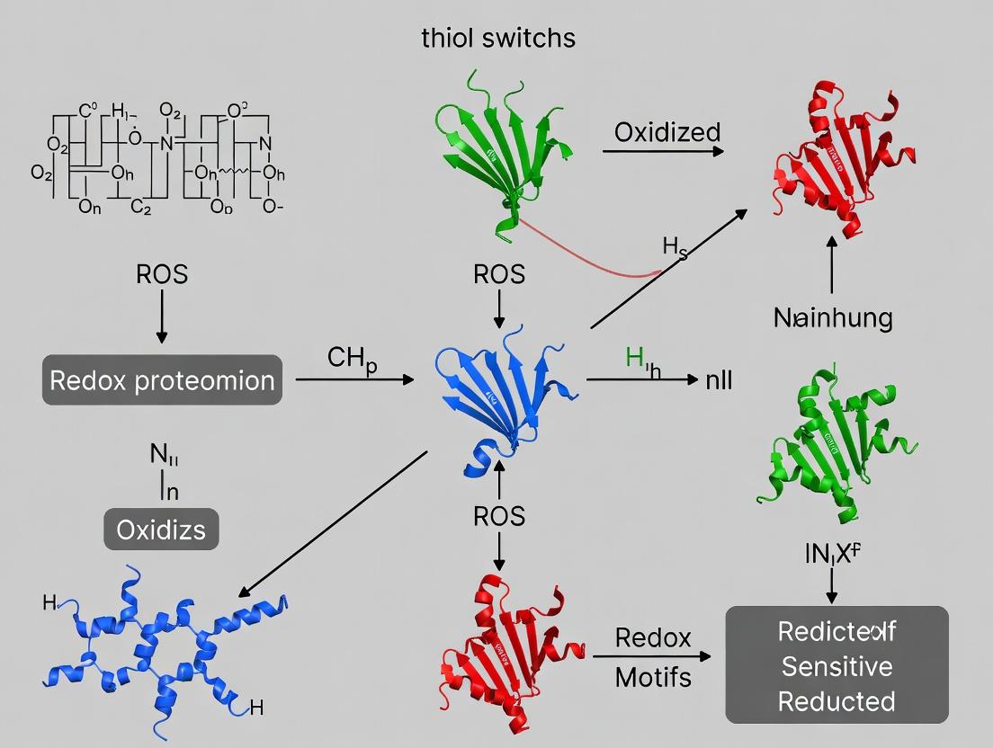
Within the broader thesis of redox proteomics in signal transduction, protein thiol switches represent a fundamental, reversible post-translational modification mechanism. Cysteine residues, owing to the unique nucleophilic and redox-active properties of their sulfur atom, serve as central sensors and transducers of cellular redox state, reactive oxygen/nitrogen species (ROS/RNS), and metabolic fluctuations. The conversion of specific cysteine thiols (-SH) to various oxidized derivatives (e.g., sulfenic acid -SOH, disulfide -S-S-, S-glutathionylation -SSG, S-nitrosylation -SNO) constitutes a "thiol switch" that can modulate protein function, localization, stability, and interactions. This guide details the core players and methodologies defining this dynamic field.
Core Chemical Players: The Cysteine Redox Landscape
The reactivity and fate of a cysteine thiol depend on its molecular microenvironment and the cellular oxidant. Key modifications are summarized in Table 1.
Table 1: Major Functional Cysteine Oxidative Modifications
| Modification | Chemical Formula | Typical Inducing Agent | Reversibility & Primary Reductase System | Functional Impact |
|---|---|---|---|---|
| Sulfenic Acid | R-SOH | H₂O₂, ROOH | Reversible, via Thioredoxin (Trx) or Glutaredoxin (Grx) | Often a transient intermediate; can regulate kinase/phosphatase activity. |
| Disulfide Bond | R-S-S-R' | ROS, oxidized environments | Reversible, via Trx or Grx/GSH | Structural stabilization or allosteric regulation. |
| S-Glutathionylation | R-S-SG | GSSG, ROS + GSH | Reversible, primarily via Grx/GSH | Neuroprotection against over-oxidation; modulates metabolic enzymes. |
| S-Nitrosylation | R-SNO | Nitric oxide (NO), N₂O₃ | Reversible, via Trx, GSNO reductase, or S-Nitrosoglutathione (GSNO) | Regulates apoptosis, vascular tone, ion channels. |
| Sulfinic Acid | R-SO₂H | Strong/Chronic ROS | Partially reversible by Sulfiredoxin (Srx) | Often considered irreversible, but Srx can reduce in some proteins. |
| Sulfonic Acid | R-SO₃H | Strong/Chronic ROS | Irreversible | Typically leads to protein degradation. |
Experimental Protocol: Identifying and Validating Thiol Switches
A comprehensive workflow integrates chemoproteomic discovery with functional validation.
Protocol: Isotopic or Isobaric Tandem Mass Tag (TMT)-Based Redox Proteomics
Objective: To quantify reversibly oxidized cysteines on a proteome-wide scale. Key Reagents: Iodoacetyl Tandem Mass Tag (iodoTMT) or Cysteine-reactive TMT (cysTMT) reagents (e.g., 6-plex, 11-plex), N-ethylmaleimide (NEM), Dithiothreitol (DTT), Mass spectrometry-grade trypsin/Lys-C.
Procedure:
- Cell Lysis and Blocking: Lyse cells/tissue under non-reducing conditions in the presence of 20-50 mM NEM and protease/phosphatase inhibitors. NEM rapidly alkylates free thiols, preventing post-lysis oxidation.
- Reduction and Labeling of Oxidized Cysteines: Remove excess NEM via protein precipitation or desalting. Reduce reversibly oxidized cysteines with 10-20 mM DTT (or TCEP). Subsequently, label the newly reduced thiols with iodoTMT reagent.
- Protein Digestion and Peptide-Level Multiplexing: Digest labeled proteins with trypsin. Pool samples labeled with different TMT channels (for relative quantification across conditions).
- Affinity Enrichment: Use anti-TMT antibody resin to enrich for labeled, cysteine-containing peptides, reducing sample complexity.
- LC-MS/MS Analysis and Data Processing: Analyze peptides by liquid chromatography-tandem mass spectrometry (LC-MS/MS). Quantify the relative abundance of each cysteine-containing peptide from different conditions based on TMT reporter ion intensities. High ratios indicate sites of increased oxidation under the test condition.
Protocol: Biotin Switch Technique (BST) for S-Nitrosylation
Objective: To specifically detect S-nitrosylated proteins. Key Reagents: Methyl methanethiosulfonate (MMTS), Sodium ascorbate, N-[6-(Biotinamido)hexyl]-3'-(2'-pyridyldithio)propionamide (Biotin-HPDP), Streptavidin-agarose.
Procedure:
- Block Free Thiols: Lyse samples in HEN buffer (HEPES, EDTA, Neocuproine) with 2.5% SDS and 20 mM MMTS at 50°C for 30 min. MMTS blocks all free thiols.
- Reduce SNO Bonds and Label with Biotin: Remove excess MMTS by acetone precipitation. Reduce S-NO bonds specifically with 1 mM sodium ascorbate. Simultaneously label the newly exposed thiols with 1 mM Biotin-HPDP for 1-3 hours.
- Affinity Capture and Detection: Remove unreacted Biotin-HPDP. Pull down biotinylated proteins with streptavidin-agarose beads. Wash thoroughly and elute with sample buffer containing β-mercaptoethanol. Detect by immunoblotting or identify by mass spectrometry.
The Scientist's Toolkit: Essential Research Reagents
Table 2: Key Research Reagent Solutions for Thiol Switch Studies
| Reagent | Function & Rationale |
|---|---|
| N-Ethylmaleimide (NEM) | Thiol-alkylating agent used to "freeze" the native redox state by covalently blocking all reduced cysteines during cell lysis. |
| Iodoacetamide (IAM) | Alternative alkylating agent; can be used for blocking or for stable isotope labeling (e.g., with heavy/light ICAT tags). |
| Iodoacetyl TMT (iodoTMT) | Isobaric mass tag that enables multiplexed quantification of cysteine oxidation states across multiple samples in a single MS run. |
| Biotin-HPDP | Thiol-reactive, cleavable biotinylation reagent used in the Biotin Switch Technique; allows for selective enrichment. |
| Dimedone & Derivatives | Nucleophilic probes that specifically and covalently react with sulfenic acid (-SOH) intermediates, enabling their detection or enrichment. |
| S-Nitrosoglutathione (GSNO) | A stable, cell-permeable NO donor used to induce S-nitrosylation experimentally. |
| Auranofin | A specific inhibitor of Thioredoxin Reductase (TrxR), used to disrupt the thioredoxin reduction system and elevate cellular oxidative state. |
| Glutaredoxin 1 (Grx1) | Enzyme used in assays to specifically reduce S-glutathionylated or mixed disulfide bonds. |
Visualization of Pathways and Workflows
Quantitative Data: Thiol Switch Prevalence and Dynamics
Table 3: Quantitative Snapshot from Recent Redox Proteomics Studies
| Study Focus (Year) | Model System | # of Quantified Cysteine Sites | # of Redox-Sensitive Sites Identified (Change >1.5x) | Major Pathway Enrichment |
|---|---|---|---|---|
| H₂O₂ Signaling (2023) | Human endothelial cells | >12,000 | ~2,100 | MAPK signaling, Actin cytoskeleton, Metabolic pathways. |
| S-Glutathionylation in Aging (2024) | Mouse liver tissue | 8,540 | 1,230 (Increased with age) | Fatty acid oxidation, TCA cycle, Antioxidant systems. |
| S-Nitrosylation in Immune Response (2023) | Macrophages (LPS/IFN-γ) | 5,780 | 892 | Inflammasome assembly, Glycolysis, NF-κB signaling. |
| Drug-induced Thiol Oxidation (2024) | Cancer cell lines (Auranofin) | 9,150 | 3,450 | Ribosome biogenesis, Proteasome, DNA repair. |
Defining the players—from specific cysteine residues to their modified states—is crucial for elucidating redox signaling networks. The integration of advanced chemoproteomic techniques, robust validation protocols, and pathway analysis, as framed within redox proteomics, provides a powerful framework. Future directions include mapping the crosstalk between thiol switches and other PTMs, developing single-cell redox assays, and designing therapeutics that target pathogenic thiol switches in cancer, neurodegeneration, and cardiovascular disease.
Cellular signal transduction is no longer viewed as a purely non-covalent protein interaction network. A critical layer of regulation is imposed by post-translational modifications driven by the cellular redox state. The "Redox Code" refers to the principle that reactive oxygen species (ROS), once considered purely detrimental, function as specific, tunable second messengers. Their opposing counterparts, antioxidant systems, precisely sculpt ROS gradients and lifetimes to modulate signaling outputs. This whitepaper frames this paradigm within the broader thesis of thiol switches and redox proteomics, which provide the mechanistic and analytical tools to decipher this code.
Thiol (-SH) groups on cysteine residues serve as prime redox sensors. Their oxidation to sulfenic acid (-SOH), disulfides (-S-S-), or further oxidized species (e.g., sulfinic -SO2H) can reversibly alter protein function, localization, and interactions—a "thiol switch." Redox proteomics, the system-wide identification and quantification of these oxidative modifications, is the indispensable methodology for mapping redox signaling networks in health, disease, and therapeutic intervention.
Core Principles of Redox Signaling
- Specificity & Reversibility: Signaling ROS (e.g., H2O2) oxidize specific cysteine residues with kinetic advantages, often via proximity to ROS-generation sites or acid-base catalysts. Reversal by dedicated antioxidant systems (Thioredoxin, Glutaredoxin) ensures transient signaling.
- Antagonistic Tuning: The cellular redox buffering capacity (glutathione, GSH/GSSG) and enzymatic antioxidants (Peroxiredoxins, Catalase) are not mere ROS scavengers but act as gatekeepers, setting thresholds for signal activation.
- Compartmentalization: Redox signaling is highly compartmentalized. Mitochondria, NADPH oxidases (NOX), and the endoplasmic reticulum generate spatially restricted ROS pools, enabling distinct signaling circuits.
Key Signaling Pathways Modulated by the Redox Code
NRF2-KEAP1 Pathway: The Master Antioxidant Response
Keap1, a cytosolic sensor protein, contains critical reactive cysteines. Under basal conditions, it targets the transcription factor NRF2 for degradation. Oxidation of Keap1 cysteines (e.g., C151) disrupts this complex, allowing NRF2 accumulation, nuclear translocation, and transcription of antioxidant and cytoprotective genes.
MAPK Pathway: Redox-Dependent Activation
Multiple nodes in the MAPK cascade are redox-sensitive. ASK1 (Apoptosis Signal-regulating Kinase 1) is held inactive via binding to reduced Thioredoxin (Trx). ROS oxidize Trx, causing its dissociation and subsequent ASK1 activation, leading to p38/JNK-mediated stress responses.
PI3K/AKT Pathway: Growth Factor Signaling Integration
H2O2 can transiently inhibit protein tyrosine phosphatases (PTPs, e.g., PTEN) via oxidation of their catalytic cysteine to sulfenic acid. This prevents dephosphorylation of receptor tyrosine kinases and downstream components like AKT, thereby potentiating growth and survival signals.
Inflammatory Signaling: NF-κB and NLRP3 Inflammasome
IKKγ (NEMO) and TNF receptor-associated factors (TRAFs) undergo redox modifications that modulate NF-κB activation. Similarly, thioredoxin-interacting protein (TXNIP) dissociates from reduced Trx upon ROS stress and activates the NLRP3 inflammasome.
Diagram 1: The NRF2-KEAP1 Redox Sensing Pathway
Diagram 2: ROS Activation of ASK1 via Thioredoxin Oxidation
Redox Proteomics: The Experimental Toolkit
This field relies on a suite of chemical probes and mass spectrometry (MS)-based methods to capture and identify labile thiol modifications.
Key Methodological Workflows
1. Biotin-Switch and Acyl-Resin Assisted Capture (Acyl-RAC): These methods rely on blocking free thiols (alkylation), selectively reducing oxidized species (e.g., S-nitrosothiols or disulfides), and labeling the newly reduced thiols with a biotin or resin-capturable tag for enrichment and MS identification.
2. Isotope-Coded Affinity Tag (ICAT) for Redox: Thiol-reactive ICAT tags in light (d0) and heavy (d8) isotopic forms allow quantitative comparison of thiol redox states between two samples (e.g., control vs. treated).
3. OxICAT: Uses a light (¹²⁵I) and heavy (¹³¹I) isotope-coded thiol-reactive probe to directly quantify the redox state of individual cysteine residues across proteomes.
4. Competitive Activity-Based Protein Profiling (ABPP): Uses reactive chemical probes to assess the reactivity/occupancy of cysteines in native proteomes, identifying those sensitive to redox modulation.
Diagram 3: Biotin-Switch Redox Proteomics Workflow
The Scientist's Toolkit: Key Research Reagent Solutions
| Reagent / Material | Primary Function in Redox Research |
|---|---|
| N-Ethylmaleimide (NEM) | Irreversible alkylating agent. Used to rapidly block all free thiols during cell lysis to "freeze" the native redox state. |
| Iodoacetamide (IAA) | Another alkylating agent, often used in-gel or in-solution after reduction to alkylate cysteines prior to digestion for MS. |
| Biotin-HPDP | Thiol-reactive, cleavable biotinylation reagent. Used in biotin-switch techniques to tag formerly oxidized cysteines for enrichment. |
| Streptavidin Agarose/Magnetic Beads | For high-affinity capture and purification of biotin-tagged peptides/proteins prior to MS analysis. |
| Dimedone & Derivatives | Electrophilic probes that specifically and irreversibly react with sulfenic acid (-SOH) modifications, allowing their detection or enrichment. |
| Tandem Mass Tags (TMT) / Isobaric Tags | Enable multiplexed (e.g., 10-16 plex) quantitative comparison of peptide abundances across multiple samples in a single MS run. |
| Recombinant Thioredoxin (Trx) / Glutaredoxin (Grx) | Used in validation experiments to test reversibility of oxidation or in enzyme-coupled assays to measure thiol status. |
| H2O2-Sensitive Probes (e.g., HyPer, roGFP) | Genetically encoded fluorescent biosensors for real-time, compartment-specific imaging of H2O2 dynamics in live cells. |
| PRDX Mimetics (e.g., BMX-001) | Pharmacological tools to manipulate endogenous antioxidant peroxidase activity and study downstream signaling effects. |
Quantitative Data in Redox Signaling
Table 1: Physiological vs. Pathological ROS Concentrations & Half-Lives
| ROS Species | Physiological [Approx.] | Pathological/Stress [Approx.] | Key Source | Estimated Half-life |
|---|---|---|---|---|
| Superoxide (O₂•⁻) | 10-100 pM | > 1 nM | NOX, ETC | Milliseconds (ms) |
| Hydrogen Peroxide (H₂O₂) | 1-10 nM | 100 nM - 1 µM | DUOX, PRDX | ~1 ms to seconds |
| Peroxynitrite (ONOO⁻) | < 50 nM | > 200 nM | O₂•⁻ + NO | < 10 ms |
| Hypochlorous Acid (HOCl) | Low (phagocytes) | High (chronic inflammation) | MPO | Stable (sec-min) |
Table 2: Redox Potentials of Key Cellular Couples
| Redox Couple | E'° (mV) at pH 7.0 | Function in Signaling |
|---|---|---|
| GSH/GSSG | -240 to -180 (varies by compartment) | Major redox buffer; sets cellular tone. |
| Trx (red/ox) | ~ -280 | Key reductase for oxidized protein thiols. |
| Cysteine/Cystine | ~ -250 | Extracellular redox buffer. |
| Peroxiredoxin (Prx-SH/Prx-SOH) | ~ -300 | High sensitivity H2O2 sensor/scavenger. |
| NRF2-KEAP1 | N/A (kinetic sensing) | Activated by Keap1 C151 oxidation. |
Detailed Experimental Protocol: Acyl-Resin Assisted Capture (Acyl-RAC) for Disulfide Mapping
Objective: To identify proteins forming reversible disulfide bonds in response to a specific stimulus (e.g., H2O2 treatment).
Materials: Lysis buffer (50 mM Tris-HCl pH 7.5, 150 mM NaCl, 1% NP-40) supplemented with 20 mM NEM and protease inhibitors. Thiopropyl Sepharose 6B resin. Dimethylformamide (DMF). Hydroxylamine hydrochloride. SDS-PAGE and Western blot or MS equipment.
Procedure:
- Lysis & Blocking: Lyse control and H2O2-treated cells in NEM-containing buffer. Incubate 30 min at 40°C with vortexing to alkylate all free thiols.
- Protein Clean-up: Precipitate proteins using acetone. Wash pellets 2x with 70% acetone to remove excess NEM.
- Reduction of Disulfides: Resuspend protein pellets in binding buffer (50 mM Tris-HCl pH 7.5, 150 mM NaCl, 1% SDS). Add 10 mM DTT (final conc.) and incubate 30 min at room temperature to reduce disulfides.
- Resin Preparation: Wash Thiopropyl Sepharose resin 3x with water, then 2x with binding buffer containing 1% SDS.
- Capture: Mix reduced protein samples with prepared resin. Add 2 mM (final) HPDP-biotin or directly incubate with resin for disulfide capture via thiol-disulfide exchange. Rotate for 3 hours at room temperature.
- Washing: Wash resin sequentially with binding buffer + 1% SDS, then high-salt buffer (2 M NaCl), and finally low-SDS buffer (0.1% SDS) to remove non-specifically bound proteins.
- Elution: Elute bound proteins (those originally in a disulfide) using elution buffer (binding buffer + 50 mM DTT or β-mercaptoethanol) for 20 min at 37°C.
- Analysis: Analyze eluates by SDS-PAGE/Western (for targets) or trypsin digest followed by LC-MS/MS for global disulfide proteomics.
Implications for Drug Development
Targeting the Redox Code offers novel therapeutic avenues:
- NRF2 Activators: (e.g., dimethyl fumarate) for chronic oxidative stress diseases (COPD, neurodegeneration).
- NOX Inhibitors: For fibrotic and inflammatory diseases.
- PRDX Mimetics: To ameliorate excessive ROS in ischemia-reperfusion injury.
- Pro-oxidant Therapies: Selectively induce lethal ROS levels in cancer cells (e.g., some chemotherapeutics).
Understanding redox networks via thiol-switch proteomics is critical for developing these targeted therapies with precise efficacy and minimized off-target effects.
Cysteine residues in proteins serve as dynamic, post-translational regulatory sites, often described as "thiol switches," that translate alterations in cellular redox state into functional changes. These modifications are central to redox signal transduction, integrating metabolic and stress responses. S-glutathionylation, S-nitrosylation, and sulfenic acid formation represent three fundamental, reversible modifications that compete for and modify reactive protein thiols. Their interplay regulates a vast array of processes, from apoptosis and metabolism to transcription and kinase signaling. This technical guide details the core mechanisms, detection methodologies, and experimental approaches essential for research in this field, framed within the broader context of redox proteomics and its implications for understanding disease and therapeutic intervention.
Core Mechanisms & Functional Consequences
Sulfenic Acid Formation (-SOH)
Sulfenic acids are the initial oxidative product of cysteine thiols, formed by reaction with hydrogen peroxide or other two-electron oxidants. This labile modification can act as a signaling intermediate, leading to further stabilization via disulfide formation or overoxidation.
Key Reaction: Protein-SH + H₂O₂ → Protein-SOH + H₂O
S-glutathionylation (-SSG)
The reversible formation of a mixed disulfide between a protein thiol and the low-molecular-weight tripeptide glutathione (GSH). This modification typically occurs via thiol-disulfide exchange with oxidized glutathione (GSSG) or via reaction of a sulfenic acid intermediate with reduced glutathione (GSH). It provides steric and charge-based regulation, protecting thiols from irreversible oxidation.
Key Reaction: Protein-SOH + GSH → Protein-SSG + H₂O or Protein-SH + GSSG → Protein-SSG + GSH
S-nitrosylation (-SNO)
The covalent attachment of a nitric oxide (NO) group to a reactive cysteine thiol, forming an S-nitrosothiol. This reaction is central to NO-mediated signaling (nitrosative signaling) and can occur through metal-catalyzed pathways or trans-nitrosylation from low-molecular-weight or protein SNO donors.
Key Reaction: Protein-SH + N₂O₃ → Protein-SNO + NO₂⁻ + H⁺
Quantitative Data & Comparative Analysis
Table 1: Comparative Properties of Key Thiol Modifications
| Property | Sulfenic Acid (-SOH) | S-glutathionylation (-SSG) | S-nitrosylation (-SNO) |
|---|---|---|---|
| Inducing Species | H₂O₂, organic peroxides, peroxynitrite | GSSG, S-glutathionyl radicals, via -SOH | NO⁺ donors, N₂O₃, metal-NO complexes, trans-nitrosylation |
| Typical Half-Life | Milliseconds to seconds | Minutes to hours | Seconds to minutes |
| Reversibility | Highly reversible | Enzymatic (Grx, Srx) & thiol exchange | Enzymatic (Trx, GSNOR) & trans-nitrosylation |
| Primary Role | Signaling intermediate, protector from overoxidation | Protective, redox buffer, steric regulation | Signaling, mimicry of phosphorylation, regulation of metals |
| Key Detectors | Dimedone-based probes | Biotinylated GSH ethyl ester, anti-GSH antibodies | Biotin-switch technique, SNO-RAC, chemiluminescence |
| Cellular Compartment | Widespread, notably mitochondria, cytosol | Cytosol, nucleus, mitochondria (GSH pools) | Membrane, cytosol, mitochondria, dependent on NOS localization |
Table 2: Select Protein Targets & Functional Outcomes
| Protein Target | Modification | Functional Consequence | Pathophysiological Context |
|---|---|---|---|
| PTP1B | -SOH / -SSG | Inhibition of phosphatase activity | Insulin signaling, receptor tyrosine kinase activation |
| Caspase-3 | -SSG / -SNO | Inhibition of protease activity | Regulation of apoptosis, ischemic preconditioning |
| NF-κB (p50) | -SNO | Inhibition of DNA binding | Anti-inflammatory effects of NO |
| GAPDH | -SNO | Increased binding to Siah1, nuclear translocation | Apoptotic signaling, neurodegenerative disease |
| Ryanodine Receptor | -SOH / -SNO | Modulation of Ca²⁺ release | Cardiac muscle contraction, arrhythmia |
| H-Ras | -SNO | Activation, promotion of GTP binding | Cell proliferation, migration |
Experimental Protocols for Detection & Analysis
Protocol: Biotin-Switch Technique for S-nitrosylation
Adapted from Jaffrey & Snyder (2001) with contemporary updates.
Principle: Selective replacement of labile SNO groups with a biotin tag for enrichment and detection.
Procedure:
- Cell Lysis & Blocking: Lyse cells in HEN buffer (250 mM HEPES pH 7.7, 1 mM EDTA, 0.1 mM Neocuproine) with 2.5% SDS. Add methyl methanethiosulfonate (MMTS) to a final concentration of 20-50 mM. Incubate at 50°C for 20 min with frequent vortexing. This blocks all free thiols.
- Acetone Precipitation: Remove excess MMTS by acetone precipitation (2-3 volumes). Wash pellet 3x with 70% acetone.
- SNO Reduction & Biotinylation: Resuspend pellet in HENS buffer (HEN + 1% SDS). For each mg of protein, add 1/3 volume of freshly prepared labeling solution: 4 mM Biotin-HPDP (or maleimide-PEG2-biotin) and 1 mM sodium ascorbate in dimethylformamide. Ascorbate selectively reduces SNO to thiol, which is immediately biotinylated. Incubate at 25°C for 1 hr in the dark.
- Pull-down & Analysis: Remove excess biotin by acetone precipitation. Resuspend in neutralization buffer. Incubate with streptavidin-agarose beads for 1 hr. Wash beads stringently, elute with Laemmli buffer containing β-mercaptoethanol, and analyze by western blot or mass spectrometry. Critical Controls: Include samples without ascorbate (negative control) and without MMTS blocking (total thiol control).
Protocol: Detection of Protein Sulfenic Acids Using Dinucleophilic Probes (e.g., DCP-Bio1/DCP-Rho1)
Adapted from Poole et al. (2007).
Principle: Cyclic 1,3-diketones (e.g., dimedone) selectively and covalently react with sulfenic acids. Derivatized probes enable detection.
Procedure:
- In Situ Labeling: Treat live cells or intact tissue with the desired oxidant stimulus. During the stimulation, add cell-permeable probe (e.g., DCP-Bio1 or DCP-Rho1) at 50-500 µM. Incubate for 5-30 min.
- Cell Lysis & Processing: Wash cells and lyse in non-reducing, detergent-based lysis buffer without thiol-scavenging agents (e.g., no DTT, β-ME).
- Detection:
- For Biotin Probes: Proceed with streptavidin pull-down (as in 4.1, step 4) or direct streptavidin-HRP western blot of total lysate.
- For Fluorescent Probes: Analyze by in-gel fluorescence scanning (Typhoon scanner) or fluorescence microscopy for cellular localization.
- Competition Assay: Confirm specificity by pre-incubating samples with unconjugated dimedone (10 mM) prior to probe addition, which should block labeling.
Protocol: Resin-Assisted Capture for S-glutathionylation (RAC-SSG)
Adapted from Guo et al. (2014).
Principle: Selective reduction of protein-glutathione mixed disulfides (-SSG) followed by covalent capture on thiol-reactive resin.
Procedure:
- Block Free Thiols & Reduce SSG: Lyse cells in buffer with 50 mM N-ethylmaleimide (NEM) to alkylate free thiols. After clearing, remove excess NEM by desalting. Incubate lysate with 10 mM sodium borohydride (NaBH₄) for 30 min at RT. Note: NaBH₄ selectively reduces -SSG but not intra-protein disulfides.
- Capture Newly Reduced Thiols: Acidify sample to quench NaBH₄. Add Thiopropyl Sepharose resin. The newly reduced protein thiols (originally -SSG) form a mixed disulfide with the resin. Incubate overnight at 4°C.
- Elution & Analysis: Wash resin thoroughly. Elute captured proteins using 20 mM DTT. Precipitate eluate and analyze by western blot or proteomics via LC-MS/MS.
Visualizing Pathways & Workflows
Diagram 1: Thiol Modification Interplay
Diagram 2: Biotin-Switch Technique Workflow
The Scientist's Toolkit: Essential Research Reagents
Table 3: Key Reagents for Thiol Redox Research
| Reagent / Kit | Primary Function | Key Considerations |
|---|---|---|
| Biotin-HPDP | Thiol-reactive, cleavable biotinylation agent for Biotin-Switch. | Disulfide-linked biotin allows gentle elution. Light-sensitive. |
| Streptavidin Agarose/Magnetic Beads | Affinity capture of biotinylated proteins. | High binding capacity (>2 µg biotin/mg beads) and low non-specific binding are critical. |
| S-Nitrosoglutathione (GSNO) | Stable, cell-permeable NO donor and trans-nitrosylating agent. | Calibrates SNO detection; induces physiological S-nitrosylation. |
| Dimedone & Derivatives (DCP-Bio1, DCP-Rho1) | Chemoselective probes for labeling protein sulfenic acids (-SOH). | DCP-Rho1 for microscopy; DCP-Bio1 for enrichment. Must use fresh. |
| Glutaredoxin-1 (Grx1) & Glutathione (GSH) | Enzymatic system for specific reduction/deglutathionylation of -SSG. | Used in activity assays and to validate -SSG modifications. |
| Anti-GSH Antibody | Direct immunodetection of protein-glutathione adducts by western/IF. | Can have variable affinity; best for abundant targets. |
| Triple Quadrupole LC-MS/MS with iTRAQ/TMT | Quantitative redox proteomics. | Enables multiplexed comparison of modification states across conditions. |
| Thiopropyl Sepharose 6B | Thiol-reactive resin for resin-assisted capture (RAC). | Specific for covalent capture of reduced thiols post-selective reduction. |
| Neocuproine & EDTA | Metal chelators in lysis buffers. | Prevent artifactual Cu²⁺-caused de-nitrosylation or oxidation during processing. |
| Cytochrome c Reduction / Amplex Red Assay | Spectrophotometric/fluorometric measurement of H₂O₂/O₂⁻ levels. | Quantifies the redox stimulus applied to cells. |
This technical whitepaper, framed within a broader thesis on thiol switches and redox proteomics in signal transduction, explores the post-translational modification of protein cysteine residues. These reversible thiol switches, including S-glutathionylation, S-nitrosation, and sulfenylation, function as central redox sensors, dynamically regulating metabolic pathways, inflammatory responses, and apoptotic signaling. Understanding these mechanisms is critical for developing targeted therapeutics for metabolic disorders, chronic inflammation, and cancer.
Cysteine thiols are unique among amino acids due to their nucleophilic sulfhydryl group, which undergoes reversible oxidation in response to reactive oxygen/nitrogen species (ROS/RNS). This forms the basis of redox signaling. Key modifications include:
- S-Glutathionylation: Formation of a mixed disulfide with glutathione (GSH).
- S-Nitrosation: Covalent attachment of a nitric oxide (NO) group.
- Sulfenylation: Formation of a sulfenic acid (-SOH). These modifications alter protein structure, activity, localization, and interactions, acting as molecular switches.
Thiol Switches in Metabolic Regulation
Cellular metabolism is exquisitely sensitive to redox state. Thiol switches directly regulate key metabolic enzymes.
Key Regulatory Nodes:
- Glyceraldehyde-3-Phosphate Dehydrogenase (GAPDH): S-Nitrosation at Cys150 inhibits its glycolytic activity, diverting glucose flux into the pentose phosphate pathway to increase NADPH production for antioxidant defense.
- Pyruvate Kinase M2 (PKM2): Oxidation or S-glutathionylation inhibits its activity, slowing glycolysis and promoting anabolic processes for cell proliferation.
- Complex I of the Mitochondrial ETC: S-Glutathionylation of specific subunits reversibly inhibits activity, reducing ROS production during ischemia/reperfusion injury.
- Phosphatase and Tensin Homolog (PTEN): Formation of a disulfide bond between the active site Cys124 and Cys71 inactivates this tumor suppressor, activating the pro-growth PI3K/AKT pathway.
Quantitative Data on Metabolic Thiol Switches
| Target Protein | Modification Site | Modification Type | Functional Consequence | % Activity Change (Typical Range) |
|---|---|---|---|---|
| GAPDH | Cys150 | S-Nitrosation | Inhibition, metabolic flux shift | 70-90% decrease |
| PKM2 | Cys358 | S-Glutathionylation | Inhibition, promotes anabolism | 50-80% decrease |
| Mitochondrial Complex I | Cys531 & Cys704 (rodent) | S-Glutathionylation | Reversible inhibition, limits ROS | 60-95% decrease |
| PTEN | Cys124-Cys71 | Intramolecular Disulfide | Inactivation, promotes growth | ~90% decrease |
| AMPK | Cys299 & Cys304 | S-Glutathionylation | Inhibition, alters energy sensing | 40-70% decrease |
Experimental Protocol: Assessing Enzyme Activity Modulation by S-Glutathionylation In Vitro
- Protein Purification: Express and purify recombinant target protein (e.g., PKM2) via affinity chromatography.
- Redox Treatment: Divide protein into aliquots.
- Reduced Control: Treat with 5mM DTT for 30 min at 4°C, then remove DTT via desalting column.
- Oxidized Sample: Treat with 0.1-1.0 mM oxidized glutathione (GSSG) or diamide in appropriate buffer (e.g., 50mM Tris-HCl, pH 7.4) for 15-30 min at 25°C.
- Removal of Oxidizing Agent: Use a desalting column (e.g., Zeba Spin) equilibrated with assay buffer to remove GSSG/diamide.
- Activity Assay: Perform standard enzymatic assay. For PKM2, monitor pyruvate production coupled to lactate dehydrogenase (LDH) and NADH oxidation (decrease in A340).
- Confirmation of Modification: Run parallel samples for non-reducing SDS-PAGE (shift in mobility) or mass spectrometry to confirm mixed disulfide formation.
Thiol Switches in Inflammatory Signaling
Redox-sensitive cysteines are pivotal in the regulation of the NF-κB and NLRP3 inflammasome pathways.
Core Mechanisms:
- IKKβ Kinase: S-Glutathionylation at Cys179 in the activation loop inhibits its kinase activity, blocking IκB degradation and NF-κB nuclear translocation.
- NLRP3 Inflammasome: Oxidation of cysteine residues in the NACHT domain is required for its assembly and activation in response to DAMPs/PAMPs.
- Keap1-Nrf2 Pathway: Oxidation of specific Keap1 cysteines (Cys151, Cys273, Cys288) disrupts its interaction with Nrf2, allowing Nrf2 translocation to the nucleus to induce antioxidant gene expression (e.g., HO-1, NQO1).
Thiol Regulation of the NF-κB Inflammatory Pathway (79 chars)
Thiol Switches in Apoptotic Control
The balance between pro-survival and pro-apoptotic signals is tightly regulated by redox modifications.
Key Apoptotic Switches:
- Caspase-3: S-Nitrosation at the catalytic Cys163 inhibits protease activity, preventing apoptosis. Denitrosation activates the executioner phase.
- Mitochondrial Permeability Transition Pore (mPTP): Oxidation of critical cysteines in the adenine nucleotide translocator (ANT) promotes pore opening, triggering cytochrome c release.
- Bcl-2 Family Proteins: Pro-apoptotic Bax can be S-nitrosated, inhibiting its translocation to mitochondria.
Quantitative Data on Apoptotic Thiol Switches
| Target Protein | Modification Site | Modification Type | Effect on Apoptosis | Associated Condition |
|---|---|---|---|---|
| Caspase-3 | Cys163 (active site) | S-Nitrosation | Inhibition, Pro-survival | Ischemic Preconditioning |
| mPTP Component (ANT) | Multiple Cys | Oxidation | Promotes Opening, Pro-apoptotic | Reperfusion Injury |
| Bax | Cys62 | S-Nitrosation | Inhibits Mitochondrial Translocation | Tumor Cell Resistance |
| ASK1 | Cys250 | S-Glutathionylation | Inhibits Kinase Activity, Pro-survival | Oxidative Stress Response |
| c-FLIP | Cys254 | S-Nitrosation | Stabilization, Inhibits DISC | Inflammatory Protection |
Experimental Protocol: Detecting S-Nitrosation (Biotin-Switch Technique)
- Cell Lysis & Blocking: Lyse cells/tissue in HEN buffer (250mM HEPES pH7.7, 1mM EDTA, 0.1mM neocuproine) with 2.5% SDS and 20mM methyl methanethiosulfonate (MMTS). Incubate 50 min at 50°C with frequent vortexing. MMTS blocks free thiols.
- Acetone Precipitation: Remove MMTS by precipitating proteins with 2 volumes of pre-chilled acetone at -20°C for 20 min. Centrifuge, wash pellet 3x with 70% acetone.
- Reduction & Biotinylation: Resuspend pellet in HENS buffer (HEN + 1% SDS). Add 1mM ascorbate (to selectively reduce S-NO bonds) and 4mM biotin-HPDP. Incubate for 1 hour at 25°C in the dark.
- Pull-Down & Detection: Remove excess biotin-HPDP by acetone precipitation. Resuspend pellet and incubate with neutralized streptavidin-agarose beads for 1 hour. Wash beads thoroughly, elute proteins with Laemmli buffer containing 2-mercaptoethanol (to break biotin-thiol bond). Analyze by immunoblotting for target protein.
Research Toolkit: Key Reagents & Solutions
| Reagent / Material | Function / Application | Key Consideration |
|---|---|---|
| Diamide | Thiol-specific oxidant. Induces S-glutathionylation and disulfide formation in vitro and in cells. | Concentration- and time-dependent effects; use with precise controls. |
| S-Nitrosoglutathione (GSNO) | Stable NO donor. Used to induce protein S-nitrosation in experimental systems. | Light-sensitive; prepare fresh. |
| Biotin-HPDP | Thiol-reactive biotinylating agent. Used in the biotin-switch technique to label reduced cysteines after ascorbate reduction of S-NO. | Compare +/- ascorbate controls to confirm specificity. |
| Streptavidin Beads (Agarose/Magnetic) | For affinity capture of biotinylated proteins in pull-down assays (e.g., biotin-switch). | High binding capacity and low non-specific binding are critical. |
| Anti-GSH Antibody | For immunodetection of protein S-glutathionylation via western blot or immunoprecipitation. | Specificity varies; confirm with positive/negative controls. |
| MS-Based Probes (e.g., Din-IAA, CIAP) | Isotope-coded or clickable probes for quantitative redox proteomics via mass spectrometry. | Requires specialized MS instrumentation and data analysis. |
| Glutaredoxin-1 (Grx1) | Enzyme to specifically reduce S-glutathionylated proteins. Used to confirm modification and study reversibility. | Assess activity before use. |
| Cellular ROS/RNS Sensors (e.g., H2DCFDA, DAF-FM) | Fluorescent dyes to measure general ROS or specific RNS (NO) in cells during experiments. | Can be non-specific; use with appropriate inhibitors. |
Biotin-Switch Technique for S-Nitrosation Detection (66 chars)
The systematic study of thiol switches via redox proteomics—integrating the biotin-switch technique, isoTOP-ABPP, and modern mass spectrometry—is central to the broader thesis of mapping redox signaling networks. Future research must focus on:
- Spatiotemporal Resolution: Developing tools to detect specific thiol modifications in real-time within cellular compartments.
- Crosstalk: Understanding how different modifications on the same or interacting proteins integrate signals.
- Therapeutic Targeting: Designing drugs that selectively modulate pathological thiol switches in metabolic disease, inflammation, and cancer, while preserving physiological redox signaling. This represents a frontier in precision medicine.
Cellular signal transduction is intricately governed by post-translational modifications. Among these, reversible oxidation of cysteine thiols (-SH) acts as a fundamental "thiol switch," modulating protein function, localization, and interactions in response to reactive oxygen species (ROS) and reactive nitrogen species (RNS). Redox proteomics, the systematic identification and quantification of these oxidative modifications, has emerged as a pivotal tool for mapping redox signaling networks. Dysregulation of these precise redox circuits—leading to either excessive oxidation (oxidative stress) or aberrant reductive signaling—is a central mechanistic node linking seemingly disparate pathologies: cancer and neurodegeneration. While cancer often exploits a pro-reductive, proliferative environment, neurodegenerative diseases are characterized by chronic oxidative damage. This whitepaper details the molecular mechanisms, experimental approaches, and therapeutic implications of redox dysregulation within the framework of thiol switch biology.
Core Mechanisms: Redox Dysregulation in Disease
Cancer: A Pro-Reductive Landscape
Cancer cells frequently sustain a hyper-reduced intracellular state to support rapid proliferation, resist apoptosis, and promote metastasis. Key mechanisms include:
- NRF2-KEAP1 Pathway Dysregulation: Gain-of-function mutations in KEAP1 or NRF2 lead to constitutive NRF2 activation, upregulating a battery of antioxidant genes (e.g., glutathione S-transferases, NADPH quinone oxidoreductase 1). This creates a chemoresistant phenotype.
- Glutathione (GSH) and Thioredoxin (Trx) System Overactivation: Elevated synthesis of GSH and increased activity of Trx, Trx reductase (TrxR), and glutathione reductase (GR) maintain a reduced thioredoxin and glutathione pool, scavenging ROS that would otherwise inhibit proliferation.
- Metabolic Reprogramming: The Warburg effect (aerobic glycolysis) and increased pentose phosphate pathway flux generate NADPH, the essential reducing equivalent for antioxidant systems.
Neurodegeneration: A State of Chronic Oxidative Damage
Neurons are post-mitotic and highly metabolically active, making them exceptionally vulnerable to cumulative oxidative insult. Key mechanisms include:
- Mitochondrial Dysfunction: Defective electron transport chains (ETC) in Alzheimer's (AD), Parkinson's (PD), and Huntington's (HD) diseases leak electrons, generating superoxide anion (O2•−).
- Metal Homeostasis Disruption: Mis-regulation of redox-active metals (e.g., Fe, Cu) catalyzes Fenton reactions, producing highly toxic hydroxyl radicals (•OH).
- Protein Aggregation: Disease-associated proteins like Aβ, tau, α-synuclein, and huntingtin can directly generate ROS or impair mitochondrial function, while their aggregates can quench antioxidant enzymes.
Table 1: Comparative Redox Signatures in Cancer vs. Neurodegeneration
| Parameter | Cancer (e.g., Non-Small Cell Lung Cancer) | Neurodegeneration (e.g., Alzheimer's Disease) |
|---|---|---|
| Primary Redox State | Generally more reduced (pro-reductive) | Chronically oxidized (oxidative stress) |
| Key Altered Pathway | Constitutive NRF2 activation; Trx/GSH upregulation | Mitochondrial ETC dysfunction; NRF2 impairment |
| GSH:GSSG Ratio | Often elevated | Typically decreased |
| NADPH:NADP+ Ratio | Increased (fueled by PPP) | Often decreased |
| Major Oxidant Source | Growth factor signaling, metabolic ROS | Mitochondrial leak, metal-catalyzed reactions |
| Key Redox-Sensitive Protein Target | PTEN (inactivated by oxidation), HIF-1α (stabilized) | PTEN (activated by oxidation), PKA (inhibited) |
| Functional Outcome | Proliferation, survival, metastasis | Synaptic dysfunction, neuronal death |
Experimental Protocols in Redox Proteomics
Protocol: Isotope-Coded Affinity Tag (ICAT) for Redox Cysteine Profiling
This method quantifies the reduced vs. oxidized state of cysteine thiols across proteomes.
- Cell Lysis and Blocking: Lyse tissue or cells in a buffer containing 50 mM Tris-HCl (pH 8.3), 150 mM NaCl, 1% NP-40, and 50 mM N-ethylmaleimide (NEM) or iodoacetamide (IAA) to alkylate and block all free (reduced) thiols.
- Reduction of Oxidized Thiols: Remove excess alkylating agent via cold acetone precipitation. Resuspend the pellet and treat with 10 mM dithiothreitol (DTT) to reduce reversibly oxidized thiols (e.g., disulfides, sulfenic acids).
- Isotopic Labeling: Label the newly reduced thiols with light (12C) or heavy (13C) ICAT reagent (containing a biotin affinity tag and a thiol-reactive group). For a case-control experiment, label the control sample with light ICAT and the treated/diseased sample with heavy ICAT.
- Combination, Digestion, and Affinity Purification: Combine the labeled samples 1:1. Digest with trypsin. Purify ICAT-labeled peptides using streptavidin chromatography.
- LC-MS/MS Analysis and Quantification: Analyze purified peptides by liquid chromatography-tandem mass spectrometry (LC-MS/MS). Relative quantification of light/heavy peptide pairs provides the redox state change for specific cysteine sites.
Protocol: Biotin Switch Technique (BST) for S-Nitrosylation Mapping
This technique specifically detects protein S-nitrosylation (SNO), a NO-mediated thiol modification.
- Block Free Thiols: Lyse samples in HENS buffer (250 mM HEPES pH 7.7, 1 mM EDTA, 0.1 mM neocuproine, 1% SDS) with 20 mM methyl methanethiosulfonate (MMTS) to block all free thiols.
- Reduce S-Nitrosothiols: Precipitate proteins to remove MMTS. Treat pellets with 1 mM ascorbate (or a more specific reagent like Cu/ascorbate or SNO-COPH) to selectively reduce S-nitrosothiols to free thiols.
- Label with Biotin-HPDP: Label the newly revealed thiols with 1 mM N-[6-(biotinamido)hexyl]-3'-(2'-pyridyldithio) propionamide (Biotin-HPDP) or similar thiol-reactive biotinylating agent.
- Affinity Capture and Detection: Precipitate proteins, resuspend, and pull down biotinylated proteins with streptavidin-agarose beads. Wash extensively. Elute proteins with DTT-containing buffer or directly analyze by western blot (using streptavidin-HRP) or mass spectrometry (after on-bead trypsin digestion).
Visualization of Key Pathways and Workflows
Title: Redox Signaling in Cancer vs Neurodegeneration
Title: Biotin Switch Technique Workflow
The Scientist's Toolkit: Key Research Reagents
Table 2: Essential Reagents for Redox Proteomics and Thiol Switch Analysis
| Reagent / Material | Function / Role | Key Example(s) |
|---|---|---|
| Thiol-Alkylating Agents | Irreversibly block free cysteine thiols to "freeze" the redox state during lysis. Prevents post-lysis oxidation/reduction. | N-ethylmaleimide (NEM), Iodoacetamide (IAA), Methyl methanethiosulfonate (MMTS) |
| Isotope-Coded Tags | Enable multiplexed, quantitative MS comparison of redox states between samples via light/heavy isotope pairs. | ICAT (Isotope-Coded Affinity Tag), iodoTMT (tandem mass tag) |
| Biotin-HPDP / Maleimide-Biotin | Thiol-reactive biotinylation agents used to tag specific, previously oxidized cysteines (after reduction) for affinity purification. | N-[6-(biotinamido)hexyl]-3'-(2'-pyridyldithio)propionamide (Biotin-HPDP), EZ-Link Maleimide-PEG2-Biotin |
| Selective Reducing Agents | Chemically reduce specific oxidative modifications to free thiols for subsequent labeling. | Ascorbate (for S-nitrosothiols), Arsenite (for sulfenic acids), DTT/TCEP (general for disulfides) |
| ROS/RNS Sensors & Probes | Detect and quantify specific reactive species in live cells or lysates. | H2DCFDA (general ROS), MitoSOX (mitochondrial superoxide), DAF-FM (NO), HyPer (H2O2 biosensor) |
| Activity-Based Probes (ABPs) | Chemically tag the active site of redox enzymes (e.g., peroxiredoxins, glutathione peroxidases) to monitor their functional state. | Cyanamides, Sulfonate esters |
| Antibodies for Oxidative PTMs | Immunodetection of specific oxidative modifications. | Anti-3-nitrotyrosine, Anti-4-hydroxynonenal (4-HNE), Anti-S-glutathionylation |
| LC-MS/MS System | The core analytical platform for identifying and quantifying labeled peptides in redox proteomics. | Orbitrap, Q-TOF, or Triple Quadrupole mass spectrometers coupled to nano-UHPLC |
Mapping the Redoxome: Essential Proteomic Workflows and Translational Applications
Redox proteomics is an indispensable field for elucidating the molecular mechanisms of cellular signal transduction. At its heart is the study of reversible post-translational modifications (PTMs) on cysteine residues, termed "thiol switches." These modifications, including S-nitrosation, S-glutathionylation, S-sulfenylation, and disulfide formation, function as dynamic molecular sensors that transduce changes in cellular redox state into functional biological outcomes. This technical guide details the core principles of chemoselective probe design and enrichment strategies that enable the systematic capture, identification, and quantification of these labile modifications, driving discovery in redox biology and drug development.
Core Chemoselective Probes for Thiol Modifications
The specificity of redox proteomics hinges on chemical probes that react selectively with a given thiol modification. These probes typically incorporate three elements: a reactive group for chemoselective ligation, a handle for enrichment (e.g., biotin), and a reporter tag (e.g., alkyne/azide for click chemistry).
Table 1: Major Classes of Chemoselective Probes for Thiol Modifications
| Thiol Modification | Probe/Reactive Group | Target Specificity | Enrichment Handle | Key Limitations |
|---|---|---|---|---|
| S-Nitrosothiol (SNO) | Biotin-HPDP (N-[6-(Biotinamido)hexyl]-3'-(2'-pyridyldithio)propionamide) | Ascorbate-dependent reduction of SNO to free thiol, followed by disulfide exchange. | Biotin | Ascorbate can reduce metal centers; risk of disulfide scrambling. |
| S-Sulfenic Acid (SOH) | Dimedone (and derivatives like DYn-2, BTD) | Cyclocondensation with the sulfenic acid, forming a stable thioether. | Alkyne (for click to biotin-azide) | Requires specific pH; can have slow kinetics. |
| Disulfide (SSR) | Iodoacetyl-PEG₂-Biotin (IAM-Biotin) | Alkylation of free thiols post-reduction of disulfides. | Biotin | Requires reducing agent; labels all reduced cysteines. |
| S-Glutathionylated (SSG) | Biotinylated Glutathione Ethyl Ester (BioGEE) | Metabolic incorporation or glutaredoxin-mediated exchange. | Biotin | Metabolic labeling efficiency varies by cell type. |
| Sulfinic/Sulfonic Acid (SO₂H/SO₃H) | No direct reversible probe. | Typically identified via differential labeling with ICAT or OxMRM. | N/A | Irreversible modifications; identified by mass shift. |
Enrichment Strategies and Workflows
Following chemoselective tagging, modified peptides/proteins are isolated for LC-MS/MS analysis.
Protocol 1: Biotin-Avidin Affinity Purification
- Step 1: Cell Lysis. Lyse cells/tissue in HEN buffer (HEPES 250 mM, EDTA 1 mM, Neocuproine 0.1 mM) with 1-2% CHAPS/Igepal, protease inhibitors, and specific alkylating agents to block free thiols (e.g., NEM for SNO, IAM for SOH workflows). Sonication on ice is often required.
- Step 2: Chemoselective Labeling. Incubate lysate with the appropriate probe (e.g., 0.2-0.4 mM biotin-HPDP for SNO, 0.5 mM dimedone-alkyne for SOH) for 2-4 hours in the dark at room temperature.
- Step 3: Protein Clean-up. Precipitate proteins using cold acetone or methanol-chloroform. Wash pellets thoroughly to remove excess probe.
- Step 4: Click Chemistry (if using alkyne probes). Resuspend protein pellet. Perform copper-catalyzed azide-alkyne cycloaddition (CuAAC) with biotin-azide (100 µM), TBTA ligand (100 µM), and CuSO₄ (1 mM) for 1 hour at RT.
- Step 5: Avidin Enrichment. Incubate solubilized protein with pre-washed, high-capacity streptavidin-agarose/beads for 2-4 hours at 4°C.
- Step 6: Stringent Washes. Wash beads sequentially with: 1) SDS wash buffer (1% SDS), 2) High-salt buffer (650 mM NaCl, 1% Triton-X), 3) Organic wash (25% isopropanol), and 4) Urea wash (6 M Urea). Perform 3-4 washes with each buffer.
- Step 7: On-bead Digestion. Reduce and alkylate proteins on beads. Digest with sequencing-grade trypsin/Lys-C (1:50 enzyme:protein) overnight at 37°C.
- Step 8: Elution & MS Analysis. Elute peptides with 50% ACN/0.1% TFA or by boiling in Laemmli buffer. Desalt and analyze by LC-MS/MS.
Protocol 2: Resin-Assisted Capture (RAC)
RAC uses thiol-reactive resin (e.g., thiopropyl sepharose) for direct capture.
- Step 1: Free Thiol Blocking. Block free thiols in lysate with NEM or IAM.
- Step 2: Selective Reduction. Reduce the target modification (e.g., use ascorbate for SNO, TCEP for disulfides) to generate new free thiols.
- Step 3: Capture. Incubate the sample with activated thiol-resin, which forms a mixed disulfide with the newly reduced cysteines.
- Step 4: Washes & Elution. Wash resin stringently. Elute captured proteins/peptides with DTT (10-20 mM) or β-mercaptoethanol. This method reduces non-specific binding compared to biotin-avidin.
Data Analysis and Quantitative Strategies
Quantification is achieved via isotopic labeling or label-free methods.
Table 2: Quantitative Strategies in Redox Proteomics
| Method | Principle | Application in Redox | Advantages | Disadvantages |
|---|---|---|---|---|
| Isobaric Tags (TMT/iTRAQ) | MS2/MS3-based reporter ions from peptides labeled post-enrichment. | Quantify redox state changes across multiple conditions. | High multiplexing (up to 18-plex). | Reporter ion compression/ratio distortion. |
| Dimethyl Labeling | Stable isotope labeling of peptides post-enrichment via reductive amination. | Simpler, cost-effective 2-plex or 3-plex comparison. | Simple chemistry, minimal side reactions. | Lower multiplexing capacity. |
| SILAC (Stable Isotope Labeling by Amino acids in Cell culture) | Metabolic incorporation of heavy isotopes. | Distinguish specific vs. non-specific binding in pull-downs. | Accurate, incorporated early in workflow. | Cannot be used for tissue samples; expensive. |
| Label-Free Quantification (LFQ) | Comparison of MS1 peak intensities or spectral counts. | Discovery studies with limited sample. | No cost for labels, unlimited sample comparisons. | Requires high reproducibility and careful normalization. |
| OxMRM | Targeted MS/MS using multiple reaction monitoring for specific modified peptides. | Validation and high-throughput screening of known redox sites. | Highly sensitive and specific, absolute quantification possible. | Requires prior knowledge of modified peptide transitions. |
The Scientist's Toolkit: Research Reagent Solutions
Table 3: Essential Materials for Redox Proteomics Experiments
| Reagent/Material | Function & Critical Notes |
|---|---|
| Neocuproine | Specific Cu(I) chelator; prevents ascorbate-mediated copper reduction and artifactual SNO formation in SNO-trapping assays. |
| Methyl Methanethiosulfonate (MMTS) | Membrane-permeable thiol alkylating agent; used for rapid blocking of free thiols in vivo prior to lysis. |
| Trialkylphosphines (e.g., Tris(2-carboxyethyl)phosphine, TCEP) | Metal-free reducing agent; used to reduce disulfides without affecting S-nitrosothiols under specific conditions. |
| Streptavidin Magnetic Beads (High Capacity) | Enables batch processing and easier washing compared to agarose resin, reducing non-specific binding. |
| Thiopropyl Sepharose 6B | Activated disulfide resin for Resin-Assisted Capture (RAC) workflows. Must be freshly reduced/activated before use. |
| Photo-caged Cysteine Modifiers | Tools for in situ temporal control of redox signaling, allowing precise perturbation studies. |
| Anti-dimedone Antibodies | Enable western blot detection and immunofluorescence imaging of S-sulfenylated proteins, orthogonal to MS. |
| Cysteine-specific ICPL (Isotope Coded Protein Label) Tags | Isobaric tags for quantitative profiling of total cysteine reactivity (redox state + abundance). |
Visualization of Key Concepts
Diagram Title: Thiol Switches in Signal Transduction
Diagram Title: S-Sulfenic Acid Capture Experimental Workflow
Within the context of redox proteomics and the study of thiol switches in signal transduction, the quantitative analysis of cysteine thiol modifications is paramount. Reversible oxidative modifications, such as S-glutathionylation, S-nitrosylation, and disulfide formation, act as molecular sensors that translate redox changes into functional cellular responses. The techniques of ICAT (Isotope-Coded Affinity Tag), OxICAT (Oxidation-state ICAT), and direct tagging with iodoacetyl Tandem Mass Tags (iodoTMT) represent cornerstone methodologies for the enrichment, quantification, and characterization of these critical post-translational modifications, providing a window into the dynamic redox landscape of the cell.
Core Principles & Comparative Framework
Each technique employs a cysteine-reactive group (iodoacetamide or iodoacetyl) linked to an enrichment handle and a quantification moiety. Their differentiation lies in the chemistry, quantification strategy, and specific application to redox states.
Table 1: Core Comparison of ICAT, OxICAT, and iodoTMT Workflows
| Feature | ICAT (Original) | OxICAT | iodoTMT (Direct Tagging) |
|---|---|---|---|
| Primary Application | General differential proteomics (e.g., control vs. treated) | Quantification of cysteine oxidation state | Multiplexed quantification of reducible thiol modifications |
| Reactive Group | Iodoacetamide | Iodoacetamide | Iodoacetyl |
| Quantification Method | Isotopic tags (light d0/heavy d8 biotin) |
Isotopic tags (light d0/heavy d8 biotin) |
Isobaric tags (TMT 6- or 11-plex reporter ions) |
| Key Redox Insight | Infers change from total protein/thiol abundance | Directly measures % oxidation for each cysteine | Identifies and quantifies sites of specific reversible modifications (e.g., S-nitrosylation) |
| Typical Workflow | 1. Reduce all thiols. 2. Label with light/heavy ICAT. 3. Combine, digest, enrich. | 1. Block reduced thiols with light ICAT. 2. Reduce oxidized thiols. 3. Label newly reduced thiols with heavy ICAT. 4. Combine, digest, enrich. | 1. Selectively reduce target modification (e.g., SNO with Ascorbate/Cu²⁺). 2. Label newly reduced thiols with iodoTMT. 3. Combine samples, digest, enrich via anti-TMT. |
| Multiplexing Capacity | Duplex (2 samples) | Duplex (2 states: reduced vs. oxidized) | Hexaplex (6) or Undecaplex (11) samples |
| MS Readout | Precursor ion intensity pairs in MS1 | Precursor ion intensity pairs in MS1 | Reporter ion intensities in MS2/MS3 |
Detailed Experimental Protocols
OxICAT Protocol for Profiling Reversible Cysteine Oxidation
The OxICAT protocol is the gold standard for determining the oxidation state of individual protein thiols on a proteome-wide scale.
Materials: Lysis buffer (100 mM Tris, 1% NP-40, 1 mM EDTA, pH 7.4 + protease inhibitors, without reducing agents), Light ICAT (d0-biotin), Heavy ICAT (d8-biotin), Tris(2-carboxyethyl)phosphine (TCEP), Sequencing-grade modified trypsin, Streptavidin beads.
Procedure:
- Cell Lysis: Rapidly lyse cells under anaerobic conditions (N₂ chamber or with degassed buffers containing alkylating agents) to quench ongoing redox reactions.
- Blocking of Reduced Thiols (Light Labeling): Add light ICAT (
d0) to the lysate to alkylate all currently reduced cysteine thiols. Incubate in the dark at 25°C for 1-2 hours. - Reduction of Oxidized Thiols: Add a high concentration of TCEP (e.g., 20 mM) to reduce all reversibly oxidized thiols (disulfides, S-hydroxylations, S-NO, etc.). Incubate for 1 hour.
- Labeling of Newly Reduced Thiols (Heavy Labeling): Add heavy ICAT (
d8) to alkylate the thiols that were oxidized at the time of lysis. Incubate as before. - Sample Combination & Cleanup: Combine light- and heavy-labeled samples (if processed separately for a comparative condition). Perform protein precipitation (e.g., acetone/methanol) to remove excess reagents.
- Proteolytic Digestion: Resuspend protein pellet in digestion buffer. Add trypsin (1:50 w/w) and digest overnight at 37°C.
- Avidin Affinity Purification: Acidify digest, load onto pre-conditioned streptavidin cartridges/beads. Wash extensively. Elute ICAT-labeled peptides with 30% acetonitrile in water with 0.4% TFA.
- LC-MS/MS Analysis: Analyze on a high-resolution LC-MS/MS system. Light/Heavy peptide pairs are chemically identical and co-elute, separated by 8 Da in MS1.
- Data Analysis: The oxidation percentage is calculated for each cysteine-containing peptide:
% Oxidation = [Heavy/(Heavy + Light)] * 100. A shift towards higher heavy signal indicates increased oxidation in the sample.
iodoTMT Protocol for Multiplexed Analysis of S-Nitrosylation
This protocol leverages the multiplexing power of TMT to compare specific thiol modifications across multiple conditions simultaneously.
Materials: HENS lysis buffer (250 mM HEPES, 1 mM EDTA, 0.1 mM Neocuproine, 1% SDS, pH 7.7), Methyl methanethiosulfonate (MMTS), Sodium ascorbate, Copper(II) sulfate, iodoTMTsixplex or eleventplex kit (including iodoTMT tags, anti-TMT antibody resin), C18 spin columns.
Procedure:
- Lysis & Blocking of Free Thiols: Lyse samples in HENS buffer with MMTS (20-50 mM) to block all free, reduced thiols. Incubate in the dark for 30-45 min with frequent vortexing.
- Acetone Precipitation: Precipitate proteins to remove excess MMTS. Wash pellets 2-3 times with 70% acetone.
- Selective Reduction of S-Nitrosothiols (SNO): Resuspend pellets in HENS buffer. For each sample, add CuSO₄ (final 0.1-1 mM) and sodium ascorbate (final 1-10 mM) to selectively reduce S-NO groups to free thiols. Incubate for 1 hour at room temperature in the dark.
- Labeling with iodoTMT: Add a unique channel of iodoTMT reagent (dissolved in anhydrous DMSO) to each sample. Incubate for 1-2 hours in the dark. The iodoacetyl group reacts with the newly revealed thiols.
- Quenching & Sample Combination: Quench the reaction with dithiothreitol (DTT). Combine equal amounts of protein from each of the up to 11 iodoTMT-labeled samples into a single tube.
- Digestion & Cleanup: Digest the pooled sample with trypsin/Lys-C. Desalt peptides using C18 spin columns.
- Immunoaffinity Enrichment: Incubate the peptide mixture with anti-TMT antibody resin for several hours or overnight at 4°C. Wash stringently. Elute TMT-labeled peptides with 0.2% TFA.
- LC-MS³ Analysis: Analyze on a mass spectrometer capable of MS³ or Synchronous Precursor Selection (SPS) to minimize reporter ion ratio compression. Peptides are identified by MS2, and quantification is derived from the MS3 reporter ion intensities (126-131 Da for 6-plex).
- Data Analysis: Normalize reporter ion intensities across channels. Ratios between conditions (e.g., stimulated/control) reveal changes in S-nitrosylation at specific cysteines.
Visualization of Workflows & Pathways
Title: OxICAT Experimental Workflow for Redox State Quantification
Title: iodoTMT Multiplexed Workflow for Specific Thiol Modifications
Title: Generalized Thiol Switch-Mediated Signal Transduction
The Scientist's Toolkit: Essential Research Reagents
Table 2: Key Reagents for Thiol Redox Proteomics
| Reagent/Solution | Primary Function in Workflow | Key Consideration |
|---|---|---|
| Iodoacetamide (IAM) | Alkylating agent for blocking free thiols. In OxICAT, used in the form of light ICAT reagent. | Must be fresh and protected from light. Alkylation is pH-dependent (optimal >7.0). |
| Tris(2-carboxyethyl)phosphine (TCEP) | Reducing agent for cleaving disulfides and other reversible oxidations. Used in OxICAT step 3. | Strong, non-thiol, water-soluble reductant. More stable than DTT in acidic conditions. |
| Isotope-Coded Affinity Tag (ICAT) | Duplex reagent containing iodoacetamide, a linker (light d0 or heavy d8), and biotin. Core of ICAT/OxICAT. |
Early cleavable versions improved MS compatibility. Modern "ICAT" often refers to the concept. |
| Iodoacetyl TMT (iodoTMT) | Isobaric tagging reagent combining a cysteine-reactive iodoacetyl group with a TMT mass tag. Enables multiplexing. | Susceptible to hydrolysis. Must be stored dry and prepared in anhydrous DMSO immediately before use. |
| Methyl Methanethiosulfonate (MMTS) | Thiol-blocking agent used in iodoTMT and related protocols to covalently modify free thiols. | Smaller and more membrane-permeable than N-ethylmaleimide (NEM). Reversible under strong reduction. |
| Cu²⁺/Ascorbate (or Cu-AT) | Selective reducing system for S-nitrosothiols (SNO). Critical for targeted iodoTMT labeling. | Specificity is concentration- and time-dependent. Can cause artifactual oxidation at high concentrations. |
| HENS Lysis Buffer | Standard buffer for S-nitrosylation studies. Chelators (EDTA, Neocuproine) prevent metal-catalyzed redox reactions. | Neocuproine is a Cu⁺ chelator. SDS ensures complete denaturation and thiol accessibility. |
| Anti-TMT Antibody Resin | Immunoaffinity matrix for highly specific enrichment of TMT-labeled peptides after digestion. | Provides superior specificity over streptavidin-biotin, reducing background in iodoTMT workflows. |
| Streptavidin Agarose/Cartridges | Affinity resin for capturing biotinylated peptides (ICAT-labeled). | High binding capacity requires stringent washing to remove non-specifically bound peptides. |
Integrating Redox Proteomics with Transcriptomics and Metabolomics for Systems Biology
Within the broader thesis on thiol switches and redox proteomics in signal transduction research, this guide details the technical integration of redox proteomics with transcriptomics and metabolomics. The reversible oxidation of protein thiols (-SH) on cysteine residues acts as a fundamental molecular switch, transducing changes in redox state into altered protein function, localization, and interactions. These post-translational modifications (PTMs), including S-glutathionylation, S-nitrosylation, and disulfide formation, are crucial for regulating metabolic pathways, stress responses, and cellular signaling networks. A systems biology approach, integrating the proteomic mapping of these redox switches with global gene expression and metabolite profiling, is essential for constructing predictive models of redox-regulated biological processes and their dysregulation in disease.
Foundational Concepts and Technological Platforms
Redox Proteomics identifies and quantifies the redox state of specific cysteine residues across the proteome. Core techniques include:
- ICAT (Isotope-Coded Affinity Tag) and OxICAT: Use thiol-reactive isotopic tags to differentiate reduced and oxidized cysteine pools.
- CPT (Cysteine-Reactive Tandem Mass Tag) Proteomics: Employs isobaric tags for multiplexed quantification of redox states across multiple samples.
- BIAM (Biotin-Conjugated Iodoacetamide) Labeling: Utilizes biotin-based alkylation to isolate and enrich reduced thiols.
- Resin-Assisted Capture (RAC): Uses thiol-reactive resins to enrich proteins/peptides containing reversibly oxidized cysteines.
Transcriptomics (e.g., RNA-seq, single-cell RNA-seq) measures global gene expression, revealing how redox signals alter transcriptional programs.
Metabolomics (e.g., LC-MS, GC-MS) profiles small-molecule metabolites, providing a functional readout of enzymatic activities influenced by redox PTMs.
The convergence of these datasets allows for the construction of comprehensive network models where a redox event on a key enzyme (e.g., GAPDH, PRDX) can be linked to downstream metabolic flux changes and the subsequent transcriptional response.
Core Experimental Workflow for Multi-Omic Integration
The following diagram outlines the sequential and integrative workflow for a systems-level analysis of redox signaling.
Detailed Methodologies and Protocols
Protocol: Resin-Assisted Capture (RAC) for Redox Proteomics coupled with Multi-Omic Analysis
Objective: To enrich and identify proteins with reversibly oxidized cysteines (S-sulfenylated, S-glutathionylated) from cell lysates, followed by preparation for downstream transcriptomic and metabolomic correlation.
I. Cell Treatment and Lysis
- Culture cells in triplicate. Treat one set with redox stimulus (e.g., 200 µM H₂O₂, 15 min), maintain another as a reduced control.
- Rapidly lyse cells in a nitrogen-filled chamber using RAC lysis buffer (50 mM Tris-HCl pH 7.5, 150 mM NaCl, 1% NP-40, 0.25% sodium deoxycholate, 1 mM EDTA) supplemented with:
- 40 mM NEM (N-ethylmaleimide) – to alkylate and block free thiols.
- Protease/phosphatase inhibitors.
- Catalase (500 U/mL) – added immediately post-lysis to quench residual H₂O₂.
- Centrifuge at 16,000 x g, 15 min, 4°C. Transfer supernatant. Determine protein concentration.
II. Reduction and Enrichment of Reversibly Oxidized Cysteines
- Remove excess NEM: Pass lysate through Zeba Spin Desalting Columns (7K MWCO).
- Reduce reversibly oxidized cysteines: Treat lysate with 10 mM DTT (or TCEP) for 30 min at room temperature in the dark.
- Label newly reduced thiols: Alkylate with 20 mM IAM-Biotin (Iodoacetamide-PEG2-Biotin) for 1 hour in the dark.
- Enrich: Incubate biotinylated lysate with pre-washed NeutrAvidin or Streptavidin agarose resin overnight at 4°C with gentle rotation.
- Wash: Wash resin stringently (3x high-salt buffer, 3x PBS).
- Elute: Elute bound proteins using 2x Laemmli buffer with 20 mM DTT and 2 mM biotin. Alternatively, perform on-bead trypsin digestion for LC-MS/MS.
III. Parallel Sample Preparation for Transcriptomics & Metabolomics
- For RNA-seq: From an aliquot of the same treated cells, isolate total RNA using TRIzol or column-based kits with DNase I treatment. Assess RNA integrity (RIN > 8.5).
- For Metabolomics: Quench metabolism of an identical cell pellet with liquid nitrogen or -80°C methanol/water. Extract metabolites in 80% cold methanol. Dry down and reconstitute in LC-MS compatible solvent.
IV. Data Acquisition
- Redox Proteomics: Analyze peptides via LC-MS/MS (e.g., Q-Exactive HF, Orbitrap Fusion). Search data against a protein database with modifications: Cys carbamidomethylation (static, from initial blocking), Cys biotinylation (dynamic, on reduced sites).
- Transcriptomics: Prepare stranded cDNA libraries and sequence on Illumina platform (e.g., NovaSeq), aiming for 30-40 million reads/sample.
- Metabolomics: Perform hydrophilic interaction liquid chromatography (HILIC) or reversed-phase LC coupled to a high-resolution mass spectrometer.
Data Integration and Bioinformatics Pipeline
- Redox Protein Quantification: Normalize MS1 intensity or spectral counts. Calculate oxidation ratio (Stimulated/Control).
- Transcriptomics: Align reads, quantify gene expression (e.g., using Salmon), perform differential expression analysis (DESeq2, edgeR).
- Metabolomics: Process raw data (MS-DIAL, XCMS), annotate metabolites, perform statistical analysis (MetaboAnalyst).
- Multi-Omic Integration:
- Pathway Overlap Analysis: Use KEGG or Reactome to identify pathways enriched in all three datasets.
- Correlation Network Analysis: Calculate pairwise correlations between significant redox protein fold-changes, gene expression changes, and metabolite abundance shifts. Visualize using Cytoscape.
- Reverse Causal Reasoning: Use tools like ClueReg to identify upstream regulatory processes that explain the observed multi-omic changes.
Key Signaling Pathways Involving Thiol Switches
The NF-κB and Nrf2 pathways are prime examples of redox-sensitive signaling, regulated by thiol switches on key intermediates like KEAP1 and IKK.
Quantitative Data from Integrated Studies
Table 1: Example Multi-Omic Data from a Study of H₂O₂-Induced Redox Signaling in Lung Epithelial Cells
| Omics Layer | Key Identified Molecule | Change (Fold, H₂O₂ vs. Ctrl) | p-value | Associated Pathway |
|---|---|---|---|---|
| Redox Proteomics | PRDX2 (Cys51-SOH) | Oxidation +8.5-fold | 1.2e-5 | Antioxidant, H₂O₂ Sensor |
| Redox Proteomics | GAPDH (Cys152-SSG) | S-glutathionylation +12.1-fold | 3.5e-6 | Glycolysis, Apoptosis |
| Transcriptomics | HMOX1 (gene for HO-1) | Expression +22.3-fold | 7.8e-10 | Nrf2 Antioxidant Response |
| Transcriptomics | IL8 | Expression +15.7-fold | 2.1e-8 | NF-κB Inflammatory Response |
| Metabolomics | Lactate | Abundance -3.2-fold | 0.003 | Glycolysis |
| Metabolomics | Fumarate | Abundance +2.1-fold | 0.015 | TCA Cycle |
| Metabolomics | Reduced Glutathione (GSH) | Abundance -4.5-fold | 0.001 | Redox Buffering |
Table 2: Correlation Matrix Between Redox PTMs and Metabolite Changes (Pearson r)
| Lactate (↓) | Fumarate (↑) | GSH (↓) | |
|---|---|---|---|
| GAPDH S-glutathionylation (↑) | -0.92 | +0.87 | -0.95 |
| PRDX2 oxidation (↑) | -0.45 | +0.32 | -0.78 |
The Scientist's Toolkit: Essential Research Reagents & Materials
Table 3: Key Reagent Solutions for Integrated Redox Multi-Omics
| Reagent / Material | Function in Experimental Workflow | Key Consideration |
|---|---|---|
| N-Ethylmaleimide (NEM) | Alkylating agent used to rapidly "block" free thiols during cell lysis, capturing the native redox state. | Must use high purity. Prepare fresh in ethanol. Quench excess before reduction step. |
| Iodoacetamide-PEG2-Biotin (IAM-Biotin) | Thiol-reactive biotin tag used to label previously oxidized cysteines after reduction, enabling affinity enrichment. | PEG spacer reduces steric hindrance. Protect from light. |
| Streptavidin/NeutrAvidin Agarose | High-affinity resin for capturing biotinylated proteins/peptides post-labeling. | NeutrAvidin has lower pI, reducing non-specific binding. Requires stringent washing. |
| Triethylammonium bicarbonate (TEAB) buffer | MS-compatible buffer used in CPT/plexTMT labeling protocols for multiplexed redox proteomics. | Preferred over Tris for LC-MS compatibility. |
| Tandem Mass Tags (TMT) or TMTpro | Isobaric tags for multiplexed (up to 18-plex) quantification of peptides, adapted for redox studies (e.g., cysTMT). | Allows parallel analysis of multiple conditions, improving throughput and quantitative accuracy. |
| RNeasy Kit (Qiagen) / TRIzol | For high-integrity total RNA isolation, essential for transcriptomics downstream of redox perturbations. | Ensure complete removal of contaminants; check RIN number. |
| Cold 80% Methanol (-80°C) | Quenching and extraction solvent for intracellular metabolomics. Rapidly halts enzymatic activity. | Must be ice-cold. Use in dry ice/ethanol bath for reliable quenching. |
| C18 & HILIC SPE Cartridges | For clean-up and concentration of metabolite samples prior to LC-MS analysis. | Removes salts and lipids that can interfere with chromatography. |
| DNase I (RNase-free) | Critical for removing genomic DNA contamination during RNA-seq library preparation. | Essential for accurate RNA quantification and sequencing. |
The broader thesis on Thiol Switches and Redox Proteomics in Signal Transduction Research posits that reactive oxygen and nitrogen species (ROS/RNS) are not merely toxic byproducts but crucial second messengers. Their signaling function is primarily mediated through the reversible oxidation of specific cysteine thiols (-SH) to sulfenic acid (-SOH) or the formation of disulfide bonds, acting as molecular "switches" that modulate protein function. This whitepaper details the application of this fundamental redox biology principle to the systematic identification of novel, druggable targets in diseases where oxidative stress is a pathogenic driver, such as neurodegenerative disorders, cardiovascular diseases, metabolic syndrome, and cancer.
Core Principles: From Thiol Switch to Druggable Target
A viable drug target in this context is a protein whose activity is pathologically altered via a redox-sensitive thiol switch. The identification process follows a logical pipeline:
- Identification: Discover proteins that undergo specific, reversible cysteine oxidation in response to a disease-relevant redox signal.
- Validation: Confirm the functional consequence of the oxidation (e.g., kinase activation/inactivation, altered binding affinity).
- Druggability Assessment: Determine if the switch or its allosteric consequences can be modulated by a small molecule or biologic.
- Therapeutic Modulation: Develop agonists or antagonists of the switch to restore physiological signaling.
Experimental Workflow & Key Protocols
The following integrated workflow is employed for target identification and validation.
Diagram 1: Redox Proteomics Target ID Workflow
Protocol: Biotin-Switch Technique (BST) for S-Nitrosylation
This classic method identifies S-nitrosylated (SNO) proteins, a key redox modification.
- Materials: Lysis buffer (HEN buffer: 250 mM HEPES, 1 mM EDTA, 0.1 mM Neocuproine, pH 7.7), Methyl methanethiosulfonate (MMTS), Ascorbate, N-[6-(Biotinamido)hexyl]-3'-(2'-pyridyldithio)propionamide (Biotin-HPDP), Streptavidin-agarose beads.
- Procedure:
- Cell Lysis & Blocking: Lyse tissue/cells in HEN buffer with 2.5% SDS. Block free thiols with 20 mM MMTS at 50°C for 30 min.
- Precipitation: Remove excess MMTS by acetone precipitation.
- Reduction & Biotinylation: Resuspend pellet in HEN buffer with 1% SDS. Reduce S-NO bonds with 1 mM ascorbate and simultaneously label newly reduced thiols with 2 mM Biotin-HPDP for 1-3 hours in the dark.
- Pull-down & Analysis: Remove excess biotin by acetone precipitation. Resuspend and incubate with streptavidin-agarose beads overnight. Wash beads extensively, elute proteins, and analyze by western blot or MS.
Protocol: OxSWATH for Global Cysteine Reactivity Profiling
A modern, quantitative mass spectrometry approach.
- Materials: Iodoacetyl Tandem Mass Tag (iodoTMT) or similar isobaric tags, Cysteine-selective resin (e.g., Thiopropyl Sepharose), LC-MS/MS system with SWATH capability.
- Procedure:
- Reduction & Labeling: Reduce disulfides with TCEP. Label free thiols from different experimental conditions (e.g., control vs. oxidative stress) with different isobaric iodoTMT channels.
- Pooling & Digestion: Pool labeled samples, trypsin digest.
- Enrichment: Enrich cysteine-containing peptides via thiol-affinity resin.
- LC-MS/MS Analysis: Analyze via data-independent acquisition (SWATH). Quantify the relative abundance of each cysteine-containing peptide across conditions based on iodoTMT reporter ions. A decrease in signal indicates increased oxidation (loss of free thiol) in that condition.
Data Presentation: Key Oxidative Stress-Associated Target Candidates
Table 1: Candidate Drug Targets Identified via Redox Proteomics
| Target Protein | Redox Modification | Associated Disease | Functional Consequence of Oxidation | Druggability Approach |
|---|---|---|---|---|
| Protein Tyrosine Phosphatase 1B (PTP1B) | Sulfenic acid formation at active-site Cys215 | Type 2 Diabetes, Obesity | Irreversible inactivation → Sustained insulin receptor signaling | Develop allosteric activators of oxidation; or inhibitors of reactivation. |
| Keap1 | Multiple sensor cysteine disulfides / S-alkylation | COPD, Neurodegeneration | Loss of Nrf2 repression → Antioxidant response element activation | Develop cysteine-directed covalent agonists to stabilize Nrf2 release. |
| Parkin (E3 Ubiquitin Ligase) | S-Nitrosylation at Cys323 | Parkinson's Disease | Inhibits ubiquitin ligase activity & mitophagy | Develop denitrosylating agents or protectors of the thiol. |
| Complex I (NDUFS2 subunit) | S-Glutathionylation at Cys206 | Ischemia-Reperfusion Injury | Reversible inhibition of electron transport | Develop mitochondrial-targeted reducing agents (e.g., MitoQ derivatives). |
| ASK1 (Apoptosis Signal-regulating Kinase 1) | Disulfide bond with Trx1 (Cys250) | Cardiovascular Disease, ASH/NASH | Trx1 dissociation → Kinase activation → Apoptosis | Develop molecules that mimic reduced Trx1 binding. |
Pathway Visualization: The Keap1-Nrf2-ARE Axis
Diagram 2: Keap1-Nrf2 Redox Signaling Pathway
The Scientist's Toolkit: Essential Research Reagents
Table 2: Key Reagent Solutions for Redox Target Identification
| Reagent / Material | Function in Experiment | Key Consideration |
|---|---|---|
| N-Ethylmaleimide (NEM) / Iodoacetamide (IAM) | Alkylating agents to irreversibly block free thiols during lysis, "freezing" the redox state. | Must use high concentration and alkaline pH (pH 8.0+) to ensure rapid blocking. Neocuproine is often added as a Cu(I) chelator to prevent artifactual oxidation. |
| Diamide & Hydrogen Peroxide (H₂O₂) | Controlled oxidants used to induce defined oxidative stress in model systems. | Concentration and time are critical. Diamide directly induces disulfides; H₂O₂ generates sulfenic acids. |
| Biotin-HPDP / Iodoacetyl-PEG₂-Biotin | Thiol-reactive biotinylation tags for labeling formerly oxidized cysteines after reduction (as in BST). | HPDP is cleavable by reducing agents, allowing gentle elution from streptavidin. |
| Iodoacetyl Tandem Mass Tags (iodoTMT/Isobaric Tags) | MS-based multiplexing reagents for quantitative comparison of cysteine oxidation states across multiple samples. | Enables high-throughput, precise quantification in a single MS run (e.g., 6- or 11-plex). |
| Streptavidin Magnetic Beads | For affinity purification of biotinylated peptides/proteins post-enrichment. | Superior to agarose beads for washing efficiency and compatibility with automated platforms. |
| Thiol-Reactive Probes (e.g., Dimedone derivatives) | Chemical probes that selectively react with sulfenic acids, allowing detection or enrichment. | Can be conjugated to biotin or fluorophores for specific detection of this elusive intermediate. |
| Recombinant Thioredoxin (Trx) / Glutaredoxin (Grx) Systems | Enzyme systems used to test specificity of reduction for distinct modifications (disulfides vs. glutathionylation). | Used in validation experiments to probe the chemical nature of the identified modification. |
This case study is situated within a broader thesis on the role of thiol-based redox switches in cellular signal transduction. The central premise posits that the reversible oxidation of protein cysteine residues acts as a fundamental post-translational mechanism, akin to phosphorylation, for sensing and transducing oxidative and nitrosative stress. In cancer cells, chemotherapy-induced oxidative stress dynamically remodels the thiol redox proteome, activating specific signaling cascades that drive adaptive survival responses or initiate cell death. Profiling these switches via redox proteomics is therefore critical for understanding chemoresistance mechanisms and identifying novel therapeutic targets.
Core Mechanisms: Thiol Switches in Chemotherapy Response
Chemotherapeutic agents such as cisplatin, doxorubicin, and paclitaxel elevate intracellular reactive oxygen species (ROS). Key cysteine residues on regulatory proteins can undergo reversible modifications, functioning as molecular switches.
Primary Thiol Modifications:
- S-glutathionylation (PSSG): Formation of a mixed disulfide with glutathione (GSH).
- S-nitrosylation (SNO): Covalent attachment of a nitric oxide (NO) group.
- Disulfide formation (PSSP): Intra- or inter-molecular disulfide bonds.
- Sulfenylation (SOH): Reversible oxidation to sulfenic acid.
These modifications can alter protein function, localization, and interactions, thereby modulating critical pathways including apoptosis (e.g., Keap1-Nrf2, NF-κB), metabolism, and DNA damage response.
Experimental Protocols for Profiling Thiol Switches
Chemotherapy Treatment & Sample Preparation
- Cell Culture: Maintain adherent cancer cell line (e.g., A549 lung carcinoma, MCF-7 breast cancer) in appropriate medium. Seed cells for 80% confluence.
- Treatment: Treat cells with chemotherapeutic agent (e.g., 10 µM cisplatin, 1 µM doxorubicin) for a time-course (e.g., 0, 2, 6, 12, 24h). Include vehicle control.
- Lysis under Alkylating Conditions: Rapidly lyse cells in a buffer containing 50 mM Tris-HCl (pH 7.5), 150 mM NaCl, 1% NP-40, 0.1% SDS, and 20 mM N-ethylmaleimide (NEM) to alkylate free thiols and "lock" the redox state. Incubate on ice for 30 min. Clarify by centrifugation.
Enrichment of Modified Cysteines (Biotin-Switch Technique Variant)
- Free Thiol Blocking: Incubate lysate with 20 mM NEM for 2h at 4°C with gentle agitation. Remove excess NEM via protein precipitation (acetone) or desalting column.
- Reduction of Reversible Modifications: Split sample. Treat one aliquot with a reducing agent specific to the modification of interest:
- For S-nitrosylation: 1 mM Ascorbate + 0.1 mM CuCl₂.
- For S-glutathionylation/Disulfides: 10 mM DTT or TCEP.
- Control: Incubate parallel aliquot in buffer without reductant.
- Labeling of Newly Exposed Thiols: Add 0.5 mM biotin-HPDP (or iodoacetyl-PEG₂-biotin) to both aliquots. Incubate for 2h in the dark.
- Enrichment: Capture biotinylated proteins/peptides using streptavidin-agarose beads. Wash stringently.
- Elution & Digestion: Elute with 20 mM DTT (reduces biotin tag) or directly digest on-bead with trypsin (2 µg/mL, 37°C, overnight).
Mass Spectrometry & Data Analysis
- LC-MS/MS: Analyze peptides on a Q-Exactive HF or Orbitrap Fusion Lumos coupled to a nano-UPLC. Use a 120-min gradient.
- Database Search: Use MaxQuant or Proteome Discoverer with a human UniProt database. Set variable modifications: +541.002 (biotin-HPDP on Cys), +57.021 (NEM on Cys), and oxidation (M).
- Quantification: Use label-free quantification (LFQ) or TMT/SILAC if multiplexed. Compare peptide intensities between reduced and non-reduced samples to identify specific redox-modified sites.
Key Data Presentation
Table 1: Quantification of Thiol Switch Dynamics in A549 Cells Post-Cisplatin Treatment (10 µM, 6h)
| Protein (Gene) | Redox Modification | Site | Fold Change (Treated/Control) | p-value | Known Function in Chemo-Response |
|---|---|---|---|---|---|
| KEAP1 | S-nitrosylation | C151 | 8.5 | 1.2E-05 | Nrf2 dissociation, antioxidant activation |
| PARK7 (DJ-1) | S-sulfenylation | C106 | 6.2 | 3.4E-04 | Oxidative stress sensor, pro-survival |
| PTP1B | S-glutathionylation | C215 | 12.1 | 5.6E-06 | Inactivation, sustained EGFR/PDGFR signaling |
| PRDX2 | Disulfide formation | C51-C172 | 15.3 | 2.1E-07 | Hyperoxidation, loss of peroxidase function |
| NFKBIA (IκBα) | S-nitrosylation | C179 | 4.8 | 9.8E-04 | Enhanced degradation, NF-κB activation |
| GAPDH | S-glutathionylation | C152 | 9.7 | 4.3E-05 | Inhibition of glycolysis, apoptotic signaling |
Table 2: Research Reagent Solutions for Thiol Redox Proteomics
| Reagent | Function in Protocol | Key Consideration |
|---|---|---|
| N-ethylmaleimide (NEM) | Irreversible alkylating agent; blocks all free thiols to "freeze" the native redox state. | Must be fresh and used in excess. Can hydrolyze. |
| Iodoacetyl-PEG₂-Biotin | Thiol-reactive biotinylation reagent; labels newly reduced cysteines after reduction step. | Alkylating agent. Light-sensitive. More specific than maleimide-based tags. |
| Biotin-HPDP | Disulfide-exchange biotinylation reagent. Labels reduced thiols, allows elution with DTT. | Can be less efficient than alkylating tags in complex lysates. |
| Streptavidin Agarose/Magnetic Beads | High-affinity capture of biotinylated proteins/peptides. | Magnetic beads allow easier handling and reduce non-specific binding. |
| Triethylammonium bicarbonate (TEAB) Buffer | MS-compatible buffer for digestions and labeling reactions. | Maintains pH without interfering with downstream MS. |
| Tandem Mass Tag (TMT) Redox Reagents | Isobaric tags for multiplexed quantification of redox changes across multiple conditions in one MS run. | Expensive but increases throughput and reduces run-to-run variance. |
Signaling Pathways & Workflow Visualizations
Title: Thiol Switch Mechanism in Chemotherapy Signaling
Title: Redox Proteomics Experimental Workflow
Solving Redox Proteomics Challenges: Best Practices for Reproducible Data
The integrity of redox proteomics, particularly in the study of thiol switches as central mediators in cellular signal transduction, hinges entirely on the fidelity of the initial sample preparation. This guide details the critical considerations for preserving the labile redox state of cysteine residues from the moment of cell lysis, framing them within the broader thesis of mapping redox-dependent signaling networks for drug discovery.
Core Principles of Redox Quenching
The primary objective is instantaneous quenching of all cellular enzymatic activity and fixation of the redox state at the moment of harvest. Any delay or use of inappropriate buffers leads to rapid post-lytic oxidation or reduction, obscuring the native in vivo redoxome profile.
Key Threats to Redox State Fidelity:
- Autoxidation: Catalyzed by trace metals (Fe²⁺, Cu²⁺) and O₂ present in standard buffers.
- Enzymatic Activity: Residual activity of oxidoreductases (e.g., peroxiredoxins, glutathione reductases).
- Artificial Oxidation from Shear Stress: Mechanical disruption can generate reactive oxygen species (ROS).
- pH Variability: Thiol pKa influences reactivity; a consistent, controlled pH (typically 7.0-7.5) is mandatory.
Composition of an Ideal Redox Lysis Buffer
An effective buffer must lyse cells while simultaneously inhibiting redox-altering processes. The table below summarizes the essential components, their optimal concentrations, and rationale.
Table 1: Essential Components of a Redox-Preserving Lysis Buffer
| Component | Typical Concentration | Function & Rationale |
|---|---|---|
| Chaotropic Agent (Urea or Guanidine HCl) | 6-8 M Urea or 4-6 M Guanidine HCl | Denatures proteins instantly, inactivating all enzymes; must be fresh and free of cyanate. |
| Metal Chelator | 20-50 mM EDTA or 10-20 mM DTPA | Chelates divalent cations (Fe²⁺, Cu²⁺) to prevent metal-catalyzed oxidation. DTPA is stronger. |
| Alkylating Agent (Iodoacetamide, NEM) | 40-100 mM Iodoacetamide (IAA) or 20-50 mM NEM | Rapidly alkylates free thiols (-SH), "locking" them in their reduced state. NEM is faster but IAA is MS-compatible. |
| Buffering Agent (HEPES, Tris) | 50-100 mM, pH 7.0-7.5 | Maintains physiological pH to stabilize thiolate anion state. HEPES is preferred for minimal temperature effect. |
| Detergent (CHAPS, SDS) | 1-4% CHAPS or 1-2% SDS | Aids membrane solubilization. SDS is a strong denaturant but must be compatible with downstream steps. |
| Protease/Phosphatase Inhibitors | Commercial Cocktail | Prevents proteolytic degradation and dephosphorylation, which can be redox-linked. |
Experimental Protocol: Rapid Acidification for Redox Proteomics
One of the most effective methods for global redox proteome analysis involves rapid acidification to preserve the in vivo state, followed by organelle isolation or direct protein processing.
Protocol: Trichloroacetic Acid (TCA) Quenching & Cold Acetone Lysis This protocol is designed for adherent mammalian cells in culture.
- Pre-chill Solutions: Chill 20% (w/v) TCA in water and acetone containing 20 mM HCl to -20°C.
- Rapid Aspiration & Quenching: Quickly aspirate culture medium. Immediately flood the plate/dish with the chilled 20% TCA solution (e.g., 1 mL per 10 cm²). Incubate on ice for 15-20 minutes.
- Cell Scraping & Collection: Using a chilled cell scraper, detach the precipitated cells. Transfer the acidified slurry to a pre-chilled microcentrifuge tube.
- Washing: Centrifuge at 17,000 x g for 10 minutes at 4°C. Carefully discard supernatant. Wash the protein pellet twice with 1 mL of ice-cold acetone/HCl (20 mM) solution, vortexing and centrifuging each time.
- Final Wash & Drying: Perform a final wash with ice-cold 80% acetone (without HCl). Air-dry the pellet for 2-5 minutes.
- Redox-Stable Lysis: Resuspend the dried pellet in a lysis buffer containing 6 M Guanidine HCl, 100 mM Tris-HCl (pH 7.5), 50 mM iodoacetamide (IAA), and 10 mM DTPA. Vortex vigorously for 1-2 hours at room temperature in the dark to fully dissolve and alkylate.
- Processing: The lysate can now be cleared by centrifugation, and the alkylated proteins can be precipitated to remove excess IAA and salts before tryptic digestion for LC-MS/MS analysis.
Table 2: Comparison of Common Redox Quenching Methods
| Method | Speed | Compatibility with Subcellular Fractionation | Suitability for MS | Key Limitation |
|---|---|---|---|---|
| Direct Lysis in Alkylating Buffer | Moderate | Low | High | Slow for large tissues; potential post-lytic artifacts. |
| Rapid Acidification (TCA) | Very High | Low (post-lysis) | High | Terminates all biology; not suitable for native complex analysis. |
| Freeze-Clamping (LN₂) | Highest | High (if homogenized under LN₂) | High | Technically challenging for cell culture; requires specialized tools. |
| NEM Alkylation in Intact Cells | High | Moderate (post-alkylation) | Moderate | NEM must be membrane-permeable; may not access all compartments equally. |
The Scientist's Toolkit: Research Reagent Solutions
Table 3: Essential Reagents for Redox Sample Preparation
| Reagent | Specific Product Example | Function in Redox Proteomics |
|---|---|---|
| Strong Alkylating Agent | Iodoacetamide (IAA), Ultra Pure | Blocks free thiols irreversibly for "snapshot" of reduced cysteines. |
| Acid-Labile Surfactant | RapiGest SF (Waters) or PPS Silent Surfactant | Aids protein solubilization in MS-compatible lysis buffers; cleaved under acidic conditions. |
| Thiol-Reactive Probe | Isotopically labeled ICAT (Isotope-Coded Affinity Tag) or IodoTMT | Enables quantification of redox state changes via differential alkylation pre- and post-reduction. |
| Redox Western Blot Control | Reduced/Oxidized Protein Ladder | Essential for validating the efficacy of redox state preservation in western blot assays (e.g., for dimerization of Prx). |
| Specialized Lysis Kit | OxiProt Redox Protein Isolation Kit (MilliporeSigma) | Commercial kit providing optimized reagents for redox-specific protein extraction and labeling. |
Visualization of Workflows and Pathways
Diagram 1: Redox proteomics sample preparation workflow.
Diagram 2: Thiol switch in a canonical signaling pathway.
Cysteine thiol-based redox switches are fundamental post-translational modifications regulating protein function, influencing signal transduction, metabolism, and stress adaptation. The core thesis of modern redox proteomics posits that specific, reversible cysteine oxidations constitute a sophisticated regulatory language, akin to phosphorylation. However, the experimental pursuit of mapping these switches is fraught with technical peril. The primary confounding factors are auto-oxidation (the non-enzymatic, artifactual oxidation of thiols during sample preparation) and false-positive labeling (the non-specific alkylation or detection of moieties other than the targeted redox form). This guide details rigorous methodologies to control for these artifacts, ensuring data accurately reflect the physiological redox state.
Core Artifacts: Mechanisms and Impact
Auto-oxidation Pathways
Auto-oxidation occurs when reactive protein thiols (RSH) are exposed to ambient oxygen or reactive oxygen species (ROS) ex vivo. The process is catalyzed by trace metal ions (e.g., Fe²⁺, Cu²⁺) and leads to the formation of disulfides (RSSR), sulfenic acids (RSOH), and higher oxidations. This artifact falsely elevates the signal for oxidative modification.
False positives arise from:
- Non-specific probe reactivity: Maleimide- or iodoacetamide-based probes reacting with amines, histidines, or sulfenic acids not of interest.
- Incomplete blocking: Residual alkylating agents from the blocking step modifying subsequent probes.
- Metabolic incorporation: In pulse-chase experiments using labeled cysteine/methionine.
- Antibody cross-reactivity: In immunoblot-based detection methods.
Table 1: Impact of Common Artifacts on Redox Proteomics Data
| Artifact Type | Typical Cause | Estimated False-Positive Rate (Range) | Primary Consequence |
|---|---|---|---|
| Auto-oxidation | O₂ exposure during lysis | 15-40% of identified "oxidized" sites | Overestimation of oxidative load |
| Metal-Catalyzed Oxidation | Contaminated buffers/chelators | Can exceed 50% without chelation | Non-physiological disulfide formation |
| Alkylating Agent Over-reaction | High pH (>8.0), prolonged incubation | 5-20% non-thiol labeling | Background noise, reduced probe dynamic range |
| Incomplete Quenching | Insufficient β-mercaptoethanol/DTT prior to 2D labeling | Variable, site-dependent | False signal in reciprocal labeling protocols |
Table 2: Efficacy of Common Antioxidant/Chelator Systems
| Reagent/System | Target | Recommended Conc. in Lysis Buffer | % Reduction in Auto-oxidation* |
|---|---|---|---|
| Desferrioxamine (DFO) | Fe³⁺ | 1-2 mM | ~60% |
| Neocuproine | Cu⁺ | 100-200 µM | ~75% |
| EDTA | Divalent cations | 1-5 mM | ~40% |
| TCEP (vs. DTT) | General reductant (more stable) | 1-10 mM | ~30% (over DTT) |
| Anaerobic Chamber (N₂ atmosphere) | Ambient O₂ | N/A | >90% |
*Representative data from comparative studies; actual values depend on cell/tissue type.
Experimental Protocols for Artifact Control
Protocol 1: Anaerobic Tissue Lysis and Trapping for Thiol State
Objective: To preserve the native redox state by eliminating oxygen during sample preparation. Materials: Pre-degassed buffers, sealed homogenizer, glove box (N₂ atmosphere), acid-quenching solution (1M sulfosalicylic acid). Procedure:
- Prepare lysis buffer (50mM Tris-HCl, 150mM NaCl, 0.1% NP-40) and degas by bubbling with argon for 30 min. Add fresh protease inhibitors and 1mM Neocuproine + 2mM DFO immediately before use.
- Process tissue/cells inside an anaerobic glove box (<2 ppm O₂).
- Homogenize samples in sealed tubes with the deoxygenated buffer.
- Immediately precipitate an aliquot of the lysate with cold 10% TCA or 1M sulfosalicylic acid for 10 min on ice to "trap" the redox state.
- Pellet proteins (14,000 x g, 10 min, 4°C), wash twice with cold acetone, and air-dry.
- Proceed with selective labeling or analysis under controlled conditions.
Protocol 2: Sequential Blocking and Labeling for S-Nitrosylation
Objective: To specifically label S-nitrosylated (SNO) cysteines while minimizing false positives from disulfides or free thiols. Materials: Methyl methanethiosulfonate (MMTS), Ascorbate, HPDP-biotin, NeutrAvidin beads. Procedure:
- Block Free Thiols: Lyse cells (with 250mM HEPES pH 7.7, 1mM EDTA, 0.1mM Neocuproine, 0.5% Triton). Incubate with 20mM MMTS for 20 min at 50°C with frequent vortexing.
- Precipitate & Wash: Acetone-precipitate proteins, wash to remove excess MMTS.
- Reduce SNO to Thiols: Resuspend pellets in HENS buffer (250mM HEPES, 1mM EDTA, 0.1mM Neocuproine, 1% SDS) with 1% (v/v) 2-Mercaptoethanol OR 20mM Ascorbate (for biotin switch technique) for 1 hr at room temperature.
- Label Newly Reduced Thiols: Add HPDP-biotin (final 1mM) and incubate for 1 hr.
- Pull-down & Analyze: Precipitate, resuspend in neutralization buffer, and incubate with NeutrAvidin beads. Wash stringently, elute, and analyze by immunoblot or mass spectrometry.
Protocol 3: Isotope-Coded Affinity Tag (OxICAT) for Quantification
Objective: To quantitatively differentiate between reduced and oxidized thiols using stable isotopes. Materials: Light (¹²C) and heavy (¹³C) ICAT reagents, Streptavidin, SCX chromatography. Procedure:
- Denature and Block: Denature proteins in 6M Guanidine-HCl, block reduced cysteines with light ICAT reagent.
- Reduce Artifactual Oxidations: Treat with a reducing agent (TCEP).
- Label Initially Oxidized Thiols: Label newly exposed thiols (the ones originally oxidized in vivo) with heavy ICAT reagent.
- Combine, Digest, and Enrich: Mix light/heavy samples 1:1, digest with trypsin, enrich ICAT-labeled peptides via streptavidin chromatography.
- Analyze by LC-MS/MS: Quantify the light/heavy ratio for each peptide. A high heavy/light ratio indicates a high degree of original oxidation.
Diagrams of Pathways and Workflows
Diagram Title: Generic Redox Proteomics Workflow with Controls
Diagram Title: Sources of Artifacts in Redox Detection
Diagram Title: OxICAT Quantitative Workflow
The Scientist's Toolkit: Research Reagent Solutions
Table 3: Essential Reagents for Controlling Redox Artifacts
| Reagent | Primary Function | Key Consideration for Artifact Avoidance |
|---|---|---|
| Trialkylphosphines (TCEP) | Thiol reduction. | More stable and specific than DTT; less likely to promote metal-catalyzed oxidation. Use at neutral pH. |
| Iodoacetamide (IAM) / N-Ethylmaleimide (NEM) | Alkylating agent for blocking free thiols. | Must be fresh and used in large molar excess. IAM can modify other residues at high pH/long time. |
| Methyl Methanethiosulfonate (MMTS) | Blocking agent for biotin-switch techniques. | Smaller, penetrates proteins better than NEM, but reversible under strong reducing conditions. |
| Metal Chelators (Neocuproine, Desferrioxamine) | Inhibit metal-catalyzed oxidation. | Must be added fresh to buffers. Neocuproine is specific for Cu⁺. Use in combination. |
| HPDP-Biotin | Thiol-specific biotinylating agent. | Forms reversible disulfide bond, allowing controlled elution. Verify specific activity. |
| Isotope-Coded Affinity Tags (ICAT) | MS-based quantification of redox states. | Clear mass differential (light/heavy) allows precise ratio determination, correcting for background. |
| Anti-Sulfenic Acid Antibodies (e.g., DCP-Rho/DCP-Bio1) | Direct detection of sulfenylation. | Requires careful validation with controls (e.g., DTT treatment) to confirm specificity. |
| Anaerobic Chamber Glove Box | Maintains O₂-free atmosphere for sample prep. | Critical for labile modifications (e.g., sulfenic acid). Must monitor O₂ levels (<1 ppm ideal). |
Optimizing MS Parameters for the Detection of Low-Abundance Redox Modifications
Within the broader thesis on thiol switches and redox proteomics in signal transduction, this technical guide addresses the critical challenge of detecting low-abundance, labile post-translational modifications (PTMs) such as S-sulfenylation (-SOH), S-nitrosylation (-SNO), and persulfidation (-SSH). These reversible modifications act as molecular switches, regulating protein function and cellular signaling pathways in response to oxidative and nitrosative stress. Their transient nature, low stoichiometry, and susceptibility to artifactual changes during sample preparation necessitate highly optimized mass spectrometry (MS) parameters. This whitepaper provides an in-depth, actionable framework for tuning MS instrumentation and experimental workflows to maximize the sensitivity, specificity, and reproducibility required for meaningful redox proteomics research and subsequent drug target identification.
Cellular signal transduction is dynamically regulated by redox biochemistry. Specific, reactive cysteine residues undergo reversible oxidative modifications, altering protein conformation, activity, localization, and interactions—functioning as "thiol switches." Mapping these modifications is essential for understanding diseases characterized by redox imbalance, including cancer, neurodegenerative disorders, and cardiovascular diseases. Mass spectrometry-based proteomics is the premier tool for this discovery, but the low abundance (often <1% modification stoichiometry) and chemical lability of these PTMs place extreme demands on instrument parameters and experimental design.
Core Mass Spectrometry Parameter Optimization
Optimal detection hinges on harmonizing parameters across the LC-MS/MS workflow. The following tables summarize critical settings.
Table 1: Liquid Chromatography (LC) Optimization for Redox Peptides
| Parameter | Recommended Setting for Redox Proteomics | Rationale |
|---|---|---|
| Column Type | C18, 1.9 µm or smaller particle size, 75 µm x 25 cm | Maximizes peak capacity and resolution for complex peptide mixtures. |
| Gradient Length | 90-180 min | Provides sufficient separation to resolve low-abundance modified peptides from dominant unmodified signals. |
| Mobile Phase A | 0.1% Formic Acid in water | Standard for positive mode ESI. Avoid TFA as it suppresses ionization. |
| Mobile Phase B | 0.1% Formic Acid in 80% Acetonitrile | Ensures efficient elution and sharp peaks. |
| Column Temperature | 50-60°C | Lowers backpressure, improves peak shape and reproducibility. |
| Sample Loading | High-pressure packing (>400 bar) | Ensures uniform column packing and optimal peak shape. |
Table 2: Mass Spectrometer (Orbitrap-Class) Parameter Optimization
| Parameter | Recommended Setting | Rationale |
|---|---|---|
| MS1 Resolution | 120,000 @ 200 m/z | High resolution enables accurate mass measurement and distinguishes isobaric interferences. |
| MS1 AGC Target | 3e6 to 1e7 |
Increased target improves signal-to-noise for low-intensity precursor ions. |
| MS1 Max IT | 100-200 ms | Allows sufficient time to reach higher AGC targets. |
| MS2 Resolution | 30,000 @ 200 m/z | Balances identification confidence, sensitivity, and scan speed. |
| MS2 AGC Target | 1e5 to 5e5 |
Prevents overfilling for low-abundance precursors, maximizing quantitative accuracy. |
| MS2 Max IT | 100-200 ms | |
| Inclusion List | Use of targeted mass lists for known modifications | Dramatically increases sampling depth for specific redox PTMs of interest. |
| Dynamic Exclusion | 20-30 s | Prevents repeated sequencing of high-abundance peptides, allowing more scans for low-abundance species. |
| Isolation Window | 1.2-1.6 m/z | Narrow window reduces co-isolation and chimeric spectra. |
| NCE/HCD | 25-30% for unlabeled peptides; Optimized (often lower) for labeled tags (e.g., Dyn-2) | Sufficient energy for fragmentation while preserving labile modification on the peptide backbone. |
Table 3: Data-Dependent Acquisition (DDA) vs. Parallel Reaction Monitoring (PRM)
| Feature | DDA (Discovery) | PRM (Validation/Targeted) |
|---|---|---|
| Principle | Top-N most intense precursors from MS1 scan. | Targets pre-defined m/z list with high specificity. |
| Sensitivity | Moderate; can undersample low-abundance ions. | High; all duty cycle devoted to targets. |
| Quantification | Label-free or isotopic labeling. | Excellent precision using heavy labeled internal standards. |
| Best For | Untargeted discovery of novel redox sites. | Validating and absolutely quantifying candidate sites. |
Detailed Experimental Protocols
Protocol 1: Chemoselective Labeling and Enrichment for S-Sulfenylation (Using DYn-2)
Principle: The probe DYn-2 (1,3-cyclohexanedione derivative) selectively reacts with sulfenic acids (-SOH), introducing a biotin handle for enrichment.
Method:
- Cell Lysis & Labeling: Lyse cells in labeling buffer (50 mM HEPES, 100 mM NaCl, 1% NP-40, pH 7.4) supplemented with 1-10 µM DYn-2. Incubate in the dark with gentle rotation for 1-2 hours at 4°C.
- Click Chemistry: Perform copper-catalyzed azide-alkyne cycloaddition (CuAAC) to conjugate biotin-azide to the alkyne of DYn-2. Use a premixed cocktail: 100 µM biotin-azide, 1 mM CuSO₄, 1 mM TBTA ligand, and 5 mM sodium ascorbate in PBS. Incubate for 1 hour at room temperature.
- Protein Clean-up: Precipitate proteins using cold acetone/methanol. Wash pellets twice with 70% methanol.
- Streptavidin Enrichment: Resolubilize pellet in PBS with 1% SDS. Dilute SDS to 0.1% and incubate with pre-washed streptavidin-agarose beads overnight at 4°C.
- Stringent Washes: Wash beads sequentially with: i) PBS + 1% SDS, ii) PBS + 6M Urea, iii) PBS + 1M NaCl, iv) PBS.
- On-Bead Digestion: Reduce with 5 mM DTT, alkylate with 10 mM iodoacetamide, and digest with trypsin (1:50 w/w) overnight at 37°C in 50 mM TEAB.
- Peptide Elution: Elute peptides with 70% ACN / 1% formic acid. Dry down and reconstitute in 0.1% FA for LC-MS/MS analysis using parameters in Tables 1 & 2.
Protocol 2: Resin-Assisted Capture for S-Nitrosylation (SNO-RAC)
Principle: S-nitrosothiols are selectively reduced by ascorbate to free thiols, which are then captured on thiol-reactive resin.
Method:
- Block Free Thiols: Lyse tissue/cells in HEN buffer (250 mM HEPES, 1 mM EDTA, 0.1 mM Neocuproine, pH 7.7) with 2.5% SDS and 20 mM methyl methanethiosulfonate (MMTS). Incubate at 50°C for 30 min with frequent vortexing.
- Acetone Precipitation: Remove excess MMTS by precipitating proteins with 2 volumes of cold acetone. Wash pellet 3x with 70% acetone.
- Selective Reduction & Capture: Resuspend pellet in HENS buffer (HEN + 1% SDS). Split input aliquot. To the capture sample, add 20 mM sodium ascorbate and 10 µl of pre-washed Thiopropyl Sepharose 6B resin. Incubate in the dark with rotation for 4 hours at room temperature.
- Stringent Washes: Wash resin 5x with HENS buffer, then 3x with 20 mM HEPES (pH 7.7).
- Elution & Digestion: Elute captured proteins/peptides with 20 mM HEPES containing 20 mM DTT (or directly digest on-bead as in Protocol 1, step 6). Analyze via LC-MS/MS.
Visualization of Workflows and Pathways
Diagram 1: Redox Signaling via Thiol Switches
Diagram 2: MS Workflow for Redox PTM Enrichment
The Scientist's Toolkit: Research Reagent Solutions
| Reagent / Material | Function in Redox Proteomics |
|---|---|
| DYn-2 (or DAz-2) | Chemoselective probe that covalently tags sulfenic acid (-SOH) modifications, introducing an alkyne handle for bioconjugation. |
| Biotin-Azide | Used in CuAAC click chemistry with alkyne-labeled proteins, enabling streptavidin-based enrichment. |
| Thiopropyl Sepharose 6B | Thiol-reactive resin used in resin-assisted capture (RAC) techniques for -SNO (SNO-RAC) or general disulfide/persulfide analysis. |
| Methyl Methanethiosulfonate (MMTS) | Thiol-alkylating agent used to block free, unmodified cysteine residues before selective reduction of a specific redox PTM. |
| Sodium Ascorbate | Selective reducing agent for S-nitrosothiols (-SNO); critical for SNO-RAC. Does not reduce disulfides or sulfenic acids under optimized conditions. |
| Neocuproine (Cu⁺ chelator) | Included in lysis buffers for SNO analysis to chelate copper ions and prevent artifunctional decomposition of -SNO. |
| High-Selectivity Streptavidin Beads | For efficient capture of biotinylated proteins/peptides; low non-specific binding is essential due to low PTM stoichiometry. |
| Heavy Labeled Synthetic Peptides | Internal standards for Parallel Reaction Monitoring (PRM) assays, enabling absolute quantification of specific redox-modified peptides. |
| Tandem Mass Tag (TMT) Reagents | Enable multiplexed quantitative comparison of redox modifications across multiple samples (e.g., time courses, dose responses) in a single MS run. |
Accurate identification and quantification of post-translational modifications (PTMs) are foundational to redox proteomics, particularly in the study of Thiol switches—reversible cysteine oxidations that regulate signal transduction. Within this field, data analysis presents significant, often underappreciated, pitfalls that can compromise biological interpretation. This guide outlines critical challenges and strategies for robust site localization and quantification, framed within the context of redox signaling research.
Core Data Analysis Pitfalls in Redox Proteomics
The workflow from mass spectrometry (MS) data to biological insight is fraught with potential errors, especially for labile modifications like S-nitrosylation, S-glutathionylation, and sulfenic acid formation.
Pitfall 1: Ambiguous Site Localization
In bottom-up proteomics, peptides containing multiple cysteine residues create ambiguity. Incorrect localization falsely assigns regulatory sites, misleading pathway mapping.
Strategy: Employ probabilistic scoring algorithms. The Andromeda and PTM-RS algorithms integrate peak intensity and fragment ion evidence to calculate localization confidence. A probability threshold of >0.95 is recommended for high-confidence assignments.
Pitfall 2: False-Positive PTM Assignment
Chemical artifacts during sample preparation (e.g., air oxidation) or non-enzymatic adducts can be misidentified as genuine redox switches.
Strategy: Implement stringent negative controls. Always include:
- Reducing agent control: Treat samples with DTT/TCEP prior to labeling.
- Blocking control: Alkylate free thiols with N-ethylmaleimide (NEM) before redox treatment.
- Isotope-labeling strategies: Use ICAT or iodoTMT tags to distinguish native modifications from artifacts.
Pitfall 3: Inaccurate Quantification in Multiplexed Experiments
Isobaric tags (e.g., TMT, iTRAQ) used in redox proteomics are susceptible to ratio compression due to co-isolated precursor interference, severely skewing fold-change measurements.
Strategy:
- Use the MS3/SPS-MS3 method to mitigate ratio compression.
- Apply interference correction algorithms (e.g., the "OT" correction in MaxQuant).
- Employ label-free quantification (LFQ) for deep, unbiased profiling, though at the cost of multiplexing.
Pitfall 4: Inconsistent Data Normalization
Global median normalization can fail if redox modifications are widespread under the experimental condition, artificially dampening real changes.
Strategy: Utilize invariant controls.
- Spike-in standards: Add known amounts of synthetic, heavy-isotope-labeled peptides with defined redox states.
- Housekeeping protein normalization: Use the total protein levels of unmodified, abundant proteins (e.g., actin) only if their expression is verified to be unchanged.
Table 1: Comparative Performance of Quantification Methods in Redox Proteomics
| Method | Principle | Advantage for Redox Studies | Key Limitation | Recommended Use Case |
|---|---|---|---|---|
| TMT/Isobaric (MS2) | Isobaric tags, reporter ions in MS2 | High multiplexing (up to 18-plex) | Severe ratio compression (>50% suppression) | Preliminary, high-level screening |
| TMT with SPS-MS3 | Isobaric tags, reporter ions in MS3 | Reduced ratio compression (~10% suppression) | Lower sensitivity, increased scan time | High-accuracy targeted redox studies |
| Label-Free (LFQ) | Peak intensity or spectral counting | Unlimited comparisons, no chemical labeling | Higher variability, requires more replicates | Discovery-phase, deep redox profiling |
| Dimethyl Labeling | Chemical labeling via reductive amination | Medium plex (2-3), minimal compression | Lower multiplexity compared to TMT | Paired case-control redox experiments |
Table 2: Effect of Localization Probability Cutoff on Identified Redox Sites
| Localization Probability Cutoff | # S-Cys Sites Identified | Estimated False Localization Rate (FLR) | Recommended for |
|---|---|---|---|
| ≥ 0.75 (Permissive) | 1,250 | ~25% | Exploratory network analysis |
| ≥ 0.95 (Strict) | 850 | ~5% | Downstream validation, mechanistic studies |
| = 1.00 (Classical) | 520 | <1% | High-confidence targets for drug development |
Detailed Experimental Protocol: OxICAT forIn VivoThiol State Quantification
The Oxidant-induced Cysteine Modification (OxICAT) method is a gold standard for quantifying reversible cysteine oxidation.
Principle: Differential labeling of reduced (light ICAT) and oxidized (heavy ICAT) thiols to calculate the oxidation percentage per site.
Procedure:
- Cell Lysis under Quenching Conditions: Lyse cells in 100 mM Tris-HCl, 1% NP-40, 100 mM NEM, 1 mM EDTA, pH 7.0, with 50 µM Deferoxamine. Incubate 30 min at 25°C in the dark to alkylate free thiols.
- Protein Precipitation: Precipitate proteins with ice-cold 20% TCA. Wash pellets twice with acetone.
- Reduction and Heavy Labeling: Redissolve pellet in 6 M Guanidine-HCl, 100 mM Tris, pH 8.0. Reduce reversibly oxidized thiols with 10 mM TCEP for 1h at 37°C. Label newly reduced thiols with heavy ICAT reagent (13C9) for 2h at 25°C in the dark.
- Control Light Labeling: As a control, label a separate aliquot of fully reduced protein (treated with excess DTT) with light ICAT (12C9).
- Proteolysis: Combine heavy- and light-labeled samples at a 1:1 protein ratio. Digest with sequencing-grade trypsin (1:50 w/w) overnight at 37°C.
- Avidin Affinity Purification: Isolate ICAT-labeled peptides using monomeric avidin chromatography.
- LC-MS/MS Analysis: Analyze on a high-resolution Q-Exactive HF Orbitrap. Use HCD fragmentation.
- Data Analysis: Process with MaxQuant (v2.0+). Set cysteine (+57.021 Da for NEM, +227.126 Da for light ICAT, +236.157 Da for heavy ICAT) as variable modifications. Oxidation ratio = Heavy / (Heavy + Light).
Visualization of Workflows and Pathways
Title: OxICAT Experimental Workflow for Thiol Redox Quantification
Title: Thiol Switch in Growth Factor Signaling (e.g., PTP1B)
The Scientist's Toolkit: Essential Research Reagent Solutions
Table 3: Key Reagents for Redox Proteomics and Thiol Switch Studies
| Reagent | Function in Redox Proteomics | Critical Consideration |
|---|---|---|
| N-Ethylmaleimide (NEM) | Thiol-alkylating agent to "freeze" the native redox state during lysis. | Use at high concentration (50-100 mM); acts rapidly but can hydrolyze. |
| Iodoacetyl Tandem Mass Tags (iodoTMT) | Isobaric tags for multiplexed quantification of redox states. | Enables 6-plex comparison; requires careful control of labeling pH and time. |
| Triethylammonium bicarbonate (TEAB) | Buffering agent for chemical labeling reactions (e.g., TMT, iodoTMT). | Preferred over HEPES for MS compatibility; maintain pH 8.5 for optimal labeling. |
| Tris(2-carboxyethyl)phosphine (TCEP) | Reducing agent to reduce reversibly oxidized thiols (disulfides, sulfenic acids). | More stable and specific than DTT; compatible with alkylation agents. |
| Dimedone & Derivatives (DCP-Bio1) | Electrophilic probes that specifically react with sulfenic acids (-SOH). | Key for chemoselective labeling of this elusive intermediate. |
| Streptavidin/NeutrAvidin Beads | For affinity enrichment of biotinylated peptides (e.g., after biotin-switch or using biotin-NEM). | Use high-capacity, ultrapure beads to minimize non-specific binding. |
| S-methyl methanethiosulfonate (MMTS) | Used in the biotin-switch technique (BST) to block free thiols. | Small and membrane-permeable, allowing for in situ blocking. |
| Deferoxamine (DFO) | Iron chelator added to lysis buffers. | Prevents Fenton chemistry and artificial hydroxyl radical generation during sample prep. |
Benchmarking and Protocol Standardization Across Laboratory Settings
Advancements in signal transduction research, particularly in the study of thiol switches and redox proteomics, are fundamentally reliant on reproducible, high-quality data. This guide focuses on the critical need for rigorous benchmarking and protocol standardization across laboratories to ensure the validity of findings in this rapidly evolving field. The redox modification of protein cysteine residues (thiol switches) serves as a central post-translational mechanism regulating cellular signaling pathways. Inconsistent methodologies for detecting reversible S-glutathionylation, S-nitrosylation, or disulfide formation, however, generate irreproducible data, impeding progress in understanding redox signaling networks and developing targeted therapeutics.
The Imperative for Standardization in Redox Proteomics
Quantitative data from recent inter-laboratory comparison studies reveal significant variability in outcomes, underscoring the necessity for standardization.
Table 1: Key Variability Metrics in Redox Proteomics Studies
| Metric | Reported Range Across Labs | Impact on Data Interpretation | Primary Source of Variance |
|---|---|---|---|
| Identification Yield (Cysteine sites) | 500 - 5,000 per experiment | Underpowered vs. overloaded statistical models | Lysis buffer stringency, alkylation efficiency |
| Quantification Precision (CV) | 15% - 65% (label-free) | Low power to detect <2-fold changes | Sample prep consistency, LC-MS stability |
| False Discovery Rate (FDR) | 1% - 5% applied thresholds | Inconsistent stringency leads to divergent candidate lists | Database search parameters, statistical validation |
| Dynamic Range | 3 - 4 orders of magnitude | Limits detection of low-abundance redox-modified proteins | Fractionation depth, enrichment specificity |
Core Experimental Protocols for Benchmarking
Protocol for Benchmarking Thiol-blocking and Reduction Efficiency
Aim: To assess the completeness of free thiol blocking and the efficiency of subsequent reduction of oxidized thiols, a critical pre-step for resin-assisted capture or biotin-switch techniques.
Detailed Methodology:
- Prepare a Control Protein Cocktail: Combine pure proteins (e.g., BSA, creatine kinase, glyceraldehyde-3-phosphate dehydrogenase) at known concentrations in a non-reducing buffer (e.g., 50 mM HEPES, pH 7.4, 1 mM EDTA). Pre-treat a portion with diamide (oxidant) and another portion with DTT (reductant) to generate defined redox states.
- Free Thiol Blocking: Treat samples with 20-40 mM iodoacetamide (IAM) or N-ethylmaleimide (NEM) in the dark for 30 min at 25°C. Critical: Maintain pH >7.0. Include a no-blocker control.
- Reduction of Oxidized Thiols: After removing excess alkylating agent via desalting, treat samples with 10-20 mM DTT (or TCEP) for 30 min at 37°C.
- Labeling and Detection: Label newly reduced thiols with a fluorescent conjugate (e.g., 1 mM Cy5-maleimide). Separate proteins by non-reducing SDS-PAGE.
- Quantification: Image gel fluorescence. Use the no-blocker control (all thiols labeled) as 100% reference. Calculate: % Blocking Efficiency = 100 - [(Signal blocked sample / Signal no-blocker) x 100]. % Reduction Efficiency = (Signal reduced sample / Signal no-blocker) x 100.
Protocol for Inter-laboratory Comparison of S-nitrosylation (SNO) Detection
Aim: To standardize the biotin-switch technique (BST) for SNO detection using a shared, pre-made protein standard.
Detailed Methodology:
- Standard Preparation: Generate a benchmark standard containing S-nitrosylated bovine serum albumin (SNO-BSA), reduced BSA, and S-glutathionylated BSA at defined ratios.
- Free Thiol Blocking: Incubate standard and test samples in blocking buffer (225 mM HEPES, pH 7.7, 0.9 mM EDTA, 0.09% CHAPS) with 20 mM methyl methanethiosulfonate (MMTS) at 50°C for 30 min with frequent vortexing.
- SNO Reduction and Biotinylation: After acetone precipitation, resuspend pellets. Reduce SNO sites with 1 mM ascorbate (negative control uses buffer only). Immediately label with 4 mM N-[6-(Biotinamido)hexyl]-3'-(2'-pyridyldithio)propionamide (Biotin-HPDP) for 1 hour at 25°C.
- Enrichment and Analysis: Remove unbound biotin via acetone precipitation. Resuspend and perform streptavidin pull-down. Elute bound proteins with Laemmli buffer containing 100 mM DTT.
- Western Blot & MS: Analyze by Western blot for BSA (all labs) and mass spectrometry (participating labs). Key benchmarking metrics: SNO-BSA signal intensity (normalized to total BSA input), signal-to-noise ratio (vs. reduced BSA control), and number of unique, ascorbate-dependent cysteine identifications in MS.
Visualization of Core Concepts
Diagram 1 Title: Standardized Redox Proteomics Workflow
Diagram 2 Title: Thiol Switch Redox Signaling Cycle
The Scientist's Toolkit: Research Reagent Solutions
Table 2: Essential Reagents for Standardized Thiol Redox Proteomics
| Reagent/Category | Function & Rationale | Key Considerations for Standardization |
|---|---|---|
| Thiol Alkylating Agents (Iodoacetamide, NEM, MMTS) | Irreversibly block free thiols to freeze redox state at moment of lysis. | Purity, freshness, concentration verification, consistent incubation time/temp/pH across experiments. |
| Selective Reducing Agents (Ascorbate for SNO, ATP for S-sulfenylation) | Chemically specific reduction of a particular oxidative modification prior to labeling. | Source and batch consistency; critical for technique specificity (e.g., ascorbate purity for SNO-BST). |
| Thiol-reactive Probes (Biotin-HPDP, Cy5-maleimide, Isotope-coded tags) | Label previously oxidized thiols post-reduction for detection/enrichment/quantitation. | Conjugate stability, labeling efficiency, and consistent supplier/specifications across labs. |
| Enrichment Matrices (Streptavidin beads, anti-GSH antibodies) | Isolate low-abundance modified peptides/proteins from complex lysates. | Bead capacity, lot-to-lot variability, and strict washing protocol adherence. |
| Benchmark Protein Standards (Pre-modified SNO-BSA, GSH-protein mixes) | Internal controls to assess enrichment efficiency, specificity, and workflow performance. | Centrally validated, aliquoted, and distributed standard for cross-lab comparisons. |
| MS-Compatible Redox Buffers (HEPES, CHAPS, Chelators) | Maintain pH and prevent metal-catalyzed oxidation or disulfide scrambling during processing. | Defined buffer recipes, pH verification protocols, and chelator concentration consistency. |
A Framework for Cross-Laboratory Standardization
Implementing a robust benchmarking culture requires a structured approach:
- Adopt Common Data Standards: Utilize established formats (mzML, mzIdentML) and public repositories (PRIDE, PeptideAtlas) with mandatory metadata submission detailing all sample processing steps.
- Establish Reference Datasets: A consortium should generate and distribute a "gold standard" dataset from a complex sample (e.g., redox-stressed cell line) analyzed with standardized workflows, against which new labs/methods can be benchmarked.
- Define Minimum Reporting Requirements (MIRR): Develop a checklist for publications, mandating disclosure of alkylation efficiency, enrichment specificity metrics, FDR thresholds, and raw data accessibility.
For the field of thiol switches and redox proteomics to mature and provide reliable insights into signal transduction, a paradigm shift towards rigorous, community-driven benchmarking is non-negotiable. By adopting standardized protocols, shared reagent standards, and transparent data reporting, researchers can transform redox proteomics from a technically variable endeavor into a robust, reproducible pillar of systems biology and targeted drug discovery.
Validating Redox Targets: From Mass Spec to Functional Confirmation
This whitepaper provides an in-depth technical guide to three orthogonal validation techniques—Western Blot, Cysteine-reactive Phosphonate Tandem Mass Tag Sequencing (CPT-seq), and Activity-Based Protein Profiling (ABPP)—within the critical research framework of thiol switches and redox proteomics. The reversible oxidation of cysteine thiols to sulfenic, sulfinic, or disulfide states serves as a fundamental post-translational modification regulating signal transduction. Validating the functional consequences of these redox modifications requires a multi-faceted, orthogonal approach to move from discovery-level omics data to mechanistic insight, a core requirement for drug development targeting redox-sensitive pathways in cancer, neurodegeneration, and inflammatory diseases.
Core Techniques: Principles and Applications
Western Blot (Immunoblotting)
Principle: A semi-quantitative technique that uses gel electrophoresis to separate proteins by molecular weight, followed by transfer to a membrane and specific detection using antibodies. In redox proteomics, it is indispensable for validating changes in protein expression, cleavage, or specific redox modifications (e.g., using antibodies against sulfenylation or glutathionylation) identified via discovery platforms.
Key Application in Thiol Switch Research: Confirmation of oxidative modification or dimerization of specific signaling proteins (e.g., PTP1B, AKT) under stimulated conditions.
Cysteine-reactive Phosphonate Tandem Mass Tag Sequencing (CPT-seq)
Principle: A quantitative chemoproteomic technique that uses cysteine-reactive phosphonate probes to covalently label and enrich redox-sensitive cysteines from complex proteomes. Isobaric Tandem Mass Tags (TMT) enable multiplexed quantification across multiple conditions (e.g., time courses, dose responses) in a single MS run.
Key Application: System-wide mapping of cysteine reactivity changes in response to redox stimuli, identifying candidate thiol switches involved in signal transduction.
Activity-Based Protein Profiling (ABPP)
Principle: Uses active site-directed chemical probes to report on the functional state of enzyme families based on their catalytic activity, not merely abundance. In redox biology, ABPP probes can target enzymes like phosphatases, proteases, or kinases whose activity is modulated by thiol oxidation.
Key Application: Direct functional readout of enzyme activity loss or gain due to redox modification, linking cysteinomic data to phenotypic consequences.
Experimental Protocols
Protocol 1: Redox Western Blot for Detecting Protein S-Sulfenylation
- Cell Lysis: Lyse control and H₂O₂-treated cells in RIPA buffer supplemented with 50 mM N-ethylmaleimide (NEM) to alkylate free thiols and block post-lysis oxidation. Incubate 30 min on ice.
- Probe Labeling: React lysates with 100 µM dimedone-based probe (e.g., DYn-2) for 1 hr at room temperature. Dimedone specifically reacts with sulfenic acid.
- Click Chemistry (if using alkyne-Dyn-2): Perform copper-catalyzed azide-alkyne cycloaddition (CuAAC) with biotin-azide for subsequent streptavidin detection, or with TAMRA-azide for direct fluorescence.
- Electrophoresis & Transfer: Run 20-50 µg protein on non-reducing SDS-PAGE (omit β-mercaptoethanol/DTT). Transfer to PVDF membrane.
- Detection: Block membrane, then incubate with Streptavidin-HRP (1:5000) or anti-TAMRA antibody. Develop with ECL. Parallel blotting with protein-specific antibody confirms loading.
- Analysis: Compare band intensity shift between control and treated samples.
Protocol 2: CPT-seq Workflow for Quantitative Cysteine Reactivity Profiling
- Sample Preparation: Prepare biological replicates (n=4) for each condition (e.g., +/- oxidative stress). Lyse cells in PBS with 1% CHAPS, protease inhibitors, and 10 mM iodoacetamide (IAM) to cap basal thiols.
- Probe Labeling: Clarify lysates. Treat with 200 µM cysteine-reactive phosphonate probe (e.g., iodoacetamide-phosphonate) for 1 hr in the dark at 25°C. The probe covalently tags reactive cysteines.
- Protein Digestion: Precipitate proteins, resuspend in urea, reduce with TCEP, alkylate with light IAM, and digest with trypsin/Lys-C overnight.
- Phosphopeptide Enrichment: Use TiO₂ or Fe-IMAC chromatography to enrich probe-labeled phosphopeptides (carrying the phosphonate tag).
- TMT Labeling: Label peptides from each condition with a unique TMT channel (e.g., TMT11-plex), pool.
- LC-MS/MS Analysis: Fractionate by high-pH reverse-phase HPLC, then analyze by LC-MS/MS on an Orbitrap Eclipse.
- Data Analysis: Search data against UniProt database. Quantify TMT reporter ions to calculate fold-change in cysteine reactivity/probe engagement across conditions.
Protocol 3: ABPP for Redox-Sensitive Enzyme Families (e.g., Serine Hydrolases)
- Preparation of Active Proteomes: Generate soluble proteomes from tissues or cells in isotonic buffer. Divide into aliquots for +/- in vitro H₂O₂ treatment.
- Activity-Based Probing: Incubate proteomes (50-100 µg) with 2 µM fluorophosphonate-rhodamine (FP-Rh) probe for 1 hr at 37°C. FP-Rh labels active serine hydrolases.
- Gel-Based Analysis: Resolve proteins by SDS-PAGE. Visualize fluorescently labeled active enzymes directly using a gel scanner (ex: 532 nm, em: 580 nm).
- Competitive ABPP (for target identification): Pre-incubate one proteome aliquot with a small-molecule inhibitor or cellular treatment, then add FP-Rh. Loss of specific fluorescent bands indicates engagement or inactivation of specific hydrolases by the treatment.
- LC-MS/MS Identification: Enlarge reaction scale, click FP-alkyne probe to biotin-azide, streptavidin-purify, digest on-bead, and identify labeled enzymes by LC-MS/MS.
Data Presentation: Quantitative Comparison of Techniques
Table 1: Orthogonal Technique Comparison for Thiol Switch Validation
| Parameter | Western Blot | CPT-seq | ABPP |
|---|---|---|---|
| Primary Readout | Protein abundance / specific modification | Cysteine reactivity quantification | Direct enzyme activity |
| Throughput | Low (1-10 targets/assay) | High (1000s of cysteines) | Medium (Entire enzyme family) |
| Quantitative Rigor | Semi-quantitative | Highly quantitative (Multiplexed TMT) | Semi-quantitative (fluorescence) / Quantitative (MS) |
| Dynamic Range | ~2 orders of magnitude | >3 orders of magnitude | ~2 orders of magnitude |
| Key Requirement | Specific, validated antibody | Cysteine-reactive probe, MS instrumentation | Activity-based probe |
| Best For | Target verification, modification-specific check | System-wide discovery of reactive cysteines | Functional validation of activity loss/gain |
| Typical Timeline | 1-2 days | 5-7 days (plus data analysis) | 2-3 days |
| Approx. Cost per Sample | $50-$200 | $300-$500 (multiplexed) | $100-$300 |
Table 2: Representative Data from a Model Study: H₂O₂-induced Redox Signaling
| Protein (Thiol Switch) | WB: Sulfenylation (Fold Δ) | CPT-seq: Reactivity (Log2 FC) | ABPP: Activity (% Remaining) | Orthogonal Conclusion |
|---|---|---|---|---|
| PTP1B (C215) | +8.5 | +3.2 | 15% | Confirmed oxidation & inactivation |
| AKT1 (C296) | +3.2 | +1.8 | 210% | Confirmed oxidation & activation |
| GAPDH (C152) | +12.1 | +4.0 | 40% | Confirmed oxidation & partial inactivation |
| KEAP1 (C151) | +5.5 | +2.5 | N/A (not ABPP target) | Oxidation validated |
Visualization of Workflows and Pathways
Title: Orthogonal Validation Workflow for Redox Signaling
Title: Example Thiol Switch in AKT Signaling Pathway
The Scientist's Toolkit: Research Reagent Solutions
Table 3: Essential Reagents for Orthogonal Redox Validation
| Reagent / Material | Supplier Examples | Function in Thiol Switch Research |
|---|---|---|
| Dimedone-based Probes (e.g., DYn-2) | Cayman Chemical, Thermo Fisher | Chemical probe for specific labeling of protein sulfenic acids (S-sulfenylation). |
| Iodoacetamide (IAM) Alkyne | Sigma-Aldrich, Click Chemistry Tools | Used in CPT-seq to tag reactive cysteines for enrichment and mass spec analysis. |
| Tandem Mass Tags (TMTpro 16-plex) | Thermo Fisher Scientific | Isobaric tags for multiplexed, quantitative mass spectrometry in CPT-seq. |
| Fluorophosphonate (FP) Probes | ActivX (Thermo), MilliporeSigma | Activity-based probes for profiling active serine hydrolases; used in ABPP. |
| Anti-Sulfenic Acid Antibodies | MilliporeSigma, Abcam | Antibodies for detection of sulfenylated proteins via Western blot (less specific than chemical probes). |
| Thiol-Reactive Crosslinkers | Pierce (Thermo) | E.g., DSP, to trap transient disulfide bonds or protein complexes for analysis. |
| TiO₂ Phosphopeptide Enrichment Tips | GL Sciences, Thermo | For enrichment of phospho/phosphonate-labeled peptides in CPT-seq. |
| Streptavidin-HRP Conjugate | Cell Signaling, Bio-Rad | For detection of biotinylated probes in Western blot or in-gel fluorescence. |
| Cell Lysis Buffer (RIPA + NEM) | Homebrew or commercial kits | Lysis buffer that includes alkylating agents to "freeze" the native redox state. |
| Recombinant Active Enzymes | R&D Systems, Enzo Life Sciences | Positive controls for ABPP and functional assays (e.g., active PTP1B, AKT). |
The integration of Western Blot, CPT-seq, and ABPP forms a powerful orthogonal framework for advancing redox proteomics and thiol switch research. This multi-technique approach moves beyond identification to deliver functional validation, a non-negotiable step for elucidating mechanisms in signal transduction and for developing targeted therapeutics that modulate redox-sensitive pathways.
Within the broader thesis on the role of Thiol switches in cellular signal transduction, redox proteomics emerges as the critical enabling technology. Post-translational modifications of cysteine residues, including S-glutathionylation, S-nitrosylation, and disulfide formation, act as molecular sensors transducing oxidative, nitrosative, and metabolic signals into functional cellular changes. Accurately profiling these reversible, labile modifications on a proteome-wide scale is fundamental to understanding redox signaling networks. This guide provides a comparative analysis of the major experimental platforms driving this field, detailing their technical principles, experimental workflows, and respective capacities to inform on thiol-based signaling mechanisms.
Core Redox Proteomic Platforms: Methodologies & Workflows
Biotin Switch Technique (BST) and Derivatives
Principle: Selective substitution of a labile redox modification (e.g., S-nitrosylation) with a biotin tag for enrichment and identification.
Detailed Protocol (S-Nitrosylation):
- Block Free Thiols: Lysate in HENS buffer (HEPES, EDTA, Neocuproine, SDS). Treat with methyl methanethiosulfonate (MMTS) or N-ethylmaleimide (NEM) at 50°C for 20 min to alkylate reduced cysteines.
- Remove Excess Alkylating Agent: Desalt via acetone precipitation or column.
- Reduce S-NO Bonds: Treat with ascorbate (or Cu/ascorbate for biotin switch technique, BST) to selectively reduce S-nitrosothiols.
- Label Newly Reduced Thiols: React with biotin-HPDP (N-[6-(biotinamido)hexyl]-3'-(2'-pyridyldithio)propionamide) at room temperature for 1-3 hours.
- Enrich & Analyze: Capture biotinylated proteins/peptides on streptavidin beads, wash stringently, elute (or on-bead digest with trypsin), and analyze via LC-MS/MS. Variants: SNO-RAC (Resin-Assisted Capture), which uses thiol-reactive resin instead of biotin.
Isotope-Coded Affinity Tag (ICAT) for Redox
Principle: Differential labeling of reduced cysteines from two samples (e.g., control vs. oxidized) with isotopically light/heavy thiol-reactive tags for quantitative comparison.
Detailed Protocol (Oxidized Thiol Profiling):
- Block Reduced Thiols (Control State): Treat one sample (e.g., reduced control) with NEM or iodoacetamide (IAM) to alkylate all reduced cysteines.
- Reduce and Label (Oxidized Thiol Pool): In parallel, treat the second sample (e.g., oxidized) with a reducing agent (DTT, TCEP) to reduce reversibly oxidized cysteines. Then label these newly reduced cysteines with the isotopically heavy form of a thiol-reactive tag (e.g.,
¹³C-ICAT,D5-NEM, or TMT/Isobaric Tags). - Combine Samples: Mix the two samples in a 1:1 protein ratio.
- Digest & Enrich: Digest with trypsin. Enrich tagged peptides using the affinity handle (e.g., biotin in ICAT) via streptavidin chromatography.
- LC-MS/MS Analysis: Analyze by LC-MS/MS. The relative abundance of light/heavy peptide pairs provides the site-specific redox state ratio.
OxICAT (Oxidative Isotope-Coded Affinity Tag)
Principle: Direct in vivo labeling of protein thiols with isotopically distinct alkylating agents before and after reduction to quantify the reversible oxidation state.
Detailed Protocol:
- In Vivo Labeling of Reduced Thiols: Rapidly lyse cells in presence of NEM-
¹²C(light) to alkylate the current reduced cysteine pool, blocking air oxidation. - Reduce Reversibly Oxidized Thiols: Treat lysate with DTT to reduce all reversibly oxidized thiols (disulfides, S-glutathionylation).
- Label Newly Reduced Thiols: Alkylate with NEM-
¹³C(heavy). - Protein Digestion: Digest with trypsin.
- Affinity Purification (Optional): Enrich NEM-labeled peptides using an anti-NEM antibody or specific chromatography.
- MS Analysis: LC-MS/MS analysis quantifies the
¹²C/¹³Cratio per cysteine site, directly reporting the percentage oxidized at baseline.
Resin-Assisted Capture (RAC)
Principle: Direct, covalent capture of reduced thiols onto a solid-phase activated disulfide resin (e.g., Thiopropyl Sepharose).
Detailed Protocol (Reduced Thiol Capture):
- Block Disulfide Re-arrangement: Lysate in buffer with NEM or IAM to alkylate pre-existing reduced thiols and prevent scrambling.
- Reduce Reversible Oxidations: Treat with DTT or TCEP to reduce oxidized cysteines of interest.
- Capture: Incubate with Thiopropyl Sepharose resin. Newly reduced thiols undergo disulfide exchange, covalently linking proteins to the resin.
- Stringent Washes: Wash with high-salt and detergent buffers to remove non-specifically bound proteins.
- Elution: Elute captured proteins/peptides with excess reducing agent (DTT). For peptide-level analysis, on-bead digestion can be performed.
- Identification: Analyze eluate by gel electrophoresis or LC-MS/MS.
Table 1: Platform Comparison - Technical Attributes
| Platform | Modifications Targeted | Quantitative Capability | Sensitivity (Dynamic Range) | Key Artifact/Interference Risks | Throughput |
|---|---|---|---|---|---|
| BST & SNO-RAC | S-Nitrosylation (Specific) | Limited (can be semi-quantitative with labels) | Moderate; false positives from ascorbate reduction of disulfides | Ascorbate reduction of disulfides; incomplete initial blocking | Medium |
| ICAT Redox | General reversible oxidation (disulfide, S-OH, S-NO, S-SG) | Excellent (Isotopic) | High | Incomplete blocking or reduction; tag-induced hydrophobicity | Low-Medium |
| OxICAT | General reversible oxidation | Excellent (Isotopic, provides % oxidation) | High | Incomplete cell lysis/blocking; side reactions of NEM | Medium |
| RAC | General reversible oxidation | Semi-quantitative (with SILAC or TMT) | Moderate-High | Non-specific binding to resin; incomplete capture/elution | High |
| CP-RAC (for disulfides) | Specific for disulfide bonds | Semi-quantitative | Moderate | Requires careful acid quenching; specific to disulfide | Medium |
Table 2: Platform Comparison - Practical & Biological Application
| Platform | Sample Compatibility (In vivo snapshot) | Site-Specific Resolution | Ability to Detect Low-Abundance Proteins | Suitability for Kinetic Studies | Major Technical Hurdles |
|---|---|---|---|---|---|
| BST & SNO-RAC | Poor (requires lysis before labeling) | Yes | Low-Moderate (enrichment dependent) | Poor | High false-positive rate; specificity controls critical |
| ICAT Redox | Poor (post-lysis labeling) | Yes | High (affinity enrichment) | Good (with time-series) | Complex workflow; potential labeling inefficiency |
| OxICAT | Excellent (in vivo alkylation) | Yes | Moderate (unless combined with enrichment) | Excellent | Rapid quenching required; specialized reagents |
| RAC | Poor (post-lysis labeling) | Yes (if digested on-bead) | High (effective enrichment) | Good | Optimization of wash stringency is critical |
| CP-RAC | Moderate (acid quenching possible) | Yes | Moderate | Good | Acid-quenching may not fully preserve all states |
Visualizing Redox Proteomic Workflows & Signaling Context
Title: Redox Signaling and Proteomic Analysis Pathway
Title: Core Platform Strategies in Redox Proteomics
The Scientist's Toolkit: Key Research Reagent Solutions
Table 3: Essential Reagents for Redox Proteomics
| Reagent/Category | Specific Example(s) | Function in Redox Proteomics |
|---|---|---|
| Thiol Alkylating Agents | N-Ethylmaleimide (NEM), Iodoacetamide (IAM), Methyl Methanethiosulfonate (MMTS) | Irreversibly block reduced thiols to "freeze" redox state and prevent post-lysis artifacts. |
| Isotopic Alkylating Agents | NEM-¹²C/¹³C, IAM-d0/d5, ICAT Reagents (Light/Heavy) |
Enable mass spectrometry-based quantitative comparison of redox states between samples. |
| Selective Reducing Agents | Ascorbate (for SNO), Tris(2-carboxyethyl)phosphine (TCEP) | Specifically reduce particular modifications (e.g., S-nitrosothiols) or broadly reduce reversibly oxidized thiols. |
| Thiol-Reactive Affinity Tags | Biotin-HPDP, Iodoacetyl Tandem Mass Tags (TMT/Isobaric) | Covalently label newly reduced thiols with a handle for streptavidin enrichment or direct MS multiplexing. |
| Capture Resins | Thiopropyl Sepharose, Arsenite Agarose | Solid-phase media for covalent (disulfide) or affinity-based capture of modified proteins/peptides. |
| Streptavidin Beads | High-Capacity Streptavidin Agarose/Magnetic Beads | High-affinity capture of biotinylated proteins/peptides following BST or similar protocols. |
| Metal Chelators & Inhibitors | Neocuproine (Cu+ chelator), EDTA, Catalase, SOD | Prevent metal-catalyzed thiol oxidation and decomposition of labile modifications during processing. |
| Lysis/Buffer Additives | HENS Buffer, SDS, CHAPS, Urea | Ensure efficient lysis, denaturation to expose buried cysteines, and inhibit modifying enzymes. |
The selection of a redox proteomic platform is dictated by the specific biological question within thiol-switch research. For in vivo snapshot quantification of overall reversible oxidation, OxICAT is unparalleled. For profiling specific modifications like S-nitrosylation, BST/SNO-RAC remains a standard, albeit with rigorous controls. For comprehensive, quantitative mapping of various reversible oxidations from complex samples, redox-adapted isobaric tagging (e.g., TMT) combined with RAC is gaining traction. The integration of these platforms, coupled with careful experimental design and robust validation, is essential to decode the complex language of redox signal transduction and its implications in physiology and drug discovery.
This guide details a core functional validation technique within the broader thesis framework investigating Thiol Switches and Redox Proteomics in Signal Transduction Research. Cysteine residues serve as central redox sensors, with their reversible oxidation (to sulfenic acid, disulfides, or glutathionylated forms) constituting critical post-translational modifications that regulate protein function. A key step in establishing a specific cysteine as a functional "thiol switch" is to demonstrate that its alteration abrogates the associated signaling phenotype. Site-directed mutagenesis, substituting the redox-active cysteine with serine (Cys-to-Ser), is the definitive experiment for this purpose. Serine mimics the size and polarity of cysteine but is incapable of redox-based modifications, thereby isolating the role of that specific thiol chemistry.
Core Principles of Cys-to-Ser Mutagenesis
- Rationale: Serine is isosteric with cysteine and can often preserve the native protein fold while removing the sulfhydryl group's reactivity.
- Control Mutants: Critical for interpretation.
- Cys-to-Ala (C-to-A): Removes polarity; used to confirm effects are not due to loss of polarity alone.
- "Pseudo-wild-type" Cys-to-Ser (C-to-S) with rescue: Re-introducing cysteine at a different, non-conserved site can test structural sufficiency.
- Redox-dead mutant (C-to-S) vs. Constitutively oxidized mimic (C-to-Asp/Glu): The latter can simulate a permanently oxidized state.
The table below summarizes exemplar findings from recent literature, demonstrating the utility of this approach across diverse pathways.
Table 1: Validation of Redox-Sensitive Cysteines in Signaling Proteins
| Protein Name | Signaling Pathway | Redox-Sensitive Cys Residue(s) | Phenotype of C-to-S Mutant | Key Readout / Assay | Reference (Year) |
|---|---|---|---|---|---|
| KEAP1 | Nrf2 Antioxidant Response | C151, C273, C288 | Constitutive Nrf2 activation, loss of stress sensing. | Nrf2 nuclear translocation, ARE-luciferase reporter. | Yamamoto et al. (2018) |
| PTP1B | Insulin & Growth Factor | C215 (Catalytic) | Ablated phosphatase activity, enhanced & sustained EGFR/p-IRS1 signaling. | In vitro phosphatase assay, p-EGFR/p-Akt immunoblot. | Tonks et al. (2021) |
| ASK1 | MAPK / Apoptosis | C250 | Loss of H2O2-induced activation and apoptosis. | ASK1 kinase assay, JNK/p38 phosphorylation, cell viability. | Fujino et al. (2022) |
| GAPDH | Metabolic Redox Sensing | C152 | Impaired inactivation by H2O2, altered glycolytic flux and cell death signaling. | Enzymatic activity assay, metabolic flux analysis. | Tristan et al. (2023) |
| NRAS | MAPK / Cell Proliferation | C118 | Reduced GTP loading and downstream ERK activation in response to oxidative stress. | RAF-RBD pulldown (GTP-RAS), p-ERK immunoblot. | Heppner et al. (2022) |
Detailed Experimental Protocol
This protocol outlines the key steps for validating a redox-sensitive cysteine's role in a hypothetical kinase signaling pathway.
A. Site-Directed Mutagenesis (SDM) & Cloning
- Primer Design: Design complementary primers (25-45 bp) encoding the serine codon (AGC/AGT) in place of the cysteine codon (TGC/TGT). Include 10-15 bp of correct sequence on each side.
- PCR Reaction: Use a high-fidelity polymerase with plasmid DNA template.
- Reaction Mix: Template DNA (10-50 ng), Forward & Reverse primers (0.1-0.5 µM each), dNTPs (0.2 mM), polymerase buffer, polymerase (1-2 U). Total volume: 25-50 µL.
- Thermocycling: Initial denaturation (98°C, 30 sec); 18 cycles of [98°C (10 sec), 55-72°C (30 sec), 72°C (2-6 min/kb)]; final extension (72°C, 5-10 min).
- Template Digestion: Treat PCR product with DpnI (10 U, 37°C, 1-2 hrs) to digest methylated parental template DNA.
- Transformation: Transform competent E. coli with 2-5 µL of DpnI-treated DNA. Plate on selective antibiotic agar.
- Sequence Verification: Pick colonies, isolate plasmid DNA, and perform Sanger sequencing across the entire mutated region.
B. Cell-Based Functional Validation
- Transfection: Transfect mammalian cells (e.g., HEK293T, HeLa) with plasmids encoding: a) Wild-Type (WT) protein, b) C-to-S Mutant, c) Empty Vector control, d) Optional rescue or other control mutants.
- Stimulation & Lysis: At 24-48h post-transfection, treat cells with relevant redox stimulus (e.g., H2O2, 100-500 µM, 5-15 min) or inhibitor (e.g., NAC, 5 mM, pre-treatment). Lyse cells in RIPA buffer supplemented with alkylating agent (e.g., 20-40 mM N-ethylmaleimide) to preserve cysteine redox states.
- Analysis:
- Immunoblotting: Probe for phospho-specific targets of the pathway (e.g., p-ERK, p-JNK, p-Akt) and total proteins.
- Co-Immunoprecipitation (Co-IP): Assess stimulus-dependent protein-protein interactions (e.g., ASK1-TRAF2 interaction).
- Reporter Gene Assay: For transcription factors (e.g., Nrf2), measure luciferase activity from a cognate response element-driven reporter.
Visualization of Core Concepts
Title: Cys-to-Ser Mutagenesis Validation Logic
Title: Experimental Workflow for Cys-to-Ser Validation
The Scientist's Toolkit: Essential Research Reagents
Table 2: Key Reagent Solutions for Cys-to-Ser Validation Experiments
| Reagent / Material | Function & Rationale | Key Considerations |
|---|---|---|
| High-Fidelity DNA Polymerase (e.g., Q5, PfuUltra) | PCR for SDM with ultra-low error rates. Essential for accurate mutation introduction. | Use vendor's primer design tool and optimized protocols for SDM. |
| DpnI Restriction Enzyme | Selectively digests methylated parental plasmid template post-PCR, enriching for mutant plasmids. | Critical step; ensure complete digestion to reduce WT background. |
| Alkylating Lysis Buffer (e.g., + NEM, IAM) | Irreversibly alkylates free thiols upon cell lysis, "freezing" the redox state of cysteines for downstream analysis. | Must be fresh and present at sufficient concentration (20-50 mM). Avoid thiol-containing reagents (e.g., DTT). |
| Phospho-Specific Antibodies | Detect activation states of signaling pathway components (e.g., p-ERK1/2, p-Akt, p-JNK). Primary readout for functional change. | Validate specificity; always probe alongside total protein antibodies. |
| Redox Stimuli (e.g., H₂O₂, DMNQ) | To induce controlled oxidative stress and trigger the putative thiol switch. | Use precise concentrations and timing. Include catalase for pulse treatments. |
| Redox Controls (e.g., NAC, DTT) | Antioxidants or reductants to prevent or reverse oxidation. Confirm redox dependence of phenotype. | Pre-treat for 1-2 hrs prior to stimulus. |
| Site-Directed Mutagenesis Kits | Commercial kits streamline primer design, PCR, and template removal. | Ideal for high-throughput or novice users, though more expensive than component-based SDM. |
| Cysteine Oxidation Detection Probes (e.g., dimedone-based) | Chemically probe sulfenic acid formation directly. Confirm the cysteine is oxidizable. | Use in parallel with C-to-S mutagenesis for comprehensive validation. |
Within the broader thesis on thiol switches and redox proteomics in signal transduction, understanding the structural basis of redox sensing is paramount. Reversible redox modifications of cysteine residues, particularly disulfide bond formation and S-glutathionylation, act as sophisticated molecular switches. They directly alter protein conformation, thereby modulating activity, stability, and protein-protein interactions in response to reactive oxygen and nitrogen species (ROS/RNS). This guide integrates structural biology methodologies to elucidate these conformational changes, providing a technical framework for researchers in signal transduction and drug development.
Core Redox Modifications and Conformational Impact
Reversible cysteine modifications induce conformational changes through alterations in side-chain chemistry, charge, and steric bulk.
| Modification Type | Chemical Change | Typical Conformational Consequence | Key Structural Method for Detection |
|---|---|---|---|
| Disulfide Bond | Formation of -S-S- bond between two cysteines. | Stabilization of tertiary/quaternary structure; large-scale domain movement. | X-ray Crystallography, NMR. |
| S-Glutathionylation | Addition of glutathione (GSH) via mixed disulfide. | Steric hindrance at active/allosteric sites; local unfolding. | Cryo-EM, Hydrogen-Deuterium Exchange MS (HDX-MS). |
| S-Nitrosylation | Addition of Nitric Oxide (NO) group to form S-NO. | Subtle local electrostatic changes; can promote further oxidative modifications. | NMR, Mass Spectrometry (MS). |
| Sulfenylation (-SOH) | Oxidation to sulfenic acid. | Transient, locally destabilizing; often a precursor to other modifications. | Chemical probing + X-ray/Cryo-EM. |
Key Experimental Protocols for Structural Redox Biology
Protocol 1: Trapping Redox States for Crystallography/Cryo-EM
Objective: Capture and stabilize a specific redox-modified protein conformation for high-resolution structural determination.
- Protein Engineering & Expression: Introduce site-specific mutations (e.g., Cys→Ser) to isolate functional cysteines. Express in E. coli or mammalian cells under controlled redox media.
- Redox State Trapping:
- For Reduced State: Purify in buffers containing 5-10 mM DTT or TCEP. Alkylate free thiols with iodoacetamide to prevent oxidation.
- For Oxidized State (Disulfide): Purify under anaerobic conditions. Induce oxidation by adding low μM CuCl₂/phenanthroline or H₂O₂. Quench with EDTA.
- For S-Glutathionylation: Treat purified protein with oxidized glutathione (GSSG) or S-nitrosoglutathione (GSNO). Use size-exclusion chromatography to remove excess reagent.
- Validation: Confirm modification state and homogeneity using non-reducing SDS-PAGE and intact mass spectrometry.
- Structure Determination: Proceed with standard crystallization or cryo-EM grid preparation and data collection.
Protocol 2: HDX-MS for Mapping Redox-Induced Dynamics
Objective: Identify regions of increased/decreased solvent accessibility upon redox modification, revealing conformational changes.
- Sample Preparation: Prepare identical aliquots of purified protein in reduced and oxidized states (as per Protocol 1, steps 1-2).
- Deuterium Labeling: Dilute protein 10-fold into D₂O-based labeling buffer (pD 7.0, 25°C). Allow labeling for varying timepoints (10s to 2hrs).
- Quenching & Digestion: Quench by lowering pH to 2.5 and temperature to 0°C. Pass sample through an immobilized pepsin column for rapid digestion.
- LC-MS Analysis: Separate peptides using ultra-performance liquid chromatography (UPLC) under quenched conditions and analyze by high-resolution mass spectrometry.
- Data Analysis: Calculate deuterium uptake for each peptide over time. Compare uptake profiles between redox states. Decreased uptake indicates protection (e.g., due to structural stabilization or binding); increased uptake indicates destabilization or unfolding.
Protocol 3: NMR for Monitoring Local & Global Structural Changes
Objective: Obtain atomic-resolution information on structural perturbations and dynamics in solution.
- Isotope Labeling: Express protein in minimal media with (^{15}\text{NH}_4\text{Cl}) and/or (^{13}\text{C})-glucose for uniform isotopic labeling.
- Sample Preparation: Prepare (^{15}\text{N})-labeled protein in reduced state in NMR buffer. Acquire reference (^{1}\text{H})-(^{15}\text{N}) HSQC spectrum.
- Titration: Titrate sub-stoichiometric amounts of oxidant (e.g., H₂O₂, diamide) directly into the NMR tube. Acquire HSQC spectra after each addition.
- Analysis: Monitor chemical shift perturbations (CSPs) of backbone amide peaks. Mapping CSPs onto a known structure identifies affected regions. Peak broadening/disappearance indicates intermediate exchange dynamics or conformational heterogeneity.
Pathway Visualization
Diagram Title: Redox Signaling via Conformational Change
Diagram Title: Integrated Structural Workflow for Redox Studies
The Scientist's Toolkit: Key Research Reagent Solutions
| Reagent / Material | Function / Role in Experiment |
|---|---|
| Tris(2-carboxyethyl)phosphine (TCEP) | Strong, stable, and water-soluble reducing agent; prevents unwanted disulfide formation during purification and analysis. |
| S-Nitrosoglutathione (GSNO) | Nitrosylating agent used to induce S-nitrosylation or, at higher concentrations, S-glutathionylation in a controlled manner. |
| Diamide | Thiol-oxidizing agent that specifically catalyzes disulfide bond formation, useful for trapping oxidized states. |
| Iodoacetamide (IAM) & N-Ethylmaleimide (NEM) | Thiol-alkylating agents used to "lock" free thiols in their current state, preventing post-lysis redox artifacts. |
| Deuterium Oxide (D₂O) | Essential for HDX-MS experiments; enables tracking of solvent accessibility through hydrogen-deuterium exchange. |
| Isotopically Labeled Media ((^{15}\text{N}), (^{13}\text{C})) | Required for NMR backbone assignment and detailed structural analysis of proteins in solution. |
| Cryo-EM Grids (e.g., UltrAuFoil) | Gold or holey carbon grids optimized for vitrification and high-resolution single-particle cryo-EM data collection. |
| Redox Buffers (e.g., GSH/GSSG gradients) | Buffered systems to maintain specific thermodynamic redox potentials during experiments, mimicking cellular conditions. |
The identification of redox-modified proteins via high-throughput proteomics has revolutionized our understanding of thiol-based signaling. However, translating these "hits" into viable therapeutic "leads" requires a stringent, multi-parametric prioritization framework. This guide details a systematic approach for evaluating redox-sensitive protein targets, integrating quantitative cysteine-reactivity profiling, functional validation, and druggability assessment within the broader thesis of thiol switches as central regulators of cellular signal transduction.
Reversible oxidation of cysteine thiols acts as a fundamental post-translational modification, akin to phosphorylation, dynamically regulating protein function, localization, and interactions in response to reactive oxygen and nitrogen species (ROS/RNS). Redox proteomics has cataloged thousands of potentially modified sites, creating a bottleneck in target validation. Effective prioritization is critical for focusing drug development efforts on the most therapeutically promising, druggable redox-sensitive nodes.
Quantitative Prioritization Framework: Key Parameters
Candidate proteins must be evaluated across the following dimensions, with data consolidated into comparative tables.
Table 1: Target Prioritization Scoring Matrix
| Parameter | Sub-Parameter | Weight | Scoring Criteria (1-5) | Measurement Method |
|---|---|---|---|---|
| Redox Sensitivity | Baseline Cysteine Reactivity (pKa) | 20% | 1 (Low) to 5 (High) | ICAT, OxICAT, qTRaC |
| Magnitude of Modification In Vivo | 15% | Fold-change upon stimulus | Biotin-switch assays, chemoproteomics | |
| Functional Impact | Effect on Protein Activity | 25% | 1 (None) to 5 (Drastic) | Enzyme kinetics, binding assays |
| Pathway Relevance | 15% | Genetic validation score | CRISPR screens, siRNA phenocopy | |
| Druggability | Ligandability of Site/Pocket | 15% | 1 (Poor) to 5 (High) | X-ray/NMR, fragment screening |
| Toxicity Risk (on-target) | 10% | 1 (High) to 5 (Low) | Essentiality scores, tissue expression |
Table 2: Example Candidate Comparison
| Target Protein | Redox Sensitivity Score | Functional Impact Score | Druggability Score | Total Priority Score |
|---|---|---|---|---|
| PTEN (Cys71) | 4.5 | 4.8 (Tumor suppressor) | 3.2 (Shallow pocket) | 4.12 |
| KEAP1 (Cys151) | 4.8 | 4.5 (NRF2 regulator) | 4.5 (Well-defined pocket) | 4.58 |
| ASK1 (Cys250) | 4.2 | 4.0 (Apoptosis kinase) | 3.8 (ATP-site competitive) | 4.00 |
| GAPDH (Cys152) | 4.7 | 3.5 (Metabolic & non-metabolic) | 2.5 (Highly conserved) | 3.52 |
Core Experimental Protocols for Validation
Protocol 1: Chemoproteomic Profiling of Reactive Cysteines (IodoTMT)
Objective: Quantitatively identify and rank reactive, redox-sensitive cysteines across the proteome. Materials: IodoTMT six-plex kit, Urea lysis buffer, Anti-TMT resin, Mass spectrometer. Procedure:
- Cell Lysis & Denaturation: Lyse cells in 8M urea, 50mM TEAB, pH 8.5 with fresh protease inhibitors. Reduce with 5mM TCEP (10 min, 55°C) and alkylate with 10mM iodoacetamide (30 min, RT, dark).
- IodoTMT Labeling: Label control vs. treatment samples (e.g., H₂O₂) with different IodoTMT channels (1:128 ratio, 1hr, RT, dark). Quench with 10mM DTT.
- Proteolysis & Enrichment: Digest with trypsin (1:50 w/w, overnight, 37°C). Enrich TMT-labeled peptides using anti-TMT antibody resin.
- LC-MS/MS & Analysis: Analyze on a high-resolution mass spectrometer. Identify and quantify modified peptides using software (e.g., Proteome Discoverer). Calculate fold-change per site.
Protocol 2: Functional Validation via Cysteine Mutagenesis & Phenotyping
Objective: Determine causal link between specific cysteine oxidation and protein/functional outcome. Materials: Site-directed mutagenesis kit, Activity assay reagents, Cell viability/pathway reporters. Procedure:
- Mutant Generation: Generate cysteine-to-serine (C->S, redox-dead) and sometimes cysteine-to-aspartic acid (C->D, phosphomimetic/oxidation mimetic) mutants via PCR-based mutagenesis.
- Reconstitution: Express wild-type and mutant proteins in a null-background cell line (CRISPR knockout).
- Functional Assay: Measure activity (e.g., kinase, phosphatase, ubiquitin ligase) under basal and oxidizing conditions. Assess pathway output (e.g., Western for phospho-targets, luciferase reporter assays).
- Phenotypic Screening: Evaluate cell proliferation, apoptosis, migration, or other relevant phenotypes post-stimulus.
Visualization of Workflows and Pathways
Diagram 1: Redox Target Prioritization Funnel
Diagram 2: KEAP1-NRF2 Redox-Sensing Pathway
The Scientist's Toolkit: Research Reagent Solutions
Table 3: Essential Reagents for Redox Target Validation
| Reagent Category | Specific Example | Function & Utility |
|---|---|---|
| Chemoproteomic Probes | Iodoacetyl Tandem Mass Tags (IodoTMT) | Isobaric tags for multiplexed, quantitative profiling of reactive cysteines. |
| BIAM (Biotinylated Iodoacetamide) | Avin-enrichable probe for labeling reduced thiols; used in biotin-switch techniques. | |
| Oxidant Donors | D,L-Buthionine-(S,R)-sulfoximine (BSO) | Inhibits glutathione synthesis, elevating endogenous ROS, testing redox vulnerability. |
| PEG-Catalase / PEG-SOD | Cell-permeable enzymes to scavenge specific ROS, used for mechanism studies. | |
| Detection Antibodies | Anti-S-glutathionylation | Specifically detects protein-SSG adducts, a major oxidative modification. |
| Anti-3-nitrotyrosine | Detects protein tyrosine nitration, a marker for peroxynitrite (ONOO⁻) damage. | |
| Activity Reporters | roGFP2-Orp1 / Grx1-roGFP2 | Genetically encoded biosensors for real-time imaging of H₂O₂ or glutathione redox potential. |
| Small Molecule Tools | Auranofin | Thioredoxin reductase inhibitor, induces oxidative stress, validates redox-dependent targets. |
| Methylene Blue | Electron cycler, can ameliorate or exacerbate redox states depending on context. |
Prioritizing redox-modified proteins requires moving beyond mere identification. A successful lead candidate must demonstrate a strong causal link between specific cysteine oxidation, a functionally significant conformational or activity change, and a disease-relevant phenotype. Integrating the quantitative scoring matrix with rigorous experimental validation creates a tractable pipeline for developing novel therapeutics that target the redoxome, moving from high-throughput hits to mechanistically understood leads. This approach solidifies the thesis of thiol switches as critical, druggable components of signal transduction networks.
Conclusion
Thiol switches represent a fundamental, dynamic layer of post-translational control integral to cellular signaling and homeostasis. Redox proteomics has evolved from a descriptive cataloging tool to a robust, quantitative discipline capable of capturing this complexity. Success requires a meticulous, integrated approach—combining optimized, artifact-free sample preparation with advanced mass spectrometry and rigorous functional validation. The convergence of these methodologies is now driving the transition from mechanistic discovery to translational impact. Future directions hinge on achieving single-cell redox profiling, dynamic imaging of thiol switches in vivo, and the rational design of small molecules that selectively target pathogenic redox-modified proteins, opening new frontiers in precision medicine for oxidative stress-related disorders.