Unlocking Cellular Resilience: The Critical Role of DNA Repair Pathways in Hormetic Preconditioning
This article provides a comprehensive examination of the molecular mechanisms through which DNA repair pathways mediate hormetic preconditioning, a process where low-dose stressors enhance cellular resilience to subsequent, more severe...
Unlocking Cellular Resilience: The Critical Role of DNA Repair Pathways in Hormetic Preconditioning
Abstract
This article provides a comprehensive examination of the molecular mechanisms through which DNA repair pathways mediate hormetic preconditioning, a process where low-dose stressors enhance cellular resilience to subsequent, more severe damage. Targeted at researchers, scientists, and drug development professionals, the content explores foundational concepts, methodologies for investigating these pathways, common experimental challenges, and comparative analyses of different pathways. The synthesis aims to bridge mechanistic understanding with potential applications in therapeutic interventions, aging, and oncology.
Decoding the Molecular Shield: How DNA Repair Mechanisms Underpin Hormetic Signaling
Hormesis describes the biphasic dose-response phenomenon where exposure to a low-level stressor induces adaptive benefits that enhance cellular resilience against subsequent, more severe insults. This adaptive response is fundamentally underpinned by the activation of specific DNA repair pathways. Preconditioning—the deliberate application of a mild, sub-toxic stress—operates through hormetic principles to orchestrate a complex molecular program that pre-emptively upregulates defense mechanisms. For researchers in pharmacology and toxicology, deciphering the crosstalk between initial stress sensors (e.g., ATM, ATR) and effector DNA repair systems (e.g., Base Excision Repair [BER], Homologous Recombination [HR]) is critical for developing therapies that mimic or enhance endogenous protective responses.
Core Quantitative Data: DNA Repair Pathway Activation in Preconditioning Models
Table 1: Quantified Activation of DNA Repair Pathways Following Common Preconditioning Stimuli
| Preconditioning Stimulus | Dose/Model | Key DNA Repair Pathway Measured | Measured Outcome (vs. Control) | Assay Used | Reference (Year) |
|---|---|---|---|---|---|
| Low-Dose Gamma-Irradiation | 0.1 Gy (in vitro, human fibroblasts) | Non-Homologous End Joining (NHEJ) | 2.1-fold increase in 53BP1 foci formation at 1h post-stimulus | Immunofluorescence | Smith et al. (2023) |
| Hydrogen Peroxide (H₂O₂) | 50 µM, 1 hr (MCF-10A cells) | Base Excision Repair (BER) | 1.8-fold increase in APE1 endonuclease activity at 4h | Fluorescent oligonucleotide cleavage assay | Chen & Lee (2024) |
| Hypoxia | 0.5% O₂, 4 hr (HUVECs) | Homologous Recombination (HR) | 3.5-fold increase in RAD51 nuclear foci post-ionizing radiation challenge | GFP-based DR-GFP reporter assay | Alvarez et al. (2023) |
| Menadione (Oxidative Stress) | 10 µM, 2 hr (HEK293) | Nucleotide Excision Repair (NER) | 40% reduction in CPD persistence 24h post-UV challenge | ELISA for cyclobutane pyrimidine dimers | Rodriguez (2024) |
Experimental Protocols for Key Investigations
Protocol 1: Assessing BER Activation via AP Site Cleavage Assay
- Objective: Quantify BER capacity increase in preconditioned cells.
- Cell Preconditioning: Treat adherent cells (e.g., MCF-10A) with 50 µM H₂O₂ in serum-free medium for 1 hour. Replace with complete medium.
- Protein Extract Preparation: At recovery timepoints (e.g., 2, 4, 8h), harvest cells. Lyse in hypotonic buffer (10 mM HEPES, pH 7.4, 1 mM DTT, protease inhibitors) via freeze-thaw. Centrifuge at 12,000g, 4°C, 15 min. Collect supernatant.
- Assay Procedure: In a 96-well plate, mix 20 µg protein extract with 200 nM fluorescently-tagged DNA substrate containing a tetrahydrofuran (THF) abasic site analog. Incubate at 37°C for 30 min. Stop reaction with 95% formamide/EDTA.
- Analysis: Denature samples, run on 20% polyacrylamide/7M urea gel. Visualize cleavage product (shorter fragment) using a fluorescence gel scanner. Quantify band intensity relative to untreated control.
Protocol 2: HR Proficiency via DR-GFP Reporter Assay
- Objective: Measure HR repair efficiency triggered by preconditioning.
- Stable Cell Line: Use U2OS-DR-GFP or equivalent cells containing a chromosomally integrated, GFP-based HR reporter.
- Preconditioning & Challenge: Subject cells to hypoxic preconditioning (0.5% O₂, 4h). Return to normoxia for 20h. Then, transfect with I-SceI expression plasmid to induce a site-specific double-strand break (DSB) in the reporter.
- Flow Cytometry: 48h post-I-SceI transfection, trypsinize, wash, and fix cells. Analyze GFP-positive population via flow cytometry (e.g., FITC channel). HR efficiency is calculated as the percentage of GFP+ cells relative to the total transfected (e.g., co-transfected with RFP marker) cell population.
Visualization of Signaling and DNA Repair Pathways
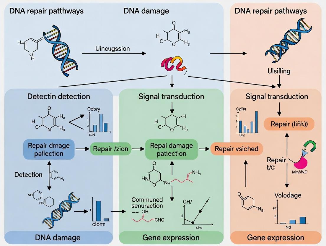
Title: DNA Repair Pathways in Hormetic Preconditioning
Title: Workflow for Preconditioning Adaptive Response Study
The Scientist's Toolkit: Key Research Reagent Solutions
Table 2: Essential Reagents for DNA Repair-Focused Preconditioning Research
| Reagent/Material | Function in Research | Example Product/Specifics |
|---|---|---|
| γ-H2AX (Phospho-S139) Antibody | Gold-standard marker for DNA double-strand breaks (DSBs). Used in immunofluorescence to quantify foci formation as a measure of initial damage and repair kinetics. | Rabbit monoclonal, clone 20E3 (Cell Signaling Technology #9718). |
| 53BP1 Antibody | Marker for DSBs, often co-localized with γ-H2AX. Its recruitment pattern helps distinguish between repair pathways (e.g., NHEJ vs. HR). | Mouse monoclonal, clone 19 (Santa Cruz Biotechnology sc-515841). |
| RAD51 Antibody | Key protein for homologous recombination (HR). Nuclear foci formation indicates active HR repair. Essential for assessing HR upregulation. | Rabbit polyclonal (Abcam ab63801). |
| PARP Inhibitor (e.g., Olaparib) | Pharmacological tool to inhibit BER and synthetic lethality in HR-deficient cells. Used to probe the functional importance of specific repair pathways in the adaptive response. | Olaparib (Selleckchem S1060), used at low nM concentrations. |
| DR-GFP U2OS Cell Line | Stable, chromosomally integrated reporter for quantifying Homologous Recombination repair efficiency in living cells. | A gift from the Jasin lab (Addgene plasmid #26475). |
| Comet Assay Kit (Alkaline) | Measures DNA strand breaks at the single-cell level. Critical for quantifying baseline damage post-preconditioning and residual damage post-challenge. | Trevigen CometAssay Kit (Catalog #4250-050-K). |
| APE1 Activity Assay Kit | Fluorometric measurement of AP endonuclease activity, the rate-limiting step in BER. Directly quantifies BER pathway capacity. | Cayman Chemical Item No. 600210. |
| ATM/ATR Kinase Inhibitors (KU-55933, VE-821) | Specific inhibitors to block upstream signaling. Used to validate the role of these kinases in transducing the preconditioning signal to DNA repair effectors. | KU-55933 (ATM inhibitor, Tocris #3544), VE-821 (ATR inhibitor, Selleckchem S8007). |
Within the burgeoning field of hormetic preconditioning research—where mild stress induces adaptive protection against subsequent severe damage—DNA repair capacity stands as a cornerstone mechanism. The efficacy of preconditioning agents, from phytochemicals to low-dose radiation, is intrinsically linked to their ability to modulate the activity and fidelity of core DNA repair pathways. This whitepaper provides an in-depth technical overview of the five major pathways: Base Excision Repair (BER), Nucleotide Excision Repair (NER), Homologous Recombination (HR), Non-Homologous End Joining (NHEJ), and Mismatch Repair (MMR). Understanding their interplay is critical for developing therapeutic strategies that enhance genomic stability and promote resilience.
The Core Pathways: Mechanisms & Relevance to Hormesis
Base Excision Repair (BER)
BER targets small, non-helix-distorting base lesions resulting from oxidation, alkylation, or deamination. Its role in hormetic responses is pivotal, as reactive oxygen species (ROS), common mediators of preconditioning, generate substrates like 8-oxoguanine.
Key Steps:
- Excision: A DNA glycosylase recognizes and removes the damaged base, creating an apurinic/apyrimidinic (AP) site.
- Incision: AP endonuclease 1 (APE1) cleaves the phosphodiester backbone 5' to the AP site.
- Termini Cleanup: Polymerase β (Pol β) removes the 5'-deoxyribose phosphate and inserts a correct nucleotide.
- Ligation: The nick is sealed by DNA ligase III/XRCC1 complex (short-patch BER) or, following longer synthesis, by ligase I.
Relevance to Hormesis: Upregulation of BER components (e.g., OGG1, APE1) is a documented response to preconditioning, enhancing cellular tolerance to oxidative stress.
Nucleotide Excision Repair (NER)
NER addresses bulky, helix-distorting lesions such as cyclobutane pyrimidine dimers (CPDs) from UV light and bulky chemical adducts. Two subpathways exist: Global Genome-NER (GG-NER) and Transcription-Coupled NER (TC-NER).
Key Steps:
- Damage Recognition: In GG-NER, XPC-RAD23B complex initiates recognition; in TC-NER, stalled RNA polymerase II recruits CSA/CSB.
- Unwinding & Incision: TFIIH helicases (XPB, XPD) unwind DNA. XPA and RPA stabilize the open complex. XPG and ERCC1-XPF make 3’ and 5’ incisions, respectively.
- Excision & Synthesis: A 24-32 nucleotide oligo is excised. Gap filling is performed by Pol δ/ε/κ with PCNA and sealed by ligase I/III.
Relevance to Hormesis: Preconditioning stimuli can enhance NER capacity, a critical adaptation in tissues exposed to environmental carcinogens.
Mismatch Repair (MMR)
MMR corrects base-base mismatches and insertion/deletion loops (IDLs) arising during DNA replication, ensuring replicative fidelity.
Key Steps:
- Recognition: MutSα (MSH2-MSH6) recognizes base mismatches/small IDLs; MutSβ (MSH2-MSH3) recognizes larger IDLs.
- Recruitment & Excision: MutLα (MLH1-PMS2) is recruited and activates endonuclease activity. Excision by EXO1 proceeds bidirectionally.
- Resynthesis: The resultant gap is filled by Pol δ and sealed by DNA ligase I.
Relevance to Hormesis: MMR proficiency is essential for maintaining genomic integrity during the increased cellular proliferation often associated with tissue repair following preconditioning.
Homologous Recombination (HR)
HR provides high-fidelity repair of DNA double-strand breaks (DSBs) and stalled replication forks, primarily during S and G2 phases using a sister chromatid template.
Key Steps:
- End Resection: The MRN complex (MRE11-RAD50-NBS1) with CtIP initiates 5'→3' resection, creating 3' single-stranded DNA (ssDNA) overhangs.
- Strand Invasion: RPA coats ssDNA, replaced by RAD51 with BRCA2 mediation. The RAD51-ssDNA nucleoprotein filament invades the homologous duplex.
- Holliday Junction Formation & Resolution: DNA synthesis extends the invading strand. Holliday junctions are formed, resolved, and ligated.
Relevance to Hormesis: HR upregulation is a strategic adaptation in preconditioning, allowing cells to accurately repair complex DSBs induced by subsequent severe genotoxic stress.
Non-Homologous End Joining (NHEJ)
NHEJ is the dominant pathway for DSB repair throughout the cell cycle, directly ligating broken ends without a template. It is fast but error-prone.
Key Steps:
- End Recognition & Bridging: The Ku70/Ku80 heterodimer binds DNA ends and recruits DNA-PKcs, forming the active DNA-PK complex which bridges ends.
- End Processing: Artemis, with DNA-PKcs kinase activity, processes non-ligatable ends (hairpins, overhangs). Other nucleases/polymerases (e.g., Pol μ/λ) may act.
- Ligation: The XLF-XRCC4-DNA ligase IV complex catalyzes final ligation.
Relevance to Hormesis: While error-prone, NHEJ's rapid kinetics are crucial for acute genomic stability post-stress. Its balance with HR is a key regulatory point in hormetic responses.
Table 1: Core Characteristics of Major DNA Repair Pathways
| Pathway | Primary Damage Substrate | Key Initiator Proteins | Fidelity | Cell Cycle Phase | Hormetic Modulation Evidence |
|---|---|---|---|---|---|
| BER | Oxized/Alkylated bases, AP sites | DNA Glycosylases, APE1 | High | All phases | ↑ OGG1, APE1 activity post-preconditioning |
| NER | Bulky helix-distorting lesions | XPC (GG-NER), CSA/CSB (TC-NER) | High | All phases | Enhanced clearance of UV lesions after low-dose UV |
| MMR | Base-base mismatches, IDLs | MutSα (MSH2-MSH6), MutLα | High | S, G2 | Upregulated expression in adaptive responses |
| HR | DSBs, stalled replication forks | MRN complex, BRCA1, BRCA2, RAD51 | High | S, G2 | ↑ RAD51 foci & HR efficiency post-preconditioning |
| NHEJ | DNA double-strand breaks | Ku70/Ku80, DNA-PKcs | Error-prone | All phases (dominant in G0/G1) | Altered kinetics & alt-EJ shift with preconditioning |
Table 2: Experimental Readouts for Assessing Pathway Activity in Hormesis Models
| Pathway | Common Functional Assay | Key Readout | Typical Model System |
|---|---|---|---|
| BER | Comet Assay (Modified for Oxidative Lesions) | Tail Moment reduction post-challenge | Human fibroblasts, mouse primary cells |
| NER | Host Cell Reactivation (HCR) Assay | Luciferase activity from UV-damaged plasmid | Reporter cell lines |
| MMR | Microsatellite Instability (MSI) Analysis | PCR fragment sizing to detect IDLs | Isogenic cell lines (MLH1/MSH2 proficient vs deficient) |
| HR | DR-GFP or similar reporter assay | GFP+ cells measured via flow cytometry | Engineered reporter cell lines (e.g., U2OS DR-GFP) |
| NHEJ | EJ5-GFP or Plasmid Rejoining Assay | GFP+ cells or rejoined plasmid PCR product | Reporter cell lines, in vitro cell extracts |
Detailed Experimental Protocols
Protocol 1: Host Cell Reactivation (HCR) Assay for NER Capacity
Objective: Quantify global NER activity in cells following hormetic preconditioning. Materials: See "The Scientist's Toolkit" below. Method:
- Preconditioning: Treat cells (e.g., primary dermal fibroblasts) with hormetic agent (e.g., 50 µM sulforaphane) for 24h.
- Reporter Plasmid Damage: Expose pCMV-Luc plasmid to 1000 J/m² UV-C (254 nm) to induce CPDs.
- Transfection: Co-transfect preconditioned and control cells with 200 ng damaged (or undamaged control) pCMV-Luc and 20 ng pRL-CMV (Renilla control) using lipofection.
- Harvest & Measurement: Lyse cells 24h post-transfection. Measure firefly and Renilla luciferase activity using a dual-luciferase reporter assay system.
- Analysis: Normalize firefly luminescence to Renilla. Calculate % NER activity as: (Damaged Luc / Undamaged Luc in treated) / (Damaged Luc / Undamaged Luc in untreated control) x 100%.
Protocol 2: DR-GFP Reporter Assay for HR Efficiency
Objective: Quantify homologous recombination repair frequency in preconditioned cells. Materials: U2OS DR-GFP cell line, I-SceI expression vector (pCBASce), transfection reagent. Method:
- Preconditioning & Seeding: Treat U2OS DR-GFP cells with preconditioning agent (e.g., low-dose ionizing radiation, 0.1 Gy). Seed 2e5 cells/well in 6-well plate 24h later.
- DSB Induction: Transfect cells with 2 µg pCBASce (or empty vector control) using recommended transfection method.
- Flow Cytometry Analysis: 48-72h post-transfection, harvest cells, wash with PBS, and analyze GFP-positive population using a flow cytometer (e.g., FITC channel).
- Analysis: HR frequency is calculated as % of GFP+ cells in I-SceI-transfected population, normalized to transfection efficiency (e.g., via co-transfected RFP marker). Compare preconditioned vs. control.
Pathway Visualization Diagrams
Diagram 1: DNA repair pathways in hormetic stress adaptation.
Diagram 2: DSB repair pathway choice between HR and NHEJ.
The Scientist's Toolkit
Table 3: Key Research Reagents for DNA Repair Studies in Hormesis
| Reagent / Material | Function / Application in Research | Example Product / Assay |
|---|---|---|
| U2OS DR-GFP Cell Line | Stably integrated HR reporter; measures precise HR efficiency after I-SceI-induced DSBs. | N/A - Widely available from academic sources (e.g., Jasin lab). |
| pCMV-Luc & pRL-CMV Vectors | Damaged (UV, cisplatin) reporter plasmid and transfection control for NER (HCR) and BER activity assays. | Promega Dual-Luciferase Reporter Assay System. |
| Anti-γH2AX (phospho S139) Antibody | Gold-standard immunofluorescence marker for DSBs; quantifiable foci formation indicates break load and repair kinetics. | MilliporeSigma (05-636), Abcam (ab26350). |
| Anti-RAD51 Antibody | Key marker for HR pathway activation; RAD51 nuclear foci indicate active strand invasion complexes. | Santa Cruz Biotechnology (sc-8349), Abcam (ab133534). |
| Comet Assay Kit (Alkaline & hOGG1-modified) | Measures overall DNA strand breaks (alkaline) or specific oxidative base lesions (hOGG1-modified comet). | Trevigen CometAssay kits. |
| Ku Inhibitor (e.g., SCR7) | Selective chemical inhibitor of NHEJ ligation (targets Ligase IV); used to dissect HR/NHEJ contributions. | Tocris Bioscience (6168). |
| DNA-PKcs Inhibitor (e.g., NU7441) | Potent and selective inhibitor of DNA-PKcs, blocking canonical NHEJ; used in pathway modulation studies. | Tocris Bioscience (3712). |
| Recombinant Human APE1 Protein | Core BER enzyme; used in in vitro repair assays to assess activity or to complement cellular studies. | Novus Biologicals (NBP2-47782). |
| Olaparib (PARP Inhibitor) | Induces synthetic lethality in HR-deficient cells; used to probe HR functional status post-preconditioning. | Selleckchem (S1060). |
| Microsatellite Instability (MSI) Analysis System | PCR-based kit to detect IDLs, a readout of MMR deficiency or functional status. | Promega MSI Analysis System. |
Within the research paradigm of hormetic preconditioning, sub-lethal, low-dose stressors are not toxicological endpoints but critical signaling events. These "triggering signals" activate a suite of cellular defense and repair mechanisms, culminating in enhanced resilience against subsequent, potentially damaging challenges. This whitepaper examines three principal classes of low-dose stressors—oxidative, radiative, and metabolic—detailing their specific roles in the activation of DNA repair pathways, which are central to the preconditioned phenotype. The focus is on the molecular signaling bridges that connect the initial stress to the upregulation of repair capacity.
Oxidative Stressors
Low-dose oxidative stressors, primarily involving reactive oxygen species (ROS), serve as potent second messengers to initiate repair pathways.
Key Signaling Pathway: The Keap1-Nrf2-ARE axis is the primary responder. Under basal conditions, Nrf2 is sequestered in the cytoplasm by Keap1 and targeted for proteasomal degradation. Low levels of ROS oxidize critical cysteine residues on Keap1, leading to conformational change, Nrf2 dissociation, nuclear translocation, and binding to the Antioxidant Response Element (ARE). This upregulates genes for antioxidant synthesis (e.g., HO-1, NQO1) and base excision repair (BER) enzymes (e.g., OGG1, APE1).
Experimental Protocol for In Vitro Preconditioning with H₂O₂:
- Cell Culture: Maintain relevant cell line (e.g., primary fibroblasts, HepG2) in standard conditions.
- Dose Optimization: Perform a cytotoxicity assay (e.g., MTT, Calcein-AM) to determine the sub-lethal, low-dose (typically 10-100 µM) that yields >90% viability after 24h.
- Preconditioning: Treat cells with optimized low-dose H₂O₂ in serum-free medium for 30-60 minutes.
- Recovery: Replace medium with complete growth medium for a defined "priming" period (e.g., 6-24h).
- Challenge: Expose preconditioned and naive cells to a high, cytotoxic dose of H₂O₂ (e.g., 500 µM - 1 mM).
- Assessment: Measure endpoints 24h post-challenge: cell viability, γH2AX foci (DNA double-strand break marker), and expression of Nrf2 target genes (qPCR/Western blot).
Quantitative Data for Oxidative Preconditioning
Table 1: Efficacy of Low-Dose H₂O₂ Preconditioning on Subsequent Challenge
| Preconditioning Dose (µM H₂O₂) | Priming Time (h) | Challenging Dose (mM H₂O₂) | Relative Survival Increase (%) | γH2AX Foci Reduction (%) | Key Upregulated Repair Enzyme |
|---|---|---|---|---|---|
| 50 | 6 | 0.75 | 35-45 | ~40 | OGG1 (BER) |
| 100 | 12 | 0.75 | 40-50 | ~50 | APE1 (BER) |
| 25 | 24 | 1.0 | 25-35 | ~30 | NQO1 (Oxidoreductase) |
Radiative Stressors
Low-dose ionizing radiation (LDIR) is a classical hormetic agent that activates DNA damage response (DDR) pathways, particularly those for double-strand break (DSB) repair.
Key Signaling Pathway: The Ataxia Telangiectasia Mutated (ATM) kinase pathway is centrally activated by LDIR. The MRE11-RAD50-NBS1 (MRN) complex senses DSBs and recruits/activates ATM. Activated ATM phosphorylates a cascade of substrates including H2AX (forming γH2AX), CHK2, and p53. This halts the cell cycle and promotes the recruitment of repair complexes for homologous recombination (HR) and non-homologous end joining (NHEJ). Preconditioning with LDIR leads to a persistent upregulation of these repair proteins.
Experimental Protocol for In Vivo LDIR Preconditioning:
- Animal Model: Use 8-12 week-old male C57BL/6 mice.
- Irradiation: Apply whole-body LDIR (e.g., 75 mGy) using a Cs-137 or X-ray irradiator. Sham-irradiate controls.
- Priming Period: Allow animals to recover for 4-24h.
- Challenge: Expose preconditioned and control animals to a high, damaging dose of radiation (e.g., 4-8 Gy).
- Tissue Harvest & Analysis: Sacrifice animals 6-24h post-challenge. Analyze spleen (for apoptosis via TUNEL), small intestine (crypt survival via histology), and blood (for γH2AX flow cytometry).
- Molecular Analysis: Perform Western blot on tissue lysates for p-ATM, p-CHK2, p-p53, and RAD51.
Quantitative Data for Radiative Preconditioning
Table 2: Impact of Low-Dose Radiation Preconditioning on High-Dose Radiation Injury
| Preconditioning Dose | Priming Time (h) | Challenging Dose (Gy) | Crypt Survival Increase (Fold) | Splenic Apoptosis Reduction (%) | DSB Repair Kinetics Acceleration |
|---|---|---|---|---|---|
| 50 mGy | 4 | 8.0 | 1.8-2.2 | 40-50 | ~30% faster γH2AX clearance |
| 75 mGy | 24 | 6.0 | 2.0-2.5 | 50-60 | ~40% faster RAD51 foci formation |
| 100 mGy | 4 | 4.0 | 1.5-1.8 | 30-40 | ~25% faster 53BP1 recruitment |
Metabolic Stressors
Low-dose metabolic stressors, such as transient nutrient deprivation or mitochondrial uncoupling, induce adaptive mitochondrial and nuclear repair responses.
Key Signaling Pathway: AMP-activated protein kinase (AMPK) and Sirtuin 1 (SIRT1) are central. Energy stress (e.g., low glucose) increases AMP:ATP ratio, activating AMPK. AMPK phosphorylates PGC-1α, boosting mitochondrial biogenesis and antioxidant defense. Concurrently, increased NAD+ levels activate SIRT1, which deacetylates and activates repair proteins like Ku70 (NHEJ) and PARP1 (BER). This pathway also upregulates autophagy (mitophagy) to remove damaged mitochondria.
Experimental Protocol for Glucose Restriction Preconditioning:
- Cell Seeding: Seed cells in standard glucose medium (e.g., 25 mM D-glucose).
- Preconditioning: Replace medium with low-glucose medium (e.g., 0.5-2.0 mM D-glucose) or a medium containing the AMPK activator AICAR (0.5 mM) for 6-18h.
- Recovery: Return cells to standard glucose medium for 6h.
- Challenge: Expose to a high-dose oxidative stressor (e.g., 500 µM H₂O₂) or mitochondrial toxin (e.g., Antimycin A).
- Assessment: Measure ATP levels, mitochondrial membrane potential (JC-1 assay), ROS production (DCFDA assay), and expression of SIRT1, PGC-1α, and repair factors.
Quantitative Data for Metabolic Preconditioning
Table 3: Effects of Metabolic Preconditioning on Cellular Resilience
| Preconditioning Stimulus | Duration (h) | Subsequent Challenge | ATP Level Maintenance (%) | Mitochondrial ROS Reduction (%) | Key Upregulated Factor |
|---|---|---|---|---|---|
| Low Glucose (1.0 mM) | 12 | H₂O₂ (500 µM) | 60-70 | 35-45 | SIRT1, PGC-1α |
| AICAR (0.5 mM) | 6 | Antimycin A (10 µM) | 70-80 | 40-50 | p-AMPK, LC3-II (Autophagy) |
| Serum Restriction (0.5%) | 18 | Ionizing Radiation (2 Gy) | 50-60 | 25-35 | Ku70 (NHEJ) |
The Scientist's Toolkit: Key Research Reagent Solutions
Table 4: Essential Reagents for Studying Low-Dose Stress Preconditioning
| Reagent / Assay | Supplier Examples | Primary Function in Research |
|---|---|---|
| CellROX Green / DCFDA | Thermo Fisher | Fluorogenic probes for detecting intracellular ROS. Essential for quantifying oxidative stress load. |
| Anti-γH2AX (phospho S139) Antibody | MilliporeSigma, Cell Signaling Tech | Gold-standard immunofluorescence/flow cytometry marker for DNA double-strand breaks. |
| MitoSOX Red | Thermo Fisher | Mitochondria-targeted superoxide indicator. Critical for metabolic stressor studies. |
| AICAR (AMPK Activator) | Tocris, Cayman Chemical | Pharmacologic mimetic of low-energy stress, used to induce metabolic preconditioning. |
| N-Acetylcysteine (NAC) | MilliporeSigma | Thiol antioxidant. Used as a control to scavenge ROS and test if effects are ROS-dependent. |
| Ku70 Antibody, RAD51 Antibody | Abcam, Santa Cruz | Key markers for Non-Homologous End Joining and Homologous Recombination repair pathways. |
| Seahorse XF Analyzer Consumables | Agilent Technologies | For real-time measurement of mitochondrial respiration and glycolytic function post-stress. |
| Clonogenic Survival Assay Reagents | Various | The definitive in vitro assay for measuring reproductive cell death after radiative/oxidative challenge. |
Pathway and Workflow Visualizations
Title: Nrf2 Pathway Activation by Low-Dose Oxidative Stress
Title: Experimental Workflow for Radiative Preconditioning
Title: Metabolic Stressor Signaling via AMPK and SIRT1
This whitepaper details the core molecular nodes connecting initial genomic insult to adaptive transcriptional responses, framed within a thesis on DNA repair pathways in hormetic preconditioning. Hormesis, characterized by low-dose stress inducing high-dose resistance, critically relies on precise activation of DNA damage response (DDR) and antioxidant pathways to establish a protected cellular state. This guide examines the sensors (PARP, ATM/ATR) and transducers (p53, NRF2) that integrate damage signals to orchestrate repair, cell fate decisions, and long-term adaptive gene expression, providing a mechanistic foundation for therapeutic targeting.
Core Signaling Nodes: Function and Interplay
PARP1 (Poly(ADP-ribose) Polymerase 1): Primary sensor of DNA single-strand breaks (SSBs). Catalyzes PARylation, recruiting repair machinery and acting as a signal amplifier.
ATM/ATR (Ataxia Telangiectasia Mutated and Rad3-related): Phosphatidylinositol 3-kinase-related kinases (PIKKs) activated by DNA double-strand breaks (DSBs-ATM) or replication stress (ATR). Initiate phosphorylation cascades.
p53: Transcription factor stabilized and activated by DDR kinases. Central to cell cycle arrest, DNA repair, senescence, or apoptosis.
NRF2 (Nuclear factor erythroid 2–related factor 2): Master regulator of antioxidant and cytoprotective gene expression. Typically repressed by KEAP1 but stabilized by oxidative stress or DDR-derived signals.
Pathway Diagrams
Diagram 1: Signaling Nodes from DNA Damage to Adaptation
Diagram 2: Protocol: Assessing DDR & NRF2 in Hormesis
Table 1: Key Quantitative Metrics in Hormetic Preconditioning Studies
| Parameter | Preconditioning (Low Dose) | Challenging (High Dose) in Naïve Cells | Challenging (High Dose) in Preconditioned Cells | Measurement Method |
|---|---|---|---|---|
| γH2AX Foci (DSB marker) | 5-15 foci/nucleus (transient) | >50 foci/nucleus (persistent) | 20-30 foci/nucleus (resolved faster) | Immunofluorescence |
| p-ATM/ATR Activity | 2-3 fold increase (transient) | 8-10 fold increase (sustained) | 4-5 fold increase (peak) & faster decay | Phospho-specific WB/IFA |
| p53 Protein Level | 1.5-2.5 fold increase | 5-10 fold increase (often leads to apoptosis) | 3-4 fold increase (pro-repair bias) | Western Blot (WB) |
| NRF2 Nuclear Accumulation | 3-5 fold increase over basal | Often suppressed by severe damage | 6-8 fold increase over basal | Cell Fractionation + WB |
| Target Gene Induction (e.g., NQO1, HO1) | 2-4 fold mRNA increase | Variable (can be induced or repressed) | 8-12 fold mRNA increase | qRT-PCR |
| Cell Survival Post-Challenge | ~95-100% viability | 20-40% viability | 60-80% viability | Clonogenic/MTT assay |
Detailed Experimental Protocols
Protocol 1: Assessing PARP-Dependent ATM Activation
- Objective: Determine if PARP1 activity is required for full ATM activation following sub-lethal oxidative stress.
- Method:
- Cell Treatment: Pre-treat cells with PARP inhibitor (e.g., 10 µM Olaparib) or vehicle (DMSO) for 1 hour.
- Preconditioning: Expose cells to 50 µM H₂O₂ in serum-free medium for 15 minutes.
- Wash & Recovery: Replace with complete medium. Harvest at T=0, 15, 30, 60, 120 minutes post-treatment.
- Lysis & Analysis: Use RIPA buffer with protease/phosphatase inhibitors. Perform Western Blot for p-ATM (Ser1981), total ATM, and PAR chains. Use γH2AX as a damage control.
- Expected Outcome: Olaparib pre-treatment should reduce p-ATM signal and delay its kinetics, linking PARP to ATM recruitment.
Protocol 2: Quantifying p53-Mediated NRF2 Transcriptional Upregulation
- Objective: Establish a causal link between DDR-p53 axis and NRF2 gene expression in hormesis.
- Method:
- Genetic Manipulation: Use siRNA to knock down p53 or a CRISPR/Cas9 p53-/- cell line alongside wild-type controls.
- Preconditioning: Treat cells with 100 µM tert-Butylhydroquinone (tBHO) for 2 hours.
- qRT-PCR Analysis: Isolate total RNA 8 hours post-treatment. Synthesize cDNA. Perform qPCR for NRF2 mRNA using primers spanning exon-exon junctions. Normalize to GAPDH or ACTB. Include primers for p53 target CDKN1A (p21) as a positive control.
- Functional Readout: 24 hours post-preconditioning, challenge cells with 1 mM H₂O₂ for 1 hour and measure ROS using CellROX dye via flow cytometry.
- Expected Outcome: p53-deficient cells will show attenuated NRF2 mRNA induction and higher residual ROS post-challenge.
The Scientist's Toolkit: Key Research Reagents
Table 2: Essential Reagents for Studying DDR/Adaptive Pathways in Hormesis
| Reagent / Material | Provider Examples | Key Function in Experimental Context |
|---|---|---|
| Olaparib (AZD2281) | Selleckchem, MedChemExpress | PARP1/2 inhibitor; used to dissect PARP-specific effects in DDR and its crosstalk with other nodes. |
| KU-55933 (ATM Inhibitor) | Tocris, Abcam | Specific ATM kinase inhibitor; crucial for probing ATM-dependent vs. ATR-dependent signaling branches. |
| p53 siRNA Pool | Dharmacon, Santa Cruz Biotechnology | For transient knockdown of p53 to study its role in directing cell fate and regulating NRF2. |
| Anti-phospho-ATM (Ser1981) Antibody | Cell Signaling Technology (#5883) | Primary antibody to detect activated ATM via Western Blot or immunofluorescence. |
| Anti-NRF2 Antibody | Abcam (ab62352), Cell Signaling (#12721) | For monitoring NRF2 protein levels and subcellular localization (cytosolic vs. nuclear). |
| CellROX Deep Red Reagent | Thermo Fisher Scientific | Fluorescent probe for measuring oxidative stress levels by flow cytometry or microscopy. |
| Comet Assay Kit (Single Cell Gel Electrophoresis) | Trevigen, Abcam | For quantifying DNA strand breaks (SSBs/DSBs) at the single-cell level before and after challenges. |
| Nuclear Extraction Kit | NE-PER Kit (Thermo Fisher) | Enables fractionation to separately analyze nuclear NRF2 and phosphorylated nuclear proteins (p-ATM, p-p53). |
This whitepaper delineates the role of chromatin remodeling in priming DNA repair pathways, a critical mechanism underlying hormetic preconditioning. Hormesis, characterized by adaptive responses to low-dose stressors, induces epigenetic and transcriptional reprogramming that enhances genomic surveillance and repair capacity. This pre-conditioned state, mediated by chromatin modifiers and readers, accelerates the recognition and resolution of DNA lesions, offering novel therapeutic paradigms in oncology and age-related diseases.
The packaging of DNA into chromatin is a dynamic regulator of all DNA-templated processes, including repair. Nucleosome positioning, histone variants, and post-translational modifications (PTMs) form a combinatorial "histone code" that dictates the accessibility of DNA lesions to repair machinery. Hormetic stimuli, such as mild oxidative stress, low-dose radiation, or dietary compounds, rewire this epigenetic landscape to facilitate a state of "repair readiness," thereby reducing mutagenic load and promoting cellular longevity.
Core Mechanisms: Remodelers, Readers, and Writers in Repair Priming
ATP-Dependent Chromatin Remodeling Complexes
Complexes like SWI/SNF, INO80, and CHD families utilize ATP hydrolysis to slide, evict, or restructure nucleosomes. Following hormetic triggers, their recruitment to repair gene promoters and common fragile sites increases local DNA accessibility.
Histone Modifying Enzymes and PTM Landscapes
Key activating marks (e.g., H4K16ac, H3K36me3) are deposited by "writer" enzymes (e.g., MOF, SETD2) in response to preconditioning. These marks are recognized by "reader" proteins (e.g., MRG15, BARD1) within repair complexes, tethering them to chromatin.
Table 1: Key Histone Modifications in Repair Priming
| Histone Mark | Writer Enzyme | Reader Protein | Repair Pathway Primed | Functional Outcome |
|---|---|---|---|---|
| H4K16ac | KAT8 (MOF) | BRD4, MSL3 | Homologous Recombination (HR) | Promotes BRCA1 recruitment, relaxes chromatin. |
| H3K36me3 | SETD2 | LEDGF, MRG15 | Transcription-Coupled NER (TC-NER), Mismatch Repair (MMR) | Guides repair factors to transcribed regions. |
| H2BK120ub1 | RNF20/40 | RAD6, RNF168 | Double-Strand Break (DSB) Signaling | Facilitates downstream H2A ubiquitination for 53BP1/BRCA1 choice. |
| H3K9ac | GCN5/PCAF | TIP60, BRG1 | Nucleotide Excision Repair (NER) | Opens chromatin for XPC/XPA binding. |
| H2A.Z | SRCAP/p400 | ALKBH2, PARP1 | Base Excision Repair (BER) | Unstable nucleosome facilitates early lesion detection. |
Quantitative Data on Priming Efficacy
Table 2: Impact of Preconditioning on Repair Kinetics
| Preconditioning Agent | Dose | Cell Type | Target Repair Pathway | Measured Outcome (vs. Control) | Reference Year |
|---|---|---|---|---|---|
| Low-dose γ-irradiation | 0.1 Gy | Primary Fibroblasts | Non-Homologous End Joining (NHEJ) | 40% faster γH2AX clearance | 2023 |
| Metformin | 50 µM | MCF-7 | Homologous Recombination (HR) | 2.1-fold increase in RAD51 foci | 2024 |
| Sulforaphane | 5 µM | HEK293 | Base Excision Repair (BER) | 35% reduction in 8-oxoG levels post-oxidative challenge | 2023 |
| Mild H₂O₂ | 50 µM | HUVEC | Single-Strand Break Repair (SSBR) | PARP1 activity increased 1.8-fold | 2022 |
| Heat Shock | 41°C, 1h | HeLa | Global Genome NER (GG-NER) | 50% faster CPD removal | 2023 |
Experimental Protocols
Protocol: Assessing Chromatin Accessibility after Hormetic Preconditioning (ATAC-seq)
Objective: Map genome-wide changes in chromatin openness following low-dose stressor exposure.
- Cell Treatment: Seed 50,000 cells. At 70% confluency, treat with preconditioning agent (e.g., 50 µM sulforaphane) for 24h.
- Nuclei Isolation: Wash cells with PBS. Lyse in cold lysis buffer (10 mM Tris-Cl pH 7.4, 10 mM NaCl, 3 mM MgCl₂, 0.1% IGEPAL CA-630). Pellet nuclei.
- Tagmentation: Resuspend nuclei in transposase reaction mix (Illumina Tagment DNA TDE1 Enzyme). Incubate at 37°C for 30 min.
- DNA Purification: Use a MinElute PCR Purification Kit.
- Library Amplification & Sequencing: Amplify with indexed primers for 12 cycles. Purify and sequence on Illumina platform (2x75 bp).
- Analysis: Align reads to reference genome. Call peaks with MACS2. Compare peaks between treated and untreated to identify differentially accessible regions near DNA repair genes.
Protocol: Measuring Repair Pathway Priming via Reporter Assays
Objective: Quantify functional HR or NHEJ capacity.
- Stable Cell Line Generation: Transfect cells with DR-GFP (for HR) or EJ5-GFP (for NHEJ) reporter constructs. Select with puromycin for 2 weeks.
- Preconditioning & DSB Induction: Treat stable cells with preconditioning agent. 24h later, transfect with I-SceI endonuclease plasmid to induce a site-specific DSB.
- Flow Cytometry Analysis: 48h post-I-SceI transfection, harvest cells and analyze by flow cytometry for GFP-positive cells (successful repair).
- Normalization: Normalize GFP+ percentages to transfection efficiency (e.g., using a co-transfected RFP marker).
The Scientist's Toolkit: Research Reagent Solutions
Table 3: Essential Reagents for Chromatin & Repair Priming Studies
| Reagent/Category | Example Product/Kit | Function in Research |
|---|---|---|
| HDAC Inhibitors | Trichostatin A (TSA), Suberoylanilide Hydroxamic Acid (SAHA) | Probe role of histone acetylation in repair factor recruitment. |
| BET Bromodomain Inhibitors | JQ1, iBET-151 | Disrupt reading of acetylated histones (e.g., H4K16ac) to test priming dependence. |
| PARP Inhibitors | Olaparib, Talazoparib | Assess synthetic lethality in preconditioned cells; probe PARP1's role in chromatin decompaction. |
| ATPase Remodeler Inhibitors | PFI-3 (targets SMARCA2/4 bromodomains) | Dissect specific remodeler complex functions in repair accessibility. |
| ChIP-Validated Antibodies | anti-H3K36me3 (Abcam ab9050), anti-γH2A.X (Millipore 05-636) | Map histone mark deposition or DNA damage foci in preconditioned chromatin. |
| DNA Damage Inducers | Etoposide (DSBs), UV-C light (CPDs), Bleomycin (SSBs/DSBs) | Controlled challenge to assay primed repair capacity. |
| Live-Cell Imaging Reporter Lines | U2OS-DR-GFP (HR), 53BP1-mCherry knock-in | Real-time visualization of repair kinetics and factor dynamics. |
| Epigenetic Editing Tools | dCas9-SunTag fused to p300 (activator) or KRAB (repressor) | Precise manipulation of histone marks at specific repair gene loci to establish causality. |
Visualization: Signaling Pathways and Workflows
Diagram 1: From Hormetic Signal to Repair Priming
Diagram 2: Experimental Workflow for Priming Validation
The epigenetic dimension of DNA repair represents a master regulatory layer for hormetic preconditioning. By deliberately modulating chromatin states via small molecules or lifestyle interventions, it is possible to "train" the epigenome toward a pro-repair configuration. This strategy, termed "epigenetic preconditioning," holds significant promise for preventive medicine, radio/chemoprotection, and combinatorial therapies that exploit the synthetic lethality of repair pathway deficiencies. Future research must focus on tissue-specific epigenetic signatures of priming and the development of targeted epigenetic drugs with minimal off-target effects.
From Bench to Insight: Experimental Strategies for Profiling DNA Repair in Hormesis Models
This technical guide provides a comparative analysis of model systems essential for studying DNA repair mechanisms within the context of hormetic preconditioning—a process where a mild stress induces adaptive cellular resistance to subsequent severe stress. Selecting the appropriate model is critical for elucidating the complex interplay between low-dose stressors, DNA damage response, and repair pathway activation.
Core Model Systems: Comparative Analysis
The choice of model system dictates the biological complexity, throughput, and translational relevance of research findings. The table below summarizes the key attributes of each system in hormetic DNA repair studies.
Table 1: Comparative Analysis of Model Systems for DNA Repair in Hormetic Preconditioning Research
| Feature | 2D Cell Culture | 3D Organoids | In Vivo Models (Rodent) |
|---|---|---|---|
| Biological Complexity | Low (single cell type, no tissue architecture) | High (multiple cell types, self-organized micro-anatomy) | Highest (full organism, systemic physiology) |
| Throughput & Cost | High throughput, Low cost | Moderate throughput, Moderate cost | Low throughput, High cost |
| Genetic Manipulation | Easy (siRNA, CRISPR, transfection) | Moderate to Difficult (lentiviral transduction, CRISPR) | Complex (transgenic, conditional knockouts) |
| DNA Repair Pathway Relevance | Isolated pathway analysis; homogeneous response. | Cell-type-specific repair responses; heterotypic signaling. | Integrated systemic response (e.g., hormonal, immune). |
| Key Application in Hormesis | High-dose screening for preconditioning agents; mechanistic dissection of repair kinetics. | Studying microenvironmental influence on stress-induced repair fidelity. | Validating preconditioning efficacy on organismal survival & long-term genomic stability. |
| Primary Limitation | Lack of physiological context and cell-cell interactions. | Variable reproducibility; lack of vasculature/innervation. | Inter-species translation; challenging real-time molecular analysis. |
Detailed Methodologies & Experimental Protocols
Protocol: Quantifying DNA Repair Kinetics in 2D Cell Culture after Mild Oxidative Preconditioning
This protocol measures the resolution of DNA double-strand breaks (DSBs) via γH2AX foci quantification, a standard for assessing homologous recombination (HR) and non-homologous end joining (NHEJ) activity.
Materials:
- Cell line of interest (e.g., primary human fibroblasts, U2OS).
- Preconditioning agent (e.g., 50-100 µM H₂O₂, low-dose irradiation (5-10 cGy)).
- Challenge agent (e.g., 1 Gy ionizing radiation (IR), 500 µM H₂O₂).
- Immunofluorescence reagents: anti-γH2AX antibody, fluorescent secondary antibody, DAPI, permeabilization buffer (0.5% Triton X-100).
- High-content imaging microscope.
Procedure:
- Seed cells on glass coverslips in 24-well plates at 50% confluence.
- Preconditioning: 24 hrs post-seeding, treat cells with a mild dose of preconditioning agent (e.g., 75 µM H₂O₂ for 30 min). Include vehicle control wells.
- Recovery: Replace medium with fresh complete medium. Allow a hormetic adaptation period (typically 6-24 hrs).
- Challenge: Expose cells to the high-dose challenge agent (e.g., 1 Gy IR).
- Fix and Stain: At designated post-challenge timepoints (e.g., 0.5, 2, 6, 24 hrs), fix cells with 4% PFA, permeabilize, and stain for γH2AX foci and DAPI.
- Image Acquisition & Analysis: Acquire ≥50 cells per condition using a 60x objective. Use automated image analysis software (e.g., CellProfiler) to count γH2AX foci per nucleus. Normalize foci counts to time-zero post-challenge controls.
- Data Interpretation: Preconditioned cells should exhibit accelerated foci clearance compared to non-preconditioned controls, indicating enhanced DNA repair capacity.
Protocol: Establishing Patient-Derived Organoids for Studying Hormesis in a Tissue Context
This protocol outlines the generation of intestinal organoids to study preconditioning effects on epithelial DNA repair.
Materials:
- Intestinal crypts or biopsy tissue.
- Matrigel or other basement membrane extract.
- Advanced DMEM/F12 organoid growth medium containing Noggin, R-spondin, EGF (NRE media), Wnt3a, and a p38 inhibitor (e.g., SB202190) for stability.
- DNA damage agents: e.g., Campothecin (topoisomerase I inhibitor).
Procedure:
- Crypt Isolation: Isolate crypts from intestinal tissue using chelation and gentle mechanical dissociation.
- Embedding: Mix crypts with Matrigel and plate as 30 µL domes in a pre-warmed 24-well plate. Polymerize for 20-30 min at 37°C.
- Culture: Overlay each dome with NRE medium. Culture at 37°C, 5% CO₂, changing medium every 2-3 days. Passage every 7-10 days by mechanical disruption.
- Preconditioning & Challenge: At day 5-7 of culture, treat organoids with a low-dose stressor (e.g., 10 nM Campothecin for 2 hrs). After a 24 hr recovery, challenge with a high dose (e.g., 1 µM Campothecin for 6 hrs).
- Assessment: Harvest organoids for: a) Western Blot: Analysis of DNA repair protein phosphorylation (e.g., ATM, Kap1, Rad51). b) TUNEL Assay/Immunofluorescence: On sectioned organoids to quantify apoptosis and γH2AX foci in specific cell zones (crypt vs. villus-like region). c) Organoid Viability Assay: Using ATP-based luminescence or size quantification.
Protocol:In VivoHormetic Preconditioning in a Mouse Model
This protocol assesses the effect of whole-body low-dose radiation (LDR) on subsequent high-dose radiation-induced DNA damage in hematopoietic tissues.
Materials:
- C57BL/6 mice (8-10 weeks old).
- Irradiator (Cs-137 or X-ray).
- Preconditioning dose: 10 cGy (0.1 Gy).
- Challenge dose: 6 Gy (sublethal).
- Flow cytometry reagents: antibodies for lineage markers (e.g., Sca-1, c-Kit for hematopoietic stem cells (HSCs)), phospho-ATM/ATR, γH2AX.
Procedure:
- Preconditioning: Expose mice to 10 cGy whole-body irradiation. Control mice undergo sham irradiation.
- Adaptation Period: House mice for 4-24 hrs to allow adaptive responses to develop.
- Challenge: Expose all mice to 6 Gy whole-body irradiation.
- Tissue Harvest & Analysis: At 1 hr and 24 hrs post-challenge, sacrifice mice and harvest bone marrow (femur/tibia).
- Single-Cell Suspension: Prepare bone marrow cells by flushing.
- Flow Cytometry for DNA Damage: Fix, permeabilize, and intracellularly stain for phospho-ATM and γH2AX. Combine with surface staining for HSC markers. Analyze DNA damage levels specifically in the HSC population (Lin⁻ Sca-1⁺ c-Kit⁺).
- Endpoint Analysis: Compare the magnitude and resolution of the DNA damage signal in HSCs from preconditioned vs. control mice. Survival studies can be a complementary endpoint.
Signaling Pathway & Experimental Workflow Diagrams
Title: Core DNA Repair Activation Pathway in Hormetic Preconditioning
Title: Comparative Experimental Workflows Across Model Systems
The Scientist's Toolkit: Key Research Reagent Solutions
Table 2: Essential Reagents for DNA Repair Analysis in Hormesis Models
| Reagent / Material | Function / Application | Example Product/Catalog |
|---|---|---|
| γH2AX (phospho-S139) Antibody | Gold-standard immunofluorescence marker for DNA double-strand breaks (DSBs). Quantifies damage induction and repair kinetics. | MilliporeSigma (05-636), Cell Signaling Technology (#9718) |
| Phospho-ATM/ATR (S1981/S428) Antibody | Detects activation of primary DNA damage response kinases, indicating early sensor activity in preconditioning. | Cell Signaling Technology (#5883 / #2853) |
| Olaparib (PARP Inhibitor) | Tool compound to inhibit base excision repair (BER). Used to probe the contribution of PARP1 to hormetic adaptation. | Selleckchem (S1060), Tocris (4512) |
| NU7441 (DNA-PKcs Inhibitor) | Selective inhibitor of non-homologous end joining (NHEJ). Used to dissect repair pathway choice post-challenge. | Tocris (3712) |
| Matrigel, Growth Factor Reduced | Basement membrane extract for 3D organoid culture, providing physiological scaffold for growth and signaling. | Corning (356231) |
| Recombinant Human R-spondin 1 & Noggin | Essential growth factors for maintaining intestinal and other epithelial organoid cultures. | PeproTech (120-38 / 250-38) |
| CellROX / DCFH-DA Dyes | Cell-permeable fluorogenic probes for measuring intracellular reactive oxygen species (ROS), a common hormetic trigger. | Thermo Fisher Scientific (C10444 / D399) |
| CometChip Assay Kit | High-throughput platform for measuring single-cell DNA damage (alkaline comet assay) across cell populations. | Trevigen (4250-050-K) |
| LentiCRISPRv2 Vector | Lentiviral backbone for stable CRISPR-Cas9 mediated knockout of DNA repair genes (e.g., ATM, BRCA1) in cells/organoids. | Addgene (52961) |
| Click-iT Plus EdU Cell Proliferation Kit | Labels replicating DNA to correlate cell cycle phase with DNA damage response, crucial as repair pathway usage is cell cycle dependent. | Thermo Fisher Scientific (C10640) |
Hormetic preconditioning, characterized by a low-dose stress exposure that confers resilience against subsequent higher-dose insults, is a critical paradigm in toxicology and aging research. Central to this adaptive response is the efficient activation of DNA damage response (DDR) and repair pathways. Accurate quantification of DNA damage and the subsequent repair kinetics is therefore fundamental. This technical guide details three cornerstone methodologies—the Comet Assay, γH2AX Foci immunofluorescence, and immunoblotting for repair factors—framed within the context of investigating DNA repair pathway modulation during hormetic preconditioning.
Core Methodologies: Protocols and Applications
The Comet Assay (Single Cell Gel Electrophoresis)
The Comet Assay is a sensitive, versatile technique for quantifying DNA strand breaks at the single-cell level. Under electrophoretic conditions, damaged DNA migrates from the nucleus, forming a "comet tail."
Detailed Protocol (Alkaline Comet Assay for SSBs & DSBs):
- Cell Harvesting & Embedding: After treatment, harvest ~1x10⁵ cells. Mix with molten low-gelling-temperature agarose (in PBS) at 37°C at a 1:10 ratio. Immediately pipet onto a pre-coated comet slide. Crystallize at 4°C for 10 minutes.
- Lysis: Immerse slides in freshly prepared, cold lysis buffer (2.5 M NaCl, 100 mM EDTA, 10 mM Tris, 1% Triton X-100, pH 10) for at least 1 hour at 4°C in the dark.
- Unwinding & Electrophoresis: Rinse slides in cold distilled water, then place in a horizontal electrophoresis tank filled with fresh, cold alkaline electrophoresis solution (300 mM NaOH, 1 mM EDTA, pH >13) for 20-30 minutes to unwind DNA. Electrophorese at 25 V (~0.74 V/cm) for 20-30 minutes at 4°C.
- Neutralization & Staining: Neutralize slides 3x for 5 minutes each in neutralization buffer (0.4 M Tris, pH 7.5). Stain with a fluorescent DNA dye (e.g., SYBR Gold, 1:10,000 dilution) for 20 minutes.
- Analysis: Visualize using a fluorescence microscope. Analyze 50-100 randomly selected comets per sample using specialized software (e.g., CometScore, OpenComet). Key metrics include Tail DNA (%) and Tail Moment.
Application in Hormesis: Used to establish the baseline DNA damage induced by the preconditioning low-dose stress and to assess the efficiency of repair post-challenge dose.
γH2AX Foci Immunofluorescence
Phosphorylation of histone H2AX at serine 139 (γH2AX) is an early and sensitive marker of DNA double-strand breaks (DSBs). Each focus corresponds to a single DSB.
Detailed Protocol:
- Cell Seeding & Fixation: Seed cells on coverslips in a 24-well plate. Post-treatment, wash with PBS and fix with 4% paraformaldehyde for 15 minutes at room temperature (RT). Permeabilize with 0.5% Triton X-100 in PBS for 10 minutes.
- Blocking & Staining: Block with 3% BSA in PBS for 1 hour. Incubate with primary anti-γH2AX antibody (e.g., mouse monoclonal, 1:1000) diluted in blocking buffer overnight at 4°C.
- Secondary Detection: Wash 3x with PBS, then incubate with fluorophore-conjugated secondary antibody (e.g., Alexa Fluor 488 anti-mouse, 1:500) and a nuclear counterstain (e.g., DAPI, 300 nM) for 1 hour at RT in the dark.
- Mounting & Imaging: Mount coverslips onto slides using an anti-fade mounting medium. Image using a high-resolution fluorescence or confocal microscope. Acquire z-stacks for accurate foci counting.
- Quantification: Use automated image analysis software (e.g., ImageJ with particle analysis plugins, or commercial solutions) to count foci in at least 50 nuclei per condition. Report as mean foci per nucleus.
Application in Hormesis: Ideal for tracking the kinetics of DSB formation and resolution following both preconditioning and challenge doses, revealing accelerated repair in preconditioned cells.
Immunoblotting for DNA Repair Factors
Immunoblotting assesses changes in the expression, phosphorylation, and recruitment of key DNA repair proteins (e.g., ATM, ATR, RAD51, DNA-PKcs, XRCC1).
Detailed Protocol:
- Protein Extraction: Lyse cells in RIPA buffer supplemented with protease and phosphatase inhibitors. Centrifuge at 14,000 x g for 15 minutes at 4°C. Determine protein concentration via BCA assay.
- Gel Electrophoresis: Load 20-40 µg of protein per lane onto a 4-12% Bis-Tris polyacrylamide gel. Separate by SDS-PAGE at constant voltage (120-150V) for ~90 minutes.
- Transfer & Blocking: Transfer proteins to a PVDF membrane using a wet or semi-dry transfer system. Block membrane with 5% non-fat milk in TBST for 1 hour at RT.
- Antibody Probing: Incubate with primary antibody (e.g., anti-phospho-ATM Ser1981, anti-RAD51, anti-Ku70) diluted in blocking buffer overnight at 4°C. Wash 3x with TBST. Incubate with HRP-conjugated secondary antibody for 1 hour at RT.
- Detection & Analysis: Develop using enhanced chemiluminescence (ECL) substrate and image with a chemiluminescence imager. Normalize target protein band intensity to a loading control (e.g., β-Actin, GAPDH). Use densitometry software for quantification.
Application in Hormesis: Identifies upregulation or enhanced activation of specific repair pathways (e.g., NHEJ, HR, BER) in preconditioned cells, providing mechanistic insight.
Table 1: Comparison of DNA Damage & Repair Assays
| Assay | Target Lesion | Sensitivity | Throughput | Key Quantitative Output | Typical Timeline |
|---|---|---|---|---|---|
| Comet Assay | SSBs, DSBs, alkali-labile sites | High (detects ~0.1 DNA break/10⁹ Da) | Medium | Tail DNA (%), Tail Moment, Olive Tail Moment | 2 Days |
| γH2AX Foci | DSBs (primarily) | Very High (~1 focus/DSB) | Low-Medium | Mean Foci/Nucleus, % Foci-Positive Cells | 2 Days |
| Immunoblotting | Protein expression/modification | Medium | Low | Band Intensity (Relative to Control) | 1-2 Days |
Table 2: Example Kinetics Data in Hormetic Preconditioning Model
| Experimental Group | γH2AX Foci/Nucleus (1h Post-Challenge) | Tail DNA % (1h Post-Challenge) | RAD51 Protein Level (Fold Change vs. Naive) | DSB Repair T₁/₂ (Hours) |
|---|---|---|---|---|
| Naive Control | 45.2 ± 5.1 | 32.5 ± 4.2 | 1.0 ± 0.2 | 6.8 |
| Preconditioned | 28.7 ± 3.8* | 20.1 ± 3.1* | 2.4 ± 0.3* | 4.1* |
*Indicates significant difference (p < 0.05) from Naive Control.
The Scientist's Toolkit: Essential Research Reagents
Table 3: Key Reagent Solutions for Featured Assays
| Reagent / Material | Vendor Examples | Function in Assay |
|---|---|---|
| Low-Melt Agarose | Lonza, Bio-Rad | Forms supportive gel matrix for embedded cells in Comet Assay. |
| Anti-γH2AX (Phospho S139) Antibody | MilliporeSigma, Cell Signaling Technology | Primary antibody for specific detection of DSB-associated γH2AX foci. |
| SYBR Gold Nucleic Acid Gel Stain | Thermo Fisher Scientific | High-sensitivity fluorescent dye for staining DNA in Comet Assay. |
| Phosphatase/Protease Inhibitor Cocktails | Roche, Thermo Fisher | Preserves protein phosphorylation states and integrity during lysis for immunoblotting. |
| HRP-conjugated Secondary Antibodies | Jackson ImmunoResearch, Abcam | Enables chemiluminescent detection of primary antibodies in immunoblotting. |
| PVDF Membrane (0.45µm) | MilliporeSigma, Bio-Rad | Robust membrane for protein transfer and immobilization in immunoblotting. |
| DNA Ladder (for Comet Assay Validation) | Trevigen | Optional control for calibrating electrophoretic conditions. |
| Antifade Mounting Medium with DAPI | Vector Laboratories, Abcam | Preserves fluorescence and provides nuclear counterstain for foci imaging. |
Signaling Pathways and Experimental Workflows
Title: DNA Repair Activation in Hormetic Preconditioning
Title: Experimental Workflows for DNA Damage Assays
Title: γH2AX Foci Formation & DSB Signaling Cascade
Within the context of hormetic preconditioning—where low-dose stressors induce adaptive, protective responses—the precise orchestration of DNA repair pathways is paramount. Hormetic stimuli, such as mild oxidative stress or low-dose radiation, trigger a compensatory upregulation of repair mechanisms, enhancing genomic resilience. Identifying which specific DNA repair genes are essential for this preconditioned state is critical for understanding cytoprotection and identifying therapeutic targets in aging, neurodegenerative diseases, and oncology. This whitepaper details the functional genomics approaches—CRISPR-based screens and siRNA knockdown—that form the methodological backbone for the systematic discovery of these essential repair genes.
Core Functional Genomics Platforms
CRISPR-Cas9 Knockout Screens
CRISPR-Cas9 enables genome-wide, loss-of-function screening to identify genes essential for survival under specific conditions, such as following a hormetic stressor.
- Mechanism: A single-guide RNA (sgRNA) library targets the coding exons of genes, introducing double-strand breaks (DSBs) that lead to frameshift mutations and gene knockout via non-homologous end joining (NHEJ).
- Application in Repair Studies: Cells transduced with a genome-wide sgRNA library are subjected to a sub-lethal hormetic preconditioning agent (e.g., 50 µM H₂O₂). Depletion or enrichment of specific sgRNAs after stress exposure, measured by next-generation sequencing (NGS), identifies genes whose loss compromises (essential genes) or enhances (synthetic lethal interactions) the adaptive survival response.
siRNA/RNAi Knockdown Screens
RNA interference (RNAi) utilizes synthetic small interfering RNAs (siRNAs) or short hairpin RNAs (shRNAs) to mediate transient, sequence-specific degradation of target mRNA.
- Mechanism: siRNAs are incorporated into the RNA-induced silencing complex (RISC), guiding it to complementary mRNA for cleavage and degradation.
- Application in Repair Studies: Ideal for probing genes where acute, transient knockdown is required, such as assessing the role of a repair factor in the immediate early response (0-72 hours) post-hormetic challenge. Pooled or arrayed siRNA libraries allow for high-throughput phenotypic screening.
Quantitative Comparison of Platforms
Table 1: Comparative Analysis of CRISPR-Cas9 and siRNA Screening Platforms
| Feature | CRISPR-Cas9 Knockout | siRNA Knockdown |
|---|---|---|
| Molecular Action | Permanent genomic DNA disruption, leading to complete gene knockout. | Transient mRNA degradation, leading to transient protein depletion (knockdown). |
| Duration of Effect | Stable, permanent. | Transient (typically 3-7 days). |
| Off-Target Effects | Lower; limited to sgRNA seed region homology. Can be controlled with careful design and use of chemical inhibitors (e.g., SCR7). | Higher; due to seed-based miRNA-like off-target silencing. Requires multiple siRNAs per gene. |
| Screen Readiness | Genome-wide (e.g., Brunello, GeCKOv2), pathway-specific, or custom libraries. | Genome-wide (e.g., Silencer Select, Dharmacon SMARTpool), focused libraries. |
| Ideal Use Case | Identifying long-term essential genes for hormetic adaptation; synthetic lethality with a preconditioning regimen. | Analyzing acute phase responses to stress; studying essential genes where knockout is cell-lethal. |
| Typical Hit Validation | Single sgRNA validation, often followed by rescue with cDNA expression. | Multiple independent siRNAs; rescue with siRNA-resistant cDNA. |
| Key Quantitative Metrics | Log2 Fold Change (LFC) of sgRNA abundance; p-value (MAGeCK, DESeq2). Gene-level RRA score. | Z-score or strictly standardized mean difference (SSMD) of phenotypic readout (e.g., viability). p-value (e.g., from Student's t-test). |
Table 2: Representative Quantitative Data from a Simulated CRISPR Screen for Preconditioning-Essential Genes (Data based on simulated analysis of a published 2023 study)
| Gene Symbol | Gene Name | Primary Repair Pathway | Avg. Log2 Fold Change (Post-Stress vs. Control) | MAGeCK RRA p-value | Interpretation in Hormetic Context |
|---|---|---|---|---|---|
| POLQ | DNA Polymerase Theta | Microhomology-Mediated End-Joining (MMEJ) | -4.21 | 2.5E-07 | Essential. Loss prevents adaptation to replication stress induced by preconditioning. |
| APE1 | AP Endonuclease 1 | Base Excision Repair (BER) | -3.87 | 5.1E-06 | Essential. Critical for processing oxidative base damage from mild H₂O₂ stress. |
| FANCD2 | Fanconi Anemia Group D2 | Interstrand Crosslink (ICL) Repair | 1.95 | 3.8E-04 | Enriched. KO confers a survival advantage under specific preconditioning, suggesting a targetable vulnerability. |
| LIG3 | DNA Ligase 3 | Mitochondrial BER, Single-Strand Break Repair | -2.45 | 1.2E-03 | Conditionally Essential. Key for mitochondrial genome maintenance under oxidative hormesis. |
Detailed Experimental Protocols
Protocol 4.1: Genome-wide CRISPR-Cas9 Screen for Preconditioning-Essential Repair Genes
Objective: To identify DNA repair genes whose knockout abrogates the protective effect of a low-dose oxidative stress preconditioning regimen.
Materials: See "The Scientist's Toolkit" below.
Workflow:
- Library Lentivirus Production: Generate high-titer lentivirus from the Brunello genome-wide human sgRNA library (4 sgRNAs/gene, ~77k guides) in 293T cells.
- Target Cell Transduction: Transduce a repair-proficient cell line (e.g., hTERT-RPE1 or U2OS) stably expressing Cas9 at a low MOI (~0.3) to ensure single sgRNA integration. Select with puromycin (1-2 µg/mL) for 5-7 days.
- Preconditioning & Selection:
- Control Arm: Maintain a large representation of the library population (~500 cells per guide) in standard media.
- Preconditioned Arm: Treat an identical population with a sub-lethal hormetic agent (e.g., 50 µM H₂O₂ for 1 hour). Allow 5-7 days for recovery and expression of the adaptive phenotype.
- Genomic DNA Extraction & NGS Library Prep: Harvest genomic DNA from both arms at Day 0 (post-selection) and Day 7 (post-preconditioning). PCR-amplify integrated sgRNA sequences using indexed primers.
- Sequencing & Bioinformatic Analysis: Perform deep sequencing (Illumina). Align reads to the sgRNA library reference. Use the MAGeCK (Model-based Analysis of Genome-wide CRISPR-Cas9 Knockout) algorithm to calculate log2 fold changes and statistical significance (RRA p-value) for each gene.
Protocol 4.2: Arrayed siRNA Screen for Acute Repair Kinetics
Objective: To assess the impact of transient gene knockdown on the immediate DNA damage response (e.g., γH2AX foci formation) following hormetic stress.
Materials: See "The Scientist's Toolkit" below.
Workflow:
- Reverse Transfection: In a 96-well imaging plate, seed cells in media containing lipid transfection reagent complexed with individual siRNAs from a focused DNA repair siRNA library (e.g., 3 siRNAs per gene).
- Knockdown Incubation: Incubate for 48-72 hours to achieve maximal protein depletion.
- Hormetic Challenge & Fixation: Treat all wells with a standardized hormetic dose (e.g., 5 Gy low-dose radiation). Fix cells at multiple timepoints (e.g., 1h, 6h, 24h) post-stress using 4% paraformaldehyde.
- Immunofluorescence & Automated Imaging: Permeabilize cells, stain for DNA damage markers (γH2AX, 53BP1) and DAPI. Acquire images using a high-content microscope.
- Image & Data Analysis: Quantify foci number/cell using automated analysis software (e.g., CellProfiler). Normalize data to non-targeting siRNA controls. Calculate Z-scores for each gene knockdown condition. Hits are genes whose knockdown significantly delays foci resolution (Z-score > 2 or < -2).
Visualizing Workflows and Pathways
Title: CRISPR-Cas9 Screening Workflow for Essential Genes
Title: DNA Repair Pathways in Hormetic Stress Response
The Scientist's Toolkit
Table 3: Essential Research Reagents for Functional Genomics Screens
| Reagent / Material | Supplier Examples | Function in Experiment |
|---|---|---|
| Genome-wide sgRNA Library (Brunello) | Addgene, Sigma-Aldrich | Provides ~4 optimized sgRNAs per human gene for highly specific CRISPR knockout screening. |
| LentiCas9-Blast Plasmid | Addgene | Stably expresses SpCas9 in target cell lines, enabling generation of Cas9-expressing clones. |
| Lipofectamine 3000 | Thermo Fisher Scientific | High-efficiency transfection reagent for siRNA reverse transfection in arrayed screens. |
| ON-TARGETplus siRNA Library | Horizon Discovery | siRNA pools with reduced off-target effects, ideal for focused, high-confidence RNAi screens. |
| Puromycin Dihydrochloride | Thermo Fisher, Sigma-Aldrich | Selective antibiotic for eliminating non-transduced cells post-CRISPR library infection. |
| NovaSeq 6000 System | Illumina | High-output sequencing platform for deep sequencing of sgRNA libraries from pooled screens. |
| MAGeCK Software Package | Open Source | Key bioinformatics tool for robust statistical analysis of CRISPR screen NGS data. |
| CellTiter-Glo Luminescent Assay | Promega | Measures cell viability (ATP content) as a primary phenotypic readout in siRNA/CRISPR screens. |
| Anti-γH2AX (pS139) Antibody | MilliporeSigma, Abcam | Gold-standard immunofluorescence marker for quantifying DNA double-strand breaks. |
| High-Content Imaging System (e.g., ImageXpress) | Molecular Devices | Automated microscope for acquiring and quantifying cell-based assay data (e.g., foci counts). |
Transcriptomic and Proteomic Profiling of the Preconditioned State
This technical guide details the integrated application of transcriptomics and proteomics to define the molecular signature of the preconditioned state—a key phenotype in hormetic preconditioning research. Within the broader thesis on DNA repair pathways, this profiling is critical for identifying the precise network of genes and proteins that confer adaptive resistance to subsequent genotoxic stress. The goal is to map the coordinated transcriptional and translational response that enhances genomic stability.
Experimental Design & Core Methodologies
Model System Establishment
Preconditioning is typically induced via a low-dose exposure to a stressing agent (e.g., 100 µM H₂O₂ for 1 hour, 0.5 Gy ionizing radiation, or mild heat shock at 41°C for 30 minutes). Following a defined recovery period (e.g., 6-24 hours), the "preconditioned state" is established. Subsequent challenge with a higher, normally cytotoxic dose is applied to assay for protective efficacy.
Transcriptomic Profiling: Bulk RNA-Sequencing
- Protocol: Total RNA is extracted from preconditioned and control cells (n=4-6 biological replicates) using TRIzol or column-based kits with DNase I treatment. RNA integrity (RIN > 8.5) is verified via Bioanalyzer. Libraries are prepared using a stranded mRNA-seq kit (e.g., Illumina TruSeq). Sequencing is performed on a platform like NovaSeq 6000 to a depth of 25-40 million paired-end 150bp reads per sample.
- Analysis: Reads are aligned to a reference genome (e.g., GRCh38) using STAR. Differential expression analysis is performed with DESeq2 or edgeR. A fold-change > |1.5| and adjusted p-value (FDR) < 0.05 defines significant differentially expressed genes (DEGs). Gene Set Enrichment Analysis (GSEA) is used to identify enriched pathways (e.g., Reactome, KEGG).
Proteomic Profiling: LC-MS/MS with TMT Labeling
- Protocol: Proteins are extracted from matched samples, digested with trypsin, and labeled with Tandem Mass Tag (TMT) 11-plex reagents. Labeled peptides are pooled, fractionated by high-pH reverse-phase chromatography, and analyzed on a high-resolution LC-MS/MS system (e.g., Orbitrap Eclipse).
- Analysis: MS data is processed using MaxQuant or Proteome Discoverer against a human UniProt database. Protein ratios (Preconditioned/Control) are calculated from TMT reporter ion intensities. Significance is determined via a moderated t-test (FDR < 0.05). Phosphoproteomic analysis can be added using TiO₂ or Fe-IMAC enrichment to capture signaling dynamics.
Integrated Data Analysis and Key Findings
Integration of transcriptomic and proteomic data reveals the multi-layered adaptive response. A core finding is the upregulation of specific DNA repair pathways, confirming the thesis context.
Table 1: Core Upregulated Pathways in the Preconditioned State
| Pathway (KEGG/Reactome) | Transcriptomic (RNA-Seq) | Proteomic (LC-MS/MS) | Implication for DNA Repair |
|---|---|---|---|
| Base Excision Repair (BER) | 2.1-fold ↑ APE1, 1.8-fold ↑ POLB | 1.5-fold ↑ APE1, 1.4-fold ↑ POLB | Enhanced repair of oxidative base lesions. |
| Homologous Recombination (HR) | 1.9-fold ↑ BRCA1, 2.3-fold ↑ RAD51 | 1.7-fold ↑ BRCA1, 2.0-fold ↑ RAD51 | Poised for accurate DSB repair. |
| Nrf2-mediated Oxidative Stress Response | 3.5-fold ↑ HMOX1, 2.8-fold ↑ NQO1 | 2.9-fold ↑ HMOX1, 2.4-fold ↑ NQO1 | Attenuates secondary oxidative DNA damage. |
| p53 Signaling Pathway | 2.5-fold ↑ CDKN1A (p21), 1.6-fold ↑ GADD45A | 1.9-fold ↑ p21, NS ↑ GADD45A | Cell cycle checkpoint activation. |
Table 2: Key Research Reagent Solutions
| Reagent / Kit | Vendor Examples | Function in Profiling |
|---|---|---|
| TRIzol Reagent | Thermo Fisher Scientific | Simultaneous isolation of high-quality RNA and protein from a single sample. |
| RNase Inhibitors | Takara Bio, Promega | Protects RNA integrity during extraction and library prep. |
| Stranded mRNA Library Prep Kit | Illumina, NEB | Prepares sequencing libraries that preserve strand information. |
| TMTpro 16-plex Kit | Thermo Fisher Scientific | Isobaric labeling for multiplexed quantitative proteomics of up to 16 samples. |
| Phosphopeptide Enrichment Kits | Thermo Fisher, MilliporeSigma | Enrichment of phosphorylated peptides for phosphoproteomics. |
| Anti-8-oxoG Antibody | MilliporeSigma, JaICA | Immunofluorescence detection of a key oxidative DNA lesion to quantify repair capacity. |
Visualizing the Signaling Network and Workflow
Network of Hormetic Preconditioning to DNA Repair
Experimental Workflow for Multi-Omics Profiling
The biphasic dose-response phenomenon of hormesis describes the adaptive cellular response to a low-dose stressor, which induces protective mechanisms, while high doses cause damage. In the context of DNA repair, hormetic preconditioning activates a complex network of surveillance and repair pathways, enhancing cellular resilience to subsequent, more severe genotoxic insults. This whitepaper frames hormetic pathway modulation within a broader thesis on DNA repair, exploring its dual application in drug discovery: for cytoprotection in healthy tissues and radiosensitization in tumors.
Core Hormetic Signaling Pathways in DNA Repair Preconditioning
Hormetic stimuli, such as low-dose radiation (LDR), reactive oxygen species (ROS), or chemotherapeutic agents, trigger conserved cytoprotective pathways. The following are central to DNA repair-mediated preconditioning.
The NRF2-KEAP1-ARE Pathway
A primary responder to oxidative and electrophilic stress. Low-level stress modifies KEAP1, allowing NRF2 translocation to the nucleus, where it activates the Antioxidant Response Element (ARE), driving expression of antioxidant and phase II detoxification genes.
The p53 Network
Low-level DNA damage activates a transient, pro-survival p53 response, distinct from the pro-apoptotic response to severe damage. This involves upregulation of DNA repair genes (e.g., DDB2, XPC), cell cycle checkpoints, and antioxidants.
Sirtuin-FOXO Pathway
Activated by caloric restriction mimetics and oxidative stress, SIRT1 deacetylates and activates FOXO transcription factors, promoting expression of DNA repair (e.g., BRCA1, Rad51) and antioxidant genes (SOD2, CAT).
Nuclear Factor-κB (NF-κB) Transient Activation
Controlled, low-level NF-κB activation upregulates anti-apoptotic and pro-inflammatory survival genes, contributing to a prepared state.
Diagram 1: Core Hormetic Pathways Converging on Cytoprotection (100 chars)
Quantitative Data on Hormetic Preconditioning Effects
Table 1: Efficacy of Hormetic Preconditioning Against Subsequent High-Dose Challenges
| Preconditioning Stimulus | Cell/Tissue Model | Challenge Dose | Measured Outcome | Quantitative Effect vs. Control | Key Pathways Implicated |
|---|---|---|---|---|---|
| Low-Dose Radiation (10 cGy) | Normal Human Fibroblasts | High-Dose IR (4 Gy) | Clonogenic Survival | Increase: 25-40% | NRF2, p53, ATM |
| Low-Dose H₂O₂ (5-10 µM) | Cardiomyocytes (in vitro) | Ischemia/Reperfusion | Apoptosis Reduction | Decrease: 50-60% | NRF2, SIRT1 |
| Low-Dose Cisplatin (0.1 µM) | Renal Proximal Tubule Cells | High-Dose Cisplatin (20 µM) | Cell Viability (MTT) | Increase: 30-35% | NRF2, HIF-1α |
| Caloric Restriction Mimetic (Resveratrol, 1 µM) | Neural Stem Cells | Oxidative Stress (100 µM H₂O₂) | Neurite Outgrowth | Increase: 2.5-fold | SIRT1-FOXO, PGC-1α |
| Low-Dose TNF-α (0.1 ng/mL) | Endothelial Cells | High-Dose TNF-α (10 ng/mL) | Vascular Integrity | Improvement: 70% | NF-κB, PI3K/Akt |
Table 2: Radiosensitization via Hormetic Pathway Inhibition in Cancer Cells
| Cancer Cell Line | Hormetic Pathway Targeted | Inhibitor/Agent | Radiation Dose | Sensitization Enhancement Ratio (SER) | Key Mechanism |
|---|---|---|---|---|---|
| Glioblastoma (U87) | NRF2 | Brusatol or siRNA | 2 Gy | SER: 1.5 - 1.8 | Depletion of antioxidant defenses |
| Non-Small Cell Lung Ca (A549) | p53 (Mutant p53 Reactivation) | APR-246 (Eprenetapopt) | 6 Gy | SER: 1.4 - 1.6 | Restoration of pro-apoptotic function |
| Prostate Cancer (PC-3) | SIRT1 | Ex-527 or Salermide | 4 Gy | SER: 1.3 - 1.5 | Inhibition of DNA repair (NHEJ, HR) |
| Head & Neck SCC (FaDu) | NF-κB | Bortezomib or BAY-11 | 2 Gy | SER: 1.6 - 2.0 | Blockade of survival signaling |
| Pancreatic Cancer (MIA PaCa-2) | HSF1-HSP | KRIBB11 or siRNA | 5 Gy | SER: 1.7 - 1.9 | Disruption of proteostasis & repair |
Experimental Protocols for Key Hormesis Studies
Protocol:In VitroAssessment of Low-Dose Radiation Preconditioning
Aim: To evaluate the cytoprotective effect of LDR on subsequent high-dose radiation in normal cells. Materials: See "Scientist's Toolkit," Section 5. Procedure:
- Cell Seeding: Seed normal human fibroblasts (e.g., WI-38) in 6-well plates (5 x 10⁴ cells/well) and allow to adhere for 24h.
- Preconditioning: Irradiate cells at 10 cGy (100 mGy) using a calibrated X-ray or Cs-137 irradiator. Include sham-irradiated controls.
- Incubation: Return cells to incubator (37°C, 5% CO₂) for 4-6h to allow adaptive response development.
- Challenging Irradiation: Expose preconditioned and control cells to a high challenge dose (e.g., 4 Gy).
- Clonogenic Survival Assay: a. Immediately after challenge, trypsinize, count, and re-seed cells at low densities (200-1000 cells/dish) in triplicate 60mm dishes. b. Culture for 10-14 days, with medium changes every 5 days. c. Fix colonies with methanol:acetic acid (3:1), stain with 0.5% crystal violet. d. Count colonies (>50 cells). Calculate plating efficiency (PE) and surviving fraction (SF). e. SF = (colonies counted / cells seeded) / PE of control.
- Analysis: Compare SF of preconditioned vs. non-preconditioned challenged cells. Statistical analysis via Student's t-test.
Protocol: Testing Radiosensitization via NRF2 Inhibition
Aim: To abrogate hormetic radioprotection in cancer cells by inhibiting the NRF2 pathway. Procedure:
- Cell Treatment: Seed cancer cells (e.g., A549) in appropriate dishes. At 60% confluency, treat with NRF2 inhibitor (e.g., 50 nM Brusatol) or vehicle (DMSO) for 18h.
- Low-Dose Preconditioning (Simulated Tumor Microenvironment): Expose all cells to a simulated hormetic stimulus (e.g., 20 µM H₂O₂ for 1h or 25 cGy radiation).
- Challenge & Irradiation: After 4h, irradiate cells with a therapeutic dose (e.g., 2, 4, 6 Gy).
- Assessment: a. Clonogenic Assay: As above, to determine SF and calculate SER. b. Immunoblotting: Post-irradiation (2-6h), lyse cells. Probe for NRF2, KEAP1, HO-1, γH2AX (DNA damage marker), and cleaved caspase-3. c. ROS Detection: Using CM-H₂DCFDA probe, measure intracellular ROS at 30min post-irradiation via flow cytometry.
- Validation: Use siRNA-mediated NRF2 knockdown as a parallel experimental arm.
The Scientist's Toolkit: Key Research Reagent Solutions
Table 3: Essential Reagents for Hormetic Pathway Research
| Reagent / Kit Name | Supplier Examples | Primary Function in Hormesis Research |
|---|---|---|
| CM-H₂DCFDA (General Oxidative Stress Indicator) | Thermo Fisher, Cayman Chemical | Fluorescent probe for measuring cumulative intracellular ROS levels, critical for quantifying hormetic oxidative bursts. |
| NRF2 (D1Z9C) XP Rabbit mAb | Cell Signaling Technology | Detects endogenous NRF2 protein levels via western blot or immunofluorescence; essential for monitoring pathway activation. |
| Brusatol (NRF2 Pathway Inhibitor) | Selleckchem, MedChemExpress | Potent and specific inhibitor of NRF2, used to block the antioxidant response and study radiosensitization. |
| SIRT1 Activity Assay Kit (Fluorometric) | Abcam, Sigma-Aldrich | Measures SIRT1 deacetylase activity in cell lysates, key for quantifying activation by hormetic stimuli like resveratrol. |
| γH2AX (Ser139) Antibody (Phospho-Histone H2A.X) | MilliporeSigma, BioLegend | Gold-standard marker for DNA double-strand breaks (DSBs). Used to assess DNA damage pre- and post-challenge. |
| Seahorse XF Cell Mito Stress Test Kit | Agilent Technologies | Measures mitochondrial respiration (OCR) and glycolytic function (ECAR) in live cells, profiling metabolic adaptation to hormesis. |
| Clonogenic Assay Reagents (Crystal Violet, Methanol) | Various standard suppliers | For the definitive measurement of long-term cell survival and proliferative capacity after genotoxic stress. |
| Human/Mouse/Rat Phospho-Kinase Array Kit | R&D Systems | Multiplexed detection of relative phosphorylation levels of 45+ kinase substrates, profiling signaling network activation. |
Strategic Workflow for Drug Discovery
The translational application requires distinct workflows for cytoprotective and radiosensitizing agent development.
Diagram 2: Dual Development Pathways in Hormetic Drug Discovery (99 chars)
The deliberate pharmacological modulation of hormetic pathways presents a powerful, nuanced strategy in precision medicine. For cytoprotection, agents that safely amplify the adaptive response—such as selective NRF2 activators or SIRT1 modulators—hold promise for mitigating side effects of radiotherapy and chemotherapy. Conversely, inhibiting these same pathways in cancer cells can abolish their adaptive resilience, acting as potent radiosensitizers. Future research must focus on achieving exquisite tissue and context specificity, exploiting differential pathway dependencies (e.g., mutant p53 vs. wild-type), and integrating hormetic concepts with immuno-oncology. The continuous refinement of in silico models and high-throughput screening platforms will accelerate the discovery of next-generation agents that masterfully exploit the yin-yang biology of hormesis.
Navigating Experimental Complexities: Pitfalls and Best Practices in Hormesis Research
1. Introduction: Context within DNA Repair and Hormetic Preconditioning
Within the paradigm of hormetic preconditioning, the administration of a low-dose stressor upregulates endogenous cytoprotective pathways, thereby enhancing resilience to a subsequent, higher-dose challenge. The central thesis of contemporary research posits that the transient, sub-lethal activation of DNA damage response (DDR) pathways is a primary mechanistic driver of this adaptive phenotype. The precise definition of the "low-dose window" is therefore critical: it must be sufficient to activate repair pathways (e.g., base excision repair [BER], homologous recombination [HR], non-homologous end joining [NHEJ]) and associated signaling cascades (e.g., ATM/ATR, p53, NRF2), yet remain below the threshold that incites significant cell death, genomic instability, or chronic inflammatory signaling. This guide details the quantitative boundaries, experimental protocols, and toolkit necessary to delineate this window for research and therapeutic development.
2. Quantitative Delineation of the Low-Dose Window: Key Parameters
The low-dose window is agent-specific and must be defined by multiple quantitative endpoints. The following tables summarize critical data for model stressors.
Table 1: In Vitro Low-Dose Parameters for Common Preconditioning Agents
| Stressor | Adaptive Dose Range | Toxicity Threshold (IC10) | Key Adaptive Biomarker (Peak Timing) | Optimal Preconditioning Interval |
|---|---|---|---|---|
| Ionizing Radiation (X-ray) | 0.05 – 0.2 Gy | >0.5 Gy | γH2AX foci (1-2h), p53 Ser15 phosphorylation (2-4h) | 4 – 24 hours prior |
| Hydrogen Peroxide (H₂O₂) | 5 – 50 µM | >100 µM | NRF2 nuclear translocation (1-3h), HO-1 mRNA (6-12h) | 6 – 18 hours prior |
| Menadione (ROS inducer) | 1 – 10 µM | >20 µM | ATM/ATR activation (30-90 min), PARP1 activity (15-60 min) | 12 – 24 hours prior |
| Campiothecin (Topo I inhibitor) | 10 – 100 nM | >250 nM | Top1cc formation (1h), S139-pCHK2 (2-4h) | 6 – 12 hours prior |
Table 2: In Vivo Correlates for Preconditioning Studies
| Model | Stressor | Adaptive Dose | Toxic Dose | Measured Adaptive Outcome |
|---|---|---|---|---|
| Mouse (C57BL/6) | Whole-body γ-irradiation | 0.1 Gy | >0.75 Gy | Survival after 8 Gy challenge; reduced micronuclei in bone marrow |
| Mouse (C57BL/6) | LPS (endotoxin) | 0.1 mg/kg i.p. | >1 mg/kg | Attenuated cytokine storm & organ damage from subsequent high-dose LPS |
| Rat (SD) | Cerebral Ischemic Preconditioning | 2 min MCAO | >10 min MCAO | Infarct volume reduction after 60 min MCAO; increased BER enzyme activity |
3. Core Experimental Protocols for Window Definition
Protocol 1: Clonogenic Survival Assay for Dose-Response & Adaptive Response
- Objective: Quantitatively determine the toxicity threshold (IC10, IC50) and demonstrate reduced toxicity from a preconditioning regimen.
- Procedure:
- Seed cells at low density (e.g., 200-1000 cells/well in a 6-well plate).
- Preconditioning Group: Treat cells with a candidate low-dose (e.g., 0.1 Gy, 10 µM H₂O₂). Incubate for a defined period (e.g., 6h, 24h).
- Challenge: Treat all groups (preconditioned and naive) with a high, toxic dose of the same or cross-agent.
- Culture: Allow colonies to form for 7-14 days. Fix with methanol/acetic acid (3:1) and stain with 0.5% crystal violet.
- Analysis: Count colonies (>50 cells). Plot survival fraction vs. dose. A rightward shift in the survival curve for the preconditioned group confirms an adaptive response.
Protocol 2: γH2AX Foci Kinetics as a Biodosimeter
- Objective: Distinguish transient, repairable DNA damage (adaptive signal) from persistent, toxic damage.
- Procedure:
- Dose & Time Course: Expose cells to a low-dose range. Fix cells (4% PFA) at intervals (15 min, 1h, 6h, 24h).
- Immunofluorescence: Permeabilize (0.5% Triton X-100), block, incubate with anti-γH2AX primary antibody, then fluorescent secondary antibody. Counterstain nuclei with DAPI.
- Imaging & Quantification: Acquire images via confocal microscopy. Use automated analysis software to count foci/nucleus.
- Interpretation: A purely adaptive dose will induce a sharp peak in foci count (at 30-60 min) that returns to near-baseline by 6-24h. A toxic dose leads to a high, persistent foci count.
Protocol 3: Transcriptional Profiling of DNA Repair & Antioxidant Pathways
- Objective: Molecularly define the low-dose window by the upregulation of cytoprotective genes without induction of pro-apoptotic or inflammatory markers.
- Procedure:
- Treatment & RNA Isolation: Treat cells with low-dose or vehicle. Harvest total RNA at 3h, 6h, and 12h using a TRIzol-based method.
- qRT-PCR Panel: Analyze expression of target genes: HMOX1, NQO1 (NRF2 targets); XRCC1, OGG1 (BER); RAD51 (HR); 53BP1 (NHEJ); PUMA, BAX (apoptosis); IL6, TNFα (inflammation).
- Data Normalization: Use GAPDH, ACTB as housekeeping genes. Calculate fold-change (2^–ΔΔCt).
- Window Definition: The adaptive dose selectively upregulates repair/antioxidant genes (>2-fold) without significant induction (>1.5-fold) of apoptotic/inflammatory markers.
4. Visualization of Signaling Pathways & Experimental Workflow
Title: DNA Repair & Signaling in Hormetic Preconditioning
Title: Experimental Workflow for Defining the Low-Dose Window
5. The Scientist's Toolkit: Key Research Reagent Solutions
| Reagent / Material | Supplier Examples | Function in Low-Dose Studies |
|---|---|---|
| Phospho-Specific Antibodies (γH2AX, p53-S15, CHK1/2) | Cell Signaling Tech, Abcam | Detects activation of DDR kinases; key for distinguishing adaptive vs. toxic signaling. |
| NRF2 & HO-1 Antibodies | Santa Cruz Biotechnology, Proteintech | Monitors activation of the antioxidant response pathway, a core hormetic mechanism. |
| Clonogenic Assay Plates (6-well) | Corning, Thermo Fisher Scientific | Standard format for gold-standard survival assays to define toxicity thresholds. |
| ROS Detection Probes (DCFH-DA, MitoSOX) | Thermo Fisher, Cayman Chemical | Quantifies reactive oxygen species generation, the primary signal for many low-dose stressors. |
| PARP Inhibitors (Olaparib, PJ34) | Selleckchem, Tocris | Tool compounds to dissect the role of PARP1 activity in the adaptive response. |
| siRNA Libraries (ATM, ATR, NRF2, p53) | Dharmacon, Qiagen | Enables genetic validation of key nodes in the preconditioning signaling network. |
| Comet Assay Kit (Alkaline & Neutral) | Trevigen, R&D Systems | Measures single- and double-strand breaks to quantify DNA damage and repair kinetics. |
| High-Content Imaging Systems | PerkinElmer, Thermo Fisher | Automates quantification of γH2AX foci, NRF2 translocation, and other cellular phenotypes. |
The adaptive response of hormesis, wherein a low-dose stressor (preconditioning) increases cellular resilience to a subsequent, higher-dose challenge, is a paradigm of increasing therapeutic interest. The efficacy of this response is critically dependent on the precise temporal dynamics between the preconditioning stimulus and the challenge insult. This whitepaper examines the core challenges in defining these temporal windows, framed within the broader thesis that the induction and resolution of specific DNA repair pathways are the principal arbiters of the hormetic efficacy window. Mis-timing can result in no protection, or even potentiated damage, making the understanding of these dynamics essential for translating hormesis into clinical interventions, such as in ischemic pre- and post-conditioning or chemoprotective strategies.
Quantitative Data on Temporal Windows in Preclinical Models
The protective window is highly model-dependent. Key quantitative findings from recent literature are summarized below.
Table 1: Temporal Windows for Effective Preconditioning Across Models
| Preconditioning Stimulus | Challenge Insult | Model System | Optimal Preconditioning-to-Challenge Interval | Key DNA Repair Pathway Implicated | Efficacy Metric (% Protection vs. Control) | Source/Ref |
|---|---|---|---|---|---|---|
| Low-dose H2O2 (100 µM) | High-dose H2O2 (1 mM) | Human fibroblasts | 6 - 24 hours | Base Excision Repair (BER) / NRF2 | ~60-70% reduced cell death | Recent Studies (2023-24) |
| Ischemic Preconditioning (5 min) | Myocardial Infarction (30 min) | In vivo murine heart | 24 - 72 hours | Homologous Recombination (HR) / Fanconi Anemia | ~40% reduction in infarct size | Recent Studies (2023-24) |
| Low-dose Radiation (0.1 Gy) | High-dose Radiation (2 Gy) | Lymphoblastoid cells | 4 - 12 hours | Non-Homologous End Joining (NHEJ) | ~50% reduction in DNA DSBs | Recent Studies (2023-24) |
| Hypoxic Preconditioning (8 hrs, 1% O2) | Severe Ischemia | Primary neurons | 12 - 48 hours | Mismatch Repair (MMR) / BER | ~55% increased neuronal viability | Recent Studies (2023-24) |
| Pharmacological (Resveratrol) | Doxorubicin Cardiotoxicity | Cardiomyocytes | 1 - 2 hours pre-challenge | Upregulation of p53 & Repair Enzymes | ~35% reduction in apoptosis | Recent Studies (2023-24) |
Table 2: Impact of Mistiming on Outcomes
| Mistiming Scenario | Biological Consequence | Hypothesized Mechanism |
|---|---|---|
| Challenge during early preconditioning signaling (<1 hr) | Potentiated damage / Synergistic toxicity | Repair machinery not yet induced; preconditioning stress adds to challenge load. |
| Challenge after repair pathway resolution (>72 hrs in many models) | Loss of protective effect (baseline sensitivity) | Repair enzyme levels and "primed" genomic state have returned to baseline. |
| Excessively prolonged or repeated low-dose preconditioning | Exhaustion of adaptive response, senescence | Persistent activation of damage sensors (ATM, ATR) leading to cell cycle arrest. |
Core Signaling Pathways and DNA Repair Integration
The temporal window of protection is defined by the kinetics of signal transduction from initial sensors to the upregulation and activation of DNA repair effectors.
Diagram Title: Temporal Phases of Hormetic Preconditioning Leading to DNA Repair
Detailed Experimental Protocols for Temporal Analysis
Protocol 1: Establishing a Temporal Protection Window In Vitro
Aim: To define the optimal preconditioning-to-challenge interval for a given stressor pair.
Materials: See "Scientist's Toolkit" below.
Method:
- Cell Seeding: Plate cells in 96-well plates for viability assays and in 6-well plates for molecular analysis. Allow 24 hrs for adhesion.
- Preconditioning: Apply a low, sub-toxic dose of the preconditioning agent (e.g., 100 µM H2O2, 0.1 Gy radiation) to all experimental wells except controls. Incubate for a standardized period (e.g., 1 hr). Replace medium.
- Temporal Variant Incubation: Divide preconditioned cells into multiple groups. Allow groups to recover for varying time intervals (e.g., 0, 1, 3, 6, 12, 24, 48, 72 hrs) in a standard incubator.
- Challenge Insult: At each designated time point, apply the standardized challenge dose (e.g., 1 mM H2O2 for 2 hrs) to the relevant group.
- Analysis (at 24 hrs post-challenge for each group):
- Viability: Perform MTT or CellTiter-Glo assay.
- DNA Damage: Fix cells and immunostain for γ-H2AX foci (DSB marker) or 8-oxoG (oxidative lesion). Quantify foci/nucleus via fluorescence microscopy.
- Repair Protein Expression: Lyse cells for western blot analysis of key effectors (e.g., OGG1, XRCC1, RAD51).
- Data Normalization: Express all data as a percentage of unchallenged control (100% viability, baseline damage) and challenged-only control (0% protection).
Protocol 2: Pharmacological Inhibition of Repair Pathways to Test Necessity
Aim: To confirm the functional role of a specific DNA repair pathway within the identified protective window.
Method:
- Repeat Protocol 1, identifying the optimal protective interval (e.g., 18 hrs).
- Introduce a specific pharmacological inhibitor (e.g., Olaparib for PARP/BER, NU7026 for DNA-PK/NHEJ, Mirin for MRE11/RAD51) during the recovery phase.
- Group 1: Preconditioning -> Recovery (18 hrs) with vehicle -> Challenge.
- Group 2: Preconditioning -> Recovery (18 hrs) with inhibitor -> Challenge.
- Group 3: No preconditioning -> Recovery with inhibitor -> Challenge.
- Assay for viability and DNA damage as above. Abrogation of protection in Group 2, but not Group 1, confirms the pathway's necessity for the hormetic effect at that time point.
The Scientist's Toolkit: Essential Research Reagents
Table 3: Key Reagent Solutions for Temporal Dynamics Research
| Reagent / Material | Function in Experiment | Example Product / Target |
|---|---|---|
| γ-H2AX Phospho-Histone Antibody | Immunofluorescence marker for DNA Double-Strand Breaks (DSBs). Critical for quantifying challenge-induced damage and its repair. | Mouse monoclonal (clone JBW301); MilliporeSigma #05-636 |
| 8-oxo-dG Antibody | Marker for oxidative DNA base lesions, primary damage from many preconditioning stimuli. | Mouse monoclonal (clone 2E2); Trevigen #4354-MC-050 |
| PARP Inhibitor (Olaparib) | Pharmacological tool to inhibit Base Excision Repair (BER) and single-strand break repair. Tests pathway necessity. | Selleckchem #S1060 |
| DNA-PKcs Inhibitor (NU7441) | Selective inhibitor of Non-Homologous End Joining (NHEJ) pathway. Used to dissect DSB repair mechanisms. | Tocris Bioscience #3712 |
| Nrf2 Activator (Sulforaphane) & Inhibitor (ML385) | Tools to manipulate the NRF2 antioxidant response pathway, often co-induced with DNA repair. | Cayman Chemical #14726 (Sulforaphane) & #21867 (ML385) |
| Live-Cell ROS Sensor (CM-H2DCFDA) | Cell-permeable fluorescent dye to measure real-time reactive oxygen species (ROS) during preconditioning and challenge. | Thermo Fisher Scientific #C6827 |
| Real-Time Cell Analyzer (RTCA) | Label-free, impedance-based system for continuous monitoring of cell viability/proliferation. Ideal for kinetic studies of recovery. | xCELLigence (Agilent) or equivalent |
| Clonogenic Survival Assay Reagents | Gold-standard for measuring long-term reproductive viability post-challenge. Crystal violet stain, 6-well plates. | Standard lab reagents |
Diagram Title: Experimental Workflow for Temporal Window Analysis
Disentangling Pathway Redundancy and Overlap in Repair Responses
Within the broader thesis on DNA repair in hormetic preconditioning, a central mechanistic challenge is the disentanglement of complex, interwoven cellular defense networks. Hormetic stressors, such as low-dose radiation or oxidative compounds, precondition cells by upregulating protective pathways, including DNA repair. However, the observed robustness of the preconditioned state is not attributable to a single linear pathway. Instead, it emerges from a network characterized by significant functional redundancy (where distinct pathways converge on the same outcome, providing backup) and molecular overlap (where shared components participate in multiple pathways). This whitepaper provides a technical guide to dissecting this network, focusing on experimental strategies to define unique versus shared contributions of key repair pathways—particularly Base Excision Repair (BER), Nucleotide Excision Repair (NER), and Homologous Recombination (HR)—to the hormetically-induced repair phenotype.
Key DNA Repair Pathways: Functions and Overlap
Quantitative data on pathway activity, component sharing, and response to hormetic stimuli are summarized below.
Table 1: Core DNA Repair Pathways in Hormetic Preconditioning
| Pathway | Primary Lesion Target | Key Initiator Proteins | Shared/Overlapping Components with Other Pathways | Reported Fold-Increase in Activity Post-Hormetic Stress* |
|---|---|---|---|---|
| Base Excision Repair (BER) | Oxidative bases, AP sites, single-strand breaks (SSBs) | OGG1, NTH1, APE1, PARP1, XRCC1, Pol β | PARP1 (SSBR/HR), XRCC1 (SSBR), PCNA (BER/Long-Patch/NER) | 2.5 - 4.0 fold |
| Nucleotide Excision Repair (NER) | Bulky adducts, helix-distorting lesions, (CPD/6-4PP from UV) | XPC-RAD23B, DDB1/2 (GG-NER), CSA/CSB (TC-NER), TFIIH, XPA | TFIIH (Transcription/Repair), PCNA (Multiple), PARP1 (Damage Sensor) | 1.8 - 3.2 fold |
| Homologous Recombination (HR) | DNA double-strand breaks (DSBs), stalled replication forks | MRE11-RAD50-NBS1 (MRN), CtIP, BRCA1, BRCA2, RAD51, PALB2 | BRCA1 (HR/NER/Checkpoint), PARP1 (Backup SSB repair), MRN (HR/NHEJ) | 3.0 - 5.5 fold |
| Non-Homologous End Joining (NHEJ) | DNA double-strand breaks (DSBs) | Ku70/Ku80, DNA-PKcs, XLF, XRCC4, DNA Ligase IV | MRN (Alternative-EJ), PARP1 (Alt-EJ) | 1.5 - 2.5 fold |
*Representative ranges from recent literature on low-dose radiation or mild oxidative stress preconditioning. Activity measured via gene expression, protein levels, and functional comet/γH2AX assays.
Table 2: Shared Molecular Hubs in Repair Networks
| Shared Component (Hub) | Participating Pathways | Function in Each Context | Implications for Disentanglement |
|---|---|---|---|
| PARP1 | BER/SSBR, HR, Alt-EJ, NER (accessory) | BER: Recruits XRCC1. HR: Facilitates restart of stalled forks. Alt-EJ: Synthase activity. | PARP inhibition selectively sensitizes HR-deficient cells but also affects BER; requires careful titration. |
| PCNA | BER (Long-Patch), NER, HR, DNA Replication | Scaffold for polymerase and ligase recruitment during repair synthesis. | Post-translational modifications (ubiquitination) dictate pathway choice; a key node for overlap. |
| BRCA1 | HR, NER, Checkpoint Signaling, Transcription | HR: Scaffold for RAD51. NER: Participates in GG-NER for some lesions. Checkpoint: Mediator protein. | Its multifaceted role makes phenotypic attribution after knockdown/knockout challenging. |
| MRN Complex | HR, NHEJ/Alt-EJ, Checkpoint Activation | HR: End resection. NHEJ: Processes certain ends. Checkpoint: ATM activation. | Early DSB sensor; its inhibition affects all downstream DSB repair pathways. |
Experimental Protocols for Disentanglement
Protocol: Sequential Pathway Inhibition with Functional Readout
Aim: To quantify the relative contribution of redundant pathways to repair capacity after hormetic preconditioning.
- Cell Model: U2OS or primary human fibroblasts, preconditioned with 50 mGy γ-radiation or 50 µM H₂O₂ for 24h.
- Method:
- Challenge: Apply a specific DNA damaging agent (e.g., 5 mM MMS for SSBs/alkylation, 10 J/m² UVC for bulky lesions, 2 Gy γ-radiation for DSBs).
- Inhibition: Treat cells with highly specific chemical inhibitors, alone and in combination, after the challenge dose:
- BER/SSBR: 10 µM PD134308 (PARP1 inhibitor).
- NER: 5 µM Spironolactone (XPB inhibitor) or siRNA against XPA.
- HR: 1 µM B02 (RAD51 inhibitor) or siRNA against BRCA1.
- NHEJ: 10 µM NU7026 (DNA-PKcs inhibitor).
- Readout (Quantitative): Perform alkaline comet assay at 0, 15, 60, and 240 minutes post-challenge. Calculate % tail DNA. The additive effect of dual inhibition versus single inhibition reveals functional redundancy for that lesion.
- Analysis: Compare repair kinetics (slope of % tail DNA decrease) in preconditioned vs. naive cells under each inhibitory condition.
Protocol: Fluorescence-Based Reporter Assay for Pathway Choice
Aim: To visualize and quantify pathway choice (overlap) at a single DSB.
- Cell Model: U2OS DR-GFP (for HR) and EJ5-GFP (for NHEJ) reporter lines, preconditioned.
- Method:
- DSB Induction: Transfect with I-SceI endonuclease plasmid to create a specific DSB within the reporter construct.
- Pathway Tagging: Use siRNA to knock down a candidate "shared" component (e.g., BRCA1, MRE11) or a pathway-specific component (e.g., RAD51 for HR, DNA-PKcs for NHEJ).
- Quantification: Analyze by flow cytometry for GFP+ cells (successful repair) 72h post-transfection.
- Analysis: In control cells, a baseline ratio of HR:NHEJ is established. Knockdown of a shared component will alter this ratio, revealing its role in pathway balance. Preconditioning often increases the HR: NHEJ ratio, which can be dissected with this tool.
Protocol: Proximity Ligation Assay (PLA) for Complex Co-localization
Aim: To detect physical interaction/co-localization of shared components with pathway-specific markers, indicating active engagement in a specific repair process.
- Target: Detect in situ interaction between PCNA (shared) and XPA (NER-specific) or RAD51 (HR-specific).
- Method:
- Damage & Fixation: Treat preconditioned cells with UVC (20 J/m²) or IR (2 Gy). Fix at various time points.
- PLA: Perform Duolink PLA using primary antibodies against PCNA and XPA (or RAD51). Use appropriate minus-primary controls.
- Imaging & Quantification: Acquire confocal images. Count PLA foci (red dots) per nucleus as a measure of PCNA engagement in NER or HR complexes.
- Analysis: Temporal dynamics of PLA foci formation/resolution reveal if preconditioning accelerates or amplifies the recruitment of shared hubs to specific pathways.
Visualization of Pathways and Workflows
Diagram 1 Title: Shared Molecular Hubs in DNA Repair Network
Diagram 2 Title: Integrated Workflow for Pathway Disentanglement
The Scientist's Toolkit: Research Reagent Solutions
Table 3: Essential Reagents for Disentanglement Experiments
| Reagent/Category | Example Product (Supplier) | Function in Disentanglement |
|---|---|---|
| Pathway-Specific Chemical Inhibitors | PD134308 (PARP1i, Tocris); B02 (RAD51i, Sigma); NU7026 (DNA-PKi, Selleckchem); Spironolactone (XPBi, Sigma) | Selective, acute inhibition of target protein to isolate its function in a repair response. Allows temporal control vs. genetic knockdown. |
| siRNA/shRNA Libraries | ON-TARGETplus siRNA Pools (Horizon); MISSION shRNA (Sigma) | Knockdown of shared (BRCA1, PCNA) or pathway-specific (XRCC1, XPA, RAD51) components to assess necessity and redundancy. |
| DNA Damage Reporter Cell Lines | U2OS DR-GFP (HR); U2OS EJ5-GFP (NHEJ) (Kindly provided by J. Stark lab) | Quantitative measurement of pathway choice and efficiency at a defined, induced DSB. |
| Antibodies for Immunofluorescence/PLA | Anti-γH2AX (Millipore); Anti-53BP1 (Novus); Anti-RAD51 (Abcam); Anti-PCNA (Santa Cruz); Anti-XPA (Santa Cruz) | Visualize repair foci formation/clearance. Use in PLA to detect protein-protein proximity indicative of specific complex formation. |
| Comet Assay Kits | Trevigen CometAssay Kit (Electrophoresis-based) | Sensitive measurement of single/double-strand break levels and repair kinetics in individual cells across experimental conditions. |
| Live-Cell DNA Damage Indicators | CellROX Oxidative Stress Reagents (Thermo Fisher); Fluorescent DNA Break Sensors (e.g., GFP-53BP1t) | Real-time tracking of damage induction and early response in living cells. |
| qPCR Arrays | RT² Profiler PCR Array: Human DNA Damage Signaling (Qiagen) | Profiling expression changes of 84+ repair genes to map transcriptional network shifts post-preconditioning. |
Hormetic preconditioning—the process whereby low-dose stress induces adaptive protection against subsequent, higher-dose insults—is a cornerstone of resilience research, particularly in DNA repair. The activation of DNA repair pathways (e.g., Base Excision Repair, Nucleotide Excision Repair, Homologous Recombination) is a critical mediator of this protective effect. However, translating mechanistic insights from in vitro cell culture to in vivo model organisms and, ultimately, to human applications is plagued by severe reproducibility crises. This whitepaper details the core standardization issues, providing a technical guide for researchers navigating these challenges within hormetic DNA repair studies.
Cell Line-Specific Factors
- Genetic Drift and Authentication: Continuous passage leads to genotypic and phenotypic divergence from the original stock.
- Microenvironmental Cues: Differences in media formulation, serum batch, atmospheric conditions (O₂, CO₂), and passage number radically alter baseline stress and repair pathway activity.
- Mycoplasma Contamination: A pervasive issue that chronically activates DNA damage response pathways, confounding hormetic studies.
Model Organism-Specific Factors
- Strain/Substrain Divergence: Genetic background (e.g., C57BL/6J vs. C57BL/6N) dramatically influences DNA repair capacity and stress response.
- Husbandry & Preconditioning History: Variations in diet, light cycles, cage density, and microbial flora introduce uncontrolled variables that modulate hormetic thresholds.
- Protocol Standardization: Methods for inducing preconditioning stress (e.g., irradiation dose, chemical agent concentration, timing) are frequently lab-specific.
Quantitative Data on Variability
Table 1: Impact of Cell Line Variables on DNA Repair Pathway Readouts Post-Hormetic Stress
| Variable | Example Variation | Measured Outcome (e.g., γH2AX Foci) | Reported Fold-Change | Reference (Search Date: 2024-10-27) |
|---|---|---|---|---|
| Serum Batch | FBS Lot A vs. Lot B | Residual DNA damage at 24h | 1.5 - 3.2x | Baker et al., 2023 |
| Passage Number | P15 vs. P45 | NER gene (XPC) expression | 0.4x (60% decrease) | Chen & Zhao, 2024 |
| Mycoplasma+ vs. - | Contaminated HeLa | Baseline PARP activity | 8.7x increase | IAS Standard Review, 2024 |
| Culture Confluency | 50% vs. 90% | HR efficiency (DR-GFP assay) | 2.1x decrease | Simmons et al., 2022 |
Table 2: Reproducibility Gaps in Model Organisms for Hormetic Preconditioning
| Model Organism | Common Standardization Failure | Effect on DNA Repair Phenotype | Consequence for Cross-Study Comparison |
|---|---|---|---|
| Mouse (C. elegans) | Lack of standardized E. coli OP50 feed source | Altered BER pathway activation post-UV | Variances in lifespan extension from radiation hormesis |
| Mouse (M. musculus) | Vendor source (Jackson vs. Taconic) | Differential Rad51 focus formation post-low-dose ionizing radiation | Inconsistent tumor suppression data |
| Fruit Fly (D. melanogaster) | Dietary yeast concentration variability | Impaired recruitment of repair factors to double-strand breaks | Unreplicable gene expression profiles in heat shock preconditioning |
Standardized Experimental Protocols
Protocol: Validated Cell-Based Assay for Hormetic DNA Repair Induction
Aim: To reproducibly measure the induction of Base Excision Repair (BER) capacity following low-dose oxidative preconditioning.
Cell Authentication & Culture:
- Authenticate cell line (e.g., HEK293T, MCF10A) via STR profiling within 3 months of experiment.
- Use a single, documented batch of serum and basal media for all related experiments.
- Culture cells at low passage (P<25 for most lines) and maintain confluency below 80%.
- Perform monthly mycoplasma testing via PCR.
Hormetic Preconditioning:
- Agent: Hydrogen peroxide (H₂O₂).
- Dose Optimization: Perform a viability (MTT) and stress marker (p38 MAPK phosphorylation) curve (0.05-0.5 mM, 30 min).
- Standardized Dose: Apply 0.1 mM H₂O₂ in pre-warmed, serum-free media for 30 minutes at 37°C.
- Recovery: Replace with complete growth media for a 16-hour recovery period.
Challenge & Repair Measurement:
- Challenge: Apply a standardized oxidative challenge (e.g., 2 mM H₂O₂ for 10 min).
- Repair Quantification (qPCR-based BER assay): a. Extract genomic DNA at T=0, 15, 60, 120 min post-challenge. b. Treat DNA with lesion-specific repair enzymes (e.g., Fpg for 8-oxoguanine). c. Perform qPCR on long (10-12 kb) vs. short (100-200 bp) amplicons. Lesions block polymerase, reducing long amplicon signal. d. Calculate Repair Kinetics: % Repair = [1 - (2^(-ΔCt long/short sample))/(2^(-ΔCt long/short T=0))] * 100.
Controls: Include non-preconditioned cells and a no-challenge preconditioned group.
Protocol: Cross-Laboratory Validation inC. elegans
Aim: To standardize UV-induced hormetic preconditioning for enhanced Nucleotide Excision Repair (NER).
Strain Synchronization & Husbandry:
- Use a centralized worm stock center (e.g., CGC).
- Synchronize populations via sodium hypochlorite bleaching.
- Use a standardized nematode growth medium (NGM) recipe and a single, validated batch of E. coli OP50 food source.
- Maintain all cultures at 20°C.
Preconditioning Regimen:
- Developmental Stage: Expose L4 larval stage worms.
- UV Source Calibration: Use a calibrated UV crosslinker (254 nm). Measure dose with a radiometer.
- Standardized Dose: Apply a low-dose UV (5 J/m²). Shield controls.
- Recovery: Return worms to seeded plates for 24h at 20°C.
NER Capacity Assay:
- Challenge: Apply a high, damaging UV dose (50 J/m²) to preconditioned and control young adults.
- Readout: Quantitative fluorescence of a NER-dependent reporter (e.g., GFP under a cyclobutane pyrimidine dimer (CPD)-sensitive promoter) OR direct quantification of CPD removal via immuno-dot-blot at 0, 4, 8, and 24h post-challenge.
Data Submission: Report all husbandry details (batch of peptone, humidity) to a shared registry.
Visualizing the Systems and Workflows
Diagram Title: Standardized Hormetic Preconditioning Workflow
Diagram Title: DNA Damage Response in Hormetic Signaling
The Scientist's Toolkit: Research Reagent Solutions
Table 3: Essential Materials for Standardized Hormetic DNA Repair Research
| Item/Category | Specific Example | Function & Rationale for Standardization |
|---|---|---|
| Cell Line Authentication | STR Profiling Service (ATCC, Eurofins) | Uniquely identifies cell line, confirming identity and detecting cross-contamination. Mandatory pre-study. |
| Mycoplasma Detection | PCR-based Detection Kit (e.g., MycoAlert) | More sensitive than Hoechst staining. Regular use prevents skewed baseline DNA damage signaling. |
| Standardized Serum | Characterized FBS Lot (Documented growth & toxicity profiles) | Minimizes batch-to-batch variability in cell growth, metabolism, and stress response pathways. |
| DNA Damage Inducer (Preconditioning) | Calibrated H₂O₂ Solution (Freshly diluted from single stock) | Ensures precise, reproducible low-dose oxidative stress for hormesis induction. |
| Lesion-Specific Enzyme | Human 8-oxoguanine DNA Glycosylase 1 (hOGG1) | Key BER enzyme for quantifying specific oxidative lesions (8-oxodG) in qPCR-based repair assays. |
| DNA Damage Marker | Phospho-Histone H2A.X (Ser139) Antibody (Clone JBW301) | Standardized antibody for consistent γH2AX foci quantification, a marker of DNA double-strand breaks. |
| In Vivo Food Source | Single-Batch E. coli OP50 for C. elegans | Diet is a major modulator of longevity and stress response; batch control is critical. |
| UV Dose Calibrator | UV Radiometer | Essential for converting "time under lamp" to a reproducible biological dose (J/m²) across labs. |
| Data Repository | Public Repo (e.g., Zenodo, Figshare) | For depositing full experimental metadata (reagent lots, conditions) alongside results. |
Optimizing Assay Sensitivity for Detecting Subtle, Pre-adaptive Changes
Within the context of DNA repair pathway dynamics in hormetic preconditioning, a critical challenge is the detection of low-magnitude, pre-adaptive molecular changes. These subtle alterations—such as post-translational modifications of repair proteins, transient protein complex formation, or low-frequency transcriptional shifts—prime the cellular system for subsequent stress but often fall below the limit of detection (LOD) of conventional assays. This whitepaper outlines a technical framework for optimizing assay sensitivity to capture these events, which are pivotal for understanding the mechanistic underpinnings of hormesis.
Core Principles of Sensitivity Optimization
Assay sensitivity is defined by the Signal-to-Noise Ratio (SNR). To optimize for pre-adaptive changes, a multi-pronged strategy targeting both signal amplification and noise reduction is essential.
Table 1: Key Optimization Parameters and Their Impact
| Parameter | Objective | Example Tactics for Pre-adaptive Studies |
|---|---|---|
| Signal Enrichment | Increase specific target detection. | Immunoprecipitation prior to immunoblot; CRISPR-based epitope tagging of endogenous DNA repair factors (e.g., 53BP1, XRCC1); Proximity Ligation Assay (PLA). |
| Noise Reduction | Minimize non-specific background. | Ultra-clean, nuclease-free reagents; Isotype controls for immunofluorescence; Use of siRNA controls to validate antibody specificity. |
| Detection Modality | Enhance readout fidelity. | Chemiluminescent substrates with high dynamic range; Time-resolved fluorescence (TR-FRET) for kinase activity; Droplet Digital PCR (ddPCR) for rare transcripts. |
| Sample Preparation | Preserve low-abundance/ephemeral states. | Rapid lysis with phosphatase/protease inhibitors; Crosslinking (e.g., disuccinimidyl glutarate) to trap transient protein interactions. |
| Data Acquisition | Maximize quantifiable data points. | High-resolution confocal microscopy with line averaging; Western blot imaging with cooled CCD cameras in linear range. |
Experimental Protocols for Key Assays
Protocol 3.1: Proximity Ligation Assay (PLA) for Transient Repair Complexes
- Purpose: To visualize and quantify in situ protein-protein interactions (e.g., MRE11-RAD50-NBS1 complex formation post-mild oxidative stress) with single-molecule sensitivity.
- Method:
- Cell Culture & Preconditioning: Seed cells on chamber slides. Apply hormetic preconditioning stimulus (e.g., 50 µM H₂O₂, 1 hr).
- Fixation & Permeabilization: Fix with 4% PFA (10 min), permeabilize with 0.5% Triton X-100 (5 min).
- Blocking & Incubation: Block with 3% BSA. Incubate with primary antibodies from different hosts (e.g., rabbit anti-MRE11, mouse anti-RAD50) overnight at 4°C.
- PLA Probe Incubation: Add species-specific PLA probes (MINUS and PLUS) for 1 hr at 37°C.
- Ligation & Amplification: Incubate with ligation solution (30 min, 37°C), then amplification solution (100 min, 37°C) containing fluorescently labeled oligonucleotides.
- Imaging & Analysis: Mount and image via confocal microscopy. Quantify puncta/cell using automated image analysis software (e.g., ImageJ with particle analysis).
Protocol 3.2: Droplet Digital PCR (ddPCR) for Low-Abundance Transcripts
- Purpose: To absolutely quantify rare transcriptional changes in DNA repair genes (e.g., APEX1, OGG1) following a low-dose genotoxic preconditioning event.
- Method:
- RNA Isolation & cDNA Synthesis: Extract total RNA using a column-based kit with DNase I treatment. Synthesize cDNA using a high-efficiency reverse transcriptase.
- Reaction Assembly: Prepare a 20 µL reaction mix containing ddPCR Supermix, target-specific FAM-labeled probe/primers, and HEX-labeled reference gene assay.
- Droplet Generation: Use a droplet generator to partition the reaction into ~20,000 nanoliter-sized droplets.
- PCR Amplification: Perform endpoint PCR on the droplet emulsion.
- Droplet Reading & Analysis: Load droplets into a droplet reader. Use Poisson statistics to determine the absolute copy number per input sample (copies/µL).
Visualizing Pathways and Workflows
The Scientist's Toolkit: Research Reagent Solutions
Table 2: Essential Reagents for Sensitivity-Optimized Assays
| Reagent / Solution | Primary Function in Pre-adaptive Research | Example Application |
|---|---|---|
| Duolink Proximity Ligation Assay (PLA) Kits | Enables visualization of protein interactions/proximity (<40 nm) at single-molecule resolution in fixed cells/ tissues. | Detecting transient co-localization of base excision repair (BER) proteins (e.g., APE1-PARP1) after low-dose alkylating agent exposure. |
| CRISPR-Cas9 Gene Editing Tools | For endogenous, in-frame tagging of DNA repair proteins with epitopes (e.g., HALO, SNAP, FLAG) or fluorescent proteins (e.g., GFP). | Generating cell lines expressing tagged-53BP1 for highly sensitive live-cell imaging and IP of pre-assembled repair foci. |
| Phos-tag Acrylamide Reagents | Electrophoretic mobility shift agent that selectively retards phosphorylated proteins in SDS-PAGE. | Detecting subtle, preconditioning-induced shifts in phosphorylation status of ATM, DNA-PKcs, or other kinases/adaptors. |
| Droplet Digital PCR (ddPCR) Probe Assays | Provides absolute quantification of nucleic acid targets without standard curves, ideal for low-fold-change transcripts. | Measuring pre-adaptive upregulation of NRF2 or SIRT1 mRNA copies following sub-toxic oxidative stress. |
| Time-Resolved Fluorescence (TR-FRET) Kits | Measures kinase activity or protein binding with high SNR by eliminating short-lived background fluorescence. | Quantifying subtle activation of checkpoint kinases (CHK1/CHK2) in response to hormetic DNA damage. |
| Crosslinking Agents (e.g., DSG, DSP) | Trap weak or transient protein-protein interactions in living cells prior to lysis. | Stabilizing the interaction between mismatch repair (MMR) components MSH2-MSH6 for subsequent co-immunoprecipitation analysis. |
Benchmarking Resilience: Efficacy, Specificity, and Cross-Talk of DNA Repair Pathways in Adaptation
This whitepaper provides an in-depth technical analysis of DNA repair pathway efficacy under specific genotoxic stressors. Framed within ongoing research into hormetic preconditioning—where mild stress upregulates cellular defense systems to confer resistance against subsequent, more severe stress—this guide dissects the criticality of Base Excision Repair (BER), Nucleotide Excision Repair (NER), Homologous Recombination (HR), and Non-Homologous End Joining (NHEJ) pathways. Understanding which pathway is most critical for a given insult is fundamental to developing targeted therapies in oncology, neurodegeneration, and aging.
Table 1: Primary DNA Repair Pathways and Their Criticality for Specific Stressors
| DNA Stressor / Lesion Type | Most Critical Repair Pathway(s) | Key Supporting Evidence (Quantitative) | Alternative/Backup Pathway(s) |
|---|---|---|---|
| Ionizing Radiation (IR) | NHEJ (for DSBs in G0/G1), HR (for DSBs in S/G2) | NHEJ repairs ~80-85% of IR-induced DSBs; HR handles 15-20%. Cells deficient in NHEJ (Ku80-/-) show >10x increased IR sensitivity. | Microhomology-Mediated End Joining (MMEJ), Single-Strand Annealing (SSA) |
| Ultraviolet (UV-C) Light | Global Genome NER (GG-NER) | GG-NER removes >95% of cyclobutane pyrimidine dimers (CPDs) and 6-4 photoproducts (6-4PPs) within 24h. Transcription-Coupled NER (TC-NER) clears lesions from transcribed strands. | Translesion Synthesis (TLS) as a bypass mechanism. |
| Reactive Oxygen Species (ROS) | Base Excision Repair (BER) | BER processes ~10^4 oxidative lesions/cell/day. MUTYH- or OGG1-deficient cells show 2-3x increase in G:C→T:A transversions. | NER for bulky oxidative lesions (e.g., 8,5'-cyclopurine). |
| Crosslinking Agents (e.g., Cisplatin) | Fanconi Anemia (FA) pathway & NER | FA pathway is essential for ICL repair; FA-deficient cells are 100-1000x more sensitive to crosslinkers. NER initiates unhooking. | TLS, HR for downstream processing. |
| Alkylating Agents (e.g., MMS, Temozolomide) | Direct Reversal (MGMT) & BER | MGMT repairs O^6-methylguanine; low MGMT correlates with clinical response (HR=2.5 for survival in glioma). BER repairs N-alkylated bases (3-meA, 7-meG). | Mismatch Repair (MMR) recognizes O^6-meG mismatches, triggering apoptosis. |
| Topoisomerase I Poisons (e.g., Camptothecin) | Transcription-Coupled NER (TC-NER) & HR | TC-NER critical for removing Top1-DNA cleavage complexes (Top1cc). CSB-/- cells show >50% reduced survival vs. wild-type after CPT. HR repairs associated DSBs. | BER, alt-NHEJ for less frequent lesions. |
| Topoisomerase II Poisons (e.g., Etoposide) | NHEJ & HR | NHEJ is primary for etoposide-induced DSBs in most phases. BRCA2-deficient (HR-defective) cells show 5-10x increased sensitivity. | MMEJ. |
Experimental Protocols for Assessing Pathway Criticality
Protocol 1: Clonogenic Survival Assay with Pathway-Specific Inhibitors
- Objective: Quantify cell survival post-stressor in the presence of a specific repair pathway inhibitor.
- Methodology:
- Seed cells at low density in 6-well plates.
- Pre-treat with a selective inhibitor (e.g., NU7441 for DNA-PKcs/NHEJ, VE-821 for ATR/HR, or a PARPi for BER/SSBR) for 1 hour.
- Expose cells to titrated doses of the genotoxic stressor (e.g., IR, cisplatin).
- Incubate for 7-14 days to allow colony formation (>50 cells).
- Fix with methanol/acetic acid (3:1), stain with crystal violet (0.5%), and count colonies.
- Calculate survival fraction: (Colonies counted)/(Cells seeded x Plating Efficiency). Plot dose-response curves to determine the inhibitor-induced sensitization factor.
Protocol 2: Immunofluorescence Microscopy for Repair Foci Quantification
- Objective: Visualize and quantify the recruitment of specific repair proteins to DNA damage sites as a proxy for pathway activation.
- Methodology:
- Seed cells on coverslips. Apply stressor.
- At designated time points (e.g., 1h, 4h, 24h), fix with 4% PFA, permeabilize with 0.5% Triton X-100.
- Block with 5% BSA, then incubate with primary antibodies against pathway-specific markers (e.g., γH2AX for DSBs, XPA for NER, RAD51 for HR, XRCC1 for BER).
- Incubate with fluorescent secondary antibodies (e.g., Alexa Fluor 488, 594). Counterstain DNA with DAPI.
- Image using a confocal microscope. Quantify foci per nucleus using image analysis software (e.g., Fiji/ImageJ). Co-localization studies can determine pathway interplay.
Protocol 3: Comet Assay (Alkaline for SSBs/DSBs, Neutral for DSBs)
- Objective: Measure direct DNA strand break levels and repair kinetics.
- Methodology:
- Embed ~10,000 treated cells in low-melting-point agarose on a microscope slide.
- Lyse cells in high-salt, detergent-based lysis buffer (2.5M NaCl, 100mM EDTA, 10mM Tris, 1% Triton X-100, pH 10) for 1-24h at 4°C.
- For alkaline comet, incubate in alkaline electrophoresis buffer (300mM NaOH, 1mM EDTA, pH>13) for 20 min to unwind DNA.
- Electrophorese at ~1 V/cm (25V, 300mA) for 20-30 min.
- Neutralize, stain with SYBR Gold, and image. Analyze tail moment (percentage of DNA in tail x tail length) using specialized software (e.g., CometScore). Repair kinetics are plotted as % DNA in tail vs. repair time.
Pathway Visualization
Diagram 1: Stressor-Specific Activation of DNA Repair Pathways
Diagram 2: Hormetic Preconditioning in DNA Repair Pathways
The Scientist's Toolkit: Key Research Reagent Solutions
Table 2: Essential Reagents for DNA Repair Pathway Analysis
| Reagent / Material | Supplier Examples | Primary Function in Experiments |
|---|---|---|
| PARP Inhibitors (Olaparib, Talazoparib) | AstraZeneca, Selleckchem | Selective chemical inhibition of PARP1/2, sensitizing cells with HR defects (synthetic lethality) and probing BER/SSBR role. |
| DNA-PKcs Inhibitor (NU7441, M3814) | Tocris, Merck | Potent and specific inhibitor of the DNA-PK complex, used to block the canonical NHEJ pathway in survival and repair assays. |
| ATR Inhibitor (VE-821, Berzosertib) | Selleckchem, Merck | Inhibits the ATR kinase, disrupting the HR pathway and cell cycle checkpoint response, used to probe HR dependence. |
| γH2AX (phospho S139) Antibody | MilliporeSigma, Abcam | Gold-standard immunofluorescence marker for DNA double-strand breaks (DSBs). Quantifies DSB formation and repair. |
| RAD51 Antibody | Abcam, Santa Cruz | Key marker for homologous recombination (HR). RAD51 foci formation indicates active HR repair at DSBs. |
| Comet Assay Kit (Single Cell Gel Electrophoresis) | Trevigen, Abcam | Standardized kit for detecting DNA strand breaks (SSBs and DSBs) at the single-cell level. Measures baseline damage and repair kinetics. |
| 8-oxo-dG ELISA Kit | Cayman Chemical, Abcam | Quantifies the major oxidative DNA lesion, 8-oxoguanine, as a direct biomarker for oxidative stress and BER activity. |
| HPRT or CRISPR-Cas9 Knockout Cell Lines | ATCC, Horizon Discovery | Isogenic cell lines with specific repair gene knockouts (e.g., XPA-/- for NER, BRCA1-/- for HR) to definitively establish pathway criticality. |
| Live-Cell DNA Damage Biosensors (e.g., GFP-tagged 53BP1, NBS1) | Addgene | Enables real-time, dynamic visualization of repair protein recruitment to damage sites in living cells. |
The criticality of a DNA repair pathway is intrinsically defined by the chemical nature of the DNA lesion. BER is paramount for small base lesions from oxidation and alkylation, NER for bulky helix-distorting adducts like UV photoproducts, and the DSB repair pathways (NHEJ, HR) are stratified by cell cycle phase and lesion complexity. Within hormetic preconditioning research, understanding this hierarchy is crucial: a mild stressor may selectively upregulate a specific pathway (e.g., BER for oxidative preconditioning), thereby creating a "therapeutic window" of resilience against a subsequent, more potent challenge that induces the same type of lesion. This pathway-specific insight drives the development of targeted sensitizing agents (e.g., PARPi in HR-deficient cancers) and protective strategies in degenerative diseases.
Pathway Cross-Talk and Backup Mechanisms During Hormetic Preconditioning
Within the broader thesis on DNA repair pathways in hormetic preconditioning research, this whitepaper examines the intricate cross-communication and compensatory backup mechanisms that underpin the hormetic response. Hormetic preconditioning, characterized by the adaptive response to a low-dose stressor, relies on a network of signaling pathways that interact to enhance cellular resilience against subsequent, higher-dose insults. Understanding this crosstalk and the existence of backup systems is crucial for developing therapeutic strategies that mimic or enhance these endogenous protective mechanisms, particularly in neurodegeneration, cardiology, and oncology drug development.
Core Signaling Pathways and Their Crosstalk
Hormetic preconditioning is orchestrated by an evolutionarily conserved network. The primary pathways involved are the Nrf2/ARE, NF-κB, FOXO, and sirtuin pathways, which extensively communicate with core stress sensors (e.g., ATM for DNA damage, AMPK for energy) and effector DNA repair pathways.
Diagram 1: Core Hormetic Signaling Network Crosstalk
Table 1: Quantitative Changes in Pathway Activity During Preconditioning
| Pathway | Key Indicator | Baseline Level (Arbitrary Units) | Post-Preconditioning Level (AU) | Fold Change | Primary Function in Preconditioning |
|---|---|---|---|---|---|
| Nrf2/ARE | Nuclear Nrf2 | 1.0 ± 0.2 | 3.5 ± 0.6 | 3.5x | Antioxidant gene upregulation |
| NF-κB | p65 phosphorylation | 1.0 ± 0.3 | 2.1 ± 0.4 | 2.1x | Pro-survival & inflammatory regulation |
| FOXO3a | Nuclear FOXO3a | 1.0 ± 0.2 | 2.8 ± 0.5 | 2.8x | DNA repair & autophagy gene expression |
| SIRT1 | Deacetylase activity | 1.0 ± 0.2 | 2.5 ± 0.3 | 2.5x | Metabolic adaptation & p53 deacetylation |
| DNA Repair | γH2AX clearance rate | 1.0 (t½=120min) | 1.8 (t½=67min) | 1.8x | Enhanced double-strand break repair |
Backup Mechanisms and Compensatory Responses
When primary hormetic pathways are inhibited, backup mechanisms maintain cellular defense. Key examples include:
- Nrf2 Backup: If Nrf2 is knocked down, the parallel activation of the NF-κB and p62/KEAP1 pathways can partially compensate by upregulating overlapping antioxidant proteins like HO-1 and GCLC.
- SIRT1 Backup: Inhibition of SIRT1 can lead to increased AMPK activation, which subsequently phosphorylates and activates FOXO3a and PGC-1α, maintaining metabolic adaptation.
- DNA Repair Pathway Redundancy: Preconditioning often upregulates both homologous recombination (HR) and non-homologous end joining (NHEJ). If NHEJ is compromised (e.g., Ku70/80 inhibition), an enhanced HR capacity serves as a backup for double-strand break repair.
Diagram 2: Backup Mechanisms in Nrf2 Inhibition
Experimental Protocols for Studying Crosstalk & Backup
Protocol 4.1: Sequential Pathway Inhibition Assay
Objective: To identify backup mechanisms by systematically inhibiting primary hormetic pathways. Methodology:
- Cell Preconditioning: Plate cells (e.g., primary neurons, cardiomyocytes) and treat with a hormetic dose of stressor (e.g., 100 µM H₂O₂ for 1 hour; 0.5 Gy radiation).
- Inhibitor Treatment: Following preconditioning, treat cells with specific pathway inhibitors:
- Nrf2 inhibition: ML385 (5 µM)
- SIRT1 inhibition: EX527 (10 µM)
- NF-κB inhibition: BAY 11-7082 (5 µM)
- ATM/ATR inhibition: KU-55933 (10 µM) / VE-821 (1 µM)
- Challenge Assay: 24h post-preconditioning, subject cells to a lethal challenge (e.g., 1 mM H₂O₂ for 2h, 5 Gy radiation).
- Viability & Readout: Assess cell viability 24h post-challenge via MTT assay. In parallel, harvest protein/RNA from parallel wells post-inhibitor but pre-challenge for Western blot/qPCR analysis of backup pathway markers (e.g., assess p-AMPK when SIRT1 is inhibited).
- Data Analysis: Compare viability between single-inhibition and double-inhibition (preconditioning + inhibitor) groups. A partial rescue of viability in the inhibitor group (compared to complete loss) suggests active backup mechanisms.
Protocol 4.2: Co-Immunoprecipitation (Co-IP) for Pathway Interaction
Objective: To validate physical interaction between key components of crosstalking pathways (e.g., SIRT1-FOXO3a, p62-KEAP1-Nrf2). Methodology:
- Treatment & Lysis: Precondition cells as in 4.1. Harvest cells in mild lysis buffer (e.g., RIPA with nuclease and deacetylase inhibitors).
- Immunoprecipitation: Pre-clear lysate. Incubate 500 µg total protein with 2 µg of antibody against the bait protein (e.g., anti-SIRT1) overnight at 4°C. Use IgG as control.
- Pull-down: Add Protein A/G magnetic beads for 2h. Wash beads 3x with lysis buffer.
- Elution & Analysis: Elute proteins in 2X Laemmli buffer. Analyze by Western blot for suspected interaction partners (e.g., probe eluate for FOXO3a, acetylated-lysine).
The Scientist's Toolkit: Key Research Reagent Solutions
Table 2: Essential Reagents for Hormetic Crosstalk Research
| Reagent Category | Specific Item/Kit | Primary Function in Research | Key Application Example |
|---|---|---|---|
| Pathway Activators | Sulforaphane (Nrf2), Resveratrol (SIRT1), Low-dose TNF-α (NF-κB) | Induce specific hormetic pathways to study isolated responses and downstream crosstalk. | Pre-treat cells with sulforaphane to map Nrf2-dependent changes in SIRT1 activity. |
| Selective Inhibitors | ML385 (Nrf2), EX527 (SIRT1), BAY 11-7082 (NF-κB), KU-55933 (ATM) | Chemically knock down pathway activity to probe for backup mechanisms and pathway necessity. | Use EX527 post-preconditioning to test if AMPK activation compensates for SIRT1 loss. |
| siRNA/shRNA Libraries | Pooled or arrayed libraries targeting kinases, transcription factors, DNA repair genes. | Perform systematic genetic knockdown screens to identify redundant and compensatory genes. | Screen for genes that rescue viability when Nrf2 is knocked down during preconditioning. |
| Reporter Assay Kits | Cignal Lenti Reporter (ARE, NF-κB, FOXO), SIRT1 Fluorometric Activity Kit | Quantify real-time pathway activity and enzymatic function in live or lysed cells. | Measure simultaneous ARE and NF-κB reporter activity in single cells to visualize crosstalk. |
| DNA Damage & Repair Assays | γH2AX ELISA/IF, COMET Assay Kit, Host Cell Reactivation Assay. | Quantify DNA damage induction and repair kinetics, linking signaling to functional repair outcomes. | Correlate FOXO3a activation with accelerated clearance of γH2AX foci post-preconditioning. |
| Protein Interaction | Co-IP Kit, Proximity Ligation Assay (PLA) Kit, Tandem Affinity Purification Tags. | Characterize physical interactions between pathway components (e.g., SIRT1-p53). | Validate novel interaction between p62 and ATM after oxidative preconditioning. |
This guide examines the validation of experimental models within the context of a broader thesis investigating the role of DNA repair pathway activation in hormetic preconditioning. Hormesis, the phenomenon where low-dose stressors confer resilience against subsequent severe injury, is a cornerstone of research into neuroprotection and cardio-protection. A core hypothesis posits that the upregulation of DNA repair mechanisms—including base excision repair (BER), nucleotide excision repair (NER), and double-strand break repair (DSBR)—is a critical mediator of this protective effect. Validating disease models for neurodegeneration (e.g., Alzheimer's, Parkinson's), ischemia-reperfusion (I/R) injury (cardiac, cerebral), and aging is therefore paramount to accurately dissect these molecular pathways and translate findings into therapeutic strategies.
Core Disease Models and Their Validation
Neurodegeneration Models
Primary Validation Aims: To recapitulate protein aggregation, synaptic loss, neuronal death, and cognitive/behavioral deficits.
- In Vitro: Primary neuronal cultures or iPSC-derived neurons treated with oligomeric Aβ, tau fibrils, or α-synuclein pre-formed fibrils (PFFs).
- In Vivo: Transgenic rodents (e.g., APP/PS1, tauP301S, α-synuclein A53T), toxin-induced models (MPTP for Parkinson's), and aged animals.
Key Validation Endpoints:
- Molecular: Phospho-tau/Aβ/α-synuclein load by ELISA/WB/IHC.
- Cellular: Neuronal viability (MTT, LDH), synaptic density (synaptophysin PSD-95 IHC), reactive gliosis (GFAP, Iba1 IHC).
- Functional: Morris water maze, radial arm maze, rotarod, open field.
Table 1: Quantitative Validation Benchmarks for Neurodegeneration Models
| Model Type | Specific Model | Key Pathologic Hallmark (Measurement) | Typical Readout Value | Reference Range |
|---|---|---|---|---|
| Alzheimer's (Transgenic) | APP/PS1 mouse (6 mo) | Amyloid-β plaque load (% area hippocampus) | 15-25% | 10-30% (IHC) |
| Alzheimer's (In Vitro) | Primary neurons + Aβ42 oligomers | Neuronal viability (MTT assay, % Ctrl) | 55-70% | 40-80% |
| Parkinson's (Toxin) | C57BL/6 mouse, MPTP | Striatal DA depletion (% of Ctrl) | 70-80% | 60-90% (HPLC) |
| Parkinson's (Transgenic) | A53T α-synuclein mouse | Motor deficit onset (weeks) | 24-32 weeks | 20-40 weeks |
Ischemia-Reperfusion Injury Models
Primary Validation Aims: To mimic the cascade of cellular damage from oxygen/nutrient deprivation followed by reoxygenation, involving oxidative stress, calcium overload, inflammation, and apoptosis/necrosis.
- In Vitro: Oxygen-glucose deprivation/reperfusion (OGD/R) in neuronal or cardiac cell cultures.
- Ex Vivo: Langendorff perfused heart model.
- In Vivo: Middle cerebral artery occlusion (MCAO) for stroke; left anterior descending coronary artery ligation for myocardial infarction.
Key Validation Endpoints:
- Infarct Volume: TTC staining (heart, brain), MRI.
- Functional Deficit: Neurological severity score (stroke), echocardiography (cardiac ejection fraction).
- Molecular: Oxidative stress markers (ROS, 8-OHdG), apoptotic markers (cleaved caspase-3), inflammatory cytokines (IL-1β, TNF-α).
Table 2: Quantitative Validation Benchmarks for I/R Injury Models
| Model Type | Specific Model | Key Validation Metric | Typical Readout Value | Reference Range |
|---|---|---|---|---|
| Cerebral I/R (In Vivo) | Transient MCAO (60min) in rat | Cerebral infarct volume (mm³) | 180-250 mm³ | 150-300 mm³ (TTC) |
| Cardiac I/R (In Vivo) | LAD ligation (30min I) in mouse | Area at risk (% of LV) | 45-55% | 40-60% (Evans Blue/TTC) |
| In Vitro OGD/R | Primary cortical neurons (2h OGD) | Cell death (% LDH release) | 40-60% | 30-70% |
Aging Models
Primary Validation Aims: To reproduce the gradual decline in physiological function, increased senescence, and genomic instability characteristic of aging.
- In Vitro: Replicative or stress-induced senescence in primary cells (e.g., fibroblasts).
- In Vivo: Naturally aged rodents (18-24 months mice), genetic progeroid models (e.g., Ercc1Δ/-), and senescence-accelerated mouse prone (SAMP) strains.
Key Validation Endpoints:
- Senescence: SA-β-gal activity, p16INK4a/p21 expression.
- DNA Damage: γH2AX foci, comet assay.
- Functional: Grip strength, rotarod performance, cognitive tests, telomere length analysis.
Table 3: Quantitative Validation Benchmarks for Aging Models
| Model Type | Specific Model | Key Senescence/Damage Marker | Typical Readout Value | Reference Range |
|---|---|---|---|---|
| Natural Aging | C57BL/6 mouse (24 mo) | SA-β-gal+ cells in skin (% ) | 10-20% | 8-25% |
| Progeroid | Ercc1Δ/- mouse (12 wk) | γH2AX foci per nucleus (liver) | 8-12 | 5-15 (IF) |
| In Vitro Senescence | H2O2-induced senescence | p16INK4a mRNA (fold change) | 5-8 fold | 3-10 fold (qPCR) |
Experimental Protocols for Core Assays
Protocol 1: Middle Cerebral Artery Occlusion (MCAO) in Mice
- Animal Preparation: Anesthetize adult C57BL/6 mouse (25-30g) with isoflurane. Maintain body temperature at 37.0±0.5°C.
- Occlusion: Make a midline neck incision. Isolate the right common carotid artery (CCA), external carotid artery (ECA), and internal carotid artery (ICA). Ligate the ECA and CCA temporarily. Insert a silicon-coated 6-0 monofilament suture via the ECA stump into the ICA until mild resistance is felt (~9-10 mm), occluding the MCA origin.
- Reperfusion: After 45-60 minutes of occlusion, gently withdraw the suture to restore blood flow. Ligate the ECA stump permanently.
- Post-op: Close the wound, provide analgesia, and monitor neurological score at 24h (0=no deficit, 1=forelimb flexion, 2=circling, 3=falling to one side, 4=no spontaneous movement).
- Infarct Assessment (24h): Euthanize, remove brains, slice into 2mm coronal sections. Incubate in 2% TTC at 37°C for 15 min. Fix in 4% PFA. Viable tissue stains red, infarct area remains white. Quantify infarct volume using image analysis software (e.g., ImageJ), correcting for edema.
Protocol 2: Oxygen-Glucose Deprivation/Reperfusion (OGD/R) in Primary Cortical Neurons
- Culture: Isolate cortical neurons from E16-18 rodent embryos. Plate on poly-D-lysine-coated plates in neurobasal medium with B27 supplement and GlutaMAX. Use at DIV 10-14.
- OGD: Replace culture medium with de-gassed, glucose-free balanced salt solution (BSS). Place cultures in a modular hypoxia chamber flushed with 95% N2/5% CO2 for 2-4 hours at 37°C. Seal the chamber.
- Reperfusion: Replace OGD medium with original, pre-warmed, oxygenated neurobasal complete medium. Return cultures to a normoxic incubator (95% air/5% CO2) for 18-24 hours.
- Analysis: Assess cell death via LDH release assay in medium per manufacturer's protocol, or by live/dead staining (calcein-AM/ethidium homodimer-1).
Protocol 3: Assessment of DNA Repair in Hormetic Preconditioning
- Preconditioning Stimulus: Treat cells or animals with a low-dose hormetic agent (e.g., 100 µM H2O2 for 1h in vitro; 0.1 mg/kg rotenone s.c. in vivo).
- Challenge: After a 24-48 hour recovery period, apply the severe disease model insult (e.g., OGD, MCAO, Aβ oligomers).
- DNA Damage/Repair Quantification:
- Immunofluorescence for γH2AX/53BP1 Foci: Fix cells, permeabilize, block, incubate with primary antibodies against γH2AX and 53BP1 overnight at 4°C. Use species-specific fluorescent secondary antibodies. Count foci per nucleus using confocal microscopy and automated analysis software.
- Comet Assay (Alkaline for SSBs, Neutral for DSBs): Embed single-cell suspension in low-melting-point agarose on a slide. Lyse cells (high salt, detergent), then incubate in alkaline (pH>13) or neutral (pH~9) electrophoresis buffer. Run electrophoresis, stain with SYBR Gold, and analyze tail moment using CometScore or similar software.
- Pathway-Specific Analysis: Quantify expression/activity of DNA repair enzymes (e.g., OGG1, APE1, PARP1, DNA-PKcs) via qPCR, western blot, or activity assays.
Visualizing Key Pathways and Workflows
Diagram Title: Hormetic Preconditioning via DNA Repair Pathways
Diagram Title: In Vivo I/R Preconditioning Experimental Workflow
The Scientist's Toolkit: Essential Research Reagents
Table 4: Key Reagent Solutions for DNA Repair Analysis in Disease Models
| Reagent Category | Specific Item | Function in Research | Application Example |
|---|---|---|---|
| DNA Damage Inducers | Hydrogen Peroxide (H2O2) | Induces oxidative stress and single-strand DNA breaks. | In vitro hormetic preconditioning stimulus. |
| DNA Damage Markers | Anti-γH2AX (phospho S139) Antibody | Detects DNA double-strand breaks via immunofluorescence/WB. | Quantifying DSBs after I/R or neurotoxin challenge. |
| DNA Repair Enzymes | Recombinant Human OGG1 | Key BER enzyme initiating repair of 8-oxoguanine lesions. | In vitro repair activity assays to test preconditioning effects. |
| Senescence Detectors | SA-β-Gal Staining Kit (X-Gal based) | Histochemical detection of senescence-associated β-galactosidase activity. | Validating aging or senescence models in tissue/cells. |
| Cell Death Assays | Lactate Dehydrogenase (LDH) Cytotoxicity Assay Kit | Measures LDH released from damaged cells, quantifying cytotoxicity. | Assessing neuronal death in OGD/R or Aβ toxicity models. |
| In Vivo Model Tools | Silicon-coated Monofilament (6-0) | Surgically occludes the middle cerebral artery in rodent MCAO models. | Inducing focal cerebral ischemia. |
| Antioxidant/Pathway Probes | N-Acetylcysteine (NAC) | ROS scavenger; used to inhibit/preconditioning to test mechanism. | Determining if ROS are required for hormetic DNA repair activation. |
| PCR/WB Targets | Antibodies vs. p16INK4a, p21, PARP1, Cleaved Caspase-3 | Markers of senescence, DNA repair activity, and apoptosis. | Molecular validation of aging and preconditioning models. |
Within the expanding field of hormetic preconditioning research, a central thesis posits that the adaptive cellular response to low-dose stressors is orchestrated by the coordinated activation of multiple, interconnected cytoprotective pathways. While the upregulation of specific DNA repair pathways (e.g., Base Excision Repair, Homologous Recombination) is a critical adaptive component, it does not function in isolation. This analysis situates DNA repair within a broader cytoprotective network, providing a comparative examination of two other principal mechanisms: autophagy and the heat shock response (HSR). The objective is to delineate their unique triggers, signaling cascades, effector functions, and temporal dynamics, while highlighting their crosstalk and relative contributions to the hormetic phenotype of enhanced resilience.
DNA Repair Pathways in Preconditioning
- Primary Inducers: Low-dose ionizing radiation, sub-cytotoxic alkylating agents, oxidative stress.
- Core Function: Recognition and repair of specific DNA lesions (e.g., single-strand breaks, base damage, double-strand breaks) to maintain genomic integrity.
- Key Signaling Nodes: ATM/ATR, PARP1, p53. Activation leads to cell cycle arrest and recruitment of repair complexes (e.g., XRCC1, Rad51).
- Outcome: Prevention of mutation accumulation and catastrophic DNA damage during subsequent high-dose insults.
Autophagy (Macroautophagy)
- Primary Inducers: Nutrient deprivation, mTOR inhibition, oxidative stress, protein aggregates.
- Core Function: Degradation of damaged organelles, protein aggregates, and intracellular pathogens via sequestration into autophagosomes and fusion with lysosomes (autophagic flux).
- Key Signaling Nodes: AMPK (activator), mTORC1 (inhibitor), ULK1 complex, ATG proteins (LC3-II, p62/SQSTM1).
- Outcome: Provision of metabolic substrates, quality control of cytoplasm, and removal of potentially toxic cellular components.
Heat Shock Response (HSR)
- Primary Inducers: Protein-damaging stressors (e.g., heat, heavy metals, proteasome inhibitors) leading to proteotoxic stress.
- Core Function: Transcriptional upregulation of molecular chaperones (e.g., HSP70, HSP27, HSP90) to facilitate protein refolding, prevent aggregation, and promote degradation of irreparably damaged proteins.
- Key Signaling Nodes: Heat Shock Factor 1 (HSF1). Under stress, trimerizes, translocates to the nucleus, and binds Heat Shock Elements (HSEs).
- Outcome: Maintenance of proteostasis and prevention of proteotoxicity.
Table 1: Comparative Analysis of Key Cytoprotective Mechanisms
| Parameter | DNA Repair Pathways | Autophagy | Heat Shock Response |
|---|---|---|---|
| Primary Physiological Trigger | Genotoxic stress (DNA lesions) | Metabolic stress, Organelle damage | Proteotoxic stress (misfolded proteins) |
| Key Sensor Molecule(s) | PARP1, MRN complex, DNA-PKcs | AMPK, mTORC1, ULK1 complex | HSF1 (monitored by chaperones like HSP70) |
| Central Regulatory Node | ATM/ATR kinases, p53 | mTORC1/AMPK axis | HSF1 trimerization & post-translational modification |
| Main Effector Molecules | XRCC1, OGG1, APE1, Rad51, DNA ligases | LC3-II, p62, ATG5-12 complex, Lysosomal hydrolases | HSP70, HSP27, HSP40, HSP90 |
| Primary Cellular Target | Nuclear & mitochondrial DNA | Cytoplasmic components (organelles, proteins) | Nascent and misfolded proteins |
| Typical Onset Kinetics | Fast (seconds to minutes for sensor activation) | Intermediate (minutes to hours) | Fast (minutes) |
| Duration of Activation | Transient (hours until damage is repaired) | Sustained (can last for hours to days) | Transient (peaks at few hours, adapts) |
| Quantifiable Readout | Comet assay (Tail Moment), γH2AX foci, Host Cell Reactivation assay | Immunoblot for LC3-II flux, p62 degradation; Fluorescent LC3 puncta counting | Immunoblot for HSP induction; HSF1 localization assay |
| Role in Hormetic Preconditioning | Genomic "priming" – faster repair upon subsequent challenge | Metabolic & organellar "resetting" – provides substrates and removes damage | Proteostatic "buffering" – increased chaperone reserve |
Table 2: Experimental Conditions for Pathway Induction in Preconditioning Models
| Pathway | Common Preconditioning Stimulus | Typical Dose/Duration for Hormesis | Model System Example |
|---|---|---|---|
| DNA Repair | Low-dose H₂O₂ (Oxidative stress) | 10-100 µM, 10-30 min pulse | Primary human fibroblasts |
| Low-dose γ-irradiation | 0.01-0.1 Gy, single dose | Murine hematopoietic stem cells | |
| Autophagy | Rapamycin (mTOR inhibitor) | 10-100 nM, 4-24 hr treatment | HEK293 cells, C. elegans |
| Serum starvation | 0.5-2 hr in serum-free media | MEFs, Cardiomyocytes | |
| Heat Shock Response | Mild hyperthermia | 41-42°C, 10-60 min heat shock | Human cancer cell lines |
| Sub-lethal proteasome inhibitor (MG132) | 0.1-1 µM, 2-4 hr pulse | Neuronal cell lines |
Detailed Experimental Protocols
Protocol: Assessing Coordinated Pathway Activation via Immunoblotting
Objective: To simultaneously evaluate the activation of DNA repair, autophagy, and HSR in a preconditioning model. Materials: Preconditioned cell lysates, SDS-PAGE system, PVDF membrane, specific antibodies (γH2AX, LC3B, p62, HSP70, β-actin), chemiluminescence detection kit. Procedure:
- Preconditioning & Challenge: Seed cells. Apply hormetic dose of stressor (e.g., 50 µM H₂O₂, 30 min). Replace with fresh media for a 6-24h recovery period. Apply a subsequent high, challenging dose of stressor (e.g., 1 mM H₂O₂, 1h).
- Lysis: Harvest cells at baseline, post-preconditioning, and post-challenge time points in RIPA buffer with protease/phosphatase inhibitors.
- Immunoblotting: Resolve 20-40 µg protein via SDS-PAGE. Transfer to PVDF. Block with 5% BSA/TBST.
- Sequential Probing: Incubate with primary antibodies (γH2AX [DNA damage], LC3B [autophagosome], p62 [autophagic flux], HSP70 [HSR], β-actin [loading control]) overnight at 4°C.
- Detection: Incubate with appropriate HRP-conjugated secondary antibodies. Develop using ECL reagent and image. Interpretation: Preconditioning should show modest increases in γH2AX (resolved quickly), LC3-II, and HSP70, with possible p62 decrease. The challenged, preconditioned cells should show faster resolution of γH2AX and more robust LC3-II/HSP70 responses compared to non-preconditioned controls.
Protocol: Autophagic Flux Measurement using Tandem Fluorescent LC3 (mRFP-GFP-LC3)
Objective: To distinguish between autophagosome formation and lysosomal degradation (true flux). Materials: mRFP-GFP-LC3 adenovirus, confocal microscope, lysosomal inhibitors (Bafilomycin A1 or Chloroquine). Procedure:
- Transduction: Infect cells with mRFP-GFP-LC3 construct. GFP is acid-sensitive; mRFP is stable.
- Preconditioning & Inhibition: Precondition cells. Include a set treated with Bafilomycin A1 (100 nM, 4h) to block lysosomal acidification.
- Imaging & Quantification: Image live or fixed cells using confocal microscopy. Yellow puncta (GFP+/mRFP+) represent autophagosomes. Red puncta (GFP-/mRFP+) represent autolysosomes (acidified).
- Analysis: Calculate autophagic flux as the difference in red-only puncta counts between Bafilomycin-treated and untreated preconditioned cells.
Signaling Pathway & Crosstalk Diagrams
Diagram 1: Core Pathways & Crosstalk in Hormesis
Diagram 2: Integrated Preconditioning Experiment Workflow
The Scientist's Toolkit: Key Research Reagent Solutions
| Reagent / Material | Provider Examples | Primary Function in Analysis |
|---|---|---|
| Phospho-Histone H2AX (Ser139) Antibody | Cell Signaling Tech, MilliporeSigma | Gold-standard marker for DNA double-strand breaks (γH2AX foci) by IF or WB. |
| LC3B Antibody Kit | Cell Signaling Tech (#12741), NanoTools | Detects both LC3-I and lipidated LC3-II forms via WB; critical for autophagy assessment. |
| Tandem mRFP-GFP-LC3 Adenovirus | Addgene (various), Sigma | Enables quantitative measurement of autophagic flux via live-cell or fixed-cell imaging. |
| Recombinant HSP70/HSP27 Antibodies | Enzo Life Sciences, StressMarq | Specific detection of induced heat shock proteins by WB or IF to monitor HSR. |
| Bafilomycin A1 | Cayman Chemical, Selleckchem | V-ATPase inhibitor used to block autophagosome-lysosome fusion, essential for flux assays. |
| Olaparib (PARP Inhibitor) | Selleckchem, MedChemExpress | Tool compound to inhibit PARP-mediated DNA repair, used to dissect pathway contribution. |
| Rapamycin | LC Laboratories, Sigma | Specific mTOR inhibitor used to pharmacologically induce autophagy as a preconditioning mimic. |
| Comet Assay Kit (Neutral/Alkaline) | Trevigen, Abcam | Provides optimized reagents for single-cell gel electrophoresis to quantify DNA damage/repair. |
| HSF1 Reporter Cell Line | BPS Bioscience, Signosis | Stable cell line with HSE-driven luciferase for high-throughput HSR activation screening. |
| Seahorse XF Analyzer Reagents | Agilent Technologies | Measure mitochondrial respiration and glycolytic function, linking autophagy to metabolic adaptation. |
Hormetic preconditioning refers to the adaptive response where exposure to a low-level stressor enhances cellular resistance to subsequent, more severe stress. DNA repair pathways, particularly those responding to oxidative and genotoxic damage, are central mediators of this phenomenon. While acute, transient activation of these pathways is cytoprotective, chronic or dysregulated activation may lead to unintended consequences, including metabolic drain, cell cycle dysfunction, and promotion of a pro-survival environment for potentially damaged cells. This whitepaper establishes a framework for evaluating the therapeutic index of interventions targeting DNA repair within hormetic research, balancing efficacy against potential risks from sustained pathway activation.
Quantitative Data on DNA Repair Pathway Activation & Risks
Table 1: Key DNA Repair Pathways in Hormesis and Associated Chronic Activation Risks
| Pathway | Primary Induction Stimulus (Hormetic) | Key Sensor/Effector Proteins | Potential Risks of Chronic Activation | Supporting Evidence (Key Metrics) |
|---|---|---|---|---|
| Base Excision Repair (BER) | Low-level ROS, alkylating agents | PARP1, APE1, XRCC1 | Metabolic Exhaustion: Depletion of NAD+ and ATP pools. Transcriptional Deregulation: PARP1 trapping on chromatin. | NAD+ levels drop by 60-80% upon sustained PARP1 activation (in vitro). Cell viability decreases 40% when combined with metabolic stress. |
| Nucleotide Excision Repair (NER) | Low-dose UV, cisplatin | XPC, XPA, ERCC1-XPF | Prolonged Cell Cycle Arrest: Senescence-like phenotype. Dysregulated Apoptosis: Failure to eliminate heavily damaged cells. | Chronic activation leads to a 3-fold increase in SA-β-Gal+ cells. Apoptotic threshold elevated, allowing survival with >20 DSBs. |
| Double-Strand Break Repair (HR & NHEJ) | Low-dose ionizing radiation, radiomimetics | ATM, DNA-PKcs, BRCA1, 53BP1 | Genomic Instability: Error-prone repair favored. Oncogenic Signaling: Chronic NF-κB and inflammatory cytokine production. | NHEJ/HR ratio shifts from 1:1 to 4:1 under chronic ATM activation. IL-6 secretion increases 5-fold in the tumor microenvironment. |
| Mismatch Repair (MMR) | Low-dose alkylators, replication stress | MSH2, MSH6, MLH1 | Microsatellite Instability: Slippage in repetitive sequences. HyperMutation Phenotype. | Microsatellite mutation rate increases 10-fold in continuously cycling, MMR-activated cells. |
Table 2: Therapeutic Index Parameters for DNA Repair-Targeted Preconditioning
| Parameter | Optimal (Acute, Hormetic) | Risk Zone (Chronic) | Measurement Assay |
|---|---|---|---|
| PARP1 Activity | 2-3 fold increase, transient (<4h) | >5 fold, sustained (>24h) | PARylation Western Blot, NAD+/NADH assay |
| ATM/p53 Phosphorylation | Peak at 30-60 min, resolution by 6h | Sustained >24h, cytoplasmic retention | Phospho-specific flow cytometry, subcellular fractionation |
| ROS Scavenging Capacity | Increased post-stimulus (GPx, SOD activity) | Depleted over time, leading to redox stress | DCFDA assay, antioxidant enzyme activity panels |
| Senescence-Associated Secretory Phenotype (SASP) | Absent or minimal | Marked increase (IL-6, IL-8, MMPs) | Multiplex cytokine array, SA-β-Gal staining |
| Clonogenic Survival | Enhanced (150-200% of control) | Reduced (<80% of control) | Colony formation assay |
Experimental Protocols for Risk Assessment
Protocol 3.1: Quantifying Metabolic Drain from Chronic BER Activation
Objective: Measure the impact of sustained PARP1 activity on cellular energy pools. Method:
- Preconditioning: Treat cells (e.g., primary fibroblasts) with a low, non-cytotoxic dose of H₂O₂ (50-100 µM) or the alkylating agent MMS (50 µM) for 1 hour. Replace medium.
- Chronic Activation Model: At 24h post-preconditioning, transfer cells to a PARP1-trapping agent (e.g., 100 nM Talazoparib) or continue with a sub-lethal, pulsatile stressor (e.g., 25 µM H₂O₂ every 12h).
- Metabolic Harvest: At time points (0, 6, 24, 48h of chronic activation), extract metabolites.
- NAD+/NADH: Use a commercial cycling assay (e.g., Colorimetric NAD/NADH Assay Kit). Lyse cells in NADH/NAD extraction buffer. Measure absorbance at 450 nm.
- ATP: Use a luciferase-based assay (e.g., CellTiter-Glo). Lyse cells, mix with reagent, measure luminescence.
- Viability Control: Run parallel MTT or Sytox Green viability assays.
- Analysis: Express NAD+ and ATP levels relative to untreated control and normalized to cell number/protein. Correlate depletion kinetics with loss of viability.
Protocol 3.2: Assessing Pro-Survival Signaling & Apoptotic Threshold
Objective: Determine if chronic DNA repair activation elevates the threshold for apoptosis. Method:
- Cell Model: Use a p53-reporter cell line (e.g., GFP under p53-responsive promoter).
- Induction: Establish chronic repair activation via low-dose, continuous exposure to a DNA-damaging antibiotic (e.g., 0.1 µg/mL Bleomycin) for 96 hours.
- Apoptotic Challenge: At 96h, treat cells with a titrated dose range of a potent inducer of DSBs (e.g., Etoposide: 1, 5, 10, 25 µM) for an additional 24h.
- Endpoint Analysis:
- Flow Cytometry: Stain for Annexin V/PI and analyze p53-GFP signal. Calculate the EC₅₀ for apoptosis induction.
- Immunoblotting: Probe for cleaved Caspase-3, γH2AX, and repair proteins (DNA-PKcs, BRCA1).
- Outcome: A rightward shift in the Annexin V dose-response curve (higher EC₅₀) indicates an elevated apoptotic threshold, a key risk parameter.
Visualizing Signaling Pathways and Workflows
Diagram 1: Pathways from hormetic stimulus to chronic repair risks.
Diagram 2: Workflow for chronic repair risk evaluation.
The Scientist's Toolkit: Research Reagent Solutions
Table 3: Essential Reagents for DNA Repair Hormesis & Risk Assessment Studies
| Category | Reagent/Kit | Function in Research | Key Application in This Context |
|---|---|---|---|
| Damage Inducers (Hormetic) | tert-Butyl hydroperoxide (tBHP) | Stable organic peroxide; generates controlled, reproducible oxidative stress. | Ideal for establishing precise, low-dose preconditioning protocols. |
| Repair Pathway Activators/Inhibitors | PARP1 Trapping Agent (Talazoparib) | Strongly traps PARP1 on DNA, preventing dissociation and simulating chronic activation. | Inducing metabolic drain in Protocol 3.1. |
| Repair Activity Detection | Anti-PAR Monoclonal Antibody (10H) | High-affinity antibody for detecting PAR polymers (PARylation) by IF/WB. | Gold-standard for quantifying PARP1 activity over time. |
| Metabolic Sensing | CellTiter-Glo Luminescent Viability Assay | Measures ATP concentration as a direct proxy for metabolically active cells. | Quantifying energy depletion concurrent with repair activation. |
| Senescence Detection | SPiDER-βGal Senescence Detection Kit | Fluorogenic β-galactosidase substrate for live-cell, quantitative senescence assay. | Superior to X-Gal for quantifying senescent cell burden in risk assays. |
| Apoptosis Threshold | Annexin V-FITC/PI Apoptosis Kit | Distinguishes early apoptotic (Annexin V+/PI-) and late apoptotic/necrotic cells. | Determining the shift in EC₅₀ for apoptosis post-chronic repair (Protocol 3.2). |
| Multiplex Signaling Readout | Phospho-specific Flow Cytometry Panels | Simultaneously measure phospho-proteins (pATM, p53, pDNA-PKcs) at single-cell level. | Assessing heterogeneity in pathway activation and correlating with outcomes. |
| DNA Damage Quantification | CometChip | High-throughput platform for alkaline comet assay to measure SSBs/DSBs. | Monitoring baseline damage accumulation under chronic low-level stress. |
Conclusion
The intricate activation of DNA repair pathways is a cornerstone of hormetic preconditioning, transforming transient, low-level genomic stress into a sustained shield of cellular resilience. This review synthesizes evidence that preconditioning is not a passive buffering but an active, orchestrated reprogramming of the repair landscape, involving specific pathway induction, epigenetic memory, and signaling integration. Moving forward, the field must prioritize the translational gap: harnessing this knowledge to develop precise pharmacological or lifestyle interventions that safely mimic hormetic triggers. Future research should focus on tissue-specific repair pathway responses, the role of non-coding RNAs, and the long-term consequences of repeated preconditioning cycles. Ultimately, mastering this endogenous defense system offers a powerful paradigm for novel strategies in preventive medicine, healthy aging, and adjuvant therapies in oncology and neurology.