The GSH/GSSG Ratio: Master Regulator of Cell Fate in Apoptosis and Proliferation
This article provides a comprehensive analysis of the glutathione (GSH) to glutathione disulfide (GSSG) ratio as a central redox hub governing cellular life-and-death decisions.
The GSH/GSSG Ratio: Master Regulator of Cell Fate in Apoptosis and Proliferation
Abstract
This article provides a comprehensive analysis of the glutathione (GSH) to glutathione disulfide (GSSG) ratio as a central redox hub governing cellular life-and-death decisions. Targeting researchers and drug development professionals, it explores the fundamental biochemistry of this ratio, its role as a sensor and effector in apoptosis and proliferation pathways, and established methods for its quantification. The piece delves into troubleshooting common experimental pitfalls, compares and validates analytical techniques, and synthesizes findings to highlight therapeutic implications. The goal is to equip scientists with a detailed, current, and practical framework for leveraging the GSH/GSSG ratio as a critical biomarker and target in disease research and intervention.
GSH/GSSG Biochemistry: The Redox Rheostat Controlling Cell Fate Decisions
This technical guide provides a foundational examination of glutathione (GSH) and its oxidized disulfide form (GSSG), with a specific focus on the critical GSH/GSSG ratio as a central redox biosensor in cellular fate decisions. Framed within contemporary research on apoptosis and cell proliferation, this whitepaper details molecular structures, biosynthesis pathways, quantitative dynamics, and experimental methodologies essential for researchers and drug development professionals.
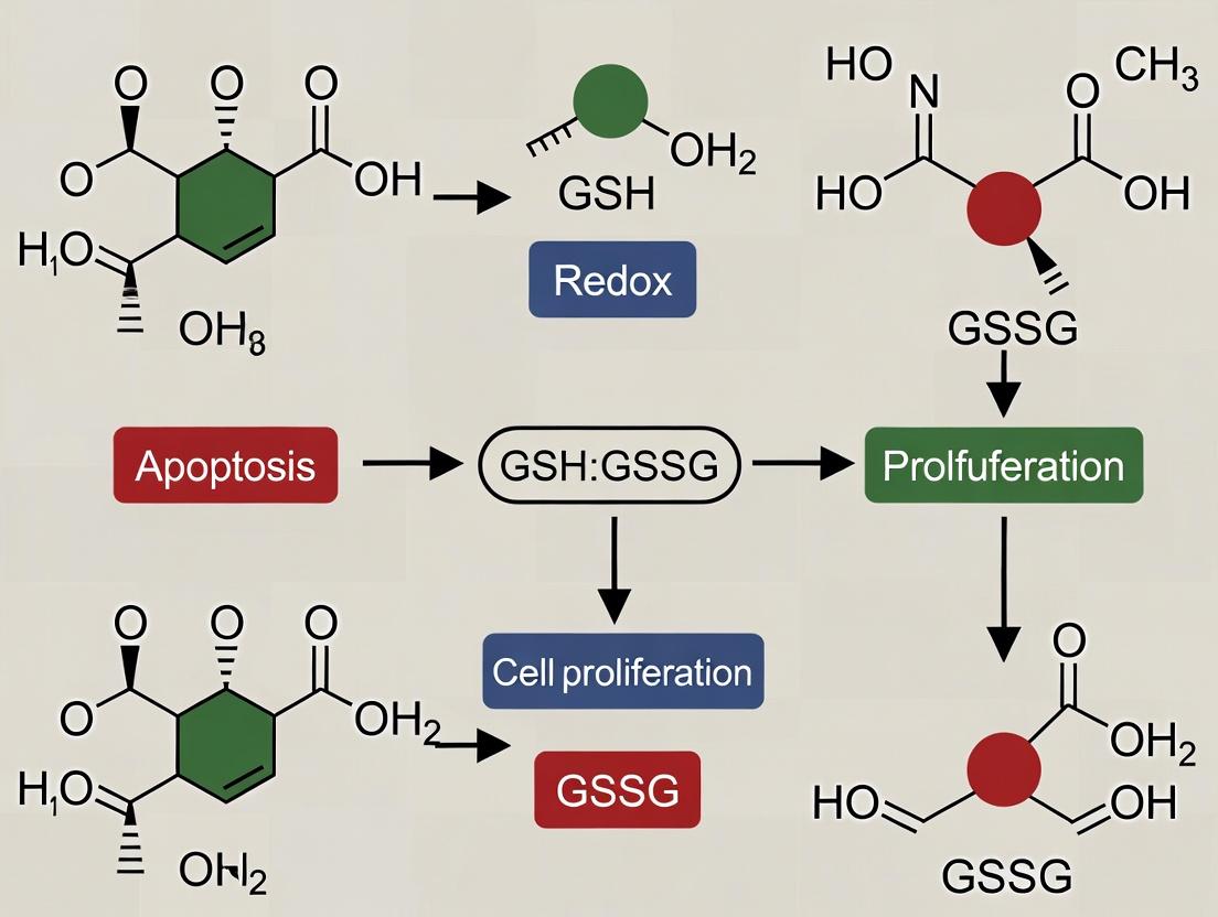
The tripeptide glutathione (γ-L-glutamyl-L-cysteinylglycine) is the predominant low-molecular-weight thiol in mammalian cells. Its redox cycling between the reduced (GSH) and oxidized (GSSG) forms constitutes the primary cellular redox buffer. The GSH/GSSG ratio is a tightly regulated metric, with a high ratio (~100:1 to 300:1 in the cytosol) indicative of a reducing environment conducive to proliferation. A significant decline in this ratio is a hallmark of oxidative stress and is intimately linked to the initiation of apoptotic signaling pathways. This document establishes the biochemical basis for monitoring this ratio as a critical parameter in cancer research, neurodegeneration, and drug discovery.
Structure and Synthesis
Molecular Architecture
- GSH (Reduced Form): A unique tripeptide with an unusual γ-peptide bond between glutamate and cysteine, conferring resistance to peptidases. The thiol (-SH) group on the cysteine residue is the functional moiety responsible for electron donation.
- GSSG (Oxidized Form): A homodimer of two GSH molecules linked via a disulfide bond (-S-S-) formed from the oxidation of their thiol groups.
Biosynthetic Pathway
GSH is synthesized in the cytosol via two ATP-dependent enzymatic steps:
- Step 1: Catalyzed by Glutamate-Cysteine Ligase (GCL), the rate-limiting enzyme. Inhibited by negative feedback from GSH.
- Step 2: Catalyzed by Glutathione Synthetase (GS).
The GSH/GSSG Redox Couple: Quantitative Dynamics
Quantitative data on glutathione status is pivotal for interpreting cellular redox health.
Table 1: Glutathione Parameters in Mammalian Cells
| Parameter | Typical Value/Range | Compartment | Significance for Apoptosis/Proliferation |
|---|---|---|---|
| Total GSH (GSH + 2xGSSG) | 1-10 mM | Cytosol | Pool size for antioxidant defense & biosynthesis. |
| GSH/GSSG Ratio | 100:1 - 300:1 | Cytosol (Healthy) | High Ratio: Reducing, pro-proliferative environment. |
| GSH/GSSG Ratio | < 10:1 | Cytosol (Under Oxidative Stress) | Low Ratio: Oxidative shift, triggers apoptosis. |
| Midpoint Potential (E°' for 2GSH/GSSG) | -240 mV (pH 7.0) | n/a | Thermodynamic reference for redox couple. |
| GSSG % of Total Pool | ~0.5-1% (Healthy) | Cytosol | Increases dramatically during oxidative challenge. |
Experimental Protocols for GSH/GSSG Analysis
HPLC-based Quantification (Gold Standard)
This protocol details the measurement of GSH and GSSG for ratio calculation.
Principle: Cell extracts are derivatized with a thiol-specific fluorescent reagent (e.g., monobromobimane, mBBr). GSSG is selectively measured by first masking GSH with N-ethylmaleimide (NEM). Separation and quantification are performed via HPLC with fluorescence detection.
Detailed Protocol:
- Sample Preparation: Rapidly lyse ~1x10^6 cells in ice-cold 1% (v/v) HClO₄ containing 2 mM EDTA (to inhibit metal oxidation). Centrifuge (10,000 x g, 10 min, 4°C) to pellet protein.
- Derivatization for Total GSH (GSH+GSSG):
- Neutralize an aliquot of supernatant with a KOH/HEPES buffer.
- Add mBBr solution (final conc. 2 mM) and NEM (final conc. 10 mM) to derivative all thiols. Incubate 20 min in the dark at room temperature.
- Quench reaction with acetic acid.
- Derivatization for GSSG Alone:
- To a separate aliquot of neutralized supernatant, add excess NEM (final conc. 50 mM) to rapidly and irreversibly derivative all free GSH. Incubate 5 min.
- Remove excess NEM by multiple ether extractions.
- Add dithiothreitol (DTT, 10 mM) to reduce GSSG to GSH.
- Add mBBr to derivative the newly formed GSH. Incubate and quench as in step 2.
- HPLC Analysis:
- Column: C18 Reverse-phase column (e.g., 5µm, 250 x 4.6 mm).
- Mobile Phase: Gradient from 0.1% (v/v) trifluoroacetic acid in H₂O to acetonitrile.
- Detection: Fluorescence (Ex: 380 nm, Em: 470 nm).
- Quantification: Compare peak areas of samples to standard curves of derivatized GSH and GSSG.
- Calculation:
- GSH concentration = [Total] - 2[GSSG]
- GSH/GSSG Ratio = [GSH] / (2[GSSG])
Enzymatic Recycling Assay (High-Throughput)
Principle: GSH reduces 5,5'-dithio-bis-(2-nitrobenzoic acid) (DTNB) to 2-nitro-5-thiobenzoic acid (TNB), producing a yellow color (412 nm). GSSG is first reduced to GSH by glutathione reductase (GR) using NADPH. The rate of TNB formation is proportional to total GSH+GSSG. GSSG alone is measured by pre-incubating samples with 2-vinylpyridine to derivative GSH.
Protocol Summary: Follow commercial kit instructions (e.g., Cayman Chemical, Sigma-Aldrich). Briefly, for total glutathione, sample is added to a reaction mix containing DTNB, GR, and NADPH. The absorbance at 412 nm is monitored kinetically. For GSSG, samples are first treated with 2-vinylpyridine, then neutralized and assayed similarly. Concentrations are determined against a standard curve.
The Scientist's Toolkit: Key Research Reagent Solutions
Table 2: Essential Reagents for Glutathione Redox Research
| Reagent | Function in Experiment | Key Consideration |
|---|---|---|
| N-Ethylmaleimide (NEM) | Thiol-alkylating agent. Rapidly masks free GSH to allow specific measurement of GSSG. | Must be used at optimal concentration/time; excess must be removed for enzymatic assays. |
| Monobromobimane (mBBr) | Thiol-specific fluorescent derivatization agent for HPLC. Forms stable adducts with GSH. | Light-sensitive. Reaction requires precise pH and incubation time. |
| 2-Vinylpyridine | Thiol-alkylating agent used in enzymatic assays to derivative GSH for GSSG-specific measurement. | Requires neutralization post-derivatization; can interfere if not properly removed. |
| Glutathione Reductase (GR) | Enzyme that reduces GSSG to GSH using NADPH, core component of enzymatic recycling assay. | Specific activity must be high; source (e.g., yeast) can affect kinetics. |
| 5,5'-Dithio-bis-(2-nitrobenzoic acid) (DTNB / Ellman's Reagent) | Colorimetric thiol probe. Reduced by GSH to yield yellow TNB (measurable at 412 nm). | Also reacts with other free thiols; specificity depends on sample preparation. |
| NADPH (Tetrasodium Salt) | Cofactor for Glutathione Reductase. Essential electron donor in the enzymatic recycling assay. | Labile; prepare fresh solutions. Oxidation compromises assay sensitivity. |
| Acivicin | Irreversible inhibitor of γ-glutamyl transpeptidase (GGT). Prevents extracellular GSH degradation in cell culture studies. | Used in media for experiments measuring extracellular glutathione flux. |
| Buthionine Sulfoximine (BSO) | Specific, irreversible inhibitor of Glutamate-Cysteine Ligase (GCL). Depletes intracellular GSH pools. | Standard tool to probe GSH dependence of cellular processes (apoptosis/proliferation). |
Integration with Apoptosis & Proliferation Research
The GSH/GSSG ratio is a functional node in cell fate signaling. A decreased ratio facilitates:
- Apoptosis: Oxidation of thiols on apoptosis signal-regulating kinase 1 (ASK1) and procaspase-3, promoting their activation. Mitochondrial permeability transition pore (MPTP) opening is also redox-sensitive.
- Proliferation Inhibition: Altered redox status affects signaling through pathways like NF-κB, MAPK, and PI3K/Akt, which are sensitive to the thiol/disulfide status of key cysteine residues.
Maintaining a high GSH/GSSG ratio is thus a hallmark of proliferating cells, particularly in tumors, making the enzymes of glutathione synthesis (GCL) and recycling potential therapeutic targets.
The glutathione (GSH) to glutathione disulfide (GSSG) ratio is a central, quantitative metric defining the redox state of a cell. Within the broader thesis of cellular fate decisions, this ratio serves as a critical node, governing the switch between proliferation and apoptosis. This whitepaper provides a technical overview of its definition, measurement, and functional implications for researchers in redox biology and therapeutic development.
Cellular redox homeostasis is maintained by the dynamic equilibrium between antioxidant and pro-oxidant systems. The tripeptide glutathione (γ-glutamyl-cysteinyl-glycine) is the most abundant non-protein thiol, functioning as a primary redox buffer. The GSH/GSSG ratio represents the thermodynamic poise of this system. A high ratio (typically >100:1 in cytosol) characterizes a reduced, proliferative state, while a decline (often to <10:1) signifies oxidative stress and can trigger apoptosis.
Quantitative Definition and Physiological Ranges
The GSH/GSSG ratio is calculated as the molar concentration of reduced glutathione (GSH) divided by the molar concentration of oxidized glutathione (GSSG). It is crucial to note that reporting the absolute concentrations alongside the ratio is essential for full interpretation.
Table 1: Representative GSH/GSSG Ratios in Mammalian Systems
| Compartment/Condition | [GSH] (mM) | [GSSG] (μM) | GSH/GSSG Ratio | Biological Implication |
|---|---|---|---|---|
| Cytosol (Resting) | 1-10 | 10-50 | ~100:1 to 200:1 | Reductive environment, supports proliferation |
| Mitochondria Matrix | 5-10 | ~10 | ~500:1 to 1000:1 | Highly reductive, protects ETC complexes |
| Endoplasmic Reticulum | ~1-5 | ~100 | ~10:1 to 50:1 | Oxidizing environment for disulfide bond formation |
| Oxidative Stress | Decreased | Increased (up to mM) | <10:1 | Activation of stress kinases (e.g., ASK1), apoptosis initiation |
| Apoptosis Execution | Severely Depleted | High | ~1:1 | Caspase activation, PARP cleavage, DNA fragmentation |
Core Methodologies for Measurement
Accurate determination requires rapid quenching of thiol-disulfide exchange to preserve the in vivo ratio.
Protocol: Metabolite Extraction for HPLC Analysis
This is the gold-standard method for specific and accurate quantification.
Reagents:
- N-ethylmaleimide (NEM) 100mM in PBS: Rapidly alkylates free thiols (GSH) to prevent oxidation during processing.
- Perchloric Acid (PCA) 5-10% (v/v): Denatures proteins and arrests enzyme activity.
- Potassium Hydroxide (KOH) 2M: Neutralizes PCA extract.
- Mobile Phases: Buffer A (0.1% Trifluoroacetic acid in water), Buffer B (0.1% TFA in acetonitrile) for reverse-phase HPLC.
- Derivatization Agent: O-phthalaldehyde (OPA) for fluorescent detection, or Ellman's reagent (DTNB) for colorimetric post-column detection.
Procedure:
- Quenching & Extraction: Aspirate culture medium. Immediately add ice-cold NEM solution (500 μL per 35mm dish) to cell monolayer. Scrape cells and transfer to a microcentrifuge tube on ice.
- Protein Precipitation: Add 50 μL of 50% PCA to the NEM-treated lysate. Vortex vigorously and incubate on ice for 10 min.
- Clarification: Centrifuge at 16,000 x g for 10 min at 4°C.
- Neutralization: Transfer supernatant to a fresh tube. Carefully add 2M KOH to neutralize to pH ~6-7. Centrifuge to remove potassium perchlorate precipitate.
- Analysis: Inject supernatant onto a C18 reverse-phase HPLC column. Derivatize with OPA for fluorescence detection (Ex 340 nm, Em 420 nm) or use electrochemical detection. Quantify using external GSH and GSSG standards.
- Normalization: Determine protein content in the initial pellet using a Bradford or BCA assay. Report data as nmol/mg protein.
Protocol: Enzymatic Recycling Assay (Commercial Kit Adaptation)
A common spectrophotometric/fluorometric method.
Principle: GSSG is first selectively masked. Then, GSH is cyclically oxidized by DTNB and reduced by glutathione reductase (GR), producing a colored (TNB) or fluorescent product proportional to total GSH. In a separate assay, GSSG is measured after derivatization of GSH.
Procedure Outline:
- Extraction without Derivatization: Lyse cells in cold 5% sulfosalicylic acid or metaphosphoric acid. Centrifuge to deproteinize.
- Total Glutathione Assay: Mix sample with assay cocktail containing DTNB, GR, and NADPH. Monitor absorbance at 412 nm or fluorescence over time.
- GSSG-Specific Assay: Treat a separate aliquot of the acid extract with a thiol-scavenging reagent (e.g., 2-vinylpyridine) to derivative all GSH. Then assay remaining GSSG as in step 2.
- Calculation: [GSH] = [Total GSH] - 2*[GSSG]. Calculate the ratio.
The GSH/GSSG Ratio in Apoptosis and Proliferation Signaling
A declining GSH/GSSG ratio is both a sensor and a mediator of cell fate decisions.
Key Pathway 1: Apoptosis Trigger via ASK1-p38/JNK
Title: ASK1 Apoptosis Activation by Low GSH/GSSG Ratio
Key Pathway 2: Proliferation Support via Nrf2-Keap1
Title: Nrf2 Inactivation by High GSH/GSSG Supports Proliferation
The Scientist's Toolkit: Essential Reagents & Materials
Table 2: Key Research Reagent Solutions
| Reagent/Material | Function/Application | Critical Note |
|---|---|---|
| N-ethylmaleimide (NEM) | Thiol-alkylating agent for rapid fixation of in vivo GSH/GSSG state during extraction. | Must be used in excess and at neutral pH; prepare fresh. |
| Metaphosphoric/Perchloric Acid | Protein-precipitating agents that quench enzymatic activity for accurate redox preservation. | Extracts require neutralization before analysis. |
| 2-Vinylpyridine | Thiol-masking agent used to derivative GSH for specific enzymatic measurement of GSSG. | Requires alkaline pH (pH 6-7.5) for efficient reaction. |
| Glutathione Reductase (GR) | Enzyme used in enzymatic recycling assays to reduce GSSG, cycling GSH. | Specific activity and purity are critical for assay sensitivity. |
| 5,5'-Dithio-bis-(2-nitrobenzoic acid) (DTNB) | Colorimetric probe (Ellman's reagent) producing TNB anion (A412) upon reaction with thiols. | Used in enzymatic assays and for direct free thiol measurement. |
| Buthionine Sulfoximine (BSO) | Specific inhibitor of γ-glutamylcysteine synthetase (GCL), depletes cellular GSH. | Essential tool for manipulating the GSH/GSSG ratio in vitro. |
| L-Buthionine-(S,R)-sulfoximine (BSO) | The specific stereoisomer used in research. | Verify isomer for study reproducibility. |
| Cell-permeable GSH Ethyl Ester (GSH-EE) | Compound used to augment intracellular GSH levels experimentally. | Can be hydrolyzed intracellularly to free GSH. |
| Cellular Glutathione Peroxidase (GPx) Mimetics (e.g., Ebselen) | Small molecules that mimic GPx activity, lowering GSH and increasing GSSG. | Useful for inducing a controlled pro-oxidant shift. |
Experimental Workflow for Apoptosis Studies
Title: Workflow for GSH/GSSG-Apoptosis Correlation Studies
The GSH/GSSG ratio is a definitive, quantifiable metric of cellular redox environment, inextricably linked to fate decisions in apoptosis and proliferation. Precise measurement requires rigorous, quenching-based methodologies. Integrating this ratio with functional apoptotic and proliferative readouts provides a powerful framework for understanding disease mechanisms and developing redox-modulating therapeutics.
The intracellular redox balance, principally defined by the ratio of reduced glutathione (GSH) to its oxidized disulfide form (GSSG), is a critical determinant of cellular fate. Within the context of apoptosis and cell proliferation research, the GSH/GSSG ratio operates as a master metabolic switch. A high GSH/GSSG ratio (reducing environment) promotes proliferation and survival signaling, while a decline in this ratio (oxidizing shift) creates a permissive environment for the activation of pro-apoptotic pathways. This whitepaper delves into the mechanistic links between this redox couple and three pivotal signaling nodes: NF-κB, p53, and MAPK, detailing how their activity is post-translationally tuned by the cellular redox state.
Table 1: Quantitative Effects of Altered GSH/GSSG Ratio on Key Signaling Pathways
| Signaling Molecule | Experimental Condition | Measured Outcome | Quantitative Change | Reference / Key Study |
|---|---|---|---|---|
| NF-κB (p50 subunit) | GSSG (20 µM) treatment in cell lysates | Inhibition of DNA binding activity | ~70% reduction | (Hansen et al., JBC, 1994) |
| p53 | Diamide (thiol oxidant) treatment in cells | Increased p53 DNA binding & transactivation | 3- to 5-fold increase | (Polyak et al., PNAS, 1997) |
| ASK1 (MAPKKK) | GSH depletion (BSO treatment) in cells | ASK1 activation & JNK/p38 phosphorylation | JNK activity increased >4-fold | (Saitoh et al., EMBO J, 1998) |
| Overall Apoptosis | GSH/GSSG ratio shift from 100:1 to 10:1 | Induction of apoptosis in Jurkat cells | Apoptosis increased from 5% to 40% | (Circu & Aw, Free Radic. Biol. Med., 2010) |
| Cell Proliferation | Maintenance of GSH/GSSG > 30:1 | Optimal proliferation rate in fibroblasts | Proliferation rate 2x higher than at ratio <10:1 | (Schafer & Buettner, Free Radic. Biol. Med., 2001) |
Mechanistic Pathways: GSH/GSSG Regulation of Signaling Molecules
NF-κB Pathway
A reducing environment (high GSH/GSSG) is required for NF-κB activity. Critical cysteine residues (e.g., Cys62 in the p50 subunit) must be in a reduced state for DNA binding. Oxidation or S-glutathionylation of these residues inhibits NF-κB, shifting the cell away from pro-survival, anti-apoptotic signaling.
p53 Pathway
p53 activation is potentiated by an oxidizing shift (lower GSH/GSSG). Oxidants promote disulfide bond formation or S-glutathionylation at specific cysteines in the DNA-binding domain, stabilizing p53 conformation and enhancing its sequence-specific DNA binding, leading to cell cycle arrest or apoptosis.
MAPK Pathway
The GSH/GSSG ratio differentially regulates MAPK branches. The JNK and p38 pathways are typically activated under oxidative stress (low GSH/GSSG), often via the redox-sensitive kinase ASK1, which is inhibited by reduced thioredoxin and activated when oxidized. In contrast, the ERK pathway, often pro-proliferative, is more active under reducing conditions.
Diagram 1: Redox Control of Key Signaling Pathways (GSH/GSSG as a Switch)
Key Experimental Protocols for Investigating Redox Signaling
Protocol: Measuring Intracellular GSH/GSSG Ratio (HPLC-based)
- Objective: Accurately quantify the concentrations of GSH and GSSG in cell lysates.
- Reagents: Perchloric acid (PCA) with EDTA, N-ethylmaleimide (NEM), Potassium phosphate buffer, O-phthalaldehyde (OPA) derivatization reagent.
- Procedure:
- Rapid Lysis & Derivatization: Wash cells (1x10^6) with ice-cold PBS. Lyse in 100 µL of 5% PCA/EDTA. For GSSG-specific measurement, immediately add 10 µL of 40mM NEM to 50 µL of lysate to alkylate free GSH. For total GSH, omit NEM.
- Neutralization: Centrifuge lysates (10,000 x g, 10 min, 4°C). Transfer supernatant to a fresh tube containing an equal volume of 0.1M potassium phosphate buffer (pH 7.0).
- Derivatization & HPLC: Mix 10 µL of neutralized sample with 10 µL OPA reagent. Incubate in the dark for 2 min. Inject onto a C18 reverse-phase column. Elute with a gradient of methanol in sodium phosphate buffer (pH 6.0). Detect fluorescence (Ex 340 nm, Em 420 nm).
- Calculation: Calculate concentrations from standard curves. Intracellular GSH = (Total GSH) - (2 x [GSSG]). Ratio = [GSH] / [GSSG].
Protocol: Assessing Redox-Sensitive Protein S-Glutathionylation (Biotin Switch Assay)
- Objective: Detect proteins that undergo S-glutathionylation under oxidative stress.
- Reagents: HEN buffer (HEPES, EDTA, Neocuproine), Methyl methanethiosulfonate (MMTS), Biotin-HPDP, Streptavidin beads, Anti-GSH antibody (optional control).
- Procedure:
- Block Free Thiols: Lyse control and treated cells (e.g., with diamide) in HEN buffer with 1% Triton X-100 and protease inhibitors. Add MMTS (20mM final) to block all free cysteine thiols. Incubate 30 min at 50°C.
- Reduce & Label S-Glutathionylated Cysteines: Remove MMTS via acetone precipitation. Resuspend pellet in HEN buffer with 1% SDS. Add sodium ascorbate (to reduce the mixed disulfide bond) and Biotin-HPDP (to label the newly freed thiol). Incubate 1 hr at RT.
- Affinity Capture & Detection: Remove excess biotin. Incubate lysate with streptavidin-agarose beads overnight at 4°C. Wash beads thoroughly. Elute bound proteins with Laemmli buffer containing β-mercaptoethanol.
- Analysis: Analyze eluates by western blot for proteins of interest (e.g., p50, p53) or by mass spectrometry for proteomic discovery.
Visualizing Key Experimental Workflows
Diagram 2: Workflow for GSH/GSSG-Mediated Signaling Analysis
The Scientist's Toolkit: Essential Research Reagents
Table 2: Key Reagents for Studying GSH/GSSG in Redox Signaling
| Reagent | Category | Primary Function in Research |
|---|---|---|
| L-Buthionine-sulfoximine (BSO) | GSH Synthesis Inhibitor | Selectively inhibits γ-glutamylcysteine synthetase (GCL), depleting intracellular GSH pools to study the effects of a low GSH/GSSG ratio. |
| N-acetylcysteine (NAC) | Thiol Antioxidant / GSH Precursor | Increases cellular cysteine levels, boosting GSH synthesis. Used to elevate the GSH/GSSG ratio and test protection against oxidative stress. |
| Diethyl maleate (DEM) | GSH Conjugating Agent | Rapidly depletes GSH by forming a conjugate via glutathione S-transferase, inducing an acute oxidizing shift. |
| Diamide | Thiol-Specific Oxidant | Selectively oxidizes thiols, converting GSH to GSSG and promoting protein disulfide formation/S-glutathionylation. |
| Monochlorobimane (mBCI) | Fluorescent Probe | Cell-permeable dye that forms a fluorescent adduct with GSH via GST; used for live-cell imaging and flow cytometry of GSH levels. |
| Grx1-roGFP2 (or similar) | Genetically Encoded Redox Sensor | A rationetric fluorescent protein biosensor that specifically reports the GSH/GSSG redox potential in specific cellular compartments. |
| Anti-Glutathione Antibody | Immunological Tool | Used in ELISA, western blot (non-reducing conditions), or immunoprecipitation to detect protein S-glutathionylation. |
| Recombinant Glutaredoxin 1 (Grx1) | Enzymatic Reductase | Specifically reduces protein-SSG mixed disulfides (deglutathionylation). Critical for validating S-glutathionylation events in assays. |
This whitepares the critical role of a lowered glutathione disulfide (GSSG) to reduced glutathione (GSH) ratio—a definitive oxidative shift—as a primary trigger for the mitochondrial (intrinsic) apoptotic pathway. Operating within the broader thesis that the GSH:GSSG redox couple is a central regulator of cell fate, this guide details the molecular mechanisms, current experimental methodologies, and key research tools for investigating this nexus between redox imbalance and programmed cell death.
The tripeptide glutathione (γ-glutamyl-cysteinyl-glycine) exists in a dynamic equilibrium between its reduced (GSH) and oxidized disulfide (GSSG) forms. The GSH:GSSG ratio functions as a fundamental cellular redox buffer, typically maintained at >100:1 in healthy mammalian cells. A significant decrease in this ratio constitutes an "oxidative shift," signaling severe redox stress. This shift is not merely a bystander but a decisive signal that permeabilizes the mitochondrial outer membrane, initiating the intrinsic apoptotic cascade—a process critical in development, homeostasis, and pathologies like cancer and neurodegeneration.
Molecular Mechanisms: From Redox Shift to Apoptosome
Primary Redox-Sensitive Targets
The oxidative shift directly modifies key mitochondrial proteins:
- Bcl-2 Family Proteins: Oxidation of pro-survival Bcl-2 and Bcl-xL at cysteine residues inhibits their function. Concurrently, pro-apoptotic Bax and Bak are activated via oxidation and translocation to the mitochondrial membrane.
- Voltage-Dependent Anion Channel (VDAC): Oxidation promotes VDAC oligomerization, facilitating cytochrome c release.
- Thioredoxin System: Oxidation of thioredoxin-2 (Trx2) in the mitochondria releases its inhibition of apoptosis signal-regulating kinase 1 (ASK1), activating downstream stress kinases.
Core Signaling Cascade
- Trigger: Depletion of GSH pool and increase in GSSG.
- Mitochondrial Permeabilization: Activated Bax/Bak form pores in the outer mitochondrial membrane (MOMP).
- Cytochrome c Release: Released into the cytosol.
- Apoptosome Formation: Cytochrome c binds Apaf-1 and procaspase-9 in the presence of dATP/ATP.
- Execution Phase: Caspase-9 activates effector caspases-3 and -7, leading to substrate cleavage and apoptotic cell death.
Diagram 1: Intrinsic Apoptosis Pathway Triggered by Oxidative Shift
Quantitative Data: Key Metrics in Redox-Dependent Apoptosis
Table 1: Threshold Values for Apoptotic Trigger
| Parameter | Normal Range (Healthy Cell) | Apoptotic Trigger Range | Measurement Method |
|---|---|---|---|
| GSH:GSSG Ratio | 100:1 to 300:1 | < 20:1 | HPLC, LC-MS, Fluorometric Kits |
| Total Glutathione | 1-10 mM | < 20% of baseline | DTNB Recycling Assay |
| Cytochrome c Localization | Mitochondrial | Cytosolic (≥ 40% release) | Cell Fractionation + WB/ELISA |
| Caspase-3/7 Activity | Low (Basal) | > 5-fold increase | Fluorogenic DEVD-peptide cleavage |
| Phosphatidylserine Exposure | Inner leaflet | Outer leaflet (≥ 15% Annexin V+ cells) | Flow Cytometry (Annexin V/PI) |
Table 2: Genetic & Pharmacological Modulators of the Pathway
| Modulator/Target | Effect on GSH:GSSG Ratio | Consequence on Apoptosis | Example Agent |
|---|---|---|---|
| GSH Synthesis Inhibitor | Drastically Lowers Ratio | Induces/Potentiates Apoptosis | Buthionine sulfoximine (BSO) |
| Glutathione Reductase Inhibitor | Lowers Ratio (↑GSSG) | Induces/Potentiates Apoptosis | Carmustine (BCNU), 2-AAPA |
| Nrf2 Activator | Increases Ratio (↑GSH) | Inhibits Apoptosis | Sulforaphane, CDDO-Me |
| Bcl-2/Bcl-xL Inhibitor | May lower ratio secondarily | Potentiates Redox Apoptosis | Venetoclax (ABT-199), ABT-737 |
| Thioredoxin Reductase Inhibitor | Disrupts related redox system | Synergistic Apoptosis Induction | Auranofin |
Experimental Protocols
Protocol: Measuring the GSH:GSSG Ratio in Apoptotic Cells
Objective: Accurately quantify reduced and oxidized glutathione to calculate the ratio during intrinsic apoptosis induction.
Materials:
- Cells undergoing apoptosis (e.g., treated with H₂O₂, BSO, or chemotherapeutic agents).
- GSH/GSSG Assay Kit (e.g., from Cayman Chemical, Sigma-Aldrich).
- Metaphosphoric Acid (MPA) / EDTA Solution (for deproteinization).
- Triethanolamine (TEA) solution (for pH adjustment).
- Microplate reader (for absorbance/fluorescence).
Procedure:
- Harvest & Deproteinize: Collect 1x10⁶ cells. Pellet and wash with PBS. Lyse in 100 µL of cold 1% MPA/EDTA solution. Vortex and incubate on ice for 5 minutes.
- Neutralize: Centrifuge at 10,000 x g for 10 min (4°C). Transfer supernatant to a new tube. Add 50 µL of TEA solution to 100 µL of supernatant to adjust pH to ~7.0.
- Assay Setup (Dual Measurement):
- Total GSH: Mix 50 µL neutralized sample with 150 µL of assay cocktail containing NADPH, DTNB, and glutathione reductase. Monitor absorbance at 412 nm for 5-10 min.
- GSSG Only: To another 50 µL sample, add 2 µL of 2-vinylpyridine. Incubate for 1 hour at room temperature to derivative GSH. Then assay as above. This measures only GSSG.
- Calculation: Generate standard curves for GSH and GSSG.
- GSH = (Total GSH) - (2 x Measured GSSG)
- Ratio = [GSH] / [GSSG]
Protocol: Correlating Redox Shift with MOMP (CytochromecRelease)
Objective: Visualize and quantify cytochrome c translocation from mitochondria to cytosol.
Materials:
- Mitochondria/Cytosol Fractionation Kit (e.g., from Abcam, BioVision).
- Protease/Phosphatase Inhibitors.
- Antibodies: Anti-cytochrome c (clone 6H2.B4), Anti-COX IV (mitochondrial loading control), Anti-β-tubulin (cytosolic loading control).
- Western Blot equipment.
Procedure:
- Cell Fractionation: Harvest 5-10x10⁶ treated/control cells. Wash with PBS. Resuspend in 1 mL of cold Cytosol Extraction Buffer with inhibitors. Homogenize on ice with 50-100 strokes in a Dounce homogenizer. Centrifuge at 700 x g for 10 min (4°C) to remove nuclei/debris.
- Separate Fractions: Transfer supernatant to a fresh tube. Centrifuge at 10,000 x g for 30 min (4°C). The supernatant is the cytosolic fraction. The pellet is the heavy membrane/mitochondrial fraction; resuspend in 100 µL Mitochondrial Extraction Buffer.
- Western Blot Analysis: Run 20-30 µg of each fraction on SDS-PAGE. Transfer to PVDF membrane. Probe with anti-cytochrome c. Loss from mitochondrial fraction and gain in cytosolic fraction indicates MOMP.
Diagram 2: Experimental Workflow for Correlating Redox Shift & Apoptosis
The Scientist's Toolkit: Key Research Reagents & Solutions
Table 3: Essential Reagents for Investigating Redox-Triggered Apoptosis
| Item | Function/Biological Role | Example Product/Assay |
|---|---|---|
| Buthionine Sulfoximine (BSO) | Irreversible inhibitor of γ-glutamylcysteine synthetase (GCL), depletes cellular GSH. | Sigma-Aldrich, B2515 |
| Monochlorobimane (mBCI) | Cell-permeable dye that forms a fluorescent adduct with GSH; measures GSH levels via flow cytometry. | Cayman Chemical, 14450 |
| CellROX Reagents | Fluorogenic probes that measure general reactive oxygen species (ROS) in live cells. | Thermo Fisher Scientific, C10422 |
| Fluorogenic Caspase-3/7 Substrate (Ac-DEVD-AMC/AFC) | Quantifies effector caspase activity upon cleavage in lysates or live cells. | Promega, G8090/G8210 |
| Annexin V-FITC / Propidium Iodide (PI) | Gold standard for detecting early (PS exposure) and late apoptosis/necrosis (PI uptake). | BioLegend, 640914 |
| MitoSOX Red | Mitochondria-targeted superoxide indicator. | Thermo Fisher Scientific, M36008 |
| BH3 Profiling Peptides | Synthetic peptides (e.g., BIM, BID) to measure mitochondrial priming and Bcl-2 family dependence. | Tocris Bioscience (Custom) |
| Anti-4-Hydroxynonenal (4-HNE) Antibody | Detects lipid peroxidation, a key consequence of severe redox imbalance. | Abcam, ab46545 |
| GSH/GSSG-Glo Assay | Bioluminescent assay for measuring GSH/GSSG ratio in a plate-based format. | Promega, V6611 |
The precise molecular sensing of the GSH:GSSG ratio and its transduction into an apoptotic signal represents a master regulatory node in cell biology. Validating this oxidative shift as a therapeutically targetable trigger offers powerful avenues in drug development: 1) Sensitizing Strategy: Depleting GSH (e.g., with BSO) to lower the ratio can sensitize resistant cancer cells to intrinsic apoptosis. 2) Protective Strategy: Pharmacologically bolstering the GSH system may protect healthy cells in degenerative diseases. Future research must focus on spatiotemporally-resolved measurements of this ratio within subcellular compartments, particularly the mitochondrial matrix, to fully decipher its role as the "redox rheostat" of life and death decisions.
The intracellular redox environment, predominantly defined by the reduced glutathione (GSH) to oxidized glutathione (GSSG) ratio, is a critical determinant of cellular fate. A high GSH:GSSG ratio is a hallmark of a reduced cytosol, a state permissive for proliferation. This paper positions itself within the broader thesis that a sustained high GSH:GSSG ratio is not merely a correlative marker but a functional proliferation enabler. It acts by two primary, interconnected mechanisms: (1) Creating a Reduced Environment that inactivates pro-apoptotic signaling and stabilizes cell cycle machinery, and (2) Directly Supporting Biosynthesis and Cell Cycle Progression by providing reducing equivalents and modulating protein activity. This whitepaper provides a technical guide for investigating this core mechanism in cancer and regenerative biology.
The Central Role of the GSH:GSSG Ratio in Redox Homeostasis
Glutathione (γ-glutamyl-cysteinyl-glycine) is the most abundant non-protein thiol. The GSH:GSSG ratio, typically >100:1 in healthy proliferating cells, sets the redox potential of the cellular milieu. A decline in this ratio (increased oxidative stress) favors apoptosis, while maintenance of a high ratio is anti-apoptotic and pro-proliferative.
Quantitative Data on GSH/GSSG in Cell States
Table 1: GSH:GSSG Ratio Across Cellular States
| Cellular State | Typical GSH:GSSG Ratio | Redox Potential (Eh, mV) | Key Implications |
|---|---|---|---|
| Rapid Proliferation | 100:1 to 300:1 | -260 to -220 | Favors reduced cysteine residues, supports biosynthesis. |
| Quiescence / Homeostasis | ~30:1 to 100:1 | -220 to -200 | Balanced redox state, maintained by GR/GPx. |
| Early Apoptosis | < 10:1 | > -180 | Oxidized environment, activation of ASK1, caspase cascades. |
| Necrosis | Near 1:1 | > -150 | Severe depletion, loss of membrane integrity. |
Mechanism I: The Reduced Environment as a Proliferation Signal
Inhibition of Pro-Apoptotic Pathways
A high GSH:GSSG ratio directly suppresses key apoptotic initiators.
- ASK1 Inactivation: Apoptosis signal-regulating kinase 1 (ASK1) is sequestered in a complex with reduced thioredoxin (Trx). Oxidation of Trx leads to its dissociation and ASK1 activation. A reduced environment maintained by GSH keeps Trx reduced, inhibiting the ASK1-p38/JNK stress kinase pathway.
- Caspase Inhibition: Direct glutathionylation of caspase-3 catalytic cysteine reversibly inhibits its activity, providing a redox checkpoint.
Stabilization of Cell Cycle Regulators
Key cyclins and transcription factors (e.g., NF-κB, c-Myc) require reduced cysteine residues for stability and DNA binding. A reduced environment prevents their oxidative degradation.
Diagram 1: Redox Control of Apoptosis vs. Proliferation Signaling
Mechanism II: Direct Support of Biosynthesis and Cell Cycle
Provision of Reducing Equivalents for Synthesis
- Ribonucleotide Reductase (RNR): This rate-limiting enzyme for deoxyribonucleotide (dNTP) synthesis requires electrons from the thioredoxin or glutaredoxin systems, both regenerated by NADPH and ultimately linked to GSH metabolism.
- Antioxidant Defense: GSH, via Glutathione Peroxidase (GPx), scavenges H₂O₂ and lipid peroxides generated during rapid metabolic activity, protecting DNA and membrane integrity essential for cycle completion.
Metabolic Rewiring for Proliferation
The pentose phosphate pathway (PPP) is upregulated to generate NADPH, which is used by Glutathione Reductase (GR) to maintain a high GSH:GSSG ratio. This creates a feed-forward loop supporting anabolism.
Quantitative Impact on Cell Cycle
Table 2: Effect of GSH Modulation on Cell Cycle Parameters
| Intervention | GSH:GSSG Ratio Change | Cell Cycle Impact (vs. Control) | Key Readout |
|---|---|---|---|
| BSO (GSH synthesis inhibitor) | ↓ > 80% | G1/S arrest; increased apoptosis. | ↓ EdU+ cells by ~70%; ↑ cleaved caspase-3. |
| NAC (GSH precursor) | ↑ ~50% | Reduced serum requirement; shortened G1. | ↑ Cyclin D1 expression; S-phase entry accelerated by ~2h. |
| GSH Ethyl Ester (cell-permeable GSH) | ↑ ~300% | Enhanced proliferation in low-glucose conditions. | ↑ dNTP pools; resistance to oxidative arrest. |
Diagram 2: Metabolic Support of Biosynthesis by High GSH Ratio
Experimental Protocols for Investigation
Protocol A: Quantifying Intracellular GSH:GSSG Ratio (DTNB/GR Recycling Assay)
Principle: Total GSH and GSSG are measured spectrophotometrically by the reaction with DTNB, catalyzed by GR. Procedure:
- Cell Lysis: Harvest 1x10⁶ cells in ice-cold 5% metaphosphoric acid. Vortex, freeze-thaw, centrifuge at 10,000xg for 10min (4°C). Use supernatant.
- Total GSH Measurement: For 96-well plate.
- Mix: 50µL sample, 150µL 0.1M sodium phosphate buffer (pH 7.5) with 1mM EDTA, 10µL 6mM DTNB, 10µL 3mM NADPH.
- Start reaction with 10µL GR (5 U/mL).
- Read kinetics at 412nm for 3 min. Calculate GSHeq from GSH standard curve.
- GSSG-Specific Measurement: Derivatize GSH in sample.
- Incubate 100µL sample with 2µL 2-vinylpyridine for 1hr at room temperature.
- Proceed as in Step 2. Value represents GSSG.
- Calculation: Total GSH = GSH + 2xGSSG. GSH = Total - (2xGSSG). Report as nmol/mg protein and ratio.
Protocol B: Assessing Proliferation Dependency (BSO/NAC Titration)
Principle: Chemically modulate GSH levels and measure proliferation/cell cycle. Procedure:
- Treatment: Seed cells in 12-well plates. At ~30% confluency, treat with:
- BSO (0.1-1.0 mM) for 24-48h to deplete GSH.
- NAC (1-5 mM) for 24h to elevate GSH.
- Include vehicle controls.
- Proliferation Assay (EdU): Add EdU (10µM) for 2h before harvest. Fix, permeabilize, and perform Click-iT reaction per manufacturer's protocol. Analyze by flow cytometry.
- Cell Cycle Analysis (PI): Fix cells in 70% ethanol, treat with RNase A, stain with Propidium Iodide (50µg/mL). Analyze DNA content by flow cytometry (FL2-A).
- Correlation: Run Protocol A on parallel samples to correlate GSH:GSSG with %S-phase and apoptosis (sub-G1 peak).
The Scientist's Toolkit: Research Reagent Solutions
Table 3: Essential Reagents for Investigating GSH-Mediated Proliferation
| Reagent / Kit | Primary Function | Application in This Context |
|---|---|---|
| L-Buthionine-sulfoximine (BSO) | Irreversible inhibitor of γ-glutamylcysteine synthetase (GCL), the rate-limiting GSH synthesis enzyme. | Experimental depletion of intracellular GSH to establish causal role in proliferation arrest. |
| N-Acetylcysteine (NAC) | Cell-permeable cysteine prodrug and antioxidant; precursor for GSH synthesis. | Augmenting intracellular GSH to test sufficiency for enhancing proliferation or conferring resistance. |
| GSH/GSSG-Glo Assay (Promega) | Luminescent-based assay for quantification of GSH and GSSG from cell lysates. | High-throughput, sensitive measurement of the GSH:GSSG ratio in multi-well plates. |
| CellROX Green / DCFH-DA | Fluorescent probes for general detection of intracellular reactive oxygen species (ROS). | Assessing the correlation between GSH depletion, ROS accumulation, and cell fate decisions. |
| Click-iT Plus EdU Alexa Fluor Flow Cytometry Assay | Detects DNA synthesis via incorporation of nucleoside analog EdU. | Accurate quantification of S-phase fraction under different redox manipulations. |
| Anti-Glutathionylation Antibody | Detects protein-SSG post-translational modifications. | Identifying specific cell cycle/pro-apoptotic proteins regulated by direct glutathionylation. |
| Recombinant Glutathione Reductase (GR) | Enzyme used in recycling assays for GSH/GSSG quantification. | Core component of the enzymatic cycling DTNB assay protocol. |
This whitepaper provides a technical examination of the reduced-to-oxidized glutathione (GSH:GSSG) ratio as a critical bioenergetic and redox sensor governing cellular fate decisions between proliferation and apoptosis. The GSH:GSSG ratio operates as a pivotal tipping point; its maintenance within a physiological range supports proliferation, while a significant decline triggers a cascade toward apoptotic commitment. We detail quantitative thresholds, experimental methodologies for their determination, and the integrated signaling pathways involved.
The cellular redox state, quantified primarily by the GSH:GSSG ratio, is not merely a homeostatic parameter but a decisive signaling modality. A high GSH:GSSG ratio (high reducing capacity) is permissive for anabolic processes and cell cycle progression. Conversely, a sustained drop below a critical threshold induces oxidative stress, leading to mitochondrial outer membrane permeabilization (MOMP) and caspase activation. This shift represents a classic bistable system where a continuous change in the ratio value passes a tipping point, resulting in a discrete, fate-altering switch.
Quantitative Thresholds: Data Compendium
The critical GSH:GSSG ratio values vary by cell type, metabolic state, and stimulus but converge within defined ranges that dictate fate switching.
Table 1: Critical GSH:GSSG Ratio Thresholds Across Cell Types & Conditions
| Cell Type / System | Physiological (Proliferation) Range | Stress/Transition Zone | Apoptotic Trigger Range | Key Experimental Context |
|---|---|---|---|---|
| Hepatocytes (Primary, Rat) | 100:1 to 50:1 | < 30:1 | < 10:1 | TNF-α induced apoptosis |
| Jurkat T-Cell Lymphocytes | 80:1 to 40:1 | < 25:1 | ≤ 5:1 | Etoposide/Fas-ligand induced apoptosis |
| HEK293 (Human Embryonic Kidney) | 60:1 to 30:1 | < 20:1 | < 7:1 | H₂O₂ exposure |
| Neuronal Progenitor Cells | 70:1 to 35:1 | < 22:1 | ≤ 8:1 | Glutamate-induced excitotoxicity |
| Cancer Cell Lines (e.g., HeLa, MCF-7) | 40:1 to 15:1* | < 12:1* | ≤ 4:1* | Chemotherapeutic agent (Cisplatin, Doxorubicin) challenge |
Note: Cancer cells often exhibit a constitutively lower GSH:GSSG ratio, reflecting chronic redox stress, yet remain sensitive to further declines.
Table 2: Key Molecular Events Correlated with Ratio Declines
| GSH:GSSG Ratio Approx. Value | Key Molecular & Phenotypic Consequences |
|---|---|
| > 30:1 | Proliferation Zone: Optimal for nucleotide synthesis, active MAPK/ERK & PI3K/Akt signaling. |
| 30:1 → 15:1 | Stress Sensing: Activation of Nrf2/ARE pathway, p38 MAPK/JNK signaling begins, cell cycle arrest. |
| 15:1 → 5:1 | Commitment Zone: Oxidation of mitochondrial pore proteins (e.g., ANT), Bax/Bak activation, Cytochrome c release. |
| < 5:1 | Execution: Caspase-3/7 activation, PARP cleavage, DNA fragmentation, phosphatidylserine exposure. |
Core Signaling Pathways: A Systems View
The transition from a high to a low GSH:GSSG ratio is transduced into fate decisions via interconnected pathways.
Experimental Protocols for Determining Critical Ratios
Protocol: HPLC-Based Quantification of GSH and GSSG
This is the gold-standard method for accurate ratio determination.
Principle: Thiol-specific derivatization followed by chromatographic separation and fluorescence/electrochemical detection. Sample Preparation:
- Rapid Quenching: Wash cells (1-2x10^6) in ice-cold PBS and lyse immediately in 100 µL of 1% (w/v) meta-phosphoric acid (MPA) containing 1 mM EDTA (pH 8.0) and 50 µM internal standard (e.g., γ-glutamyl glutamate). Use ice-cold microtubes and process samples within 30 seconds.
- Derivatization (for Total GSH): Mix 50 µL of supernatant with 5 µL of 4-vinylpyridine (for GSSG protection) and incubate in the dark for 60 min. Then, add 20 µL of 10 mM dithiothreitol (DTT) to reduce GSSG to GSH, followed by 10 µL of iodoacetic acid (for GSH derivatization) and incubation in the dark for 30 min.
- Derivatization (for GSSG-specific): Immediately after lysis, add 2 µL of 2-vinylpyridine to 50 µL of supernatant to derivative GSH specifically, leaving GSSG intact. Incubate for 60 min in the dark. HPLC Analysis:
- Column: C18 reversed-phase column (250 x 4.6 mm, 5 µm).
- Mobile Phase: Buffer A: 0.1% (v/v) trifluoroacetic acid in water. Buffer B: 0.1% TFA in acetonitrile. Gradient: 0-15% B over 20 min.
- Detection: Fluorescence detector (Ex 385 nm, Em 515 nm) for OPA-derivatized samples, or electrochemical detector.
- Calculation: Quantify peaks against internal and external standards. GSH (from total assay) - 2xGSSG (from specific assay) = free GSH. Calculate molar ratio.
Protocol: Live-Cell Monitoring of Redox Potential (roGFP)
This allows dynamic, compartment-specific tracking of the glutathione redox potential (E_GSSG/2GSH), which is directly related to the ratio.
Principle: Genetically encoded redox-sensitive GFP (roGFP) fused to human glutaredoxin-1 (Grx1) equilibrates with the GSH:GSSG pool. Workflow:
- Transfection/Infection: Introduce plasmid or viral vector encoding roGFP-Grx1 (targeted to cytosol or mitochondria) into cells 24-48h prior.
- Calibration: Perform a two-point calibration in situ for each cell/field:
- Oxidized State: Treat with 10 mM H₂O₂ for 5 min.
- Reduced State: Treat with 10 mM DTT for 5 min.
- Ratiometric Imaging: Acquire fluorescence images at two excitation wavelengths (typically 405 nm and 488 nm) with a common emission (510 nm). Use a confocal or widefield microscope with environmental control (37°C, 5% CO₂).
- Data Analysis: Calculate the ratio (I405/I488) for each pixel/cell. Normalize ratios from 0 (fully reduced, DTT) to 1 (fully oxidized, H₂O₂). Convert normalized ratio to E_GSSG/2GSH using Nernst equation: E = E0 - (RT/nF)ln([GSH]^2/[GSSG]), where E0 for roGFP2 is -280 mV.
- Fate Correlation: Continuously image cells while applying an apoptotic stimulus. Correlate the time point at which E_GSSG/2GSH crosses a threshold (e.g., -250 mV to -220 mV, depending on cell type) with subsequent apoptotic markers (Annexin V, caspase activation).
The Scientist's Toolkit: Essential Research Reagents & Solutions
Table 3: Key Reagents for GSH:GSSG & Redox Fate Research
| Reagent / Kit | Function & Critical Application |
|---|---|
| Meta-Phosphoric Acid (MPA) Lysis Buffer | Instant protein precipitation and thiol stabilization for accurate GSH/GSSG measurement. Prevents auto-oxidation. |
| Monochlorobimane (mBCL) | Cell-permeable, non-fluorescent dye that conjugates with GSH via GST, yielding a fluorescent adduct for flow cytometry. Measures total GSH. |
| roGFP2-Grx1 (Plasmid or Viral Particles) | Genetically encoded biosensor for real-time, compartment-specific measurement of glutathione redox potential (E_GSSG/2GSH). |
| GSH/GSSG-Glo Assay (or similar luminescent kit) | Homogeneous, high-throughput assay measuring total/oxidized glutathione based on luciferase-coupled enzymatic recycling. |
| Buthionine Sulfoximine (BSO) | Specific, irreversible inhibitor of γ-glutamylcysteine synthetase (GCL), the rate-limiting enzyme in GSH synthesis. Used to deplete intracellular GSH. |
| N-Acetylcysteine (NAC) | Cell-permeable cysteine precursor that boosts intracellular GSH synthesis. Used as a redox control/rescue agent. |
| Mitochondria-Targeted Antioxidants (MitoTEMPO, MitoQ) | Compounds that selectively scavenge mitochondrial ROS, used to dissect the source of redox changes in fate switching. |
| Annexin V-FITC/PI Apoptosis Detection Kit | Standard flow cytometry assay to quantify early/late apoptotic and necrotic cells, for correlation with GSH ratios. |
| Caspase-3/7 Glo Assay | Luminescent assay for measuring executioner caspase activity, a key downstream event of the redox tipping point. |
Cross-talk with Other Antioxidant Systems (Thioredoxin, Nrf2)
The cellular redox environment is a critical determinant of cell fate, governing the switch between proliferation and apoptosis. A central metric in this regulation is the ratio of reduced glutathione to oxidized glutathione (GSH/GSSG), a primary indicator of cellular redox potential. This whitepaper situates its examination of antioxidant system cross-talk within the broader thesis that dynamic shifts in the GSH/GSSG ratio are not merely correlative but are instrumental in executing and modulating apoptotic signaling and proliferative pathways. The Thioredoxin (Trx) and Nuclear factor erythroid 2–related factor 2 (Nrf2) systems are not parallel, isolated pathways; they engage in extensive, context-dependent cross-talk with the glutathione system. This interplay creates a layered redox control network, where perturbation in one system can be compensated or amplified by another, ultimately converging to fine-tune the cellular response to oxidative stress and dictate survival outcomes. Understanding this network is paramount for developing targeted therapeutic strategies in diseases characterized by redox dysregulation, such as cancer and neurodegenerative disorders.
The Core Antioxidant Systems: Glutathione, Thioredoxin, and Nrf2
The Glutathione (GSH/GSSG) System
Glutathione (γ-glutamyl-cysteinyl-glycine) is the most abundant low-molecular-weight thiol in cells. The GSH/GSSG ratio, typically maintained >100:1 in a reduced state, is crucial for maintaining protein thiols in a reduced state, detoxifying peroxides, and conjugating xenobiotics. The ratio is regulated by glutathione reductase (GR), which uses NADPH to reduce GSSG back to GSH, and glutathione peroxidases (GPx), which use GSH to reduce peroxides.
The Thioredoxin (Trx) System
The Thioredoxin system comprises Trx, Thioredoxin Reductase (TrxR), and NADPH. Trx is a small redox protein with a conserved active site (Cys-Gly-Pro-Cys) that reduces disulfide bonds in target proteins. When oxidized, it is reduced back by TrxR. This system is essential for DNA synthesis (via ribonucleotide reductase), apoptosis regulation (through interaction with ASK1 and TXNIP), and peroxide reduction (in conjunction with Peroxiredoxins, Prx).
The Nrf2-Keap1 System
Nrf2 is a master transcriptional regulator of the antioxidant response. Under basal conditions, Nrf2 is bound by its inhibitor Keap1 in the cytoplasm and targeted for proteasomal degradation. Upon oxidative or electrophilic stress, specific cysteine residues on Keap1 are modified, leading to Nrf2 stabilization, nuclear translocation, and transactivation of genes containing Antioxidant Response Elements (ARE). These genes include those for GSH synthesis (GCLC, GCLM), GR, GPx, TrxR1, and many other phase II detoxifying enzymes.
Quantitative Data on System Interdependence
Table 1: Key Quantitative Parameters of Antioxidant Systems in Mammalian Cells
| Parameter | Glutathione System | Thioredoxin System | Nrf2-Regulated Response |
|---|---|---|---|
| Typical Concentration | 1-10 mM (GSH+GSSG) | ~10 µM (Trx1) | N/A (Transcription Factor) |
| Redox Potential (E°') | -240 mV (GSH/GSSG) | -270 mV (Trx-(SH)2/Trx-S2) | N/A |
| Primary Cofactor | NADPH (for GR) | NADPH (for TrxR) | N/A |
| Key Enzymes | GR, GPx, GST, GCL | TrxR, Trx, Prx | N/A |
| Half-life of Core Component | GSH: 1-4 hrs | Trx1: ~48 hrs | Nrf2 protein: ~20 min (basal) |
| Fold Induction by Oxidants (Gene/Protein) | GCLC: 2-5x | TrxR1: 3-10x | NQO1: 10-50x |
| Impact of System Knockdown on GSH/GSSG Ratio | Drastic decrease (Direct) | Moderate decrease (30-50%) | Decrease (40-70%) |
Table 2: Experimental Outcomes Demonstrating Cross-talk in Apoptosis Models
| Experimental Model | Intervention | Effect on GSH/GSSG | Effect on Trx System | Effect on Apoptosis | Implication for Cross-talk |
|---|---|---|---|---|---|
| HeLa Cells + H₂O₂ | siRNA vs. Trx1 | 45% decrease | Trx1 activity abolished | 2.5x increase | Trx supports GSH pool under mild stress. |
| Liver Cancer Cells + Erastin | Nrf2 knockout | 80% decrease | TrxR1 activity down 60% | Severe ferroptosis | Nrf2 coordinately upregulates both systems. |
| Neuronal Cells + 6-OHDA | GSH synthesis inhibition (BSO) | >90% decrease | Trx1 oxidation increased | Accelerated apoptosis | GSH depletion stresses Trx system. |
| Lung Fibroblasts + TNF-α | Auranofin (TrxR inhibitor) | 30% decrease | TrxR inhibited | Sensitized to apoptosis | TrxR activity buffers GSH/GSSG ratio. |
Molecular Mechanisms of Cross-talk
Redox Substrate Exchange and Compensation
The Trx and GSH systems can reduce overlapping substrates. For example, Peroxiredoxins (Prxs) are primarily reduced by Trx but can also be reduced by glutaredoxin (Grx), which uses GSH as a cofactor. Inhibition of TrxR can shunt peroxides to GPx/GSH for detoxification, depleting GSH and lowering the GSH/GSSG ratio. Conversely, GSH depletion increases the oxidation of Trx.
Shared NADPH Pool
Both GR and TrxR are NADPH-dependent. A high demand on one system can deplete the available NADPH, limiting the capacity of the other, thereby coupling their activities and creating competition under severe oxidative stress.
Nrf2 as the Transcriptional Integrator
Nrf2 activation directly upregulates genes from all major antioxidant systems, creating a coordinated defense:
- GSH Synthesis & Recycling: GCLC, GCLM, GR.
- Thioredoxin System: TrxR1, Trx1, Prx1.
- NADPH Generation: Glucose-6-phosphate dehydrogenase (G6PD), Malic enzyme (ME1). This ensures that a redox threat triggers a harmonized upregulation of both the GSH and Trx systems to restore homeostasis.
Direct Protein-Protein Interactions and Regulation
- TXNIP (Trx Interacting Protein): Binds to reduced Trx, inhibiting its function. Under oxidative stress, TXNIP dissociates, freeing Trx. TXNIP expression is also linked to cellular GSH levels.
- p53 & Apoptosis: p53 activation can transcriptionally repress GCLC and induce oxidative stress, affecting both systems. Conversely, both reduced GSH and Trx can regulate p53 activity via redox modifications.
- ASK1 Apoptosis Signal Kinase: Reduced Trx binds to and inhibits ASK1. Upon Trx oxidation, ASK1 is released and activates the JNK/p38 apoptosis pathway. The GSH/GSSG ratio can influence this switch indirectly.
Experimental Protocols for Investigating Cross-talk
Protocol: Simultaneous Measurement of GSH/GSSG Ratio and Trx Redox Status
Objective: To correlate real-time changes in the major thiol redox couples during an apoptotic stimulus. Materials: See "The Scientist's Toolkit" (Section 7.0). Method:
- Cell Treatment & Harvest: Seed cells in 6-well plates. Apply apoptotic inducer (e.g., 500 µM H₂O₂, 50 µM Etoposide). At time points (0, 15, 30, 60, 120 min), rapidly aspirate medium and lyse cells directly in 500 µL of ice-cold 5% (w/v) metaphosphoric acid (for GSH) or 100 µL of alkylation buffer (40 mM NEM, 50 mM Tris-HCl, pH 7.5) (for Trx).
- GSH/GSSG Assay (Enzymatic Recycling):
- Neutralize metaphosphoric acid lysates with 0.1M Na-phosphate/5mM EDTA buffer, pH 7.5.
- For total GSH (GSH+GSSG): Add sample to reaction mix containing DTNB, GR, and NADPH. Measure absorbance at 412 nm every 30s for 2 min.
- For GSSG alone: Pre-treat sample with 2-vinylpyridine (1hr, RT) to derivative GSH. Perform the assay as above.
- Calculate GSH = Total GSH - (2 x GSSG).
- Trx Redox Status (Modified Redox Western Blot):
- Incubate NEM-alkylated lysates (to freeze in vivo redox state) with 50 µM DTT to reduce all free thiols.
- Remove excess DTT by acetone precipitation. Resuspend pellet.
- Label newly reduced thiols with 1 mM 4-acetamido-4'-maleimidylstilbene-2,2'-disulfonic acid (AMS), a thiol-alkylating agent that adds 0.5 kDa per SH group.
- Perform non-reducing SDS-PAGE and Western blot for Trx1. The more oxidized Trx (disulfide) will have incorporated 2 AMS molecules, causing a larger upward gel shift than the reduced form.
- Data Analysis: Plot GSH/GSSG ratio and percentage of oxidized Trx vs. time. Calculate correlation coefficients.
Protocol: Assessing Nrf2-Dependent Compensation upon GSH System Inhibition
Objective: To determine if pharmacological inhibition of GSH synthesis induces Nrf2-mediated upregulation of the Trx system. Method:
- Treatment: Treat cells with 100 µM L-Buthionine-(S,R)-sulfoximine (BSO), a GCL inhibitor, for 0, 6, 12, 24, and 48 hours.
- GSH/GSSG Measurement: As per Protocol 5.1 at each time point.
- qRT-PCR for Nrf2 Target Genes: Isolate RNA, synthesize cDNA. Perform qPCR for NQO1 (Nrf2 activity control), GCLC, TrxR1, and Trx1. Use β-actin for normalization.
- Functional Enzyme Assays:
- TrxR Activity: Use the insulin disulfide reduction assay. Monitor NADPH consumption at 340 nm.
- Total Antioxidant Capacity (TAC): Use a kit (e.g., based on Cu²⁺ reduction) to assess global compensatory capacity.
- Validation with Nrf2 Knockdown: Repeat BSO time course in cells transfected with control or Nrf2 siRNA. The loss of TrxR induction confirms Nrf2-mediated cross-talk.
Visualization of Signaling Pathways and Workflows
Diagram 1: Integrated Nrf2, Trx & GSH Pathways in Redox Control
Diagram 2: Dual Redox State Measurement Workflow
The Scientist's Toolkit: Key Research Reagent Solutions
Table 3: Essential Reagents for Investigating Antioxidant System Cross-talk
| Reagent / Kit | Supplier Examples | Primary Function in Cross-talk Research |
|---|---|---|
| L-Buthionine-sulfoximine (BSO) | Sigma-Aldrich, Cayman Chemical | Selective inhibitor of γ-glutamylcysteine ligase (GCL). Depletes cellular GSH, allowing study of compensatory Trx/Nrf2 activation. |
| Auranofin | Tocris, MedChemExpress | Potent, cell-permeable inhibitor of Thioredoxin Reductase (TrxR). Used to dissect Trx system's role in maintaining GSH/GSSG ratio. |
| tert-Butylhydroquinone (tBHQ) | Sigma-Aldrich, Abcam | Classic Nrf2 activator (Keap1 alkylator). Used to induce coordinated upregulation of GSH and Trx system genes. |
| Glutathione Assay Kit (Colorimetric/Fluorometric) | Cayman Chemical, Sigma-Aldrich, Abcam | Reliably measures total GSH and GSSG levels for calculating the GSH/GSSG ratio, a key output variable. |
| NADPH/NADP+ Assay Kit | BioVision, Abcam | Quantifies the shared cofactor pool that fuels both GR and TrxR, linking system activities. |
| Thioredoxin Reductase Activity Assay Kit | Cayman Chemical, Abcam | Measures TrxR enzyme activity via insulin reduction or DTNB reduction, assessing Trx system capacity. |
| Nrf2 Transcription Factor Assay Kit (ELISA-based) | Cayman Chemical, Abcam | Quantifies Nrf2 binding to ARE sequences, directly measuring the transcriptional integrator's activity. |
| CellROX / DCFDA / MitoSOX Redox Probes | Thermo Fisher Scientific | General or compartment-specific fluorescent indicators of overall oxidative stress load in live cells. |
| TXNIP Antibody (for Western/IF) | Cell Signaling Technology, Abcam | Detects TXNIP protein levels, a critical node linking Trx activity, inflammation, and cellular metabolism. |
| AMS (4-Acetamido-4'-maleimidylstilbene-2,2'-disulfonic acid) | Thermo Fisher Scientific | Membrane-impermeant thiol-alkylating agent used in redox Western blots to trap and differentiate oxidized/reduced protein states (e.g., Trx). |
Measuring the Ratio: Best Practices in GSH and GSSG Quantification for Reliable Data
The accurate measurement of the reduced glutathione (GSH) to oxidized glutathione (GSSG) ratio is a critical parameter in biomedical research, particularly within the context of investigating apoptosis, oxidative stress, and cell proliferation dynamics. A central thesis in this field posits that a declining GSH:GSSG ratio is a pivotal metabolic switch promoting apoptotic pathways, while a high ratio supports proliferative and survival signaling. However, the inherent lability of the thiol group in GSH makes it prone to auto-oxidation during sample collection and processing, artificially lowering the GSH:GSSG ratio and compromising experimental validity. This technical guide details the mechanisms of auto-oxidation and provides robust, current methodologies to preserve the in vivo redox state.
Auto-oxidation of GSH is catalyzed by transition metal ions (e.g., Fe²⁺, Cu²⁺) present in buffers or leached from tissue homogenizers. The process generates reactive oxygen species (ROS), initiating a chain reaction. Key factors include:
- pH: Higher pH (alkaline conditions) accelerates thiolate anion formation, increasing oxidation rates.
- Temperature: Elevated temperatures dramatically increase oxidation kinetics.
- Sample Dilution: Dilution decreases GSH concentration, reducing its protective, self-buffering antioxidant capacity.
- Metalloprotein Release: Homogenization releases intracellular metalloproteins that catalyze oxidation.
Critical Methodologies for Preventing Oxidation
The following protocols are designed to rapidly inactivate redox enzymes and chelate catalytic metals.
Protocol 1: Acidic Deproteinization with N-ethylmaleimide (NEM) Derivatization
This is the gold-standard method for GSSG measurement, as it instantly derivatizes free GSH, preventing its oxidation during subsequent processing.
Detailed Protocol:
- Reagent Preparation: Prepare ice-cold 5% (w/v) metaphosphoric acid (MPA) or 5% sulfosalicylic acid (SSA) containing 0.1 M EDTA. Separately, prepare a 100 mM N-ethylmaleimide (NEM) solution in ethanol or water.
- Sample Collection: Rapidly wash adherent cells (e.g., with ice-cold PBS) and immediately add the cold MPA/EDTA solution directly to the culture dish. For tissues, snap-freeze in liquid N₂ and pulverize while frozen, then transfer powder to the MPA/EDTA solution.
- GSH Derivatization: For GSSG-specific analysis, take an aliquot of the acidified sample and mix with an equal volume of the 100 mM NEM solution. Incubate on ice for 60 minutes. NEM covalently binds to free GSH, forming a stable adduct.
- Neutralization: Centrifuge the acidified sample (or NEM-treated aliquot) at 12,000 x g for 10 min at 4°C. Collect the supernatant and neutralize to pH 6-7.5 using a suitable buffer (e.g., 0.1 M phosphate buffer containing 5 mM EDTA).
- Analysis: The neutralized extract is now stable for analysis via HPLC or enzymatic recycling assays. The NEM-treated aliquot measures GSSG, while a non-NEM-treated aliquot can be used for total glutathione (GSH+GSSG).
Protocol 2: Rapid Freezing with Cryoprotective Alkylating Agents
This method is preferred for tissue samples where immediate acidification is impractical.
Detailed Protocol:
- Solution Preparation: Prepare a "quenching buffer" containing 50 mM Tris-HCl (pH 7.5), 0.1% Triton X-100, 20 mM NEM, and 1 mM EDTA, kept on ice.
- Processing: Immediately submerge freshly excised tissue (<100 mg) into a 10-fold volume of the ice-cold quenching buffer.
- Homogenization: Homogenize the tissue on ice using a pre-chilled rotor-stator homogenizer (Teflon/glass is preferred over metal probes). Complete homogenization within 60 seconds.
- Acidification: Transfer homogenate to a tube containing an equal volume of 10% MPA. Vortex and centrifuge as in Protocol 1.
- Analysis: Proceed with neutralization and assay.
Table 1: Impact of Sample Processing Conditions on Measured GSH:GSSG Ratio in HeLa Cells
| Processing Condition | Measured GSH (nmol/mg protein) | Measured GSSG (nmol/mg protein) | Calculated GSH:GSSG Ratio | Artifact vs. Optimal |
|---|---|---|---|---|
| Optimal (Snap-freeze, NEM+MPA) | 45.2 ± 3.1 | 0.8 ± 0.1 | 56.5 | Reference |
| Room Temp Homogenization (No Chelator) | 28.7 ± 5.2 | 3.4 ± 0.9 | 8.4 | -85% |
| Delayed Acidification (60 sec on ice) | 39.1 ± 2.8 | 1.5 ± 0.3 | 26.1 | -54% |
| Neutral pH Homogenization (with EDTA) | 43.5 ± 2.5 | 1.1 ± 0.2 | 39.5 | -30% |
Table 2: Efficacy of Common Thiol Blocking and Chelating Agents
| Reagent | Primary Function | Optimal Concentration in Lysis Buffer | Key Consideration |
|---|---|---|---|
| N-ethylmaleimide (NEM) | Thiol alkylating agent | 10-20 mM | Must be used at controlled pH/pH and time; can inhibit some assays if not removed. |
| Iodoacetic Acid (IAA) | Thiol alkylating agent | 10-50 mM | Alkylates at a broader pH range than NEM. |
| Ethylenediaminetetraacetic Acid (EDTA) | Metal chelator | 1-5 mM | Effective at chelating catalytic metals; standard in most buffers. |
| Desferrioxamine (DFO) | Iron-specific chelator | 1-2 mM | Highly effective at chelating redox-active iron. |
| Metaphosphoric Acid (MPA) | Protein precipitant / Acidifier | 5% (w/v) | Preserves thiols, but sample must be neutralized prior to many assays. |
Visualizing the Workflow and Impact
GSH Preservation Experimental Workflow
Consequence of GSH Auto-oxidation Artifact
The Scientist's Toolkit: Essential Research Reagents
| Item | Function | Key Consideration |
|---|---|---|
| Metaphosphoric Acid (MPA) | Protein precipitant that acidifies samples (pH <2), instantly stabilizing thiols and inhibiting enzymatic oxidation. | Must be fresh or properly stored; neutralization is required before enzymatic assays. |
| N-ethylmaleimide (NEM) | Thiol-specific alkylating agent. Binds free GSH, preventing its oxidation and allowing specific measurement of pre-existing GSSG. | Reaction time and pH must be controlled to prevent non-specific protein modification. |
| Ethylenediaminetetraacetic Acid (EDTA) | Broad-spectrum metal chelator. Binds Fe²⁺/Cu²⁺ ions that catalyze Fenton reactions and auto-oxidation. | Standard component (1-5 mM) of all collection/homogenization buffers. |
| Desferrioxamine (DFO) | High-affinity iron(III)-specific chelator. More effective than EDTA at suppressing iron-mediated oxidation. | Useful in tissues with high free iron content. More expensive than EDTA. |
| Sulfosalicylic Acid (SSA) | Alternative protein precipitant/acidifier. Easier to handle than MPA but may interfere with some downstream assays. | Check compatibility with your analytical method. |
| Cryogenic Vials & Labels | For rapid snap-freezing of samples in liquid nitrogen. Essential for preserving metabolic state. | Use pre-chilled, sterile vials and labels that adhere at ultra-low temperatures. |
| Teflon or Ceramic Homogenizers | Mechanical disruption without leaching redox-active metal ions, unlike metal probes. | Critical for tissue samples processed in neutral pH buffers. |
The quantification of reduced glutathione (GSH) and its disulfide form (GSSG) is a cornerstone in redox biology, particularly in studies of apoptosis and cell proliferation. The GSH:GSSG ratio serves as a pivotal indicator of cellular redox status, shifting towards oxidation during apoptotic stimuli and modulating proliferation signaling pathways. Accurate, specific, and sensitive measurement of these metabolites is therefore critical. The enzymatic recycling assay, utilizing glutathione reductase (GR), remains the gold-standard method for this purpose. This guide details the principles, a robust protocol, and calculations for this assay, framed within its essential role in elucidating redox dynamics in cell fate decisions.
Core Principles
The assay is based on a cyclic, enzymatically-driven reaction. The core principle is the reduction of GSSG to GSH by glutathione reductase (GR), using NADPH as a cofactor. The generated GSH then reacts with 5,5’-dithio-bis-(2-nitrobenzoic acid) (DTNB) to produce 2-nitro-5-thiobenzoic acid (TNB), a yellow-colored chromophore measurable at 412 nm. The rate of TNB formation, proportional to the total glutathione (GSH + 2GSSG) present, is monitored spectrophotometrically.
For specific GSSG measurement, GSH in the sample must first be derivatized with 2-vinylpyridine, preventing its participation in the recycling reaction. The GSH concentration is then derived by subtracting the GSSG contribution from the total glutathione measurement.
Detailed Protocol
Reagent Preparation
- Sodium Phosphate Buffer (0.1 M, pH 7.5): Contains 1 mM EDTA.
- NADPH Solution (0.16% w/v): 1.6 mg/mL in 0.5% (w/v) sodium bicarbonate. Prepare fresh and keep on ice.
- DTNB Solution (0.04% w/v): 0.4 mg/mL in 0.1 M phosphate buffer (pH 7.5).
- Glutathione Reductase (GR): Diluted in phosphate buffer to approximately 6 U/mL.
- 2-Vinylpyridine (2-VP): For GSH derivatization. Use in a fume hood.
- Triethanolamine (TEA): Used to neutralize 2-VP reaction.
Sample Preparation (for adherent cells in a 6-well plate)
- Wash cells twice with ice-cold PBS.
- Lyse cells directly in 200-300 µL of cold 1-2% sulfosalicylic acid (SSA) or metaphosphoric acid (MPA) by scraping.
- Transfer lysate to a microcentrifuge tube, vortex vigorously, and incubate on ice for 10 minutes.
- Centrifuge at 12,000 x g for 10 minutes at 4°C.
- Collect the acid-soluble supernatant for assay. The pellet contains protein for normalization (e.g., Bradford assay).
Total Glutathione (GSH + GSSG) Assay
- Prepare a master mix for n+2 samples: For each 1 mL reaction, combine 700 µL phosphate buffer, 100 µL DTNB, 100 µL NADPH, and 100 µL GR solution. Keep on ice.
- Piper 10-50 µL of sample (or standard) into a cuvette or a well of a 96-well plate.
- Add master mix to a total volume of 1 mL (cuvette) or 200 µL (well). Start the reaction with the addition of GR or NADPH.
- Immediately measure the absorbance at 412 nm every 30 seconds for 2-3 minutes. The change in absorbance (ΔA412/min) should be linear.
GSSG-Specific Assay
- Take a portion of the acid-soluble supernatant and neutralize with TEA (e.g., 2 µL TEA per 100 µL supernatant).
- Add 2-vinylpyridine to a final concentration of 1-2% (v/v). Vortex thoroughly.
- Incubate at room temperature for 60 minutes, vortexing intermittently. This derivatives all GSH.
- Perform the enzymatic recycling assay as described above on the derivatized sample. The reading now corresponds to GSSG only (as GSH is blocked).
Standard Curve
Prepare a serial dilution of GSSG (e.g., 0, 0.5, 1, 2, 4, 8 µM) in the same acid solution used for samples. Treat the standards exactly as the samples (including derivatization for the GSSG curve). Plot the ΔA412/min versus GSSG concentration.
Calculations
The concentration of glutathione in the sample is determined from the standard curve linear equation: y = mx + c, where y is ΔA412/min, m is slope, and x is concentration.
- Total Glutathione (from GSSG Standard Curve):
[Total] = (ΔA_sample / slope) x (Dilution Factor) - GSSG Concentration:
[GSSG] = (ΔA_derivatized_sample / slope) x (Dilution Factor) - GSH Concentration:
[GSH] = [Total] - (2 x [GSSG])- Note: Factor of 2 because one GSSG molecule yields two GSH molecules.
- GSH:GSSG Ratio:
Ratio = [GSH] / [GSSG] - Normalization: Express all values per mg of protein from the pellet or per number of cells.
Table 1: Typical Assay Parameters and Performance
| Parameter | Specification / Value |
|---|---|
| Detection Principle | Enzymatic recycling with DTNB chromogen |
| Linear Range | 0.1 - 10 µM GSSG in assay volume |
| Absorbance Maximum | 412 nm |
| Key Enzymes | Glutathione Reductase (GR) |
| Coefficient of Variation (Intra-assay) | < 5% |
| Sample Volume | 10-50 µL (acid extract) |
| Critical Step | Complete derivatization of GSH with 2-VP for GSSG assay |
Table 2: Representative GSH:GSSG Ratios in Cell Research
| Cell Type / Condition | Approx. GSH:GSSG Ratio | Biological Context |
|---|---|---|
| Healthy, Proliferating Cells | 100:1 to 50:1 | Reduced intracellular environment |
| Early Apoptotic Trigger | 50:1 to 10:1 | Initial redox shift, pro-apoptotic signaling |
| Late Apoptosis / Necrosis | < 10:1 | Severe oxidative stress, loss of viability |
| Drug-Treated (Pro-oxidant) | Drastically lowered | Mechanism of action for many chemotherapeutics |
The Scientist's Toolkit
Table 3: Essential Research Reagent Solutions
| Item | Function / Explanation |
|---|---|
| Glutathione Reductase (GR) | Core enzyme that recycles GSSG to GSH using NADPH. |
| 5,5’-Dithio-bis-(2-nitrobenzoic acid) (DTNB) | "Ellman's Reagent"; reacts with GSH to produce yellow TNB. |
| β-Nicotinamide adenine dinucleotide phosphate (NADPH) | Reducing cofactor for GR; its oxidation is coupled to the reaction. |
| 2-Vinylpyridine (2-VP) | Thiol-scavenging agent used to selectively mask GSH for GSSG assay. |
| Sulfosalicylic Acid (SSA) / Metaphosphoric Acid (MPA) | Protein-precipitating acids that stabilize glutathione from oxidation. |
| Triethanolamine (TEA) | Neutralizing agent for acid samples post-2-VP derivatization. |
Experimental Workflow and Pathway Diagrams
Title: Enzymatic Recycling Assay Workflow for GSH and GSSG
Title: GSH:GSSG Ratio in Apoptosis vs. Proliferation Pathways
Title: Core Enzymatic Recycling Reaction Cycle
High-Performance Liquid Chromatography (HPLC) is a cornerstone analytical technique in modern biochemical research, enabling the precise separation, identification, and quantification of complex mixtures. In the context of redox biology and cellular fate determination, the accurate measurement of reduced glutathione (GSH) and its oxidized dimer (GSSG) is critical. The GSH/GSSG ratio is a pivotal biomarker of cellular redox status, intimately linked to processes such as apoptosis and cell proliferation. This whitepaper provides an in-depth technical guide to HPLC-based methodologies for analyzing these thiols, focusing on separation principles, detection modalities (UV, Fluorescence, Mass Spectrometry), and their respective advantages, framed explicitly within redox biology research.
Separation Principles in HPLC for Thiol Analysis
The separation of GSH and GSSG by HPLC leverages differences in their physicochemical properties. GSH is a polar, hydrophilic tripeptide (γ-Glu-Cys-Gly), while GSSG is its larger, more hydrophobic disulfide-linked dimer. Common separation modes include:
- Reversed-Phase (RP-HPLC): The most prevalent mode. Uses a non-polar stationary phase (e.g., C18, C8) and a polar mobile phase (e.g., water/acetonitrile or methanol with an ion-pairing agent). GSSG, being less polar, elutes later than GSH. The addition of ion-pairing reagents like trifluoroacetic acid (TFA) or alkyl sulfonates improves peak shape.
- Ion-Exchange Chromatography: Useful for separating charged molecules. GSH and GSSG can be separated on cationic or anionic exchangers based on their net charge at a given pH.
- Hydrophilic Interaction Liquid Chromatography (HILIC): Employed for highly polar compounds. Uses a polar stationary phase (e.g., silica, amide) and a mobile phase gradient starting with high organic solvent content. GSH and GSSG are well-retained and separated.
Optimal separation requires careful control of mobile phase pH, ionic strength, and gradient profile to achieve baseline resolution, which is mandatory for accurate ratio determination.
Detection Methods: Principles, Protocols, and Applications
Ultraviolet (UV) Detection
Principle: Measures the absorption of ultraviolet light by analytes. GSH and GSSG have weak native absorbance near 200-215 nm (peptide bond), leading to non-specific detection and potential matrix interference. Protocol (Derivatization for UV Detection): To enhance sensitivity and specificity, pre-column derivatization is often employed.
- Sample Preparation: Deproteinize cell lysates (e.g., with perchloric acid or metaphosphoric acid) and neutralize.
- Derivatization: React the sample with Ellman's reagent (5,5'-dithio-bis-(2-nitrobenzoic acid), DTNB) or similar thiol-specific agents. DTNB reacts with GSH to produce 2-nitro-5-thiobenzoic acid (TNB), which absorbs strongly at 412 nm.
- HPLC Conditions:
- Column: C18 (150 x 4.6 mm, 5 µm).
- Mobile Phase: A: 0.1% TFA in water; B: 0.1% TFA in acetonitrile. Gradient: 5% B to 25% B over 15 min.
- Detection: UV-Vis at 412 nm. Advantage: Simple, cost-effective, and widely available.
Fluorescence Detection
Principle: Offers superior sensitivity and selectivity over UV by detecting the emitted light from excited analytes. Native fluorescence of thiols is poor, necessitating derivatization with fluorescent tags. Protocol (Derivatization for Fluorescence Detection):
- Sample Derivatization: The most common reagent is o-phthalaldehyde (OPA) in the presence of a reducing agent (e.g., 2-mercaptoethanol) for GSSG measurement. For total GSH (GSH+GSSG), samples are treated with a reductant like dithiothreitol (DTT) before OPA derivatization. Monobromobimane (mBrB) is another popular, more stable fluorogenic reagent.
- HPLC Conditions (OPA method):
- Column: C18 (150 x 4.6 mm, 3 µm).
- Mobile Phase: A: 50 mM sodium acetate buffer (pH 6.2); B: Methanol. Gradient: 5% B to 60% B over 20 min.
- Detection: Fluorescence with λex = 340 nm, λem = 450 nm. Advantage: Extremely high sensitivity (low fmol-pmol), excellent selectivity reducing background noise.
Mass Spectrometric (MS) Detection
Principle: The gold standard for specificity and identification. Molecules are ionized, separated by their mass-to-charge ratio (m/z), and detected. Coupled with HPLC (LC-MS/MS), it allows for unambiguous identification and highly sensitive quantification. Protocol (LC-MS/MS for GSH/GSSG):
- Sample Prep: Rapid deproteinization and stabilization with agents like N-ethylmaleimide (NEM) to alkylate and preserve reduced GSH, preventing auto-oxidation.
- LC Conditions:
- Column: HILIC or Polar-embedded C18 (e.g., 100 x 2.1 mm, 1.7 µm).
- Mobile Phase: For HILIC: A: 10 mM ammonium formate in water (pH 3.5), B: Acetonitrile. Gradient from high B to low B.
- MS Conditions:
- Ionization: Electrospray Ionization (ESI) in positive mode.
- MRM Transitions: Monitor specific precursor→product ion transitions.
- GSH-NEM: m/z 433 → 304
- GSSG: m/z 613 → 355
- Use stable isotope-labeled internal standards (e.g., GSH-¹³C₂,¹⁵N) for precise quantification. Advantage: Unparalleled specificity, ability to identify unknown metabolites, multiplexing capability, and high sensitivity.
Comparative Advantages and Data Presentation
The choice of detection method depends on the research goals, required sensitivity, specificity, and available resources.
Table 1: Comparison of HPLC Detection Methods for GSH/GSSG Analysis
| Parameter | UV Detection | Fluorescence Detection | Mass Spectrometry (MS) |
|---|---|---|---|
| Sensitivity | Low (nmol-pmol) | Very High (fmol-pmol) | Extremely High (amol-fmol) |
| Specificity | Low (requires deriv.) | High (with deriv.) | Very High (structural identity) |
| Requires Derivatization | Often (e.g., DTNB) | Always (e.g., OPA, mBrB) | Not always (preferred with NEM) |
| Identification Power | Low | Low | High (MS/MS spectra) |
| Throughput | High | Medium-High | Medium (sample prep can be lengthy) |
| Cost | Low | Medium | High |
| Ideal for Thesis Research on Apoptosis | Initial screening, high sample number | High-sensitivity quantification in limited samples (e.g., micro-dissected tissues) | Definitive identification, complex matrices, multiplex redox metabolomics |
Table 2: Example Quantitative Data from a Cell Apoptosis Study
| Cell Treatment | GSH (nmol/mg protein) | GSSG (nmol/mg protein) | GSH/GSSG Ratio | Detection Method |
|---|---|---|---|---|
| Control | 35.2 ± 2.1 | 0.85 ± 0.10 | 41.4 ± 3.5 | LC-Fluorescence (OPA) |
| H₂O₂ (200 µM, 2h) | 18.7 ± 1.8* | 3.22 ± 0.25* | 5.8 ± 0.6* | LC-Fluorescence (OPA) |
| Staurosporine (1 µM, 6h) | 12.5 ± 1.4* | 5.15 ± 0.41* | 2.4 ± 0.3* | LC-MS/MS |
| GSH Monoethyl Ester (Pre-treatment) | 42.5 ± 3.0 | 0.91 ± 0.12 | 46.7 ± 4.1 | LC-MS/MS |
*P < 0.01 vs. Control.
The Scientist's Toolkit: Research Reagent Solutions
Table 3: Essential Materials for HPLC-Based GSH/GSSG Analysis
| Item | Function/Benefit |
|---|---|
| N-Ethylmaleimide (NEM) | Thiol-alkylating agent. Rapidly reacts with GSH to prevent oxidation during sample workup, crucial for accurate ratio measurement. |
| Metaphosphoric Acid | Deproteinization agent. Precipitates proteins while stabilizing labile thiols like GSH. |
| o-Phthalaldehyde (OPA) | Fluorogenic derivatization reagent. Reacts with primary amines (of GSH) to form highly fluorescent isoindole products. |
| Monobromobimane (mBrB) | Fluorogenic thiol-specific reagent. Forms stable fluorescent adducts, suitable for intracellular staining and HPLC. |
| Stable Isotope-Labeled Internal Standards (e.g., GSH-¹³C₂,¹⁵N) | Allows for correction of matrix effects and recovery losses in LC-MS/MS, ensuring high accuracy and precision. |
| C18 Reverse-Phase Columns | Workhorse column chemistry for separating derivatized or underivatized thiols with appropriate mobile phase modifiers. |
| HILIC Columns | Ideal for separating highly polar underivatized thiols in native LC-MS/MS approaches. |
Experimental Workflow and Pathway Context
HPLC GSH/GSSG Analysis Workflow
GSH/GSSG Ratio in Cell Fate Decision
HPLC, coupled with UV, fluorescence, or MS detection, provides a versatile and powerful platform for quantifying the GSH/GSSG ratio—a master regulator of cellular redox environment. For thesis research focused on apoptosis and proliferation, the choice of method balances the need for sensitivity, specificity, and throughput. While fluorescence detection offers an excellent blend of sensitivity and practicality for many biological samples, LC-MS/MS represents the definitive technique for complex, high-stakes analyses. Accurate measurement of this ratio via robust HPLC methods is indispensable for elucidating the redox mechanisms governing cell fate.
This technical guide details the application of fluorescent probes, specifically monochlorobimane (mBCI) and redox-sensitive green fluorescent proteins (roGFPs), for live-cell imaging and dynamic tracking of the GSH/GSSG redox potential. The redox state of glutathione is a central biomarker in cellular health, pivotal for research in apoptosis and cell proliferation. This whitepaper provides current methodologies, data analysis, and practical protocols to integrate these tools into experimental workflows focused on oxidative stress dynamics.
The intracellular glutathione pool, primarily reduced glutathione (GSH) and its oxidized disulfide form (GSSG), maintains cellular redox homeostasis. A shift towards a more oxidized state (decreased GSH/GSSG ratio) is a hallmark of oxidative stress and is intrinsically linked to signaling pathways governing apoptosis and proliferation. Accurate, real-time measurement of this ratio in living cells is therefore critical. Fluorescent probes like mBCI and genetically encoded roGFPs enable non-invasive, dynamic tracking of this key parameter with high spatiotemporal resolution.
Probe Mechanisms & Selection Criteria
Monochlorobimane (mBCI)
mBCI is a cell-permeable, non-fluorescent compound that reacts specifically with GSH, catalyzed by glutathione S-transferase (GST), to form a fluorescent adduct (GS-BIM). This reaction is essentially irreversible, and fluorescence intensity is proportional to total GSH content. It is ideal for tracking GSH depletion but does not directly report on GSSG or the redox potential.
Redox-Sensitive Green Fluorescent Protein (roGFP)
roGFPs are genetically encoded sensors with two engineered surface cysteines that form a disulfide bond upon oxidation. This conformational change alters the excitation spectrum of the protein. By rationetrically measuring fluorescence intensity at two excitation wavelengths (typically ~400 nm and ~480 nm, with emission at ~510 nm), the redox state of the sensor—and by proxy, the glutathione redox potential—can be precisely quantified. roGFPs are often coupled to glutaredoxin (Grx) to equilibrate specifically with the GSH/GSSG couple (roGFP2-Grx1).
Table 1: Key Characteristics of Featured Fluorescent Probes
| Probe | Target | Readout Mode | Key Advantage | Primary Limitation | Best For |
|---|---|---|---|---|---|
| mBCI | Total GSH | Intensity-based (Ex~380 nm, Em~460 nm) | High sensitivity, rapid kinetics | Not rationetric; insensitive to GSSG | Tracking GSH depletion in apoptosis |
| roGFP2 | General Redox | Rationetric (Ex400/Ex480, Em510) | Rationetric, quantitative redox potential | Requires genetic manipulation | Steady-state GSH/GSSG ratio |
| roGFP2-Grx1 | GSH/GSSG Couple | Rationetric (Ex400/Ex480, Em510) | Specific to glutathione redox potential | Slower response time | Dynamic tracking of GSH/GSSG in proliferation/apoptosis |
Detailed Experimental Protocols
Protocol: Live-Cell GSH Tracking with mBCI
Objective: To monitor depletion of cellular GSH during apoptosis induction. Materials:
- Cell culture (e.g., HeLa, HEK293)
- Monochlorobimane (stock solution in DMSO, e.g., 10 mM)
- Apoptosis inducer (e.g., Staurosporine, 1 µM)
- Live-cell imaging medium (phenol-red free)
- Confocal or widefield fluorescence microscope with DAPI filter set.
Procedure:
- Cell Preparation: Seed cells in a glass-bottom imaging dish 24-48 hours prior to reach 60-80% confluency.
- Probe Loading: Replace medium with imaging medium containing 10-50 µM mBCI. Incubate for 20-30 minutes at 37°C, 5% CO₂.
- Washing: Gently wash cells 2x with warm imaging medium to remove excess probe.
- Baseline Imaging: Acquire baseline fluorescence images (Ex~355-385 nm, Em~430-470 nm) using low laser intensity to minimize phototoxicity.
- Treatment & Time-Course: Add apoptosis inducer directly to the dish. Image the same field of view at regular intervals (e.g., every 5-15 minutes) for 2-6 hours.
- Analysis: Quantify mean fluorescence intensity (MFI) in the cytoplasmic region of individual cells over time. Normalize to initial (t=0) intensity. A decrease in MFI indicates GSH depletion.
Protocol: Quantifying GSH/GSSG Redox Potential with roGFP2-Grx1
Objective: To measure dynamic changes in glutathione redox potential during oxidative stress or growth factor stimulation. Materials:
- Stable cell line expressing roGFP2-Grx1 (or transiently transfected)
- Positive controls: H₂O₂ (1-10 mM, oxidant) and DTT (1-10 mM, reductant)
- Live-cell imaging medium
- Rationetric fluorescence microscope or plate reader capable of dual-excitation.
Procedure:
- Cell Preparation: Use stably expressing cells seeded in an imaging-appropriate format.
- Calibration (In-situ): At the end of each experiment, perfuse cells with 10 mM DTT (fully reduced state, Rmin) followed by 10 mM H₂O₂ (fully oxidized state, Rmax). Image after 5-10 minutes incubation with each.
- Experimental Imaging: Acquire dual-excitation image pairs (Ex400 and Ex480, Em510) at defined intervals before and after experimental treatment.
- Data Processing:
- Calculate the ratio R = I(Ex400)/I(Ex480) for each cell and time point.
- Calculate the degree of oxidation (OxD) for each ratio: OxD = (R - Rmin) / (Rmax - R)
- The glutathione redox potential (EG) is calculated using the Nernst equation: EG = E0 - (RT/nF) * ln([GSH]²/[GSSG]), where the sensor OxD relates to the [GSH]²/[GSSG] ratio. The apparent E0 for roGFP2-Grx1 is approximately -280 mV at pH 7.0.
- Analysis: Plot OxD or calculated E_G over time. A rising OxD indicates oxidation (shift towards GSSG), typical of apoptosis initiation.
The Scientist's Toolkit: Research Reagent Solutions
Table 2: Essential Materials for GSH/GSSG Live-Cell Imaging
| Reagent/Material | Function & Role in Experiment | Example Vendor/Product Note |
|---|---|---|
| Monochlorobimane (mBCI) | Cell-permeable, GST-dependent probe for total GSH conjugation and detection. | Cayman Chemical, Item 14415 |
| roGFP2-Grx1 Plasmid | Genetically encoded sensor for specific, rationetric measurement of GSH/GSSG redox potential. | Addgene, Plasmid #64980 |
| Phenol-Red Free Imaging Medium | Minimizes background autofluorescence during live-cell imaging. | Gibco FluoroBrite DMEM |
| Glass-Bottom Culture Dishes | Provides optimal optical clarity for high-resolution microscopy. | MatTek, P35G-1.5-14-C |
| Glutathione Ethyl Ester (GSH-EE) | Cell-permeable GSH precursor used to augment intracellular GSH pools (positive control). | Sigma-Aldrich, G1404 |
| Buthionine Sulfoximine (BSO) | Inhibitor of GSH synthesis (γ-glutamylcysteine synthetase), used for negative control. | Sigma-Aldrich, B2515 |
| Hoechst 33342 | Cell-permeable nuclear stain for viability assessment and image segmentation. | Thermo Fisher, H3570 |
Signaling Pathways & Experimental Workflow
Diagram 1: GSH/GSSG in Apoptosis Signaling
Diagram 2: Live-Cell Imaging Workflow
Data Interpretation & Pitfalls
Quantitative Analysis: Data from roGFP experiments should be presented as both OxD and calculated E_G. mBCI data is presented as normalized fluorescence intensity. Use statistical tests (e.g., ANOVA) to compare time points or treatment groups.
Common Pitfalls:
- mBCI: Overloading can cause non-specific fluorescence. GST activity variability between cell types affects signal.
- roGFP: Incomplete calibration (Rmin, Rmax) leads to inaccurate OxD. pH sensitivity (roGFP is pH-stable, but controls are advised). Sensor overexpression can buffer the redox pool.
- General: Photobleaching must be minimized. Controls for cell viability (e.g., propidium iodide) are essential.
Fluorescent probes such as mBCI and roGFP provide complementary, powerful approaches for investigating the dynamic role of the GSH/GSSG redox couple in live cells. mBCI offers a straightforward readout of GSH depletion, while roGFP-based sensors enable precise, rationetric quantification of redox potential. Integrated into the study of apoptosis and proliferation, these tools illuminate the critical redox signaling events that govern cellular fate, offering valuable insights for mechanistic research and drug development targeting oxidative stress pathways.
The precise quantification of the reduced glutathione (GSH) to oxidized glutathione (GSSG) ratio is a cornerstone metric in redox biology, serving as a critical indicator of cellular oxidative stress. Within the context of apoptosis and cell proliferation research, this ratio is pivotal. A high GSH/GSSG ratio is generally associated with a reduced cellular state conducive to proliferation and survival, while a pronounced decrease often precedes and facilitates apoptotic pathways. Accurate measurement of this dynamic ratio in complex biological matrices demands analytical techniques of the highest sensitivity and specificity. Targeted Liquid Chromatography-Tandem Mass Spectrometry (LC-MS/MS) has emerged as the premier platform for this task, enabling robust, multiplexed, and absolute quantification of thiol redox couples to inform mechanistic studies and therapeutic development.
Fundamentals of Targeted LC-MS/MS for Redox Analytics
Targeted LC-MS/MS, specifically in Selected Reaction Monitoring (SRM) or Multiple Reaction Monitoring (MRM) mode, is optimized for the detection and quantification of predefined analytes with unparalleled precision. The workflow involves liquid chromatographic separation of analytes followed by ionization and two stages of mass filtering in the tandem mass spectrometer.
- Specificity: Achieved via two tiers of selectivity: 1) Chromatographic retention time, and 2) the unique parent ion > product ion transition(s) for each analyte.
- Sensitivity: Enhanced through the reduction of chemical noise, as the detector monitors only specific ion transitions. This is crucial for detecting low-abundance metabolites like GSSG in the presence of a much larger GSH pool.
Detailed Experimental Protocol for GSH/GSSG Ratio Analysis
The following protocol is optimized for cultured mammalian cells, a common model in apoptosis/proliferation studies.
2.1. Sample Preparation (Critical for Redox State Preservation)
- Reagent: Ice-cold acidic extraction buffer (e.g., 50-100 mM HCl containing 0.1% Triton X-100 or similar surfactant, and a metal chelator like 5 mM diethylenetriaminepentaacetic acid - DTPA).
- Procedure: Rapidly aspirate culture medium and immediately add extraction buffer (e.g., 500 µL per 10⁶ cells). Scrape cells on ice and transfer the lysate to a pre-cooled microcentrifuge tube.
- Derivatization (Optional but Recommended): To stabilize the labile thiol group of GSH and prevent auto-oxidation, add a derivatizing agent like N-ethylmaleimide (NEM) or iodoacetic acid to a final concentration of 10-50 mM directly to the acidic lysate. Incubate on ice for 30-60 min. For GSSG-specific measurement, pre-treat a separate aliquot of lysate with a thiol-blocking agent (e.g., 2-vinylpyridine) at a neutral pH to mask GSH, then re-acidify.
- Protein Removal: Centrifuge at 16,000 x g for 10 min at 4°C. Transfer the clear supernatant to a fresh vial for analysis. Store at -80°C if not analyzed immediately.
2.2. LC-MS/MS Analysis Parameters
- Chromatography:
- Column: HILIC (e.g., BEH Amide, 2.1 x 100 mm, 1.7 µm) or reverse-phase (e.g., C18) with ion-pairing agents.
- Mobile Phase: For HILIC: (A) 50 mM ammonium formate in water, pH 3-4, (B) acetonitrile. Gradient from high to low organic content.
- Flow Rate: 0.3 mL/min.
- Injection Volume: 5-10 µL.
- Column Temp: 40°C.
- Mass Spectrometry (Triple Quadrupole):
- Ion Source: Electrospray Ionization (ESI), positive mode.
- Source Parameters: Capillary Voltage: 3.0 kV; Desolvation Temp: 400°C; Source Temp: 150°C.
- MRM Transitions: Optimized values must be determined experimentally. Representative examples are provided in Table 1.
Table 1: Representative MRM Transitions for GSH and GSSG
| Analytic | Precursor Ion (m/z) | Product Ion (m/z) | Cone Voltage (V) | Collision Energy (eV) | Purpose |
|---|---|---|---|---|---|
| GSH | 308.1 (M+H)⁺ | 179.1 (γ-Glu-Cys) | 20 | 15 | Primary Quantifier |
| GSH | 308.1 (M+H)⁺ | 233.1 (Glu-Cys-Gly) | 20 | 12 | Qualifier Ion |
| GSSG | 613.2 (M+H)⁺ | 355.1 (Cys-Gly from one side) | 25 | 18 | Primary Quantifier |
| GSSG | 613.2 (M+H)⁺ | 484.1 (loss of Glu) | 25 | 16 | Qualifier Ion |
| Internal Standard (IS) | 313.1 (³⁴S-GSH) | 184.1 | 20 | 15 | For GSH Quantification |
| Internal Standard (IS) | 618.2 (³⁴S-GSSG) | 360.1 | 25 | 18 | For GSSG Quantification |
2.3. Data Analysis and Ratio Calculation
- Calibration: Use a matrix-matched calibration curve from serially diluted authentic standards (including GSH and GSSG). A stable isotope-labeled internal standard (e.g., ³⁴S-GSH and ³⁴S-GSSG) is essential for correcting for matrix effects and ionization efficiency variations.
- Quantification: Peak area ratios (analyte/IS) are plotted against concentration to generate a linear regression curve.
- Ratio Calculation:
GSH/GSSG Ratio = (GSH concentration) / (2 x GSSG concentration)The factor of 2 accounts for the fact that one GSSG molecule yields two GSH equivalents upon reduction.
The Scientist's Toolkit: Key Research Reagent Solutions
Table 2: Essential Materials for Targeted LC-MS/MS of Glutathione
| Item | Function & Rationale |
|---|---|
| Stable Isotope-Labeled Internal Standards (e.g., ³⁴S-GSH, ¹³C₂¹⁵N-GSSG) | Critical for accurate quantification. Corrects for sample loss during preparation, matrix suppression/enhancement during ionization, and instrument drift. |
| Acidic Extraction Buffer with Chelator (HCl/DTPA) | Rapidly denatures enzymes (especially glutathione reductase) to "freeze" the in vivo redox state. The chelator sequesters metal ions that catalyze thiol auto-oxidation. |
| Thiol Blocking Reagent (N-Ethylmaleimide - NEM) | Alkylates free thiols of GSH, preventing its oxidation to GSSG during sample workup. Essential for accurate GSSG measurement when using non-derivatizing methods. |
| Reductant (e.g., Dithiothreitol - DTT) | Used in separate sample aliquots to reduce all GSSG to GSH for measurement of "total glutathione" (GSH + 2xGSSG). |
| HILIC or Ion-Pairing LC Columns | Provides robust separation of highly polar glutathione molecules from matrix interferences, improving sensitivity and specificity. |
| Mass Spectrometry Calibration Kits (e.g., Pierce Triple Quad Calibration Solution) | Ensures the mass accuracy and resolution of the instrument are optimal before analytical runs. |
Visualizing the Role of GSH/GSSG in Apoptotic Signaling
The shift in the GSH/GSSG ratio is intricately linked to key apoptotic pathways. The following diagram illustrates this logical relationship.
Title: GSH/GSSG Ratio Influences Apoptosis vs. Proliferation Pathways
Experimental Workflow Diagram
The complete analytical process from cell culture to data interpretation is summarized below.
Title: Targeted LC-MS/MS Workflow for Glutathione Quantification
Targeted LC-MS/MS stands as an indispensable tool in modern redox biology and drug discovery research. By providing high-fidelity, simultaneous quantification of GSH and GSSG, it allows researchers to precisely measure the GSH/GSSG ratio—a sentinel metric of cellular redox status. Integrating this powerful analytical approach with robust, redox-preserving sample protocols enables the generation of reliable data that can elucidate mechanisms linking oxidative stress, apoptotic induction, and proliferative responses, ultimately accelerating therapeutic innovation.
In the study of cellular redox states, particularly the reduced-to-oxidized glutathione (GSH/GSSG) ratio, assay selection is a critical determinant of research success. This technical guide examines core assay characteristics through the lens of apoptosis and cell proliferation research, where the GSH/GSSG ratio serves as a pivotal indicator of oxidative stress and cellular fate.
Core Assay Comparison for GSH/GSSG Analysis
Selecting an assay requires balancing throughput, sensitivity, and compatibility with your sample matrix. The table below summarizes key methodologies.
Table 1: Comparative Analysis of Primary GSH/GSSG Assay Platforms
| Assay Principle | Throughput | Sensitivity (GSH Detection Limit) | Optimal Sample Type | Key Consideration for Apoptosis/Proliferation Studies |
|---|---|---|---|---|
| Enzymatic Recycling (DTNB) | Medium (96-well plate) | ~0.1 nmol | Cell lysates, tissue homogenates | Subject to interference by thiol-containing proteins; requires rapid deproteinization to arrest metabolism. |
| HPLC with Fluorescent Detection | Low | ~1 pmol | Deproteinized cell/tissue extracts, biological fluids | Provides definitive separation of GSH and GSSG; ideal for complex samples but low throughput. |
| LC-MS/MS | Low | ~0.1 pmol | Deproteinized cell/tissue extracts, biological fluids | Gold standard for specificity and sensitivity; measures multiple thiols simultaneously but is costly and specialized. |
| Commercial Colorimetric/Fluorimetric Kits (e.g., Tietze-based) | High (384-well possible) | ~0.5 pmol | Cell lysates, serum | Optimized for convenience and throughput; may require validation for specific cell models (e.g., cancer vs. primary). |
| Electrochemical (Biosensor) | Medium-High | ~10 nM (in solution) | Real-time cell culture monitoring | Enables kinetic measurement of redox changes during apoptosis; requires specialized equipment. |
Experimental Protocols for Key Methodologies
Protocol 1: Enzymatic Recycling Assay for GSH/GSSG in Adherent Cell Cultures
Application: Measuring redox shifts during staurosporine-induced apoptosis.
Reagents: Phosphate-EDTA buffer (pH 8.0), Metaphosphoric acid (MPA), Sodium citrate, Tris-HCl buffer (pH 8.9), 5,5'-Dithio-bis(2-nitrobenzoic acid) (DTNB), Glutathione reductase (GR), β-NADPH.
Procedure:
- Sample Preparation: Wash cells (e.g., HeLa, Jurkat) with PBS. For Total GSH (GSH+GSSG): Lyse cells in 100 µl of cold 0.1% Triton X-100 with 0.6% sulfosalicylic acid. For GSSG-only: Derivatize reduced GSH by adding 2-vinylpyridine (2% final) to the lysate and incubating for 1 hour at room temperature.
- Deproteinization: Centrifuge lysates at 12,000 x g for 10 minutes at 4°C. Collect supernatant.
- Assay Execution: In a 96-well plate, mix:
- 50 µl sample or GSH standard (0-20 µM)
- 150 µl assay mixture (0.3 mM DTNB, 0.2 mM NADPH, 1 unit/ml GR in phosphate-EDTA buffer).
- Measurement: Immediately monitor absorbance at 412 nm for 5 minutes. The rate of change is proportional to GSH concentration.
- Calculation: Calculate GSH and GSSG concentrations from standard curves. The GSH/GSSG ratio = (Total GSH - 2*GSSG) / GSSG.
Protocol 2: LC-MS/MS Quantification of GSH/GSSG from Tumor Tissue
Application: High-fidelity analysis in heterogeneous tissue samples.
Reagents: Isotopically labeled internal standards (GSH-¹³C₂,¹⁵N and GSSG-³⁴S), Formic acid, Methanol, Acetonitrile.
Procedure:
- Rapid Homogenization: Snap-freeze tissue in liquid N₂. Homogenize in 10 volumes of ice-cold 70:30 methanol:water containing 0.1% formic acid and internal standards.
- Protein Precipitation: Incubate on ice for 15 min, then centrifuge at 15,000 x g for 20 min at 4°C.
- Sample Analysis: Inject supernatant onto a reverse-phase C18 column. Use mobile phase A (0.1% formic acid in water) and B (0.1% formic acid in acetonitrile).
- Mass Spectrometry: Operate in positive electrospray ionization (ESI+) mode. Monitor transitions: GSH: m/z 308 → 179; GSSG: m/z 613 → 231.
- Quantification: Use the internal standard method to calculate absolute concentrations from peak area ratios.
Signaling Pathways in Apoptosis and the Glutathione System
Diagram 1: GSH/GSSG Ratio in Cell Fate Decisions
Experimental Workflow for Integrated Redox Analysis
Diagram 2: Integrated Redox & Phenotype Workflow
The Scientist's Toolkit: Key Research Reagent Solutions
Table 2: Essential Reagents and Kits for GSH/GSSG Research in Apoptosis
| Item | Function in Experiment | Key Consideration |
|---|---|---|
| Cell Permeable Thiol-reactive Probes (e.g., Monochlorobimane) | Live-cell imaging of GSH dynamics. | Fluorescence intensity correlates with GSH concentration; use with flow cytometry or microscopy. |
| GSH Synthesis Inhibitor (e.g., Buthionine Sulfoximine - BSO) | Chemically depletes intracellular GSH to probe its necessity in cell death pathways. | Essential for establishing causality between GSH depletion and apoptosis induction. |
| Rapid Quenching/Deproteinization Reagents (Metaphosphoric Acid, N-Ethylmaleimide - NEM) | Arrests metabolic activity and prevents GSH auto-oxidation during sample prep. | NEM alkylates and traps reduced GSH for accurate GSSG measurement. |
| Commercial GSH/GSSG Assay Kit (e.g., Colorimetric DTNB-based) | Provides optimized reagents for high-throughput screening of sample arrays. | Validate kit performance in your specific cell model; check for interference from drugs or media. |
| Apoptosis Induction Controls (e.g., Staurosporine, ABT-263) | Positive controls for inducing apoptosis to observe accompanying redox shifts. | Use dose and time courses to correlate GSH/GSSG ratio changes with caspase activation. |
| Redox-sensitive GFP (roGFP) Probes | Genetically encoded sensors for real-time, compartment-specific (e.g., mitochondrial) redox monitoring. | Enables live-cell kinetic studies of glutathione redox potential (EGSH) during apoptosis. |
The glutathione redox couple, comprising reduced glutathione (GSH) and its oxidized disulfide form (GSSG), is a central regulator of cellular redox homeostasis. The GSH:GSSG ratio is a critical metric, with a high ratio (typically >100:1 in cytosol) indicative of a reducing, proliferative state, and a declining ratio signaling oxidative stress and a shift towards apoptosis. This whitepaper frames specific application case studies within the broader thesis that the dynamic shift in the GSH:GSSG ratio is a master regulatory node, mechanistically linking the cellular decision between proliferation and apoptosis. Perturbations in this ratio are exploitable biomarkers and therapeutic targets in disease states and pharmacological interventions.
Core Quantitative Data: Compiled Case Study Findings
Table 1: GSH:GSSG Ratio Shifts Across Disease and Treatment Models
| Case Study Category | Specific Model / Condition | Reported GSH:GSSG Ratio (vs. Control) | Key Method of Measurement | Primary Implication for Apoptosis/Proliferation | Citation (Recent Search) |
|---|---|---|---|---|---|
| Cancer | Triple-Negative Breast Cancer (MDA-MB-231 cells) | ~5:1 (vs. ~30:1 in non-tumorigenic MCF-10A) | Enzymatic Recycling Assay (DTNB) | Severely depleted ratio creates a pro-oxidant state, yet cells adapt; targeting residual GSH synthesis induces apoptosis. | Kumar et al., Redox Biol., 2023 |
| Neurodegeneration | iPSC-derived neurons, Alzheimer's model (APP mutation) | Decreased by ~60% | LC-MS/MS | Redox imbalance precedes amyloid plaque formation, sensitizing neurons to apoptotic stimuli. | Tonnies & Trushina, Antioxid Redox Signal, 2023 |
| Drug-Treated Cells | A549 Lung Cancer cells treated with Cisplatin (20µM, 24h) | Decreased from 15:1 to ~3:1 | Fluorescent probe (mBCI) | Drug-induced ratio collapse directly activates JNK/p38 MAPK apoptosis pathways. | O’Brien et al., Cell Death Dis., 2022 |
| Drug-Treated Cells | HepG2 cells treated with Tert-Butyl Hydroperoxide (tBHP, oxidative stressor) | Decreased from 25:1 to <5:1 within 1h | HPLC with electrochemical detection | Rapid oxidation triggers necrosis at extreme depletion, apoptosis at moderate depletion. | Smith et al., Free Radic. Biol. Med., 2024 |
Detailed Experimental Protocols
Protocol 1: HPLC-Based Quantification of GSH and GSSG (Gold Standard)
- Principle: Separation and precise quantification of thiols and disulfides via High-Performance Liquid Chromatography.
- Sample Preparation: Rapidly lyse cells in ice-cold acidic extraction buffer (e.g., with 1% metaphosphoric acid) to prevent auto-oxidation. Centrifuge (10,000 x g, 10 min, 4°C) to deproteinize.
- Derivatization: Mix supernatant with a thiol-specific fluorescent derivatizing agent (e.g., monobromobimane, mBBr). For GSSG-specific measurement, first mask GSH with 2-vinylpyridine.
- HPLC Conditions:
- Column: C18 Reverse-Phase Column.
- Mobile Phase: Gradient of Solvent A (0.1% trifluoroacetic acid in water) and Solvent B (0.1% TFA in acetonitrile).
- Detection: Fluorescence detector (Ex/Em ~380/470 nm for mBBr derivatives).
- Calculation: Quantify using external standard curves for GSH and GSSG. Calculate the ratio: [GSH] / (2 x [GSSG]).
Protocol 2: Enzymatic Recycling Assay for High-Throughput Screening
- Principle: GSH reduces DTNB (5,5'-dithio-bis-2-nitrobenzoic acid) to TNB, yielding a yellow color (412 nm). GSSG is recycled to GSH by glutathione reductase (GR) and NADPH.
- Procedure (Total Glutathione - GSH + GSSG):
- Lyse cells in 2% sulfosalicylic acid.
- Prepare reaction mix: 0.1M phosphate buffer (pH 7.5), 1mM EDTA, 0.3mM DTNB, 0.2mM NADPH, 1U/ml GR.
- Mix sample supernatant with reaction mix in a 96-well plate.
- Monitor absorbance at 412 nm kinetically for 5 minutes.
- Procedure (GSSG alone): Pre-treat sample with 2-vinylpyridine to derivative GSH, then assay as above.
- Calculation: Calculate concentrations from a GSH standard curve. Derive GSH by subtracting (2 x [GSSG]) from total glutathione.
Visualizing Key Pathways and Workflows
The Scientist's Toolkit: Essential Research Reagents
Table 2: Key Reagent Solutions for GSH:GSSG Research
| Reagent / Material | Function & Rationale |
|---|---|
| Metaphosphoric Acid (MPA) / Sulfosalicylic Acid (SSA) | Protein-precipitating acids used in lysis buffers. Rapidly quenches metabolism and prevents ex vivo oxidation of GSH to GSSG. |
| 2-Vinylpyridine | A thiol-scavenging agent used to specifically derivative and mask GSH in a sample, allowing for the selective measurement of GSSG without interference. |
| Monochlorobimane (mBCI) / Monobromobimane (mBBr) | Cell-permeable, fluorescent dyes that selectively conjugate with GSH (catalyzed by GST). Used for live-cell imaging (mBCI) or HPLC derivatization (mBBr). |
| DTNB (Ellman's Reagent) | A colorimetric compound reduced by thiols (like GSH) to form 2-nitro-5-thiobenzoate (TNB), which is detected at 412 nm. Core of enzymatic recycling assays. |
| Glutathione Reductase (GR) & NADPH | Enzymatic recycling system. GR uses NADPH to reduce GSSG to GSH, enabling amplification of the GSH signal in kinetic assays. |
| GSH & GSSG Analytical Standards | Pure compounds of known concentration essential for creating calibration curves to convert assay signals (absorbance, fluorescence, peak area) into molar concentrations. |
| Butylated Hydroxytoluene (BHT) / EDTA | Common additives to lysis/buffers. BHT inhibits lipid peroxidation, EDTA chelates metal cations that can catalyze GSH oxidation, improving assay fidelity. |
Pitfalls and Solutions: Ensuring Accuracy in GSH/GSSG Ratio Analysis
The accurate measurement of the glutathione (GSH) to glutathione disulfide (GSSG) ratio is a critical endpoint in cellular redox biology research. Within the broader thesis investigating the role of the GSH:GSSG ratio as a decisive rheostat in apoptosis signaling and cell proliferation pathways, the fidelity of this measurement is paramount. A major, well-documented confounder is the artificial oxidation of reduced GSH to GSSG during sample processing, primarily at the lysis stage. This artifact can lead to a significant underestimation of the reducing redox potential, corrupting data interpretation and leading to false conclusions about cellular redox status during programmed cell death or proliferation assays. This whitepaper provides an in-depth technical analysis of this artifact and presents current, validated prevention strategies.
Mechanisms of Artificial Oxidation During Lysis
The lysis process exposes the reduced thiol of GSH to a pro-oxidant environment via several mechanisms:
- Enzymatic Oxidation: Release of lysosomal or mitochondrial oxidases (e.g., cytochrome c) upon membrane disruption.
- Transition Metal Catalysis: Release of free iron (Fe²⁺/Fe³⁺) and copper (Cu⁺/Cu²⁺) ions from intracellular pools, which catalyze Fenton-like reactions and thiol oxidation.
- Atmospheric Oxygenation: Increased surface area and mixing during homogenization enhance exposure to atmospheric O₂.
- Reactive Species Generation: Disruption of compartmentalization can lead to fleeting bursts of reactive oxygen species (ROS).
Quantitative Impact of the Artifact
The following table summarizes data from recent studies quantifying GSH loss under different lysis conditions without stabilization.
Table 1: Impact of Lysis Conditions on GSH Recovery and GSSG Artifact
| Lysis Condition / Omission | % GSH Loss After 5 Min Lysis | Approximate False Increase in GSSG Level | Reported Redox Potential (Eh) Shift |
|---|---|---|---|
| Standard Detergent Lysis (RT) | 40-60% | 200-400% | +30 to +50 mV |
| Mechanical Homogenization (on ice) | 20-30% | 80-150% | +15 to +25 mV |
| Freeze-Thaw in Buffer (no inhibitor) | 25-40% | 100-200% | +20 to +35 mV |
| With NEM Alkylation (optimized) | <5% | <10% | < +5 mV |
| With Acidic Lysis + Serine Borate | <8% | <15% | < +7 mV |
Data compiled from current literature (2023-2024). RT = Room Temperature; NEM = N-ethylmaleimide.
Core Prevention Strategies & Protocols
Strategy 1: Thiol Alkylation with N-Ethylmaleimide (NEM)
Principle: NEM rapidly and irreversibly alkylates free thiols (-SH) of GSH, forming a stable thioether adduct (GS-NEM) that is immune to further oxidation. Critical: NEM must be added immediately to the lysis buffer to outcompete oxidation.
Detailed Protocol: NEM-Based Lysis and Derivatization
- Preparation of NEM-Lysis Buffer: To your standard ice-cold lysis buffer (e.g., 50-100mM phosphate buffer, pH 6.5-7.0), add N-ethylmaleimide to a final concentration of 20-40mM. Prepare fresh or aliquot and store at -20°C protected from light.
- Cell/Tissue Processing: Place culture dish or tissue sample on an ice bath. Aspirate media and wash quickly with ice-cold PBS.
- Immediate Lysis: Add the ice-cold NEM-containing lysis buffer directly (e.g., 100-200 µL per 10⁶ cells). Scrape and transfer the lysate to a pre-cooled microcentrifuge tube within 10-15 seconds of buffer addition.
- Incubation: Vortex briefly and incubate on ice for 5-10 minutes to ensure complete alkylation.
- Deproteinization & Neutralization: Add an equal volume of ice-cold 10% (v/v) perchloric acid (PCA) or 5% (w/v) metaphosphoric acid (MPA) containing 2mM EDTA. Vortex vigorously.
- Clearing: Centrifuge at 16,000 × g for 10 minutes at 4°C. Transfer the clear acid-soluble supernatant to a new tube.
- Neutralization for Assay: For enzymatic recycling assays, neutralize the acidic supernatant with a solution of 2M KOH / 0.3M MOPS. Remove the precipitated KClO₄ by centrifugation. The sample is now stable for GSH(GS-NEM) and GSSG measurement.
Strategy 2: Acidic Lysis with Thiol Scavengers
Principle: Lysis directly into strong acid (pH < 2.0) denatures oxidoreductase enzymes and stabilizes thiols. Scavengers like serine-borate complex inhibit γ-glutamyltranspeptidase (GGT), which can metabolize GSH.
Detailed Protocol: Acidic Lysis with Serine-Borate
- Lysis Buffer: 5% (w/v) Metaphosphoric Acid (MPA) or 10% (v/v) Perchloric Acid (PCA), supplemented with 2mM EDTA and 20mM serine-borate complex (pre-mixed 1:1 serine:sodium borate).
- Rapid Quenching: Aspirate media from cells and immediately add the ice-cold acid lysis buffer. For tissues, homogenize directly in the acid buffer.
- Processing: Vortex, incubate on ice for 10 min, then centrifuge at 16,000 × g for 10 min at 4°C.
- Supernatant Handling: The clear supernatant contains stabilized GSH and GSSG. It can be used directly in HPLC-based assays or neutralized for enzymatic assays as described above.
Validation Experiment: Comparing Artifact Prevention Methods
Protocol: Side-by-Side Comparison for Apoptosis Research
Objective: To assess the efficacy of different stabilization methods during lysis of cells treated with an apoptotic stimulus (e.g., 500nM Staurosporine, 4h).
- Treat & Harvest: Split a flask of HeLa or Jurkat cells into 4 aliquots. Treat with stimulus or vehicle. Harvest by trypsinization (adherent) or centrifugation (suspension).
- Four Parallel Lysis Conditions: Process each pellet immediately.
- Condition A (Artifact Control): Lysis in 100µL standard RIPA buffer, room temperature, hold for 5min before acidification.
- Condition B (Ice-only): Lysis in 100µL ice-cold RIPA, on ice for 5min.
- Condition C (NEM): Lysis in 100µL ice-cold NEM-lysis buffer (40mM NEM), on ice for 5min.
- Condition D (Acid): Direct lysis in 100µL ice-cold 5% MPA + 2mM EDTA + serine-borate.
- Common Processing: Acidify samples from Conditions A-C with 100µL 10% PCA. Centrifuge all samples (A-D).
- Analysis: Measure total glutathione (GSH+GSSG) and GSSG (via the 2-vinylpyridine derivatization method for non-NEM samples; for NEM samples, GSSG is measured directly as GS-NEM blocks reduced GSH). Calculate GSH, GSSG, and redox potential (Eh).
- Expected Outcome: Condition C (NEM) and D (Acid) will show a significantly higher GSH:GSSG ratio (more reduced state) compared to A and B, revealing the magnitude of the prevented artifact. The shift will be more pronounced in apoptotic samples.
The Scientist's Toolkit: Essential Reagents for Reliable GSH:GSSG Analysis
Table 2: Research Reagent Solutions for Artifact Prevention
| Reagent / Material | Function / Purpose | Critical Consideration |
|---|---|---|
| N-Ethylmaleimide (NEM) | Thiol alkylating agent. "Locks" GSH in reduced form by covalent modification. | Must be used at sufficient concentration (≥20mM) and added instantly upon lysis. Light-sensitive. |
| Metaphosphoric Acid (MPA) / Perchloric Acid (PCA) | Strong acid for rapid protein denaturation and thiol stabilization. Halts enzyme activity. | Requires neutralization before enzymatic assays. Precipitate must be removed by centrifugation. |
| Serine-Borate Complex | Inhibitor of γ-glutamyltranspeptidase (GGT). Prevents enzymatic degradation of GSH. | Used in conjunction with acid lysis. Prepare fresh from serine and sodium borate. |
| EDTA (Ethylenediaminetetraacetic acid) | Chelating agent. Binds free Fe²⁺/Cu²⁺ ions, preventing metal-catalyzed oxidation. | Standard component of all stabilization buffers (1-2mM). |
| 2-Vinylpyridine | Derivatizing agent for GSSG-specific assay. Derivatives GSH in sample, allowing selective measurement of pre-existing GSSG. | Used only on acid-stabilized samples, not with NEM-lysed samples. Must be neutralized first. |
| Cryogenic Vials & Pre-cooled Mortars | For tissue samples. Enable rapid freezing in liquid N₂ and pulverization before oxidation occurs. | "Snapshot" of in vivo redox state before any processing artifact. |
Visualization of Pathways and Workflows
Diagram Title: Pathways of GSH Oxidation Artifact and Its Prevention
Diagram Title: Experimental Workflow to Compare Lysis Methods
Thesis Context: Accurate measurement of the glutathione (GSH) to glutathione disulfide (GSSG) ratio is a critical parameter in redox biology research, particularly in studies of apoptosis and cell proliferation. The GSH/GSSG balance is a key determinant of cellular redox status, influencing signaling pathways that control cell survival, proliferation, and programmed cell death. A shift towards a more oxidized state (lower GSH/GSSG ratio) is a hallmark of oxidative stress and is intimately linked to the initiation of apoptosis. Conversely, a reduced state (high GSH/GSSG ratio) is often associated with proliferative capacity. Therefore, precise and artifact-free quantification is essential for meaningful biological interpretation.
A primary technical challenge in GSH/GSSG measurement is the rapid (auto-)oxidation of GSH to GSSG during sample processing, which artificially lowers the ratio. Inhibitor cocktails containing N-Ethylmaleimide (NEM) and acidification are the established solution to instantly "lock" the in vivo redox state at the moment of lysis.
Core Principles and Quantitative Data
NEM is a thiol-reactive alkylating agent that covalently binds to free sulfhydryl groups (-SH) on GSH, forming a stable adduct (GS-NEM) and preventing its oxidation. Acidification (typically with sulfosalicylic acid, phosphoric acid, or perchloric acid) serves to denature and precipitate proteins, including glutathione-metabolizing enzymes like glutathione reductase and glutathione peroxidase, which would otherwise alter GSH/GSSG levels post-lysis.
The efficacy of different inhibitor cocktails is summarized in the table below.
Table 1: Comparison of Common Inhibitor Cocktails for GSH/GSSG Stabilization
| Component | Concentration in Lysis Buffer | Primary Function | Key Consideration | Reported GSH/GSSG Ratio Preservation vs. No Inhibitor* |
|---|---|---|---|---|
| N-Ethylmaleimide (NEM) | 10-50 mM | Alkylates free GSH, preventing oxidation. | Must be used at optimal concentration; excess can interfere with assay enzymes. | ~5-20x higher ratios maintained |
| Sulfosalicylic Acid (SSA) | 2-5% (w/v) | Protein precipitation & acidification (pH 1-2). Inactivates enzymes. | Sample requires centrifugation; supernatant is used for assay. Compatible with most assays. | Essential for accurate measurement; combined with NEM yields best results. |
| Metaphosphoric Acid | 3-5% (w/v) | Protein precipitation & acidification. | Less stable in solution than SSA; prepare fresh. | Similar efficacy to SSA when fresh. |
| HCl with EDTA | e.g., 0.1M HCl / 1mM EDTA | Acidification and chelation of metal ions that catalyze oxidation. | Less effective at enzyme denaturation compared to strong acid precipitants. | Moderate improvement; often used in plasma/serum prep. |
| Complete Cocktail (NEM + SSA) | e.g., 40mM NEM in 5% SSA | Simultaneous alkylation, enzyme inactivation, and protein removal. | Gold standard for tissue/cell culture samples. | Most reliable, preserving near-native ratios (e.g., >15:1 in healthy cells vs. artificially low <3:1). |
*Data synthesized from recent literature on redox sampling protocols. The preservation factor is sample-dependent.
Detailed Experimental Protocols
Protocol 1: Standard Cell Culture Sample Preparation for GSH/GSSG HPLC or Spectrophotometric Assay
This protocol is designed for adherent or suspension cells.
Key Research Reagent Solutions:
- NEM Stock Solution: 400 mM N-Ethylmaleimide in ultrapure water. Prepare fresh or aliquot and store at -20°C for short-term (1 week). Caution: NEM is toxic and a skin irritant.
- Acid Precipitation Solution: 10% (w/v) Sulfosalicylic Acid (SSA) in ultrapure water. Store at 4°C for up to 1 month.
- Complete Inhibitor Lysis Buffer: Combine NEM stock and SSA solution to final concentrations of 40 mM NEM in 5% SSA. Must be prepared fresh and kept on ice.
- Neutralization Buffer: 0.1M Potassium Phosphate buffer, pH 7.5, containing 0.5% (w/v) SSA. Alternatively, use Triethanolamine (TEAM) solution as per assay kit instructions.
Methodology:
- Preparation: Pre-chill complete inhibitor lysis buffer on ice. Have ice-cold PBS ready.
- Wash: For adherent cells, rapidly aspirate media and wash plate once with ice-cold PBS.
- Lysis: Immediately add the cold NEM/SSA lysis buffer directly to the cells on the plate (e.g., 500 µL for a 6-well plate). For suspension cells, pellet cells, wash with PBS, and lyse with buffer.
- Harvest: Use a cell scraper to dislodge cells and transfer the lysate to a pre-cooled microcentrifuge tube.
- Incubation: Keep samples on ice for 10-15 minutes to ensure complete protein precipitation and alkylation.
- Clarification: Centrifuge at 12,000-16,000 x g for 10 minutes at 4°C. The clear supernatant contains acid-soluble thiols (GS-NEM, free GSSG).
- Neutralization (for most assays): Transfer a known volume of supernatant to a new tube. For enzymatic recycling assays (e.g., DTNB-based), immediately neutralize with an appropriate volume of Neutralization Buffer (typically a 1:1 to 1:10 dilution) to bring the pH to ~6-7.5, suitable for enzyme activity.
- Analysis: Proceed with GSSG measurement first (requires derivatization of any remaining free GSH), followed by total glutathione (GSH+GSSG) measurement on a separate, parallel aliquot.
Protocol 2: Tissue Sample Preparation
The core principle is identical: achieve instantaneous lysis in inhibitor cocktail.
Methodology:
- Rapid Collection: Snap-freeze tissue in liquid nitrogen immediately upon excision.
- Homogenization: Weigh frozen tissue and add it directly to a tube containing a 10x volume of ice-cold complete inhibitor lysis buffer (40mM NEM in 5% SSA).
- Disruption: Homogenize using a pre-cooled mechanical homogenizer (e.g., bead mill or rotor-stator) while keeping the tube on ice.
- Processing: Follow steps 5-8 from Protocol 1.
Signaling Pathways and Experimental Workflow
Diagram 1: Redox State in Apoptosis & Role of Inhibitors
Diagram 2: GSH/GSSG Sample Prep Workflow
The Scientist's Toolkit: Key Research Reagent Solutions
Table 2: Essential Materials for GSH/GSSG Analysis with Inhibitor Cocktails
| Reagent/Material | Function & Critical Notes |
|---|---|
| N-Ethylmaleimide (NEM) | Thiol-alkylating agent. Core component to irreversibly bind GSH. Must be fresh; light-sensitive. |
| Sulfosalicylic Acid (SSA) | Strong acid for protein precipitation and enzyme denaturation. Preferred for stability and compatibility. |
| Metaphosphoric Acid | Alternative precipitating acid. Slightly better reducing agent preservation but less stable in solution. |
| Perchloric Acid (PCA) | Powerful precipitant. Requires neutralization with KOH/KHCO₃, forming KClO₄ precipitate (cold). |
| 2-Vinylpyridine | Thiol-scavenging derivatizing agent used specifically to mask residual GSH for GSSG-selective assay. |
| Potassium Phosphate Buffer | For neutralization of acid lysates to a pH suitable for enzymatic assay components. |
| Glutathione Reductase (GR) | Enzyme used in enzymatic recycling assays (DTNB/NADPH) to reduce GSSG to GSH. |
| 5,5'-Dithio-bis-(2-Nitrobenzoic Acid) (DTNB) | Ellman's Reagent. Chromogen that reacts with GSH to produce yellow 2-nitro-5-thiobenzoic acid (TNB). |
| β-Nicotinamide adenine dinucleotide phosphate (NADPH) | Cofactor for Glutathione Reductase. Essential for enzymatic recycling assay. Light and temperature-sensitive. |
| Microcentrifuge Tubes (Pre-cooled) | For sample processing. Pre-chilling minimizes thawing/activity during transfer. |
| Bead Mill or Mechanical Homogenizer | For effective disruption of tissue or cell pellets in viscous acid lysis buffer. |
In the study of cellular redox homeostasis, the glutathione (GSH) to glutathione disulfide (GSSG) ratio is a critical parameter. Research within the broader thesis on apoptosis and cell proliferation has established that a high GSH/GSSG ratio is generally indicative of a reducing, proliferative state, while a shift towards oxidation (lower ratio) promotes apoptotic pathways. Crucially, this ratio is not uniform across cellular compartments. The mitochondrial matrix maintains a distinct pool of glutathione, separate from the cytosol, with a ratio typically 10-100 times lower, playing a decisive role in initiating the mitochondrial pathway of apoptosis. Therefore, accurate measurement of compartment-specific GSH/GSSG ratios is paramount, presenting significant technical challenges in subcellular fractionation.
Core Challenges in Subcellular Fractionation for Redox Analysis
Achieving pure, cross-contamination-free fractions of mitochondria and cytosol is the primary obstacle. Key challenges include:
- Cross-Contamination: Cytosolic GSH (1-10 mM) can easily contaminate the mitochondrial fraction during homogenization or fractionation, artificially elevating the measured mitochondrial ratio.
- Redox State Preservation: The GSH/GSSG ratio is labile. Procedures must rapidly arrest thiol-disulfide exchange using appropriate alkylating agents like N-ethylmaleimide (NEM) or iodoacetic acid (IAA).
- Compartment Integrity: Isolated mitochondria must be intact and functionally coupled. Leakage from damaged organelles skews ratios.
- Marker Validation: Rigorous assessment of fraction purity using enzymatic or immunoblot markers is non-negotiable.
Table 1: Reported GSH/GSSG Ratios in Mammalian Cell Compartments
| Cell Type / Tissue | Cytosolic Ratio | Mitochondrial Ratio | Assay Method | Key Citation |
|---|---|---|---|---|
| Isolated Rat Liver | 100 - 200 : 1 | 10 - 30 : 1 | Enzymatic Recycling (Tietze) | Reed et al., 1980 |
| HeLa Cells | ~50 : 1 | ~5 : 1 | HPLC, Monochlorobimane | Brüne, 2013 |
| Primary Neurons | 60 - 80 : 1 | 4 - 10 : 1 | LC-MS/MS with Derivatization | Valente, 2017 |
| Apoptotic Shift (Example): Jurkat Cells (Staurosporine) | Decrease from 40:1 to ~15:1 | Precipitous Drop from 8:1 to <2:1 | Fluorescent Probes (roGFP) | Gutscher et al., 2008 |
Detailed Experimental Protocol for Compartment-Specific GSH/GSSG Analysis
The following protocol is adapted from current best practices for preserving redox states.
A. Cell Harvesting & Alkylation (Critical Step)
- Rapid Alkylation: Aspirate medium and immediately quench cells on culture plate with ice-cold PBS containing 10-50 mM N-ethylmaleimide (NEM) and protease inhibitors. NEM alkylates free thiols, "freezing" the GSH/GSSG status.
- Scrape & Pellet: Scrape cells, transfer to a microtube, and pellet at 500 x g for 5 min at 4°C.
- Wash: Wash pellet once with NEM-containing PBS.
B. Subcellular Fractionation (Differential Centrifugation)
- Homogenization: Resuspend cell pellet in isotonic homogenization buffer (e.g., 250 mM sucrose, 10 mM HEPES, 1 mM EGTA, pH 7.4, with NEM). Use a tight-fitting Dounce homogenizer (15-30 strokes). Avoid detergents.
- Nuclear Pellet: Centrifuge homogenate at 800 x g for 10 min at 4°C. Pellet (nuclei/debris) is discarded. The supernatant (S1) contains cytosol, mitochondria, and lighter organelles.
- Mitochondrial Pellet: Centrifuge S1 at 10,000 x g for 15 min at 4°C. The resulting pellet (P2) is the crude mitochondrial fraction.
- Cytosolic Supernatant: The supernatant from step 3 is centrifuged at 100,000 x g for 60 min to yield the pure cytosolic fraction (S3).
- Mitochondrial Wash: Wash the P2 pellet twice in homogenization buffer and repellet at 10,000 x g to reduce cytosolic contamination.
C. Validation of Fraction Purity
- Western Blot: Probe for compartment-specific markers:
- Mitochondria: Cytochrome c oxidase subunit IV (COX4), Voltage-Dependent Anion Channel (VDAC).
- Cytosol: Lactate dehydrogenase (LDH), GAPDH.
- Enzymatic Assay: Measure activity of cytosolic LDH in the mitochondrial fraction. Contamination >5% often invalidates redox ratios.
D. Sample Processing & Quantification
- Protein Precipitation: Add an equal volume of 10% (v/v) perchloric acid (PCA) or 5% (w/v) metaphosphoric acid to each fraction to extract analytes and precipitate proteins. Centrifuge at 15,000 x g for 10 min.
- Derivatization (for HPLC/LC-MS): Neutralize the acidic supernatant. For GSSG-specific protection, samples can be derivatized with 2-vinylpyridine before GSH measurement. Common derivatives include NEM (for MS) or dansyl chloride (for fluorescence).
- Quantification:
- LC-MS/MS (Gold Standard): Provides highest specificity and sensitivity for simultaneous GSH and GSSG measurement.
- Enzymatic Recycling Assay: Requires careful optimization to avoid GSSG underestimation. Must be performed on both native and derivatized (for GSH-only) samples.
- Redox-Sensitive GFP (roGFP): Genetically encoded sensor for in vivo measurement, but requires transfection and calibration.
Visualizations: Pathways and Workflows
Diagram 1 Title: GSH/GSSG Ratio Role in Apoptosis Signaling
Diagram 2 Title: Experimental Workflow for Redox Fractionation
The Scientist's Toolkit: Essential Research Reagents
Table 2: Key Reagents for Redox-Sensitive Fractionation
| Reagent / Material | Function & Critical Notes |
|---|---|
| N-Ethylmaleimide (NEM) | Thiol-alkylating agent. Critical for "snap-shot" fixation of redox state by blocking free GSH. Must be used in excess and included in all initial buffers. |
| Digitonin | Mild detergent. Can be used for selective plasma membrane permeabilization to release cytosolic content prior to mitochondrial isolation, reducing cross-contamination. |
| Sucrose-based Homogenization Buffer | Provides isotonic medium to preserve organelle integrity during cell rupture. Typically contains 250 mM sucrose, buffered with HEPES, plus chelators (EGTA). |
| Protease/Phosphatase Inhibitor Cocktails | Prevent post-homogenization degradation of proteins, including redox-regulatory enzymes. |
| Antibodies for Markers (COX4, LDH, VDAC) | Essential for validating fraction purity via Western Blot. High-quality, specific antibodies are required. |
| Metaphosphoric or Perchloric Acid | Strong acids used to rapidly precipitate proteins and stabilize acid-labile GSH and GSSG during sample extraction. |
| 2-Vinylpyridine | Derivatizing agent used to selectively mask GSH, allowing specific measurement of GSSG in enzymatic assays. |
| GSH & GSSG Analytical Standards (stable isotope-labeled) | Required for absolute quantification and calibration curves in LC-MS/MS analysis. |
The intracellular redox state, often operationalized as the reduced glutathione to oxidized glutathione (GSH/GSSG) ratio, is a central thesis in modern cell fate research. A high GSH/GSSG ratio is generally associated with a reduced cellular environment conducive to proliferation, while a marked decline is a hallmark of oxidative stress and a trigger for apoptosis. A critical interpretive error arises from conflating changes in this ratio with changes in total glutathione ([GSH] + 2[GSSG]). A stable ratio can mask parallel depletion of both pools, while a shifting ratio can be driven by changes in total glutathione biosynthesis or export, not solely by redox cycling. Misattribution can lead to flawed conclusions about redox signaling in disease models or drug mechanisms.
Table 1: Interpreting Glutathione Data in Hypothetical Experimental Conditions
| Condition | [GSH] (μM) | [GSSG] (μM) | GSH/GSSG Ratio | Total Glutathione (μM) | Common Misinterpretation | Correct Interpretation |
|---|---|---|---|---|---|---|
| Healthy Control | 1000 | 10 | 100.0 | 1020 | Baseline state. | Baseline redox buffer capacity. |
| Apoptosis Induction | 200 | 40 | 5.0 | 280 | Solely a "redox shift" towards oxidation. | Combined severe depletion of total pool (~73% loss) and a profound redox shift. |
| Proliferation Stimulus | 2000 | 40 | 50.0 | 2080 | A "less reduced" state vs. control (ratio halved). | Massive pool expansion (2x total GSH) with maintained high reduction capacity. |
| Export/Inhibition | 400 | 8 | 50.0 | 416 | No significant redox change (ratio stable). | Severe pool depletion (~60% loss) with proportional loss of both forms. |
| Mild Oxidant Challenge | 900 | 30 | 30.0 | 960 | Significant redox stress (ratio down 70%). | Modest total pool change (~6% loss) with a genuine redox shift towards oxidation. |
Essential Methodologies for Accurate Assessment
Protocol 1: Sequential Assay for GSH, GSSG, and Total Glutathione
- Principle: Use of thiol-scavenging agent to mask GSH prior to GSSG measurement.
- Sample Prep: Snap-freeze cells in liquid N₂. Homogenize in 5% sulfosalicylic acid (SSA) or metaphosphoric acid. Centrifuge (10,000 x g, 10 min, 4°C). Use supernatant.
- GSH Assay: Use supernatant directly with DTNB (Ellman's reagent) in the presence of glutathione reductase (GR) and NADPH. Measure absorbance at 412 nm.
- GSSG-Specific Assay: Derivatize GSH in a separate aliquot: Add 2-vinylpyridine (2VP) or N-ethylmaleimide (NEM) to supernatant, incubate (1 hr, RT). Neutralize. This derivatization blocks GSH. Then assay for GSSG as above using GR/NADPH/DTNB.
- Total Glutathione: Assay a non-derivatized aliquot with GR/NADPH/DTNB; this measures GSH + GSSG.
- Calculation: GSH concentration = Total - (2 x GSSG). Ratio = GSH / GSSG.
Protocol 2: HPLC-Based Separation with Fluorescent Detection
- Principle: Precise chromatographic separation of thiols and disulfides.
- Derivatization: Derivatize sample with iodoacetic acid (for carboxyl groups) followed by 1-fluoro-2,4-dinitrobenzene (Sanger's reagent, for amine groups) to create UV/vis or fluorescent adducts.
- Separation: Inject onto a reverse-phase C18 column. Use a gradient elution (e.g., methanol/sodium acetate buffer).
- Detection: Use a UV detector (355 nm) or fluorescence detector (excitation 385 nm, emission 515 nm). Quantify via peak area against authentic standards.
The Scientist's Toolkit: Key Reagent Solutions
Table 2: Essential Research Reagents for Glutathione Analysis
| Reagent / Kit | Primary Function |
|---|---|
| DTNB (Ellman's Reagent) | Colorimetric thiol detector; forms yellow 2-nitro-5-thiobenzoate (TNB⁻) measured at 412 nm. |
| NADPH | Cofactor for glutathione reductase; essential for enzymatic recycling assays. |
| Glutathione Reductase (GR) | Enzyme that reduces GSSG to GSH using NADPH, enabling cycling assays. |
| 2-Vinylpyridine (2VP) | Thiol-scavenging agent used to specifically derivative and mask GSH for GSSG-specific assays. |
| N-Ethylmaleimide (NEM) | Alternative thiol-alkylating agent for GSH masking. Must be removed before assay. |
| Metaphosphoric Acid / SSA | Protein precipitating agents that stabilize labile thiols and prevent auto-oxidation during prep. |
| Monochlorobimane (MCB) | Cell-permeable fluorescent dye forming adduct with GSH; used for live-cell imaging via flow cytometry or microscopy. |
| GSH/GSSG-Glo Assay | Commercial luminescent assay measuring GSH and GSSG based on glutathione S-transferase reaction. |
Visualizing the Interplay: Pathways and Workflows
Diagram 1: Glutathione Dynamics & Cell Fate Decision Pathway
Diagram 2: Experimental & Interpretive Workflow for Glutathione
The glutathione redox couple, comprising reduced glutathione (GSH) and its oxidized disulfide form (GSSG), is a critical regulator of cellular redox homeostasis. Within the thesis context of apoptosis and cell proliferation, the GSH:GSSG ratio serves as a pivotal metabolic and signaling node. A high ratio is indicative of a reduced, proliferative state, while a pronounced shift toward GSSG promotes oxidative stress, triggering signaling cascades that can lead to cell cycle arrest or apoptotic pathways. Accurate quantification of GSH and GSSG is therefore fundamental. This whitepaper details the systematic optimization of the enzymatic recycling assay—the most common method for this quantification—focusing on pH, temperature, and reaction time to establish linear reaction kinetics, the cornerstone of reliable and reproducible data.
The Principle of the Enzymatic Recycling Assay
The assay relies on two consecutive enzymatic reactions:
- GSH + DTNB → GSSG + TNB (yellow)
- GSSG + NADPH
(GR)→ 2 GSH The cycle repeats, amplifying the signal. The rate of TNB formation, measured at 412 nm, is proportional to the total GSH (GSH + 2×GSSG) concentration. For GSSG-specific measurement, GSH is first derivatized. The kinetic curve must be linear during the measurement period for accurate extrapolation to concentration.
Systematic Optimization of Core Parameters
Optimization requires holding two parameters constant while varying the third, using a purified GSH standard.
Optimization of pH
The activity of glutathione reductase (GR) is highly pH-dependent. The optimal pH balances enzyme activity with the stability of DTNB and NADPH.
Protocol:
- Prepare 0.1M sodium phosphate buffers at pH values 6.5, 7.0, 7.2, 7.5, and 7.8.
- For each pH, prepare a master mix: buffer, 0.33 mg/mL DTNB, 0.24 U/mL GR, 0.24 mg/mL NADPH.
- Initiate the reaction in a 96-well plate by adding GSH standard (final 20 µM) to the master mix.
- Immediately measure absorbance at 412nm every 30 seconds for 5 minutes at 25°C.
- Calculate the slope (ΔA412/min) for the linear phase at each pH.
Table 1: Effect of pH on Assay Initial Velocity
| pH | Mean Initial Velocity (ΔA412/min) | Linearity (R² over 3 min) | Recommended |
|---|---|---|---|
| 6.5 | 0.045 | 0.985 | Suboptimal |
| 7.0 | 0.078 | 0.997 | Acceptable |
| 7.2 | 0.095 | 0.999 | Optimal |
| 7.5 | 0.089 | 0.998 | Acceptable |
| 7.8 | 0.070 | 0.990 | Suboptimal |
Conclusion: pH 7.2 in 0.1M sodium phosphate buffer provides maximal initial velocity and excellent linearity.
Optimization of Temperature
Temperature affects enzyme kinetics and the stability of the reaction components.
Protocol:
- Using the optimized pH 7.2 buffer, set up the assay as in 3.1.
- Perform the reaction at temperatures: 20°C, 25°C, 30°C, 37°C.
- Monitor A412 every 20 seconds for 4 minutes.
- Record the slope and assess the duration of linearity.
Table 2: Effect of Temperature on Assay Kinetics
| Temperature | Initial Velocity (ΔA412/min) | Duration of Linear Phase (sec) | Recommended for |
|---|---|---|---|
| 20°C | 0.065 | >240 | High Precision |
| 25°C | 0.095 | ~180 | Standard |
| 30°C | 0.132 | ~120 | High Throughput |
| 37°C | 0.175 | <60 | Not Recommended |
Conclusion: 25°C offers an ideal balance between robust signal and a sufficiently long linear phase for reliable measurement. 37°C, while fast, causes rapid NADPH degradation and loss of linearity.
Optimization of Reaction Time & Measurement Interval
Defining the linear window is critical for assigning the correct rate.
Protocol:
- Perform the optimized assay (pH 7.2, 25°C) with a range of GSH standards (0, 5, 10, 20, 40 µM).
- Monitor A412 every 15 seconds for 10 minutes.
- For each standard, plot absorbance vs. time and determine the time segment where the curve is linear (R² > 0.995) for all concentrations.
Table 3: Linearity Duration by GSH Concentration
| [GSH] (µM) | Linear Range Start (sec) | Linear Range End (sec) |
|---|---|---|
| 5 | 30 | 360 |
| 10 | 30 | 300 |
| 20 | 30 | 240 |
| 40 | 30 | 180 |
Conclusion: A universal linear window for most physiological samples (typically <20 µM in assay) is between 30 seconds and 180 seconds after reaction initiation. Readings must be taken within this window.
The Scientist's Toolkit: Key Research Reagent Solutions
| Reagent/Solution | Function & Critical Note |
|---|---|
| 0.1M Sodium Phosphate Buffer, pH 7.2 | Maintains optimal pH for GR activity and chemical stability. Must be prepared fresh or stored at 4°C to prevent microbial growth. |
| 6 mM DTNB (Ellman's Reagent) in Buffer | Chromogen. Reacts with GSH to produce yellow TNB. Light-sensitive; store in amber vials, prepare weekly. |
| 2 mM NADPH in 0.1% NaHCO₃ | Enzymatic cofactor. Highly unstable in solution; prepare immediately before use and keep on ice. |
| Glutathione Reductase (GR), ~100 U/mL | Key recycling enzyme. Source (e.g., yeast, E. coli) can affect kinetics. Aliquot and store at -20°C; avoid freeze-thaw cycles. |
| 10% Metaphosphoric Acid (MPA) / 1% Triton X-100 | Standard cell lysate preparation for GSH. MPA precipitates proteins and acidifies lysate to prevent GSH auto-oxidation. |
| 2-Vinylpyridine (2-VP) | Derivatizing agent for GSH. Used in GSSG-specific assays to mask all reduced GSH. Must be used in a fume hood. |
| GSH & GSSG Calibration Standards | Prepared daily in the same matrix as samples (e.g., MPA) to account for matrix effects. |
Integrated Experimental Workflow & Pathway Context
Diagram 1: Redox State in Cell Fate & Assay Workflow (94 chars)
Diagram 2: Logic of Assay Parameter Optimization (89 chars)
Recommended Optimized Protocol
- Assay Buffer: 0.1M Sodium Phosphate, 1mM EDTA, pH 7.2.
- Master Mix (per well): 140 µL buffer, 20 µL 6mM DTNB, 20 µL 2mM NADPH, 10 µL GR (0.24 U). Pre-incubate at 25°C for 5 min.
- Initiation: Add 10 µL of sample or standard (in MPA matrix, neutralized) to master mix.
- Measurement: Immediately read plate at 412 nm every 30 seconds for 5 minutes, maintained at 25°C.
- Calculation: Use the slope from 30 to 180 seconds for standard curve generation.
Rigorous optimization of pH, temperature, and timing is non-negotiable for generating kinetically valid data in the GSH/GSSG enzymatic assay. The conditions established here—pH 7.2, 25°C, and measurement within the 30-180 second window—provide a robust framework. When applied within apoptosis and proliferation research, this optimized assay yields the precise, reproducible redox data necessary to delineate the causative role of the GSH:GSSG ratio in cellular fate decisions, thereby strengthening the core thesis.
Accurate measurement of the glutathione (GSH) to glutathione disulfide (GSSG) ratio is a cornerstone in redox biology research, particularly within the thesis framework investigating cellular fate decisions. This ratio is a critical determinant in signaling pathways that regulate apoptosis and cell proliferation. A dysregulated GSH/GSSG ratio, favoring oxidation, can trigger apoptotic pathways, while a reduced environment supports proliferative signaling. Consequently, reliable assay data is paramount. Low signal intensity in these assays compromises data integrity, leading to false interpretations of cellular redox status. This guide systematically addresses primary technical culprits: enzyme instability, cofactor depletion, and sample degradation.
The following table summarizes common causes of low signal, their mechanistic impact, and indicative data patterns.
Table 1: Primary Causes of Low Signal in Glutathione Assays
| Category | Specific Issue | Impact on GSH/GSSG Assay | Typical Data Indicator |
|---|---|---|---|
| Enzyme Issues | Glutathione Reductase (GR) Activity Loss | Reduced recycling of GSSG to GSH, lowering final chromophore/fluorophore generation. | Low signal for both GSSG and total GSH standards. |
| Enzyme Lot-to-Lot Variability | Inconsistent kinetics lead to unreliable standard curves and sample readings. | High CV% between replicates or assay runs. | |
| Non-optimal Reaction pH | Divergence from pH optimum (often ~7.0-7.5) reduces GR and coupled enzyme efficiency. | Signal plateau lower than expected. | |
| Cofactor Issues | NADPH Degradation (Oxidation/ Hydrolysis) | Limits reducing power for GR, halting the enzymatic recycling cycle. | Signal increases then rapidly plateaus or declines. |
| Inadequate Cofactor Concentration | Reaction becomes cofactor-limited before substrate exhaustion. | Lower maximum signal (Vmax) for standards. | |
| Endogenous NADPH in Lysates | Causes background reduction of DTNB/ probe, elevating background, effectively lowering net signal. | Artificially high "blank" or "0" standard values. | |
| Sample Degradation | Auto-oxidation of GSH to GSSG | Alters the true in vivo ratio, typically increasing GSSG signal and decreasing GSH signal. | Inflated GSSG/Total GSH calculation. |
| Protease Activity | Degrades glutathione-related enzymes (e.g., GR, GST) if added in assay. | Unpredictable, non-linear kinetics. | |
| Inadequate Acidification/ Derivatization | Failure to instantly trap redox state during cell lysis. | Results not representative of physiological state. |
Experimental Protocols for Diagnosis and Mitigation
Protocol 1: Verification of Glutathione Reductase (GR) Activity
- Purpose: To confirm the specific activity of the GR enzyme lot.
- Reagents: Assay buffer (e.g., 100mM potassium phosphate, 1mM EDTA, pH 7.0), NADPH solution (0.16mM in buffer), GSSG solution (2mM in buffer), GR enzyme (reconstituted per vendor).
- Method:
- Mix 890 µL assay buffer, 50 µL NADPH, and 50 µL GSSG in a cuvette.
- Blank the spectrophotometer at 340nm.
- Initiate reaction by adding 10 µL of GR. Mix immediately.
- Record the decrease in absorbance at 340nm (ΔA/min) for 2-3 minutes.
- Calculation: Activity (U/mL) = (ΔA/min * Total Volume (µL)) / (6.22 * Sample Volume (µL) * 0.1). One unit reduces 1 µmol of GSSG per minute. Compare to vendor specification.
Protocol 2: Assessment of NADPH Stability
- Purpose: To detect cofactor degradation.
- Reagents: NADPH stock (fresh and old lots), 100mM Tris-HCl pH 8.0.
- Method:
- Prepare a 0.3 mM dilution of each NADPH lot in Tris buffer.
- Measure absorbance at 340nm (A340) and 260nm (A260) immediately.
- Interpretation: Pure NADPH has an A340/A260 ratio of ~0.44. A lower ratio indicates degradation (oxidation to NADP+). A fresh lot with higher A340 yields stronger assay signal.
Protocol 3: Protocol for Redox State Preservation During Cell Sampling
- Purpose: To prevent artifificial GSH oxidation during lysis.
- Reagents: Ice-cold PBS, Perchloric Acid (PCA, 5-10%) with 1mM EDTA or commercially available acidification/derivatization kits (e.g., with N-ethylmaleimide).
- Method:
- Rapidly wash cells on ice with cold PBS.
- Immediately lyse with ice-cold acid-containing solution (e.g., 100 µL PCA per 1e6 cells).
- Scrape and transfer to a pre-chilled microtube.
- Vortex and centrifuge (10,000g, 10min, 4°C) to pellet protein.
- Neutralize the acidic supernatant (e.g., with potassium carbonate) before assay. For GSSG-specific measurement, use a derivatizing agent during lysis to mask free GSH.
Signaling Pathway and Experimental Workflow Visualization
Title: GSH/GSSG Ratio in Cell Fate Decisions (57 chars)
Title: GSH Assay Workflow & Critical Checkpoints (54 chars)
The Scientist's Toolkit: Essential Research Reagent Solutions
Table 2: Key Reagents for Robust GSH/GSSG Analysis
| Reagent / Material | Function & Importance | Troubleshooting Tip |
|---|---|---|
| Glutathione Reductase (GR), Lyophilized | Key enzyme that recycles GSSG, driving signal amplification. | Purchase in small aliquots; reconstitute fresh in recommended buffer; avoid freeze-thaw cycles. Test activity per Protocol 1. |
| NADPH, Tetrasodium Salt | Essential cofactor providing reducing equivalents to GR. | Prepare fresh solution in ice-cold buffer (pH ~9-10) just before use. Monitor A340/A260 ratio (Protocol 2). Store desiccated at -20°C. |
| Acidifying Agent (PCA, MPA) | Instantaneously denatures enzymes and traps the in vivo GSH/GSSG ratio upon lysis. | Ensure solution is ice-cold and added in sufficient volume for immediate acidification. |
| Thiol Scavenger (NEM, 2-VP) | Derivatizes free GSH during GSSG-specific measurement to prevent its re-oxidation artifact. | Optimize concentration to fully scavenge GSH without interfering with the GR enzyme reaction. |
| DTNB (Ellman's Reagent) or Fluorogenic Probe | Chromogenic (412nm) or fluorogenic molecule that reacts with thiols (GSH) to generate signal. | Protect from light. Prepare fresh in DMSO or ethanol. High background may indicate contaminating thiols. |
| GSH & GSSG Calibration Standards | Provides the standard curve for absolute quantification. Critical for diagnosing assay performance. | Prepare fresh from a certified stock for each assay. Include a "0" standard with cofactors/enzymes to assess background. |
| Microplate Reader with Kinetic Capability | Enables monitoring of reaction kinetics (ΔA/min or ΔRFU/min), which is more reliable than single endpoint reads. | Confirm wavelength/ filter accuracy and temperature control stability (often 25-30°C). |
Standard Curve Best Practices and Quality Control for Reproducible Results
Within the context of research on the glutathione (GSH) to glutathione disulfide (GSSG) ratio—a critical redox couple governing cellular oxidative stress, apoptosis, and proliferation—the generation of robust and reproducible standard curves is non-negotiable. Accurate quantification of GSH and GSSG via assays like DTNB (Ellman's reagent) or enzymatic recycling hinges on precise calibration. This guide details best practices and quality control (QC) measures to ensure standard curve integrity, thereby validating findings in redox biology and drug development.
Fundamental Principles of Standard Curve Construction
A standard curve is a plot of known analyte concentrations against their corresponding assay response (e.g., absorbance, fluorescence). Its reliability directly impacts the accuracy of unknown sample quantification.
Key Quality Parameters:
- Linear Range: The concentration interval where response is proportional to concentration. Extrapolation outside this range invalidates results.
- Coefficient of Determination (R²): A statistical measure ≥0.99 is typically required for high-confidence quantification.
- Accuracy (% Recovery): Should be within 80-120% for bioanalytical assays.
- Precision: Both intra-assay (repeatability) and inter-assay (intermediate precision) coefficient of variation (CV) should be <15%.
Experimental Protocols for GSH/GSSG Ratio Analysis
Protocol 1: Standard Curve Preparation for DTNB-based GSH Assay
Principle: GSH reduces DTNB to produce 2-nitro-5-thiobenzoic acid (TNB), measurable at 412 nm.
- GSH Stock Solution (10 mM): Dissolve 3.07 mg of reduced glutathione in 1 mL of 0.1 M phosphate buffer with 1 mM EDTA (pH 7.5). Prepare fresh daily.
- Serial Dilutions: Perform serial dilutions in assay buffer to create at least six non-zero standard points (e.g., 0, 2, 4, 6, 8, 10 µM). Include a true zero (buffer only).
- Reaction: In a 96-well plate, mix 50 µL of each standard with 150 µL of working DTNB reagent (0.2 mg/mL in buffer).
- Measurement: Incubate for 5 minutes at 25°C and measure absorbance at 412 nm.
- Curve Fitting: Plot absorbance vs. concentration. Apply linear regression. Force the line through zero only if justified by the assay chemistry.
Protocol 2: GSSG Derivatization and Standard Curve
Principle: GSSG is measured after masking GSH with 2-vinylpyridine.
- GSSG Stock Solution (5 mM): Dissolve 3.07 mg GSSG in 1 mL of 0.1 M phosphate-EDTA buffer.
- Sample Pretreatment: To 100 µL of cell lysate, add 2 µL of 2-vinylpyridine and 6 µL of triethanolamine. Incubate for 60 minutes at room temperature to derivative GSH.
- Standard Preparation: Prepare GSSG standards in the same derivatization matrix (0, 1, 2, 3, 4, 5 µM) to match the sample matrix.
- Assay: Follow enzymatic recycling assay kit instructions (e.g., using glutathione reductase and NADPH) on both treated samples and GSSG standards.
Quality Control Measures
- Replicate Standards: Run all standard points in duplicate or triplicate.
- QC Samples: Include independently prepared calibration verification samples at low, mid, and high concentrations within the standard curve range.
- Blank Management: Account for reagent and matrix blanks separately.
- Carryover Prevention: Use fresh pipette tips for each standard concentration.
Data Presentation: Standard Curve Performance Metrics
The following table summarizes expected performance metrics for a high-quality GSH/GSSG assay standard curve.
Table 1: Acceptable QC Parameters for GSH/GSSG Standard Curves
| Parameter | Target Value | Acceptable Range | Comment |
|---|---|---|---|
| Coefficient (R²) | 1.000 | ≥ 0.990 | For linear regression. |
| Slope CV (Inter-assay) | < 5% | < 10% | Measures day-to-day reproducibility. |
| Y-Intercept | Not Significant | p > 0.05 vs. Zero | Should be statistically indistinguishable from the blank. |
| QC Sample % Recovery | 100% | 85-115% | For all QC levels (Low, Mid, High). |
| Calibrator Back-Calculated Accuracy | 100% | 80-120% | Especially at the Lower Limit of Quantification (LLOQ). |
Visualization of Key Workflows
Diagram 1: GSH/GSSG Analysis and Standard Curve Workflow
Diagram 2: Redox Role of GSH/GSSG in Apoptosis Signaling
The Scientist's Toolkit: Research Reagent Solutions
Table 2: Essential Reagents for GSH/GSSG Ratio Analysis
| Item | Function & Importance | Recommended Type / Note |
|---|---|---|
| Reduced Glutathione (GSH) | Primary standard for GSH curve. Defines the calibration scale. | High-purity (>98%), lyophilized. Weigh accurately in an inert atmosphere. |
| Oxidized Glutathione (GSSG) | Primary standard for GSSG curve. | High-purity (>98%). Prepare in buffer with minimal reducing agents. |
| DTNB (Ellman's Reagent) | Chromogenic thiol-reactive compound for direct GSH detection. | Prepare fresh in DMSO or buffer, protected from light. |
| 2-Vinylpyridine | Thiol-masking agent for specific GSSG measurement. | Use in a fume hood. Must be fresh or under nitrogen to prevent polymerization. |
| Glutathione Reductase | Enzyme for enzymatic recycling assays. Converts GSSG to GSH. | Check specific activity. Aliquot and store at -20°C to avoid freeze-thaw. |
| NADPH | Cofactor for glutathione reductase. Source of reducing equivalents. | Light and temperature-sensitive. Prepare solution immediately before use. |
| Protein Removal Agent | (e.g., metaphosphoric acid, sulfosalicylic acid). Prevents GSH oxidation and removes protein interference. | Compatible with downstream derivatization and assay chemistry. |
| Phosphate-EDTA Buffer (pH 7.5) | Assay buffer. Chelates metals to prevent catalysis of GSH oxidation. | pH must be precisely 7.5 ± 0.1 for optimal DTNB reaction. |
Benchmarking Techniques: Validating GSH/GSSG Measurements Across Platforms
The quantification of reduced glutathione (GSH) and oxidized glutathione (GSSG), and particularly their ratio (GSH:GSSG), is a cornerstone in redox biology research. Within the broader thesis exploring the role of the GSH:GSSG ratio as a central regulator of apoptosis and cell proliferation, the choice of analytical methodology is critical. This ratio serves as a dynamic biomarker of cellular oxidative stress, shifting towards oxidation to promote apoptosis or maintaining reduction to support proliferation. Accurate determination is therefore non-negotiable for validating mechanistic hypotheses. This whitepaper provides a comparative technical analysis of the three primary methodologies: Enzymatic Recycling Assay, High-Performance Liquid Chromatography (HPLC), and Mass Spectrometry (MS).
Methodology Comparison: Protocols and Data
2.1 Enzymatic Recycling Assay
- Protocol: Cell lysates are deproteinized with metaphosphoric acid or similar. For total glutathione (GSH + 2xGSSG), the assay uses glutathione reductase (GR) to reduce GSSG to GSH in the presence of NADPH. The newly formed GSH then reacts with DTNB (5,5'-dithio-bis-(2-nitrobenzoic acid)) to produce 2-nitro-5-thiobenzoic acid (TNB), measured at 412 nm. For specific GSSG measurement, GSH is first derivatized with 2-vinylpyridine or N-ethylmaleimide to prevent its participation in the reaction.
- Key Considerations: Speed and cost-effectiveness are advantages. Specificity for GSSG can be compromised by incomplete derivatization of GSH. Sensitivity is limited compared to chromatographic methods.
2.2 High-Performance Liquid Chromatography (HPLC) with UV/FLD/ECD Detection
- Protocol: Samples are derivatized pre- or post-column to enable detection. A common protocol uses iodoacetic acid for carboxymethylation of thiols, followed by derivatization with 1-fluoro-2,4-dinitrobenzene (DNFB) to form UV-absorbing dinitrophenyl derivatives. Separation is achieved on a reverse-phase C18 column using a gradient of aqueous and methanol buffers. Detection is via UV at 355 nm. Electrochemical (ECD) or fluorescence (FLD) detection offers higher sensitivity.
- Key Considerations: Provides direct separation and quantification of GSH and GSSG, improving specificity over enzymatic methods. Throughput is lower than enzymatic assays due to run times.
2.3 Mass Spectrometry (LC-MS/MS)
- Protocol: The gold-standard approach uses liquid chromatography coupled with tandem mass spectrometry (LC-MS/MS). After deproteinization, samples are often derivatized with N-ethylmaleimide (NEM) to stabilize GSH, preventing auto-oxidation. Separation is on a HILIC or reverse-phase column. Detection uses multiple reaction monitoring (MRM) in negative or positive ion mode. Common transitions: GSH: m/z 306 > 143 or 272; GSSG: m/z 611 > 306 or 355.
- Key Considerations: Offers the highest specificity and sensitivity, capable of measuring low-abundance glutathione derivatives and isotopologues for flux studies. Highest cost and operational complexity.
Table 1: Quantitative Comparison of Methodologies
| Parameter | Enzymatic Assay | HPLC-UV/FLD | LC-MS/MS |
|---|---|---|---|
| Sensitivity (LOD) | ~0.1-1 µM | ~0.01-0.1 µM (FLD/ECD) | ~0.1-1 nM |
| Precision (CV) | 5-10% (inter-assay) | 3-8% | 2-5% |
| Specificity | Moderate (Interference possible) | High | Very High |
| Throughput | High (96-well plate) | Medium-Low | Low-Medium |
| Sample Volume | Low (10-50 µL) | Medium (20-100 µL) | Low (5-20 µL) |
| Capital Cost | Low ($1k-$10k) | Medium ($20k-$80k) | Very High ($150k-$500k+) |
| Per-Sample Cost | Very Low ($1-$5) | Low-Medium ($5-$20) | High ($30-$100+) |
| Ability to Multiplex | No | Limited (few related compounds) | Yes (Full redox metabolome) |
| Ease of Use | Simple | Requires technical skill | Requires expert skill |
Table 2: Cost-Benefit Analysis Summary
| Method | Best Suited For | Key Benefit | Primary Limitation |
|---|---|---|---|
| Enzymatic | High-throughput screening, initial ratio trending, limited budgets. | Low cost & high throughput. | Potential for artifactual ratio skew, moderate specificity. |
| HPLC | Targeted, validated assays for GSH/GSSG where MS is unavailable. | Direct quantification, good specificity. | Lower sensitivity than MS, limited multiplexing. |
| LC-MS/MS | Definitive research, low-abundance samples, flux analysis (¹³C tracing), discovery. | Unmatched sensitivity & specificity, multiplexing. | High cost and operational complexity. |
The Scientist's Toolkit: Essential Reagent Solutions
| Item | Function | Example/Note |
|---|---|---|
| Metaphosphoric Acid | Deproteinizing agent; stabilizes thiols in samples. | Often used at 5-10% in extraction buffers. |
| DTNB (Ellman's Reagent) | Chromogen that reacts with GSH to produce yellow TNB. | Core of enzymatic recycling assay. |
| 2-Vinylpyridine | Thiol-scavenging agent for derivatizing GSH in GSSG assays. | Must be used in a well-ventilated fume hood. |
| N-Ethylmaleimide (NEM) | Thiol alkylating agent; blocks GSH for GSSG assay or stabilizes for MS. | Common in both HPLC and MS protocols. |
| Glutathione Reductase (GR) | Enzyme that recycles GSSG to GSH in the enzymatic assay. | Requires NADPH as a cofactor. |
| NADPH | Cofactor for Glutathione Reductase. | Light-sensitive; prepare fresh. |
| Iodoacetic Acid | Alkylating agent for carboxymethylation in HPLC protocols. | Used before derivatization with DNFB. |
| 1-Fluoro-2,4-dinitrobenzene (DNFB) | Derivatizing agent for UV detection in HPLC (Sanger's reagent). | Forms dinitrophenyl derivatives. |
| Stable Isotope Glutathione (e.g., ¹³C₂-¹⁵N-GSH) | Internal standard for LC-MS/MS quantification. | Essential for accurate MS quantitation via isotope dilution. |
| HILIC Chromatography Column | Stationary phase for polar compound separation (e.g., underivatized GSH/GSSG). | Useful for LC-MS/MS applications. |
Visualizing the Workflow and Biological Context
Title: GSH:GSSG Ratio in Cell Fate Decision Pathways
Title: Comparative Analytical Workflows for Glutathione
Within the critical research axis of cellular redox homeostasis, the glutathione (GSH) to glutathione disulfide (GSSG) ratio serves as a master quantitative indicator of cellular health, oxidative stress, and fate decisions. The central thesis framing this guide posits that a precise and dynamic decline in the GSH:GSSG ratio is a causal metabolic driver, not merely a correlate, of the switch from cell proliferation to apoptotic commitment. Validating this thesis demands cross-validation studies that rigorously correlate ratio data acquired from disparate analytical methodologies. This whitepaper provides a technical guide for designing and executing such studies to generate credible, reproducible data for research and drug development.
The Imperative for Cross-Validation
Single-method quantification of the GSH:GSSG ratio is prone to methodology-specific artifacts. Spectrophotometric assays may lack sensitivity in complex samples, HPLC methods vary by derivatization agent, and LC-MS/MS setups differ in ionization efficiency. Cross-validation—systematically comparing results from orthogonal methods on identical biological samples—is essential to confirm the biological signal, establish accurate reference ranges, and provide actionable data for therapeutic targeting of redox pathways in oncology and degenerative diseases.
Core Methodologies for GSH:GSSG Quantification
The following table summarizes key quantitative parameters and challenges for prevalent methodologies.
Table 1: Comparison of Primary Methodologies for GSH:GSSG Ratio Analysis
| Methodology | Principle | Approx. LOD (GSH) | Key Advantage | Primary Source of Discrepancy | Best Suited For |
|---|---|---|---|---|---|
| Spectrophotometric (DTNB) | Enzymatic recycling; detection at 412nm | ~0.1 nmol | Low cost, high-throughput | Auto-oxidation during assay, less specificity | Initial screening, large sample cohorts |
| HPLC with Fluorescence | Derivatization (e.g., OPA, mBrB); separation & FL detection | ~0.05 pmol | Excellent sensitivity, separates thiols | Derivatization efficiency & stability | Precise ratio determination in tissues |
| LC-MS/MS (MRM) | Direct separation & tandem mass spec detection | ~0.01 pmol | Highest specificity & sensitivity, gold standard | Ion suppression, isotopic standards required | Absolute validation, complex matrices |
Experimental Protocol for a Cross-Validation Study
This protocol outlines a direct correlation study between HPLC-fluorescence and LC-MS/MS.
Sample Preparation (Universal)
- Cell Culture: Treat adherent cells (e.g., HepG2, primary hepatocytes) with apoptosis-inducing agents (e.g., 100 µM H₂O₂, 1 µM Staurosporine) and proliferation stimuli (e.g., 10% FBS, growth factors). Include controls.
- Quenching & Extraction: At precise timepoints, rapidly wash cells with ice-cold PBS and quench metabolism with 1% (v/v) HClO₄ containing 2 mM EDTA or with a solution containing 40 mM N-ethylmaleimide (NEM) and 1% sulfosalicylic acid to alkylate free GSH and prevent oxidation. Scrape, centrifuge (15,000g, 10min, 4°C).
- Aliquot: Split the acid-soluble supernatant from each sample into three equal aliquots for parallel analysis.
Parallel Analysis Tracks
Track A: HPLC-Fluorescence (using OPA derivatization)
- Neutralize an aliquot with a KOH/HEPES buffer.
- Centrifuge to remove KClO₄ precipitate.
- Derivatize supernatant with o-phthalaldehyde (OPA) for 2 minutes at room temperature (OPA reacts with GSH, not GSSG).
- For total glutathione (GSH+GSSG), pre-treat sample with DTT to reduce GSSG, then derivatize.
- Inject onto C18 reverse-phase column. Isocratic or gradient elution with methanol/buffer.
- Detect GSH-OPA adduct at Ex 340nm / Em 420nm.
- Calculate GSSG from (Total GSH) - (Free GSH).
Track B: LC-MS/MS (MRM Mode)
- Dilute a separate aliquot with internal standard solution (e.g., stable isotope-labeled GSH-¹³C₂,¹⁵N and GSSG-³⁴S).
- Direct injection or minimal clean-up.
- Separate on a HILIC or polar-modified C18 column.
- MS/MS detection in positive ion mode. Monitor transitions: GSH: 308→179, 308→233; GSSG: 613→355, 613→484; corresponding transitions for ISTD.
- Quantify via internal standard calibration curves.
Track C: Spectrophotometric (DTNB) Assay
- Use a commercial GSH/GSSG assay kit per manufacturer instructions.
- This provides a rapid, third data set for broader comparison.
Data Correlation & Statistical Analysis
- For each sample, plot the GSH:GSSG ratio from Method B (LC-MS/MS, y-axis) against the ratio from Method A (HPLC-FL, x-axis).
- Perform linear regression (Passing-Bablok or Deming regression recommended for method comparison).
- Calculate the Pearson correlation coefficient (r), slope, intercept, and 95% confidence intervals.
- Assess agreement using Bland-Altman plot (difference vs. average of the two methods).
Visualizing the Workflow and Pathway Context
Figure 1: Cross-Validation Experimental Workflow for GSH/GSSG Analysis
Figure 2: GSH/GSSG Ratio in Cell Fate Decision Context
The Scientist's Toolkit: Key Research Reagent Solutions
Table 2: Essential Reagents for GSH:GSSG Ratio Analysis
| Reagent / Kit | Primary Function | Critical for Cross-Validation Because... |
|---|---|---|
| N-Ethylmaleimide (NEM) | Thiol-alkylating agent. Rapidly reacts with free GSH to "lock" it and prevent auto-oxidation to GSSG during sample processing. | Ensures the in vivo redox state is preserved at the moment of quenching, a prerequisite for any method comparison. |
| Stable Isotope-Labeled Internal Standards (e.g., GSH-¹³C₂,¹⁵N) | MS/MS internal standards. Correct for variability in ionization efficiency, recovery, and matrix effects in LC-MS/MS. | Provides the gold-standard quantification against which other methods are correlated; essential for absolute accuracy. |
| o-Phthalaldehyde (OPA) | Fluorescent derivatization agent for primary amines (and GSH under specific pH). | The standard for sensitive HPLC-FL detection. Batch-to-batch consistency is crucial for reproducible cross-study comparisons. |
| 5,5'-Dithio-bis-(2-nitrobenzoic acid) (DTNB) | Colorimetric thiol reagent (Ellman's reagent). Used in enzymatic recycling assays. | Provides a cost-effective, high-throughput data stream for initial screening and trend confirmation. |
| Commercial GSH/GSSG Assay Kits | Optimized, packaged protocols (often DTNB-based). | Standardizes a set of conditions, allowing labs to generate comparable baseline data before orthogonal validation. |
| Glutathione Reductase (GR) | Enzyme used in enzymatic recycling assays to reduce GSSG, enabling total GSH measurement. | Activity must be high and consistent; lot variations can affect results in spectrophotometric and some fluorometric kits. |
This whitepaper provides an in-depth technical guide for validating genetically encoded redox probes, specifically reduction-oxidation sensitive Green Fluorescent Proteins (roGFPs), against classical biochemical assays for glutathione (GSH/GSSG) quantification. The core thesis framing this discussion posits that accurate, compartment-specific measurement of the glutathione redox potential (Eh) is critical for delineating its dual role in apoptosis (oxidizing shift) and cell proliferation (reducing shift). While roGFPs offer real-time, subcellular resolution, their readings require rigorous validation against the biochemical "gold standard" to ensure data integrity in mechanistic studies and drug discovery.
Core Principles: roGFP2 as a Paradigm Probe
roGFP2 is a genetically encoded sensor where two surface cysteine residues form a disulfide bond upon oxidation, altering the fluorescence excitation spectrum.
- Measurement: The ratio of fluorescence intensity upon excitation at 400 nm (protonated form, sensitive to oxidation) and 488 nm (deprotonated form) provides a ratiometric readout independent of probe concentration.
- Calibration: The ratio is converted to a degree of oxidation (OxD), and subsequently to Eh, using in situ calibration with dithiothreitol (DTT) and hydrogen peroxide (H2O2).
- Advantage: Enables live-cell, compartment-specific (e.g., cytosol, mitochondria) kinetic studies.
- Key Limitation: The probe reports on the local glutathione redox couple but is also influenced by the glutaredoxin (Grx) system that catalyzes electron exchange. Its reading is a proxy for the GSH/GSSG redox potential.
The Biochemical Gold Standard: Enzymatic Recycling Assay
The quantitative biochemical assay for GSH and GSSG remains the benchmark for validation.
- Principle: Total glutathione (GSH + 2xGSSG) and GSSG alone are measured via a kinetic enzymatic recycling reaction, where glutathione reductase (GR) reduces GSSG to GSH, consuming NADPH. The rate of NADPH oxidation is proportional to concentration.
- Critical Step: For GSSG-specific measurement, free GSH must be rapidly and reliably derivatized during sample preparation using agents like 2-vinylpyridine or N-ethylmaleimide (NEM), followed by removal of excess NEM.
- Output: Absolute concentrations of GSH and GSSG, allowing calculation of:
- GSH/GSSG Ratio
- Redox Potential (Eh) using the Nernst equation: Eh = E0 + (RT/nF) ln([GSSG]/[GSH]2) where E0 is -240 mV for pH 7.0.
Experimental Protocol for Parallel Validation
A definitive validation experiment requires parallel measurement from the same biological system.
Protocol A: Live-Cell roGFP2 Imaging
Key Reagents: Cells expressing roGFP2 (e.g., roGFP2-Grx1 for glutathione-specific readout), DTT, H2O2, suitable imaging medium.
- Culture & Seed: Plate cells stably or transiently expressing roGFP2 in an appropriate chamber for live imaging.
- Image Acquisition: Acquire time-series images using a confocal or widefield microscope with rapid excitation switching at 400 nm and 488 nm, emission ~510 nm.
- In situ Calibration: At experiment end, treat cells sequentially with 10 mM DTT (full reduction) and 1-10 mM H2O2 (full oxidation). Acquire images after each treatment.
- Data Analysis:
- Calculate ratio R = I400/I488 for each pixel/cell.
- Calculate OxD = (R - Rred) / (Rox - Rred), where Rred and Rox are ratios under DTT and H2O2, respectively.
- Calculate Eh = E0 - 59.1 * log((1 - OxD)/OxD) mV at 37°C (for roGFP2-Grx1, E0 ≈ -280 mV).
Protocol B: Biochemical GSH/GSSG Assay from Cell Lysates
Key Reagents: Acidic extraction buffer (e.g., with 5% meta-phosphoric acid), Neutralization buffer, 2-vinylpyridine or NEM, Glutathione Reductase, NADPH, 5,5'-Dithio-bis(2-nitrobenzoic acid) (DTNB), GSSC standard.
- Rapid Extraction: Wash parallel cell cultures identically to imaging samples. Quench and extract using cold acidic buffer to inhibit thiol oxidation and inactivate enzymes. Centrifuge to obtain clear supernatant.
- Derivatization for GSSG: For the GSSG-specific aliquot, immediately mix extract with 2-vinylpyridine or NEM to block free GSH. Incubate, then neutralize.
- Enzymatic Assay: In a 96-well plate, mix sample with assay cocktail containing GR, NADPH, and DTNB. Monitor absorbance at 412 nm for 5-10 minutes.
- Calculation: Generate a standard curve with known GSH concentrations. Calculate total GSH and GSSG from respective wells. Derive GSH concentration by subtraction: [GSH] = [Total] - 2*[GSSG].
Data Correlation & Interpretation Table
The following table summarizes typical outcomes and correlation metrics from validation studies.
Table 1: Comparison of roGFP2 and Biochemical Assay Outputs in a Hypothetical Apoptosis Model
| Experimental Condition (e.g., Apoptosis Induction) | Biochemical Assay Result (Mean ± SD) | roGFP2 Imaging Result (Mean ± SD) | Correlation & Validation Notes |
|---|---|---|---|
| Control (Healthy Proliferation) | [GSH] = 8.2 ± 0.9 mM[GSSG] = 0.12 ± 0.03 mMRatio = 68.3Eh = -260 ± 3 mV | OxD = 0.18 ± 0.04Eh = -272 ± 5 mV | Strong linear correlation (R² > 0.9) between probe OxD and log([GSSG]/[GSH]²). roGFP Eh is typically 10-20 mV more negative due to Grx coupling. |
| Early Apoptosis (2h post-stimulus) | [GSH] = 4.1 ± 0.7 mM[GSSG] = 0.25 ± 0.05 mMRatio = 16.4Eh = -225 ± 4 mV | OxD = 0.45 ± 0.06Eh = -235 ± 6 mV | Direction and magnitude of shift (~+35 mV) are concordant. Biochemical assay confirms the absolute concentration changes underlying the roGFP signal. |
| Late Apoptosis/Necrosis | [GSH] = 1.1 ± 0.3 mM[GSSG] = 0.45 ± 0.08 mMRatio = 2.4Eh = -180 ± 5 mV | OxD = 0.82 ± 0.07Eh = -190 ± 7 mV | Correlation may weaken if compartmentalization is lost (e.g., mitochondrial rupture). Biochemical assay reflects total cellular collapse. |
| Proliferation (Growth Factor Stim.) | [GSH] = 12.5 ± 1.2 mM[GSSG] = 0.09 ± 0.02 mMRatio = 138.9Eh = -275 ± 2 mV | OxD = 0.09 ± 0.02Eh = -290 ± 4 mV | Validates roGFP's ability to detect a more reduced state. Highlights importance of dynamic range in calibration. |
The Scientist's Toolkit: Essential Research Reagents
Table 2: Key Reagent Solutions for Redox Validation Experiments
| Item | Function & Brief Explanation |
|---|---|
| roGFP2 Plasmid (e.g., pLPC-roGFP2-Grx1) | Genetic construct for expressing the glutathione-specific redox probe in mammalian cells. Targeted versions (mito-roGFP2, nuclear-roGFP2) enable compartment-specific analysis. |
| Meta-Phosphoric Acid (MPA) Extraction Buffer | Rapidly acidifies cell lysate (pH < 3.0), denatures proteins, and stabilizes thiols to prevent auto-oxidation of GSH during sample processing for biochemical assay. |
| N-Ethylmaleimide (NEM) or 2-Vinylpyridine | Thiol-alkylating agents. Used to rapidly and irreversibly derivative free GSH in a sample aliquot, allowing subsequent specific measurement of pre-existing GSSG. |
| Glutathione Reductase (GR) & NADPH | Core enzymes for the enzymatic recycling assay. GR reduces GSSG to GSH, oxidizing NADPH to NADP⁺. The rate of this reaction is the assay's readout. |
| 5,5'-Dithio-bis(2-nitrobenzoic acid) (DTNB) | "Ellman's Reagent." Reacts with GSH to produce 2-nitro-5-thiobenzoic acid (TNB), a yellow chromophore measured at 412 nm. This reaction recycles GSH in the assay. |
| Dithiothreitol (DTT) & H2O2 | Reductant and oxidant used for in situ calibration of roGFP. DTT fully reduces the probe; H2O2 fully oxidizes it, defining the dynamic range (Rred, Rox). |
Visualizing Pathways and Workflows
Title: roGFP Sensing & Validation Workflow in Apoptosis Thesis
Title: Biochemical GSH/GSSG Assay Protocol Flow
Within the broader thesis on the role of the GSH/GSSG redox couple in cellular fate, establishing biological validation is a critical step. The quantification of the glutathione disulfide (GSSG) to reduced glutathione (GSH) ratio provides a sensitive indicator of cellular oxidative stress. However, its biological significance is only confirmed by correlating this ratio with definitive functional readouts of apoptosis and proliferation. This guide details the experimental frameworks and methodologies for establishing these essential correlations, moving from a biochemical measurement to a validated biomarker of cell fate decisions.
The Central Role of the GSH/GSSG Ratio in Cell Fate
The intracellular redox environment, largely governed by the GSH/GSSG couple, is a key regulator of signaling pathways that determine whether a cell proliferates, differentiates, or undergoes programmed cell death. A high GSH/GSSG ratio maintains a reduced state, supporting proliferation and survival. A significant decrease in this ratio, indicating oxidative stress, can trigger apoptosis through multiple mechanisms, including the direct modulation of cysteine proteases (caspases) and stress kinase pathways.
Key Signaling Pathways Linking Redox State to Function
The following diagram illustrates the core pathways connecting a decreased GSH/GSSG ratio to the activation of apoptosis and the inhibition of proliferation.
Title: Pathways Linking Low GSH/GSSG Ratio to Apoptosis and Arrest
Quantitative Correlation Data: Key Studies
The table below summarizes findings from pivotal studies that quantitatively correlate the GSH/GSSG ratio with apoptosis and proliferation endpoints.
Table 1: Correlation of GSH/GSSG Ratio with Functional Readouts in Model Systems
| Cell Type / Model | Inducer of Oxidative Stress | Measured GSH/GSSG Ratio | Apoptosis Readout & Result | Proliferation Readout & Result | Reference (Key Finding) |
|---|---|---|---|---|---|
| Jurkat T-Cells | H₂O₂ (200 µM, 2h) | Control: ~15 | Annexin V+: ~8% | CFSE Dilution: High | Circu et al. (2008). Baseline reduced state supports proliferation. |
| Treated: ~3 | Annexin V+: ~65% | CFSE Dilution: Low | A shift to ~3 triggers apoptosis and inhibits division. | ||
| Primary Hepatocytes | Acetaminophen (5 mM, 12h) | Control: ~20 | Caspase-3 Act.: 1.2 fold | BrdU Incorp.: 100% (norm) | Reid et al. (2005). Ratio collapse precedes caspase activation. |
| Treated: ~2 | Caspase-3 Act.: 8.5 fold | BrdU Incorp.: <15% | |||
| MCF-7 Breast Cancer | γ-Irradiation (10 Gy, 24h) | Control: ~12 | PARP Cleavage: Absent | Ki67 Staining: 45% positive | Voehringer et al. (1998). Ratio predicts radio-sensitivity. |
| Treated: ~4 | PARP Cleavage: Present | Ki67 Staining: 10% positive | |||
| HL-60 Leukemia | Buthionine Sulfoximine (BSO, 24h) | Control: ~10 | Sub-G1 Peak: 5% | S-Phase Fraction: 35% | Armstrong et al. (2002). Direct GSH depletion causal. |
| Treated: ~1.5 | Sub-G1 Peak: 40% | S-Phase Fraction: 10% |
Essential Experimental Protocols
Protocol 1: Concurrent Measurement of GSH/GSSG Ratio and Apoptosis (Annexin V/PI)
Objective: To correlate real-time changes in redox state with early and late apoptotic markers.
- Cell Treatment & Harvest: Seed cells in 6-well plates. Apply redox modulator (e.g., H₂O₂, chemotherapeutic agent). Harvest cells (floating + adherent) at multiple time points (e.g., 0, 3, 6, 12h).
- Sample Split: Divide cell suspension into two equal aliquots (A & B).
- Aliquot A - GSH/GSSG Assay:
- Lyse cells in cold 1% meta-phosphoric acid. Centrifuge (10,000 x g, 10 min, 4°C).
- For Total GSH: Use supernatant with DTNB (Ellman's reagent) in the presence of glutathione reductase and NADPH. Monitor absorbance at 412nm.
- For GSSG: Derivatize GSH in an aliquot of supernatant with 2-vinylpyridine for 1h. Then assay as above for GSSG content.
- Calculate GSH = Total GSH - (2 x GSSG). Compute ratio = GSH/GSSG.
- Aliquot B - Annexin V/Propidium Iodide (PI) Staining:
- Wash cells with cold PBS. Resuspend in 100 µL Annexin V binding buffer.
- Add FITC-conjugated Annexin V (e.g., 5 µL) and PI (e.g., 2 µL of 50 µg/mL). Incubate 15 min in dark.
- Add 400 µL buffer and analyze immediately by flow cytometry.
- Quantify viable (Annexin V-/PI-), early apoptotic (Annexin V+/PI-), and late apoptotic/necrotic (Annexin V+/PI+) populations.
- Correlation: Plot GSH/GSSG ratio against the percentage of Annexin V+ cells for each time point/treatment.
Protocol 2: Correlating Redox State with Proliferation (BrdU/EdU Incorporation)
Objective: To link the GSH/GSSG ratio to active DNA synthesis.
- Treatment & Labeling: Seed cells on coverslips in 24-well plates. Treat as required. Add 10 µM BrdU or EdU to culture medium for the final 2-4 hours of treatment.
- Fixation & Permeabilization: Rinse cells with PBS. Fix with 4% paraformaldehyde (15 min). Permeabilize with 0.5% Triton X-100 (20 min).
- Immunostaining (for BrdU):
- Denature DNA with 2M HCl (30 min). Neutralize with 0.1M Borate Buffer (pH 8.5).
- Block with 5% BSA. Incubate with anti-BrdU primary antibody (1h), then fluorescent secondary antibody (1h). Alternatively, for EdU, perform click-chemistry reaction per kit instructions.
- Counterstain & Mount: Stain nuclei with DAPI (300 nM, 5 min). Mount coverslips.
- Microscopy & Analysis: Image using fluorescence microscopy. Calculate proliferation index as (BrdU+/EdU+ nuclei / Total DAPI nuclei) x 100%.
- Parallel GSH/GSSG Measurement: Run a parallel set of identically treated cells in a separate well plate for GSH/GSSG analysis as per Protocol 1, Step 3.
- Correlation: Plot GSH/GSSG ratio against the proliferation index for each treatment condition.
Workflow for Integrated Biological Validation
The following diagram outlines the sequential and parallel experimental steps required to robustly correlate the GSH/GSSG ratio with functional outcomes.
Title: Integrated Workflow for Correlating GSH/GSSG with Function
The Scientist's Toolkit: Research Reagent Solutions
Table 2: Essential Materials for GSH/GSSG and Functional Correlation Studies
| Item / Reagent | Function / Application | Key Consideration |
|---|---|---|
| GSH/GSSG-Glo Assay (Promega) | Luminescent-based kit for direct, plate-based measurement of GSH, GSSG, and ratio from the same well. | Minimizes artifactual oxidation during sample prep; suitable for high-throughput screening. |
| DTNB (Ellman's Reagent) | Colorimetric detection of thiols in enzymatic recycling assays for GSH/GSSG. | Cost-effective; requires careful sample deproteinization and GSH derivatization for GSSG. |
| Annexin V-FITC/PI Apoptosis Kit | Flow cytometry-based detection of phosphatidylserine exposure (early apoptosis) and membrane integrity. | Gold standard for apoptosis quantification; requires fresh, unfixed cells. |
| Caspase-Glo 3/7 Assay (Promega) | Luminescent assay for activity of executioner caspases-3 and -7 in a homogeneous format. | Highly sensitive and specific marker of apoptosis commitment. |
| Click-iT Plus EdU Kit (Invitrogen) | Fluorescent detection of newly synthesized DNA via click chemistry for proliferation measurement. | Superior to BrdU; no DNA denaturation required, better epitope preservation. |
| CellTiter-Glo Luminescent Assay (Promega) | Measures ATP levels as a marker of metabolically active, viable cells. | Useful as a complementary viability readout alongside redox and specific apoptosis assays. |
| 2-Vinylpyridine | Derivatizing agent used to selectively mask reduced GSH for specific measurement of GSSG. | Critical step in traditional assays; reaction time and pH must be tightly controlled. |
| Meta-Phosphoric Acid (MPA) | Protein precipitant and acidifying agent used to stabilize thiols during cell lysis for GSH analysis. | Prevents rapid auto-oxidation of GSH to GSSG post-lysis. |
| N-Ethylmaleimide (NEM) | Alternative to 2-vinylpyridine; alkylates and blocks free GSH. | Must be removed via filtration or chromatography before GSSG measurement. |
| BSO (Buthionine Sulfoximine) | Specific inhibitor of γ-glutamylcysteine synthetase, the rate-limiting enzyme in GSH synthesis. | Essential pharmacological tool for depleting intracellular GSH to study causal effects. |
Establishing a causal or tightly predictive link between the GSH/GSSG ratio and functional outcomes of apoptosis and proliferation is non-negotiable for validating its role as a mechanistic biomarker. By employing the integrated workflows, precise protocols, and critical reagents outlined in this guide, researchers can move beyond simple correlation to demonstrate biological significance. This rigorous validation strengthens the thesis that the GSH/GSSG redox couple is a central regulator and a actionable diagnostic indicator of cellular fate in health, disease, and therapeutic intervention.
Reliable measurement of the reduced-to-oxidized glutathione ratio (GSH:GSSG) is a critical yet challenging endpoint in cellular redox biology research. Within the broader thesis exploring the mechanistic role of the GSH:GSSG ratio as a central switch governing the cellular decision between proliferation and apoptosis, the issue of inter-laboratory reproducibility becomes paramount. Inconsistent methodologies can obscure subtle but biologically significant redox shifts, leading to contradictory findings and hindering translational drug development. This whitepaper details the current standardization efforts and reference material landscape essential for validating findings in this field.
Core Challenges in GSH:GSSG Measurement
Quantifying GSH and GSSG is fraught with technical pitfalls that compromise reproducibility:
- Auto-oxidation: Rapid oxidation of GSH to GSSG during sample preparation.
- Matrix Effects: Interference from cellular components (proteins, metabolites) in assays.
- Derivatization Efficiency: Inconsistent yield from reagents like N-ethylmaleimide (NEM) or 2-vinylpyridine used to trap GSH.
- Assay Linearity & Sensitivity: Variability across fluorometric, colorimetric, and LC-MS/MS platforms.
- Data Normalization: Use of varying denominators (protein content, cell number, total glutathione), impacting cross-study comparisons.
Standardization Efforts: Methodological Harmonization
Key organizations like the National Institute of Standards and Technology (NIST) and the LIPID MAPS consortium have pioneered workflows applicable to redox metabolite quantification. The following protocol represents a consensus approach for adherent cell cultures, derived from current best practices.
Detailed Experimental Protocol for GSH:GSSG Ratio Determination
A. Sample Preparation & Stabilization (Critical Phase)
- Culture & Treatment: Plate cells in triplicate. Perform treatments in a time-course relevant to apoptosis/proliferation induction.
- Rapid Aspiration & Washing: Quickly aspirate medium and wash cells once with 2 mL of ice-cold PBS (pH 7.4).
- Metabolite Stabilization: Immediately add 500 µL of ice-cold extraction buffer containing:
- 40 mM N-ethylmaleimide (NEM) in 0.1% PBS (for instant GSH derivatization).
- 0.5% (v/v) Triton X-100 (for lysis).
- Internal Standard: 10 µM deuterated GSH (GSH-d3) and GSSG (GSSG-¹³C₄,¹⁵N₂).
- Harvesting: Scrape cells on ice, transfer the lysate to a pre-cooled microcentrifuge tube.
- Deproteinization: Centrifuge at 16,000 x g for 10 minutes at 4°C. Transfer the clear supernatant to a new tube.
- Acidification & Storage: Acidify with 5% (v/v) 5-sulfosalicylic acid, vortex, and store at -80°C until analysis (within 24 hours is optimal).
B. Analysis via LC-MS/MS (Gold Standard)
- Instrument: Triple quadrupole LC-MS/MS with electrospray ionization (ESI).
- Column: HILIC column (e.g., Acquity UPLC BEH Amide, 1.7 µm, 2.1 x 100 mm).
- Mobile Phases:
- A: 95% Acetonitrile / 5% 20 mM Ammonium formate (pH 3.0).
- B: 50% Acetonitrile / 50% 20 mM Ammonium formate (pH 3.0).
- Gradient: 0-4 min: 5% B to 45% B; 4-4.5 min: 45% B to 100% B; 4.5-6 min: Hold at 100% B; 6-6.1 min: 100% B to 5% B; 6.1-9 min: Re-equilibrate at 5% B.
- Detection: Multiple Reaction Monitoring (MRM) in positive ion mode. Key transitions:
- GSH-NEM: m/z 433 → 304
- GSSG: m/z 613 → 355
- Corresponding deuterated/internal standard transitions.
- Quantification: Use calibration curves prepared in an identical extraction buffer matrix with stable isotope-labeled internal standards for both GSH-NEM and GSSG.
C. Data Calculation
- GSH (nmol/mg protein) = [Calculated GSH-NEM] x Dilution Factor / Protein Concentration.
- GSSG (nmol/mg protein) = [Calculated GSSG] x Dilution Factor / Protein Concentration.
- GSH:GSSG Ratio = GSH / (2 x GSSG). The factor of 2 accounts for the two GSH moieties in GSSG.
Reference Materials and Their Application
The use of certified reference materials (CRMs) and quality control (QC) materials is non-negotiable for inter-laboratory comparability.
Table 1: Key Reference & Quality Control Materials for Glutathione Analysis
| Material Name / Type | Source (Example) | Function & Purpose in Standardization |
|---|---|---|
| GSH & GSSG Certified Reference Material (CRM) | NIST SRM 4239 | Provides traceable, certified concentrations for calibrant preparation, ensuring accuracy across labs. |
| Stable Isotope-Labeled Internal Standards | Cambridge Isotopes, C/D/N Isotopes | Corrects for matrix effects and ionization efficiency losses during LC-MS/MS; essential for precise quantification. |
| Lyophilized Human Plasma QC Pools | BioreclamationIVT, Utak | Serves as a consistent, complex-matrix material for long-term inter-assay and inter-laboratory precision monitoring. |
| Synthetically Derived Glutathione Peroxidase (GPx) Activity Control | Sigma-Aldrich, Cayman Chemical | Validates functional assays linked to glutathione metabolism, connecting concentration to biological activity. |
| Characterized Cell Lysate (Redox-stressed) | In-house preparation | A process control to monitor the entire workflow from extraction to analysis under defined stress conditions (e.g., H₂O₂ treatment). |
Quantitative Data on Reproducibility
Table 2: Impact of Standardization on Inter-Laboratory Variability (Hypothetical Data Based on Current Literature)
| Parameter | Non-Standardized Workflow (CV%) | Standardized Workflow with CRM & Internal Standards (CV%) | Improvement Factor |
|---|---|---|---|
| Intra-Assay GSH Concentration | 15-25% | 3-7% | 3-5x |
| Inter-Assay GSH Concentration | 20-40% | 5-10% | 4-8x |
| Inter-Lab GSH:GSSG Ratio (Same Cell Line, Treatment) | 50-150% | 15-25% | 3-10x |
| Reported Absolute GSH (nmol/mg protein) in HepG2 cells | 10 - 45 | 28 ± 3 | N/A |
The Scientist's Toolkit: Essential Research Reagent Solutions
Table 3: Key Reagents for Reproducible GSH:GSSG Research
| Item | Function & Critical Note |
|---|---|
| N-Ethylmaleimide (NEM), >99% purity | Thiol alkylating agent. Must be freshly prepared in degassed buffer to prevent GSH auto-oxidation during quenching. |
| Deuterated GSH/GSSG Internal Standards (e.g., GSH-d3, GSSG-¹³C₄,¹⁵N₂) | Allows for isotope-dilution mass spectrometry, the gold standard for accurate quantification. |
| Mass Spectrometry-Grade Solvents & Acids | Minimizes background noise and ion suppression in LC-MS/MS, improving sensitivity and repeatability. |
| Matrix-Matched Calibration Standards | Calibration curves must be prepared in a solution mimicking the sample matrix (e.g., NEM-containing extraction buffer) to correct for recovery differences. |
| Validated, Apoptosis-Inducing Positive Control (e.g., Staurosporine) | Ensures the biological model is functioning as expected, contextualizing redox changes within the thesis framework. |
Visualizing the Integrated Workflow and Pathway Context
Diagram 1: Integrated workflow from standardized sample processing to biological interpretation of the GSH:GSSG ratio in cell fate decisions.
Diagram 2: Decision tree for achieving reproducible inter-laboratory GSH:GSSG ratio data.
Within the critical research axis connecting the GSH:GSSG ratio to cellular fate, robust inter-laboratory reproducibility is achievable only through rigorous methodological standardization and the adoption of certified reference materials. The implementation of the stabilized extraction protocols, isotope-dilution LC-MS/MS, and consistent QC practices outlined here provides a foundational framework. This enables the generation of comparable, high-fidelity data essential for validating the GSH:GSSG ratio as a reliable biomarker and therapeutic target in drug development for cancer and proliferative diseases.
Within the critical research axis of the glutathione (GSH)/glutathione disulfide (GSSG) redox couple's role in regulating apoptosis and cell proliferation, technological innovation is a primary driver of discovery. This whitepaper provides an in-depth technical evaluation of emerging sensor technologies and high-throughput screening (HTS) platforms designed to quantify this pivotal biochemical ratio and its downstream cellular consequences. We focus on the practical implementation, advantages, and limitations of these systems for researchers and drug development professionals.
The tripeptide glutathione (γ-glutamyl-cysteinyl-glycine) exists in reduced (GSH) and oxidized (GSSG) states. The cellular GSH/GSSG ratio is a fundamental marker of the intracellular redox environment, directly influencing signaling pathways that govern cell fate. A high ratio promotes proliferation and survival, while a shift towards oxidation (lower ratio) is a canonical trigger for apoptosis. Accurate, dynamic, and high-throughput measurement of this ratio is therefore paramount for research in cancer biology, neurodegeneration, and drug toxicity.
Emerging Novel Sensor Technologies
Genetically Encoded Fluorescent Biosensors (GEFBs)
GEFBs provide real-time, subcellular resolution of redox dynamics.
- Grx1-roGFP2: The current gold standard. It comprises redox-sensitive green fluorescent protein (roGFP) coupled to human glutaredoxin-1 (Grx1), which equilibrates specifically with the GSH/GSSG pool. Oxidation/reduction causes a shift in excitation peaks.
- RealThiol (nuoRoGFP2): A newer variant with brighter fluorescence and reduced pH sensitivity, improving signal-to-noise ratio in challenging compartments like the mitochondrial matrix.
Table 1: Comparison of Key Genetically Encoded Redox Sensors
| Sensor Name | Target | Excitation/Emission Peaks (nm) | Dynamic Range (Ratioox/red) | Key Advantage | Primary Limitation |
|---|---|---|---|---|---|
| Grx1-roGFP2 | GSH/GSSG | 400/510 & 490/510 | ~6-8 | Specific, rationetric, compartment-targetable | Moderate brightness, pH sensitive |
| RealThiol | GSH/GSSG | 400/515 & 490/515 | ~8-10 | Brighter, reduced pH sensitivity | Requires careful calibration |
| HyPer | H₂O₂ | 420/500 & 500/520 | ~4-5 | Highly specific to H₂O₂ | Not direct GSH reader; can be saturated |
| rxYFP | General Thiol Redox | 515/527 | N/A (intensity-based) | Broad redox sensitivity | Not rationetric, prone to artifacts |
Protocol 2.1: Live-Cell Imaging with Grx1-roGFP2
- Transfection: Seed cells in glass-bottom dishes. Transfect with plasmid encoding Grx1-roGFP2 (optionally with organelle-targeting sequences) using preferred method (e.g., lipofection, electroporation).
- Calibration: 24-48h post-transfection, perform a two-point calibration.
- Full Oxidation: Treat cells with 10 mM H₂O₂ for 5 min.
- Full Reduction: Treat cells with 10 mM DTT (dithiothreitol) for 5 min.
- Imaging: Use a confocal or widefield microscope with capability for rapid excitation switching. Capture images using 405 nm and 488 nm excitation lasers/light sources, with emission collected at 500-540 nm.
- Analysis: Calculate the 405/488 excitation ratio for each pixel/cell. Normalize ratios using the formula: Oxidation Degree = (R - R_red) / (R_ox - R_red), where R is the measured ratio, Rred is the average ratio under DTT, and Rox is the average ratio under H₂O₂.
Electrochemical Nanosensors
These offer continuous, label-free monitoring in micro-environments.
- Technology: Carbon nanotubes or graphene-based electrodes functionalized with redox-active mediators (e.g., methylene blue, Prussian blue) and enzymes like glutathione reductase. The electron transfer current correlates with GSH or GSSG concentration.
- Recent Advance: Multiplexed, flexible microelectrode arrays (MEAs) can now be integrated into organ-on-a-chip systems, allowing parallel redox monitoring in different cell types.
High-Throughput Screening (HTS) Platforms
Luminescence & Fluorescence-Based HTS Assays
These are workhorses for drug discovery screens targeting redox metabolism.
- GSH-Glo Assay: A homogeneous, luciferase-based assay. GSH conjugates with a luciferin derivative, forming a substrate for luciferase. The luminescent signal is proportional to GSH concentration.
- GSSG/GSH Ratio Detection Assays: Employ glutathione reductase cycling coupled to a colorimetric or fluorescent probe (e.g., DTNB, TNB). Sequential kits first mask GSH, measure GSSG, then reduce total glutathione to measure GSH by difference.
Protocol 3.1: 384-Well HTS for Modulators of GSH/GSSG Ratio
- Cell Plating: Plate adherent cells (e.g., HepG2) at 5,000 cells/well in 384-well plates. Incubate overnight.
- Compound Treatment: Using an acoustic liquid handler, transfer 50 nL of library compounds (from 10 mM stock) to wells. Include controls: DMSO (vehicle), BSO (buthionine sulfoximine, GSH synthesis inhibitor, positive control for depletion), and NAC (N-acetylcysteine, GSH precursor).
- Incubation: Incubate plates for 6-24h at 37°C, 5% CO₂.
- Assay Reagent Addition: Use a multichannel dispenser to add a commercial GSH/GSSG ratio detection kit reagent mix (e.g., 20 μL/well).
- Readout: Measure fluorescence (e.g., Ex/Em ~490/520 nm) on a plate reader for both GSSG and total GSH timepoints.
- Data Analysis: Calculate GSH = Total - (2 x GSSG). Compute ratio. Z'-factor for the plate should be >0.5 for a robust screen.
Mass Spectrometry (MS)-Based Metabolomics
The gold standard for absolute quantification and untargeted discovery.
- Technology: Liquid Chromatography (LC) coupled to tandem MS (MS/MS). Multiple Reaction Monitoring (MRM) mode provides high sensitivity and specificity for GSH and GSSG.
- HTS Integration: Automated sample preparation robots (e.g., Hamilton STAR) coupled with rapid UPLC systems and high-speed MS detectors (e.g., Sciex 7500, Thermo Exploris 240) enable analysis of 1000s of samples per day.
Table 2: Comparison of HTS Platform Modalities for GSH/GSSG Analysis
| Platform | Throughput (Samples/Day) | Approx. Cost per Sample | Quantitative? | Key Strength | Key Weakness |
|---|---|---|---|---|---|
| Luminescence (GSH-Glo) | >50,000 | Low | Yes (GSH only) | Extreme simplicity & speed | Single analyte, indirect ratio |
| Fluorescence Kit (Sequential) | 10,000 - 20,000 | Medium | Yes | Direct ratio measurement | More steps, potential interference |
| LC-MS/MS (Targeted) | 1,000 - 5,000 | High | Yes (Absolute) | Gold-standard specificity, multiplexing | High capital cost, complex data |
| Flow Cytometry (Sensor-expressing cells) | 5,000 - 10,000 | Medium | Semi-quantitative | Single-cell resolution | Requires transgenic cells, slower |
Integrated Workflow for Apoptosis/ Proliferation Research
The power of novel sensors and HTS is realized in an integrated experimental cascade.
Title: Integrated Redox Screening & Validation Workflow
The Scientist's Toolkit: Essential Research Reagent Solutions
| Item / Reagent | Function in GSH/GSSG/Apoptosis Research | Example/Note |
|---|---|---|
| Grx1-roGFP2 Plasmid | Genetically encoded sensor for live-cell, compartment-specific GSH/GSSG ratio imaging. | Available from Addgene (#64985). Cytosolic, mitochondrial, and nuclear targeted versions exist. |
| GSH-Glo Assay | Homogeneous, luminescence-based assay for high-throughput quantification of GSH levels. | Promega, Cat.# V6911. Ideal for 96/384/1536-well formats. |
| GSSG/GSH Quantification Kit | Sequential fluorescence-based assay for direct measurement of both GSH and GSSG. | Cayman Chemical, Cat.# 703002. Includes GSH masking agent. |
| BSO (Buthionine Sulfoximine) | Irreversible inhibitor of γ-glutamylcysteine synthetase, the rate-limiting enzyme in GSH synthesis. | Standard positive control for GSH depletion. Use at 100-500 μM. |
| NAC (N-Acetylcysteine) | Cell-permeable cysteine precursor that boosts intracellular GSH synthesis. | Redox control compound; used at 1-5 mM. |
| Auranofin | Thioredoxin reductase inhibitor, indirectly perturbs GSH system and induces oxidative stress. | Useful positive control for apoptosis via redox disruption. |
| CellTiter-Glo Assay | Luminescent assay for ATP quantification as a marker of cell viability/proliferation. | Promega. Correlate GSH/GSSG changes with metabolic activity. |
| Caspase-Glo 3/7 Assay | Luminescent assay for caspase-3/7 activity, a key apoptosis marker. | Promega. Links redox shift to apoptotic execution. |
| C11-BODIPY⁵⁹¹/⁵⁹³ | Fluorescent lipid peroxidation sensor for imaging oxidative membrane damage. | Indicator of downstream oxidative consequences. |
| H₂O₂ & DTT | Oxidizing and reducing agents for calibration of redox biosensors. | Critical for quantitative imaging with roGFP-based sensors. |
Title: Core GSH/GSSG Sensing & Signaling Logic
The precision of research into the GSH/GSSG nexus in cell fate decisions is now inextricably linked to the sophistication of the tools employed. Novel sensors like RealThiol provide unprecedented spatial and temporal resolution, while next-generation HTS platforms—from ultra-rapid luminescence to high-speed MS—enable systematic discovery of redox-modulating agents. The integration of these technologies into a coherent workflow, as outlined, represents the current state-of-the-art approach for target identification and validation in this critical field.
Conclusion
The GSH/GSSG ratio stands as a master integrative sentinel, quantitatively linking cellular redox environment to the fundamental decisions of proliferation and apoptosis. A nuanced understanding of its biochemistry, coupled with rigorous and validated measurement techniques, is non-negotiable for meaningful research. As methodological standardization improves, this ratio is poised to transition from a research biomarker to a robust diagnostic and pharmacodynamic indicator. Future directions should focus on developing tools for real-time, compartment-specific ratio monitoring in vivo and designing targeted therapeutics that selectively modulate the ratio in diseased cells (e.g., pushing cancer cells toward apoptosis). Successfully harnessing this redox hub holds immense promise for advancing treatments in oncology, neurodegeneration, and aging-related disorders.