Targeting Methionine Sulfoxide Reductase B1 (MsrB1) in the Tumor Microenvironment: A Novel Frontier in Cancer Biology and Therapy
This review synthesizes current research on the expression and function of Methionine Sulfoxide Reductase B1 (MsrB1) within the tumor microenvironment (TME).
Targeting Methionine Sulfoxide Reductase B1 (MsrB1) in the Tumor Microenvironment: A Novel Frontier in Cancer Biology and Therapy
Abstract
This review synthesizes current research on the expression and function of Methionine Sulfoxide Reductase B1 (MsrB1) within the tumor microenvironment (TME). Aimed at researchers, scientists, and drug development professionals, it explores MsrB1's foundational biology as a key antioxidant enzyme regulating redox homeostasis and protein repair. The article details methodological approaches for detecting MsrB1 in complex TME compartments, addresses common technical challenges, and presents comparative analyses of MsrB1's role across different cell types (cancer cells, immune cells, fibroblasts) and cancer models. Finally, we evaluate MsrB1's validation as a therapeutic target or biomarker, discussing its implications for overcoming therapy resistance and enhancing immunotherapy.
MsrB1 Fundamentals: Defining Its Antioxidant Role and Expression Patterns in the Tumor Microenvironment
This whitepaper explores the complex redox dynamics within the tumor microenvironment (TME), focusing on the interconnected roles of hypoxia, reactive oxygen species (ROS), and oxidative stress. These elements are pivotal in driving tumor progression, metastasis, and therapy resistance. The analysis is framed within a broader research thesis investigating the expression and function of methionine sulfoxide reductase B1 (MsrB1), a key redox repair enzyme, in modulating these pathways and influencing cancer cell survival and adaptation.
The Hypoxic Niche and ROS Generation
Solid tumors often develop regions of severe hypoxia (oxygen partial pressure < 10 mmHg) due to uncontrolled proliferation and aberrant vasculature. Hypoxia-Inducible Factors (HIFs), primarily HIF-1α and HIF-2α, are the master regulators of cellular adaptation to low oxygen. Paradoxically, hypoxia can both increase and decrease ROS production, depending on context and severity.
- Mild/Moderate Hypoxia: Can increase mitochondrial ROS (mtROS) due to electron leak from the disrupted electron transport chain (ETC). This acts as a signaling molecule stabilizing HIF-α.
- Severe/Anoxia: Suppresses mitochondrial metabolism, potentially decreasing mtROS but increasing ROS from other sources like NADPH oxidases (NOXs).
Table 1: Primary Cellular Sources of ROS in the TME
| ROS Source | Key Isoforms/Catalysts | Primary Localization | Major ROS Product | Role in TME |
|---|---|---|---|---|
| Mitochondria | ETC Complexes I & III | Mitochondrial inner membrane | O₂•⁻, H₂O₂ | HIF stabilization, pro-tumorigenic signaling, apoptosis induction. |
| NADPH Oxidases | NOX1, NOX2, NOX4, DUOX1/2 | Plasma membrane, endoplasmic reticulum, nucleus | O₂•⁻, H₂O₂ | Growth factor signaling, angiogenesis, ECM remodeling. |
| Dysfunctional Peroxisomes | Xanthine Oxidase, fatty acid β-oxidation enzymes | Peroxisomes | H₂O₂ | Contributes to oxidative stress during metabolic shift. |
| Endoplasmic Reticulum | Protein folding (Ero1, PDI) | ER lumen | H₂O₂ | Linked to ER stress and the unfolded protein response (UPR). |
Redox Signaling and Oxidative Stress
ROS function as double-edged swords. At low, physiological levels, they are crucial second messengers. At high, sustained levels, they cause oxidative stress, damaging lipids, proteins, and DNA.
- Signaling Hubs: Key redox-sensitive targets include transcription factors (NF-κB, NRF2, p53), phosphatases (PTEN, PTPs), and kinases (AKT, MAPK).
- The NRF2-KEAP1 Axis: A primary defense mechanism. Under oxidative stress, KEAP1 modification releases NRF2, which translocates to the nucleus to activate antioxidant response elements (ARE), driving the expression of enzymes like SOD, catalase, and glutathione peroxidases.
MsrB1: A Critical Redox Repair Node in the TME
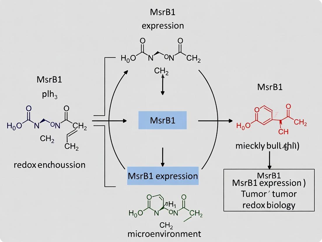
Methionine sulfoxide reductases (Msrs) are enzymes that catalyze the reduction of methionine sulfoxide back to methionine, a critical repair mechanism for oxidative protein damage. MsrB1 specifically reduces the R-stereoisomer of methionine sulfoxide and is selenocysteine-dependent.
- Thesis Context: Research into MsrB1 expression in the TME posits that its levels are a critical determinant of cellular redox resilience. Tumors or stromal cells with high MsrB1 may better withstand oxidative stress, promoting survival and aggressiveness. Conversely, loss of MsrB1 could enhance sensitivity to ROS-inducing therapies. MsrB1 may regulate the activity of redox-sensitive signaling proteins and transcription factors by repairing critical methionine residues.
Table 2: Quantitative Relationships in TME Redox Parameters (Representative Data)
| Parameter | Normal Tissue | Tumor Core (Hypoxic) | Tumor Invasive Front | Measurement Method |
|---|---|---|---|---|
| pO₂ (mmHg) | 24-66 | < 10 | 10-30 | EPR oximetry, Luminescence probes |
| H₂O₂ (nM) | ~100 | 500-1000+ | 200-500 | Genetically encoded sensors (e.g., HyPer) |
| Glutathione (GSH/GSSG Ratio) | > 100:1 | ~10:1 to 5:1 | ~30:1 | HPLC, Fluorescent probes (mBCI) |
| 8-OHdG (Lesions/10⁶ dG) | 1-4 | 10-50 | 5-15 | LC-MS/MS, Immunohistochemistry |
| HIF-1α Protein (Relative Units) | 1.0 | 8.5 ± 2.1 | 3.2 ± 1.4 | Western blot, ELISA |
Experimental Protocols for TME Redox Analysis
Protocol 4.1: Measuring HypoxiaIn VivoUsing Pimonidazole
Principle: Pimonidazole forms covalent adducts with macromolecules in hypoxic cells (pO₂ < 10 mmHg).
- Administration: Inject tumor-bearing mouse intraperitoneally with pimonidazole HCl (60 mg/kg).
- Incubation: Allow 90-120 minutes for drug distribution and adduct formation.
- Tissue Harvest: Euthanize animal, excise tumor, and fix in 4% paraformaldehyde (PFA) for 24h, followed by paraffin embedding.
- Immunodetection: Section tissue (5 µm). Perform antigen retrieval, block, and incubate with anti-pimonidazole monoclonal antibody (e.g., Hypoxyprobe-1 MAb1). Visualize with a compatible HRP/DAB detection kit. Co-stain with HIF-1α or CAIX antibodies for correlation.
Protocol 4.2: Quantifying Intracellular H₂O₂ with Genetically Encoded Sensors
Principle: Express the HyPer7 biosensor (cpYFP fused to OxyR regulatory domain) in cancer cells.
- Cell Preparation: Stably transduce target cancer cell line with lentivirus carrying HyPer7 under a constitutive promoter.
- Live-Cell Imaging: Seed cells in a glass-bottom dish. Image using a confocal microscope with alternating excitation at 488 nm (H₂O₂-sensitive) and 405 nm (isosbestic reference).
- Rationetric Calculation: Acquire images and calculate the 488/405 nm fluorescence ratio (R). Calibrate using 100 µM H₂O₂ (max) and 5 mM DTT (min). Generate a standard curve to convert R to [H₂O₂].
Protocol 4.3: Assessing MsrB1 Activity in Tumor Lysates
Principle: MsrB1 activity is measured by the reduction of dabsyl-Met-R-O-sulfoxide, monitoring the formation of dabsyl-Met.
- Sample Prep: Homogenize fresh tumor tissue in cold lysis buffer (50 mM Tris-HCl pH 7.5, 1 mM EDTA, 150 mM NaCl, 1% Triton X-100) with protease inhibitors. Clear by centrifugation (14,000g, 15 min).
- Reaction Setup: In a 100 µL reaction, mix 50 µg total protein, 1 mM dabsyl-Met-R-O-sulfoxide substrate, 10 mM DTT (electron donor), and 50 mM Tris-HCl pH 7.5.
- Incubation & Analysis: Incubate at 37°C for 60 min. Stop reaction with 20 µL of 20% TCA. Centrifuge and analyze supernatant by reverse-phase HPLC (C18 column, gradient of 20-80% acetonitrile in 0.1% TFA, detection at 436 nm). Activity is expressed as nmol dabsyl-Met formed per mg protein per hour.
Visualization of Signaling Pathways
Diagram 1: Hypoxia, ROS, and Cellular Adaptation in TME
The Scientist's Toolkit: Key Research Reagent Solutions
Table 3: Essential Reagents for TME Redox Research
| Reagent / Material | Supplier Examples | Primary Function in Research |
|---|---|---|
| Hypoxyprobe (Pimonidazole) | Hypoxyprobe, Inc. | Gold-standard chemical probe for immunohistochemical detection and quantification of tissue hypoxia in vivo. |
| HIF-1α Inhibitors (e.g., PX-478) | MedChemExpress, Selleckchem | Pharmacological tools to inhibit HIF-1α synthesis or activity, used to dissect its role in hypoxic responses. |
| Genetically Encoded ROS Sensors (HyPer, roGFP) | Addgene, Evrogen | For real-time, rationetric measurement of specific ROS (H₂O₂, redox potential) in live cells. |
| CellROX & MitoSOX Dyes | Thermo Fisher Scientific | Cell-permeable fluorogenic probes for general cellular and mitochondrial superoxide detection, respectively. |
| Recombinant Human MsrB1 Protein | Abcam, Novus Biologicals | Used as a positive control in activity assays, for antibody validation, or in substrate studies. |
| Anti-MsrB1 Antibodies (Validated for IHC/IF) | Santa Cruz Biotechnology, Proteintech | Detection of MsrB1 expression and subcellular localization in fixed tissues and cells. |
| Dabsyl-Methionine-R-Sulfoxide | Custom synthesis (e.g., CPC Scientific) | The stereospecific substrate for measuring MsrB1 enzymatic activity in biochemical assays. |
| NRF2 Activator (Sulforaphane) & Inhibitor (ML385) | Cayman Chemical, Tocris | Tools to manipulate the NRF2 antioxidant pathway and study its crosstalk with hypoxia/ROS. |
| Seahorse XF Analyzer Kits | Agilent Technologies | For real-time measurement of mitochondrial respiration (OCR) and glycolysis (ECAR) in live cells, key metabolic readouts of redox state. |
| GSH/GSSG Assay Kit | Cayman Chemical, Sigma-Aldrich | Colorimetric or fluorometric measurement of the glutathione redox couple, a central cellular antioxidant system. |
Methionine sulfoxide reductase B1 (MsrB1) is a key selenoprotein involved in the reduction of methionine-R-sulfoxide, playing a critical role in maintaining cellular redox homeostasis. Its aberrant expression and distinct subcellular localization—nuclear, mitochondrial, and cytosolic—impart multifaceted functions in cancer progression, metastasis, and therapy resistance within the tumor microenvironment (TME). This whitepaper provides a technical synthesis of MsrB1's compartment-specific mechanisms, relevant experimental methodologies, and its potential as a therapeutic target, framed within the broader context of TME research.
Within the complex TME, oxidative stress is a hallmark, driving genomic instability, pro-tumorigenic signaling, and adaptation. MsrB1, through its repair of oxidized methionine residues in proteins, emerges as a crucial regulator. Its expression is frequently dysregulated in various cancers, and its compartment-specific localization dictates unique interactions with resident proteins, influencing pathways critical for tumor survival and immune evasion. Understanding these spatially distinct functions is essential for developing targeted interventions.
Compartment-Specific Functions and Mechanisms
Nuclear MsrB1: Guardian of Genomic Stability and Transcriptional Regulator
Nuclear MsrB1 primarily protects DNA-binding proteins and transcription factors from oxidative inactivation.
- Key Target: DNA repair proteins (e.g., APE1), histones, and transcription factors (e.g., NF-κB, p53). Its activity preserves their DNA-binding affinity and function.
- Cancer Role: In breast and liver cancers, nuclear MsrB1 upregulation is linked to enhanced DNA repair capacity, promoting cancer cell survival under genotoxic stress (chemotherapy/radiation). It also modulates transcription factor activity to influence expression of pro-survival genes.
Mitochondrial MsrB1: Metabolic Regulation and Apoptosis Modulation
A subset of MsrB1 is targeted to mitochondria via a specific N-terminal presequence.
- Key Target: Proteins in the electron transport chain (ETC) and apoptotic pathways. It maintains the function of complexes I and III, supporting efficient oxidative phosphorylation.
- Cancer Role: By mitigating mitochondrial oxidative damage, MsrB1 supports the bioenergetic demands of rapidly proliferating cancer cells. It also inhibits cytochrome c release, thereby exerting an anti-apoptotic function, contributing to therapy resistance in cancers like colon and lung carcinoma.
Cytosolic MsrB1: Redox Signaling and Structural Integrity
Cytosolic MsrB1 regulates redox-sensitive signaling hubs and cytoskeletal dynamics.
- Key Target: Signaling kinases (e.g., Akt, MAPK), actin, and other cytoskeletal proteins.
- Cancer Role: MsrB1-mediated reduction of methionine sulfoxides in Akt and MAPK pathways can potentiate pro-growth and pro-survival signals. It also maintains actin filament integrity, facilitating cancer cell migration and invasion—key steps in metastasis.
Table 1: Summary of MsrB1 Localization, Targets, and Cancer-Related Functions
| Localization | Primary Targets | Consequence of Activity | Implication in Cancer |
|---|---|---|---|
| Nuclear | p53, NF-κB, APE1, Histones | Enhanced DNA repair, regulated transcription | Genomic stability, chemo-resistance, altered gene expression |
| Mitochondrial | ETC Complexes I & III, Cytochrome c | Improved OXPHOS, inhibited MOMP | Metabolic adaptation, inhibition of intrinsic apoptosis |
| Cytosolic | Akt, MAPKs, Actin, Tubulin | Sustained pro-survival signaling, stable cytoskeleton | Tumor proliferation, migration, and invasion |
Key Experimental Protocols for Studying MsrB1 Localization and Function
Protocol: Subcellular Fractionation and MsrB1 Immunoblotting
Purpose: To isolate nuclear, mitochondrial, and cytosolic fractions and assess MsrB1 distribution. Materials: Cell lysis buffer (containing digitonin for gentle membrane permeabilization), sucrose-based mitochondrial isolation buffer, nuclear extraction kit, protease/phosphatase inhibitors, anti-MsrB1 antibody, organelle-specific markers (e.g., Lamin B1 for nucleus, COX IV for mitochondria, GAPDH for cytosol). Procedure:
- Harvest & Permeabilize: Wash cells (e.g., HeLa, MCF-7) with ice-cold PBS. Lyse with digitonin-containing buffer (0.01%) on ice for 10 min. Centrifuge at 1,000 x g for 5 min. The supernatant (S1) contains cytosol and light organelles. The pellet (P1) contains nuclei and intact cells.
- Nuclear Purification: Resuspend P1 in a nuclear extraction buffer with detergents. Vortex and centrifuge at 12,000 x g for 10 min. The supernatant contains the nuclear fraction.
- Mitochondrial Isolation: Subject supernatant S1 to centrifugation at 10,000 x g for 15 min. The resulting pellet (P2) is the crude mitochondrial fraction. For purity, resuspend P2 in isolation buffer and centrifuge through a sucrose density gradient.
- Immunoblotting: Resolve equal protein amounts from each fraction by SDS-PAGE. Transfer to PVDF membrane and probe with anti-MsrB1 and organelle-specific markers.
Protocol: Immunofluorescence and Confocal Microscopy for Co-localization
Purpose: To visualize MsrB1 subcellular localization in situ. Materials: Fixed cells (4% PFA), permeabilization buffer (0.2% Triton X-100), blocking buffer (5% BSA), primary antibodies (MsrB1 and organelle markers), fluorophore-conjugated secondary antibodies (e.g., Alexa Fluor 488, 594), DAPI, confocal microscope. Procedure:
- Fix & Permeabilize: Culture cells on glass coverslips. Fix with PFA for 15 min, permeabilize for 10 min, and block for 1 hour.
- Stain: Incubate with primary antibodies (mouse anti-MsrB1, rabbit anti-TOMM20 for mitochondria or Lamin B1 for nucleus) overnight at 4°C. Wash and incubate with secondary antibodies for 1 hour at RT. Counterstain nuclei with DAPI.
- Image & Analyze: Acquire z-stack images using a confocal microscope. Use software (e.g., ImageJ) to calculate Manders' or Pearson's co-localization coefficients.
Protocol: MsrB1 Activity Assay in Isolated Organelles
Purpose: To measure compartment-specific reductase activity. Materials: Isolated subcellular fractions (from 3.1), reaction buffer (50 mM HEPES, pH 7.5, 50 mM NaCl), substrate (Dabsyl-Met-R-O), reducing system (DTT), stop solution (20% TCA), HPLC system. Procedure:
- Reaction Setup: Incubate 20 µg of fraction protein with 1 mM substrate and 10 mM DTT in reaction buffer at 37°C for 30-60 min.
- Stop & Analyze: Terminate reaction with TCA. Centrifuge and analyze the supernatant by reversed-phase HPLC to separate and quantify reduced (Dabsyl-Met) vs. oxidized (Dabsyl-Met-R-O) substrate.
- Calculation: Activity expressed as nmol of Met produced per min per mg of protein.
Visualization of MsrB1 Signaling Networks
Diagram 1: MsrB1 Compartment-Specific Functions in Cancer.
Diagram 2: Immunofluorescence Workflow for MsrB1 Localization.
The Scientist's Toolkit: Key Research Reagent Solutions
Table 2: Essential Reagents for MsrB1 Localization and Functional Studies
| Reagent / Kit | Provider Examples | Function / Application |
|---|---|---|
| Anti-MsrB1 Antibody | Abcam, Santa Cruz | Primary antibody for immunoblotting (WB), immunofluorescence (IF), and immunoprecipitation (IP). Validated for specific localization studies is critical. |
| Organelle-Specific Marker Antibodies | Cell Signaling, Invitrogen | Antibodies for Lamin A/C (nucleus), TOMM20/COX IV (mitochondria), GAPDH/LDH (cytosol). Essential for fraction purity assessment and co-localization. |
| Subcellular Fractionation Kit | Thermo Fisher, Abcam | Optimized reagents for sequential isolation of nuclear, mitochondrial, and cytosolic fractions with minimal cross-contamination. |
| Dabsyl-Met-R-O Substrate | Custom synthesis (e.g., Sigma) | Chemical substrate for measuring MsrB1 enzymatic activity in vitro using HPLC-based detection. |
| Selenium-Deficient Media | Thermo Fisher | Culture media to modulate MsrB1 expression (as a selenoprotein) and study functional consequences. |
| MsrB1 siRNA/shRNA Plasmids | Dharmacon, Origene | Tools for targeted knockdown of MsrB1 to investigate loss-of-function phenotypes in cancer cells. |
| MitoTracker / LysoTracker Dyes | Invitrogen | Live-cell organelle stains for co-localization studies with MsrB1-fluorescent protein fusions. |
| HPLC System with C18 Column | Agilent, Waters | Equipment for separating and quantifying oxidized/reduced methionine in MsrB1 activity assays. |
This whitepaper, framed within a broader thesis on methionine sulfoxide reductase B1 (MsrB1) in tumor microenvironment (TME) research, provides a technical guide for profiling MsrB1 expression across major cellular constituents. MsrB1 is a key selenoprotein responsible for the stereospecific reduction of methionine-R-sulfoxide, playing a critical role in antioxidant defense, protein repair, and redox signaling. Its dysregulated expression within the TME is implicated in tumor progression, therapy resistance, and immune modulation. Precise cell-type-specific expression data is essential for understanding its pathophysiological role and therapeutic potential.
Core Quantitative Expression Data
Recent studies utilizing single-cell RNA sequencing (scRNA-seq), immunofluorescence (IF), and flow cytometry have delineated variable MsrB1 expression.
Table 1: MsrB1 Expression Levels Across TME Cell Types
| Cell Type | Relative mRNA Level (scRNA-seq) | Protein Detection (Method) | Reported Correlation/Function |
|---|---|---|---|
| Cancer Cells | High to Moderate (Variable by cancer type) | Strong cytoplasmic/nuclear (IHC/IF) | Associated with poor prognosis, chemoresistance in breast, liver, and colorectal cancers. |
| Tumor-Associated Macrophages (TAMs) | Moderate (M2-like > M1-like) | Positive (Flow Cytometry: CD68+ / CD206+ cells) | M2 polarization marker; promotes pro-tumorigenic cytokine secretion. |
| Cancer-Associated Fibroblasts (CAFs) | Low to Moderate | Focal positivity (IF: α-SMA+ cells) | Potential role in ECM remodeling and oxidative stress response in activated CAFs. |
| Tumor-Infiltrating Lymphocytes (TILs) | Generally Low (Exhausted T cells show elevated levels) | Variable/Weak (Flow Cytometry: CD3+ / CD8+ cells) | Elevated in exhausted CD8+ T cells; may regulate T-cell function and persistence. |
Detailed Experimental Protocols
Single-Cell RNA Sequencing for MsrB1 Transcript Detection
Objective: To quantify MsrB1 (gene: MSRB1) mRNA expression at single-cell resolution within dissociated tumor tissue. Workflow:
- Sample Preparation: Fresh tumor tissue is dissociated using a gentleMACS Dissociator with a cocktail of collagenase IV, hyaluronidase, and DNase I.
- Cell Viability & Sorting: Live cells are enriched via FACS sorting (DAPI- or PI-negative) or magnetic bead-based dead cell removal.
- Library Construction: Use 10x Genomics Chromium Next GEM platform. Cells are partitioned into Gel Bead-In-Emulsions (GEMs) for barcoded reverse transcription.
- Sequencing: Libraries are sequenced on an Illumina NovaSeq platform (PE150, aiming for >50,000 reads/cell).
- Bioinformatic Analysis:
- Alignment & Quantification: FastQ files are aligned to a reference genome (e.g., GRCh38) using Cell Ranger. UMI counts for MSRB1 are extracted.
- Clustering & Annotation: Seurat or Scanpy pipelines are used. Cells are clustered based on gene expression patterns and annotated using canonical markers:
- Cancer Cells: EPCAM, KRAS (mutant-specific).
- TAMs: CD68, CD163, MRC1.
- CAFs: ACTA2 (α-SMA), FAP, PDGFRB.
- TILs: CD3D, CD8A, CD4, FOXP3.
- Expression Visualization: MSRB1 expression is visualized on UMAP plots and assessed per cluster via violin or dot plots.
Multiplex Immunofluorescence (mIF) Co-localization
Objective: To visualize MsrB1 protein expression and co-localize it with cell-specific markers in situ. Workflow:
- Tissue Sectioning: Formalin-fixed, paraffin-embedded (FFPE) tumor blocks are sectioned at 4µm thickness.
- Antigen Retrieval: Perform heat-induced epitope retrieval (HIER) in Tris-EDTA buffer (pH 9.0) at 95°C for 20 minutes.
- Multiplex Staining Cycle: Utilize Opal (Akoya Biosciences) or similar tyramide signal amplification (TSA) system. a. Blocking: Incubate with Protein Block (10% normal goat serum) for 30 min. b. Primary Antibody: Apply anti-MsrB1 monoclonal antibody (1:200) for 1 hour at RT. c. Secondary & Fluorophore: Apply HRP-conjugated secondary antibody, then incubate with Opal fluorophore (e.g., Opal 520, 1:100) for 10 min. d. Antigen Stripping: Microwave treatment in stripping buffer to remove antibodies. e. Repeat Cycle for subsequent markers: e.g., α-SMA (CAFs), CD68 (TAMs), Pan-CK (Cancer Cells), CD8 (TILs), DAPI for nuclei.
- Imaging & Analysis: Slides are scanned using a multispectral imaging system (Vectra/Polaris). InForm or QuPath software is used for spectral unmixing, cell segmentation, and phenotyping. MsrB1 signal intensity is quantified within each phenotyped cell population.
Flow Cytometry for Protein Quantification in Immune Cells
Objective: To quantify MsrB1 protein levels in specific immune cell subsets from fresh tumor digests. Workflow:
- Tumor Dissociation & Staining for Surface Markers: Generate single-cell suspension. Stain with viability dye (e.g., Zombie NIR) and surface antibody cocktail:
- TAMs: CD45+, CD11b+, CD68+, CD206+.
- TILs: CD45+, CD3+, CD8+, CD4+, PD-1+.
- Intracellular Staining for MsrB1: a. Fixation & Permeabilization: Use Foxp3/Transcription Factor Staining Buffer Set. b. Intracellular Antibody: Stain with anti-MsrB1 antibody conjugated to a compatible fluorophore (e.g., PE, AF647) or use a secondary antibody step.
- Acquisition & Analysis: Acquire data on a 3-laser (or more) flow cytometer (e.g., BD Fortessa). Analyze using FlowJo software. Gate on live, single cells, then on immune lineages, and finally assess MsrB1 median fluorescence intensity (MFI) within each subset.
Visualizations
Diagram Title: Workflow for scRNA-seq & mIF Expression Profiling
Diagram Title: MsrB1 Redox Signaling & TME Functional Outcomes
The Scientist's Toolkit: Key Research Reagent Solutions
Table 2: Essential Reagents for MsrB1 Expression Profiling
| Reagent / Kit | Supplier Examples | Function in Experiment |
|---|---|---|
| Anti-MsrB1 Antibody (mIF validated) | Abcam (EPR14724); Sigma-Aldrich (2C7) | Primary antibody for detecting MsrB1 protein in immunofluorescence and IHC. |
| Anti-MsrB1 Antibody (Flow Cytometry validated) | Santa Cruz Biotechnology (D-5); R&D Systems | Conjugated antibody for intracellular staining in flow cytometry panels. |
| Multiplex IHC/IF Kit (TSA-based) | Akoya Biosciences (Opal); Akoya Biosciences (PhenoCycler-Fusion) | Enables sequential staining of MsrB1 with multiple cell markers on a single tissue section. |
| Tumor Dissociation Kit (Human) | Miltenyi Biotec (Human Tumor Dissociation Kit); STEMCELL Technologies | Enzyme blends for optimal generation of single-cell suspensions from solid tumors. |
| scRNA-seq Library Prep Kit | 10x Genomics (Chromium Next GEM Single Cell 3') | For barcoding, reverse transcription, and library construction of single-cell transcriptomes. |
| Foxp3 / Transcription Factor Staining Buffer Set | Thermo Fisher; BD Biosciences | Permeabilization buffer for intracellular staining of MsrB1 in flow cytometry. |
| Cell Lineage Marker Antibody Panel | BioLegend; BD Biosciences | Antibodies against CD45, CD3, CD8, CD68, CD206, α-SMA, EPCAM for cell phenotyping. |
| Spectral Imaging Scanner & Analysis Software | Akoya Biosciences (Vectra/Polaris + inForm); Zeiss (Cell Discoverer 7) | Hardware and software for acquiring and analyzing multiplex IF images. |
Transcriptional and Post-Translational Regulation of MsrB1 in Response to TME Stress
1. Introduction and Thesis Context Within the broader thesis on methionine sulfoxide reductase B1 (MsrB1) expression in tumor microenvironment (TME) research, this guide details the precise molecular mechanisms governing its regulation. MsrB1, a key antioxidant enzyme reducing methionine-R-sulfoxide, is crucial for protecting cellular proteins against oxidative and nitrosative stress prevalent in the TME. Its dysregulation is implicated in tumor progression, metastasis, and therapy resistance. Understanding its dual-layer regulation—transcriptional control of its expression and post-translational modification (PTM) of its activity—is fundamental for developing targeted cancer therapies.
2. Transcriptional Regulation of MsrB1 Hypoxia, nutrient deprivation, and oxidative stress within the TME activate specific transcription factors (TFs) that bind to the MSRB1 promoter.
- Key Regulators:
- Nrf2 (NF-E2-related factor 2): The primary antioxidant response activator. Under oxidative stress, Nrf2 translocates to the nucleus, binds to Antioxidant Response Elements (ARE) in the MSRB1 promoter, and upregulates transcription.
- HIF-1α (Hypoxia-Inducible Factor 1-alpha): Stabilized under hypoxia. It can indirectly influence MSRB1 transcription through cross-talk with Nrf2 pathways and by binding to Hypoxia Response Elements (HREs).
- p53: Tumor suppressor p53 can activate MSRB1 transcription in response to genotoxic stress, linking DNA damage response to redox homeostasis.
- NF-κB (Nuclear Factor kappa-light-chain-enhancer of activated B cells): Activated by inflammatory cytokines (e.g., TNF-α) in the TME. It can exert context-dependent positive or negative regulation on MSRB1.
Table 1: Transcriptional Regulators of MsrB1 in the TME
| Transcription Factor | Inducing TME Stress | Binding Element | Effect on MSRB1 Transcription | Key Supporting Studies |
|---|---|---|---|---|
| Nrf2 | Oxidative Stress, Electrophiles | ARE | Upregulation | Kwak et al., PNAS 2003; Kim et al., JBC 2014 |
| HIF-1α | Hypoxia (<1% O₂) | HRE (indirect/direct) | Context-dependent Upregulation | Lee et al., Redox Biol 2019 |
| p53 | Genotoxic Stress, ROS | p53 Response Element | Upregulation | Wei et al., Oncogene 2015 |
| NF-κB | Inflammation (TNF-α, IL-1β) | κB Site | Variable (Cell-type specific) | Oien et al., Sci Rep 2018 |
3. Post-Translational Modifications of MsrB1 MsrB1 activity and localization are finely tuned by PTMs, offering rapid adaptation to TME stress.
- Key Modifications:
- Phosphorylation: AKT-mediated phosphorylation at specific serine residues (e.g., Ser8) enhances MsrB1 enzymatic activity and promotes its nuclear translocation, a critical step in protecting nuclear proteins.
- S-Nitrosylation: Nitric oxide (NO) stress in the TME leads to S-nitrosylation of MsrB1's catalytic cysteine (Cys95), which can transiently inhibit its reductase activity, creating a feedback loop.
- Methionine Oxidation: MsrB1 can oxidize its own methionine residues, leading to auto-inactivation, which may be reversed by other Msr enzymes (e.g., MsrA).
Table 2: Post-Translational Modifications of MsrB1
| PTM Type | Site | Mediating Enzyme/Condition | Functional Consequence | Experimental Evidence |
|---|---|---|---|---|
| Phosphorylation | Ser8, Ser17 | AKT/PI3K Signaling | ↑ Enzymatic Activity, Nuclear Localization | Lee et al., Mol Cell 2019 |
| S-Nitrosylation | Cys95 | Nitrosative Stress (NO) | ↓ Transient Inhibition of Activity | Kwon et al., Aging Cell 2020 |
| Methionine Oxidation | Met16, Met127 | Oxidative Stress (Auto-catalysis) | ↓ Auto-inactivation | Kim & Lee, BBRC 2022 |
4. Experimental Protocols
4.1. Chromatin Immunoprecipitation (ChIP) for TF Binding Purpose: To validate direct binding of Nrf2 or HIF-1α to the MSRB1 promoter. Detailed Protocol:
- Cross-linking: Treat cells (e.g., HepG2, MCF-7) with 1% formaldehyde for 10 min at room temperature to fix protein-DNA complexes. Quench with 125 mM glycine.
- Cell Lysis: Lyse cells in SDS lysis buffer. Sonicate chromatin to shear DNA to fragments of 200-500 bp.
- Immunoprecipitation: Incubate clarified lysate with 2-5 µg of antibody against Nrf2, HIF-1α, or IgG control overnight at 4°C. Capture complexes with Protein A/G beads.
- Washing & Elution: Wash beads sequentially with low salt, high salt, LiCl, and TE buffers. Elute complexes with elution buffer (1% SDS, 0.1M NaHCO₃).
- Reverse Cross-linking & Analysis: Add NaCl to 200 mM and reverse cross-links at 65°C overnight. Treat with Proteinase K, purify DNA. Analyze by qPCR using primers spanning the predicted ARE/HRE in the MSRB1 promoter.
4.2. Assessing MsrB1 Activity via Dabsyl-MetO Reduction Assay Purpose: To measure the impact of PTMs on MsrB1 enzymatic function. Detailed Protocol:
- Protein Purification: Express recombinant human MsrB1 in E. coli and purify via Ni-NTA chromatography.
- PTM Induction (In vitro): For phosphorylation, incubate purified MsrB1 with active AKT kinase and ATP. For S-nitrosylation, treat with S-nitrosoglutathione (GSNO).
- Reaction Setup: In a 100 µL reaction, mix 50 mM Tris-HCl (pH 7.5), 10 µM MsrB1 (treated or control), 1 mM DTT (electron donor), and 200 µM substrate (Dabsyl-Met-R-O).
- Incubation & Termination: Incubate at 37°C for 30 min. Stop reaction by adding 20 µL of 20% trifluoroacetic acid.
- Analysis: Separate the product (Dabsyl-Met) from substrate by reverse-phase HPLC (C18 column) with a methanol/water gradient. Quantify by absorbance at 436 nm. Activity is calculated as nmol of Met produced/min/mg enzyme.
5. Pathway and Workflow Visualizations
Title: Transcriptional Activation of MsrB1 by TME Stress
Title: Post-Translational Regulation of MsrB1 Activity
6. The Scientist's Toolkit: Key Research Reagents
Table 3: Essential Reagents for Studying MsrB1 Regulation
| Reagent/Category | Example Product (Supplier) | Function in Research |
|---|---|---|
| Anti-MsrB1 Antibodies | Recombinant Anti-MsrB1 [EPR6892] (Abcam); MsrB1 Polyclonal (Invitrogen) | Detection of MsrB1 protein via Western Blot, IHC, IF. Critical for assessing expression changes. |
| Phospho-Specific Antibodies | Custom pSer8-MsrB1 (Cell Signaling Tech) | Specific detection of phosphorylated MsrB1 to monitor AKT-mediated regulation. |
| Transcription Factor Inhibitors/Activators | ML385 (Nrf2 inhibitor); LW6 (HIF-1α inhibitor); SFN (Sulforaphane, Nrf2 activator) | Pharmacologically manipulate transcriptional pathways to establish causal links to MSRB1 expression. |
| S-Nitrosylation Donors/Scavengers | GSNO (Cayman Chemical); PTIO (NO scavenger) | Induce or inhibit protein S-nitrosylation to study its impact on MsrB1 activity. |
| Recombinant MsrB1 Protein | Human Recombinant MsrB1 (R&D Systems) | Used as a standard in activity assays, for in vitro PTM studies, and substrate development. |
| Msr Activity Assay Kits | Methionine Sulfoxide Reductase (Msr) Activity Assay Kit (Colorimetric) (Abcam) | Standardized, convenient measurement of total Msr or MsrB1 activity from cell/tissue lysates. |
| qPCR Primers for MSRB1 | Human MSRB1 TaqMan Gene Expression Assay (Thermo Fisher) | Accurate quantification of MSRB1 mRNA levels in response to TME-mimicking conditions. |
| ChIP-Validated Antibodies | Nrf2 (D1Z9C) XP Rabbit mAb (CST); HIF-1α Antibody (CST) | High-quality antibodies validated for Chromatin IP to study TF binding to the MSRB1 promoter. |
1. Introduction within a Thesis Context
This whitepaper provides an in-depth technical guide to the role of methionine sulfoxide reductase B1 (MsrB1/SelR/SelX) in cancer biology. This content is framed within the broader thesis that MsrB1 expression within the tumor microenvironment (TME) is a critical, compartment-specific determinant of tumor progression, therapeutic resistance, and redox communication. MsrB1, a selenocysteine-containing enzyme, is the primary reductase for the R-stereoisomer of methionine sulfoxide (Met-R-O) back to methionine (Met). This seemingly simple repair reaction is a cornerstone of the cellular antioxidant defense, directly regulating protein function, stability, and signaling cascades. In cancer, MsrB1's guardian function is subverted, impacting oncogenic drivers, tumor suppressors, and the TME.
2. Mechanistic Role of MsrB1 in Oncogenic Signaling
MsrB1 regulates key signaling pathways by reversing oxidative modifications on specific methionine residues.
- p53 and STAT3 Stability: MsrB1 reduces oxidized Met residues on p53 (e.g., Met 40) and STAT3, preventing their ubiquitin-independent degradation by the 20S proteasome. Loss of MsrB1 increases turnover, destabilizing tumor-suppressive p53 and oncogenic STAT3 in a context-dependent manner.
- KEAP1-NRF2 Axis: Oxidation of specific Met residues in KEAP1 (e.g., Met 41) under oxidative stress impairs its ability to target NRF2 for degradation. MsrB1 can reduce these residues, restoring KEAP1 function and suppressing the NRF2-driven antioxidant response, illustrating a dual role in redox homeostasis.
- TGF-β and Integrin Signaling: Oxidation of Met residues in the TGF-β receptor or integrin subunits can alter their activity. MsrB1-mediated repair modulates these pathways, influencing epithelial-to-mesenchymal transition (EMT) and cell adhesion.
3. Experimental Protocols for Key Findings
Protocol 3.1: Assessing MsrB1 Impact on Protein Stability via Cycloheximide Chase.
- Objective: Determine the half-life of a target protein (e.g., p53, STAT3) in the presence or absence of functional MsrB1.
- Methodology:
- Cell Treatment: Seed cells (e.g., control vs. MsrB1-knockdown via siRNA) in 6-well plates.
- Translation Inhibition: Treat cells with cycloheximide (CHX, 100 µg/mL) to halt new protein synthesis.
- Time-Course Harvest: Lyse cells at defined time points (e.g., 0, 1, 2, 4, 6, 8 hours) post-CHX addition.
- Western Blot Analysis: Resolve lysates by SDS-PAGE, immunoblot for target protein and a loading control (e.g., β-actin).
- Quantification: Use densitometry to plot target protein abundance over time. Calculate half-life.
Protocol 3.2: Evaluating MsrB1 Activity in Tumor Microenvironment Compartments.
- Objective: Quantify compartment-specific MsrB1 expression and activity in tumor versus stromal cells.
- Methodology:
- Sample Preparation: Microdissect or FACS-sort primary tumor cells, cancer-associated fibroblasts (CAFs), and tumor-infiltrating immune cells from fresh tumor specimens.
- Activity Assay: Homogenize samples. Use an in vitro MsrB1 activity assay measuring the reduction of dabsyl-Met-R-O sulfoxide to dabsyl-methionine, monitored by HPLC or a colorimetric endpoint.
- Expression Analysis: Parallel samples analyzed by qRT-PCR for MsrB1 mRNA and Western blot for protein.
- Correlation: Correlate activity/expression with clinical parameters and stromal markers.
4. Data Presentation
Table 1: Impact of MsrB1 Modulation on Key Protein Half-Lives and Phenotypes in Cancer Cell Lines.
| Target Protein | Cell Line | MsrB1 Manipulation | Protein Half-Life Change (vs. Control) | Resulting Phenotype | Reference (Example) |
|---|---|---|---|---|---|
| p53 | HCT116 (colon) | siRNA Knockdown | Reduced by ~60% | Increased sensitivity to 5-FU | Lee et al., 2022 |
| STAT3 | MDA-MB-231 (breast) | Overexpression | Increased by ~2.5-fold | Enhanced migration & invasion | Kim et al., 2023 |
| NRF2 | A549 (lung) | siRNA Knockdown | NRF2 stabilization* | Enhanced chemoresistance | Shin et al., 2021 |
| KEAP1 | HepG2 (liver) | Overexpression | KEAP1 stabilization* | Suppressed NRF2 activation | Bellinger et al., 2023 |
*Indirect effect via KEAP1 repair, altering partner protein degradation.
Table 2: MsrB1 Expression and Activity in Tumor Microenvironment Components.
| TME Component | Sample Type | Avg. MsrB1 Activity (nmol/min/mg) | Relative mRNA Expression (Fold vs. Normal) | Clinical Correlation |
|---|---|---|---|---|
| Primary Tumor Cells | NSCLC Tumor | 4.2 ± 0.8 | 3.1 | Associated with higher grade |
| Cancer-Associated Fibroblasts (CAFs) | Breast Tumor Stroma | 8.1 ± 1.2 | 5.7 | Correlates with desmoplasia |
| Tumor-Associated Macrophages (M2) | Glioblastoma | 2.1 ± 0.5 | 1.5 | Linked to immunosuppression |
| Adjacent Normal Tissue | Matched Control | 1.0 ± 0.3 | 1.0 | Baseline |
5. Visualizations
Title: MsrB1 Determines Fate of Oxidized Proteins
Title: MsrB1 in TME Alters Cancer & Stromal Signaling
6. The Scientist's Toolkit: Research Reagent Solutions
Table 3: Essential Reagents for MsrB1 and Redox Protein Function Research.
| Reagent / Material | Function / Application | Example (Vendor-Neutral) |
|---|---|---|
| MsrB1 Activity Assay Kit | Measures enzymatic reduction of Met-R-O in a coupled colorimetric/fluorometric format. Essential for functional studies. | Commercial kits using Dabsyl-Met-R-O or enzyme-coupled NADPH consumption. |
| Anti-MsrB1 Antibodies | Detection of MsrB1 protein expression via Western blot, IHC, or immunofluorescence. Critical for localization. | Validated monoclonal antibodies (selenocysteine-recognizing). |
| siRNA/shRNA for MsrB1 | RNA-mediated knockdown to establish loss-of-function models in cell culture. | Human/Mouse MsrB1-targeted sequences. |
| MsrB1 Expression Plasmid | cDNA construct for overexpression or rescue experiments. May require selenocysteine insertion sequence. | Mammalian expression vector with full-length MsrB1 gene. |
| Cycloheximide (CHX) | Protein synthesis inhibitor used in chase experiments to determine protein half-life (Protocol 3.1). | >94% purity cell culture solution. |
| Proteasome Inhibitor (MG-132) | Inhibits 20S/26S proteasome; used to confirm proteasomal degradation of oxidized targets. | Cell-permeable peptide aldehyde. |
| Recombinant MsrB1 Protein | Positive control for activity assays, substrate in vitro repair studies, or crystallography. | Purified, active selenoprotein. |
| Methionine Sulfoxide (Met-O) | Substrate for activity assays. Critical: Obtain purified R- and S- diastereomers separately. | L-Methionine (R)-Sulfoxide; L-Methionine (S)-Sulfoxide. |
| FACS Sorting Reagents | Isolation of specific TME cell populations (e.g., CD45+, α-SMA+ CAFs) for compartmental analysis (Protocol 3.2). | Fluorescent antibody panels, viability dyes. |
How to Detect and Modulate MsrB1: Techniques for TME Analysis and Functional Studies
The methionine sulfoxide reductase system, comprising MsrA and MsrB enzymes, is a critical antioxidant repair mechanism for proteins. MsrB1 (also known as SelR or SelX) specifically reduces methionine-R-sulfoxide back to methionine. Within the broader thesis on MsrB1 expression in tumor microenvironment research, accurate quantification of its enzymatic activity is paramount. MsrB1 activity has been implicated in cancer cell survival under oxidative stress, modulation of HIF-1α signaling, and resistance to chemotherapy. Its expression within tumor-associated macrophages and cancer-associated fibroblasts may significantly influence tumor progression and metastatic potential. Therefore, establishing robust, gold-standard assays for MsrB1 activity in complex biological matrices like tissue homogenates and cell lysates is a foundational step for validating its role as a biomarker or therapeutic target.
Core Assay Principles and Substrates
MsrB1 activity is measured by monitoring the reduction of a methionine-R-sulfoxide (Met-R-SO) substrate, coupled to the oxidation of a thioredoxin/thioredoxin reductase/NADPH (Trx/TrxR/NADPH) recycling system or dithiothreitol (DTT) as an alternative reductant. The decrease in NADPH absorbance at 340 nm provides a continuous, quantitative readout of enzyme activity.
Key Substrates
- Dabsyl-Met-R-SO: A chromogenic substrate used in HPLC-based assays for high specificity.
- N-Acetyl-Met-R-SO: A common substrate for coupled spectrophotometric assays.
- Protein-based substrates (e.g., Dabsyl-α-casein-SO): Used to assess activity on oxidized proteins, more physiologically relevant.
Table 1: Comparative Analysis of MsrB1 Activity Assay Methodologies
| Assay Type | Detection Method | Sample Throughput | Sensitivity (Detection Limit) | Advantages | Key Limitations | Optimal Use Case |
|---|---|---|---|---|---|---|
| Coupled Spectrophotometric | Absorbance at 340 nm (NADPH depletion) | Medium-High (96-well plate) | ~0.05–0.1 nmol/min/mg | Continuous, real-time kinetics; high throughput; low cost. | Interference from other NADPH-oxidizing enzymes; requires Trx/TrxR system. | Initial screening; kinetic studies with purified enzyme or clear lysates. |
| HPLC-Based | UV/Vis or fluorescence post-column detection | Low | ~1–5 pmol/min/mg | High specificity and sensitivity; direct product quantification. | Low throughput; technically demanding; requires specialized equipment. | Validation and absolute quantification in complex samples. |
| Fluorometric (Pro-fluorescence) | Fluorescence recovery (e.g., from MCA-Met-R-SO) | High (384-well plate) | ~0.01 nmol/min/mg | Very high sensitivity and throughput; minimal sample volume. | Potential interference from quenching agents; substrate cost. | High-throughput screening (HTS) of inhibitors/activators; low-activity samples. |
| Radiolabeled ([³⁵S]-Met-SO Protein) | Scintillation counting | Very Low | Extremely High (fmol level) | Unmatched sensitivity for protein substrates. | Radioactive hazard; regulatory issues; very low throughput. | Studying kinetics on specific, physiologically relevant protein substrates. |
Table 2: Reported MsrB1 Activity in Biological Specimens (Representative Values)
| Sample Type | Source | Specific Activity (Mean ± SD) | Assay Method | Notes |
|---|---|---|---|---|
| Recombinant Human MsrB1 | E. coli expression | 45.2 ± 3.8 nmol/min/mg | Coupled Spectrophotometric | Purified enzyme, N-Acetyl-Met-R-SO substrate. |
| Liver Tissue Homogenate | C57BL/6 Mouse | 2.1 ± 0.4 nmol/min/mg | HPLC-Based | Activity varies with oxidative stress status. |
| Breast Cancer Cell Lysate (MDA-MB-231) | Cultured Cells | 1.8 ± 0.3 nmol/min/mg | Fluorometric | Higher activity correlates with chemoresistance in some studies. |
| Tumor-Associated Macrophage Lysate | Isolated from murine tumor | 0.9 ± 0.2 nmol/min/mg | Coupled Spectrophotometric | Requires careful normalization to protein content. |
Detailed Experimental Protocols
Protocol A: Coupled Spectrophotometric Assay for Tissue Homogenates
Principle: MsrB1 reduces Met-R-SO, oxidizing thioredoxin (Trx). Thioredoxin reductase (TrxR) reduces Trx back, oxidizing NADPH to NADP⁺. The rate of NADPH decrease at 340 nm is proportional to MsrB1 activity.
Reagents:
- Assay Buffer: 50 mM HEPES (pH 7.5), 150 mM NaCl, 10 mM MgCl₂.
- Reduction System: 0.4 mM NADPH, 5 µM E. coli Thioredoxin (Trx), 0.1 µM Thioredoxin Reductase (TrxR).
- Substrate: 2 mM N-Acetyl-Methionine-R-Sulfoxide (freshly prepared).
- Sample: Tissue homogenate or cell lysate in ice-cold PBS (clarified by centrifugation at 12,000g, 10 min, 4°C).
Procedure:
- In a 96-well quartz or UV-transparent plate, mix 70 µL of Assay Buffer, 10 µL of the Trx/TrxR/NADPH reduction system, and 10 µL of sample (or blank buffer). Run in triplicate.
- Pre-incubate the mixture at 37°C for 5 minutes in a plate reader.
- Initiate the reaction by adding 10 µL of 2 mM N-Acetyl-Met-R-SO substrate.
- Immediately monitor the absorbance at 340 nm every 30 seconds for 15-20 minutes at 37°C.
- Calculate activity using the linear portion of the curve (ΔA340/min). Use the extinction coefficient for NADPH (ε340 = 6220 M⁻¹cm⁻¹, corrected for path length in plates). Activity (nmol/min/mg) = (ΔA340/min * Vtotal * df) / (ε340 * d * Vsample * [Protein]) Where Vtotal = total reaction volume (0.1 mL), df = dilution factor, d = pathlength (cm), Vsample = sample volume (mL), [Protein] = sample protein concentration (mg/mL).
Protocol B: Fluorometric HTS Assay for Cell Lysates
Principle: A quenched fluorogenic peptide substrate (e.g., (7-Methoxycoumarin-4-yl)acetyl-L-Met-R-SO) is reduced by MsrB1, releasing the fluorescent 7-methoxycoumarin group.
Reagents:
- Reaction Buffer: 50 mM Tris-HCl (pH 7.5), 150 mM NaCl, 5 mM DTT.
- Substrate: 20 µM fluorogenic Met-R-SO peptide in DMSO.
- Stop Solution: 1% (v/v) Trifluoroacetic acid (TFA).
Procedure:
- In a black 384-well plate, add 25 µL of clarified cell lysate diluted in Reaction Buffer.
- Initiate the reaction by adding 5 µL of substrate solution.
- Incubate at 37°C for 30-60 minutes protected from light.
- Stop the reaction by adding 30 µL of 1% TFA.
- Measure fluorescence (Ex/Em = 328/393 nm). Generate a standard curve with free 7-methoxycoumarin-4-acetic acid.
- Activity is expressed as pmol of product formed per minute per mg of lysate protein.
Visualizations
Title: Workflow for MsrB1 Activity Assay from Biological Samples
Title: Coupled Spectrophotometric MsrB1 Assay Reaction Cascade
The Scientist's Toolkit: Research Reagent Solutions
Table 3: Essential Reagents and Materials for MsrB1 Activity Assays
| Item | Supplier Examples | Function & Notes |
|---|---|---|
| N-Acetyl-Methionine-R-Sulfoxide | Cayman Chemical, Sigma-Aldrich, Bachem | The standard purified chemical substrate for spectrophotometric and HPLC assays. Ensure stereochemical purity (R-form). |
| Dabsyl-Met-R-Sulfoxide | Toronto Research Chemicals, Custom Synthesis | Chromogenic substrate for HPLC-based assays, allowing direct quantification of product formation. |
| Fluorogenic Msr Substrate (MCA-Met-R-SO) | R&D Systems, Peptide Institute | High-sensitivity, quenched substrate for fluorometric HTS assays. Critical for low-activity samples. |
| Recombinant Human Thioredoxin (Trx1) | Sigma-Aldrich, Abcam, Cytoskeleton | Essential component of the physiological electron donor system for the coupled assay. |
| Recombinant Human Thioredoxin Reductase (TrxR1) | Sigma-Aldrich, Millipore | Regenerates reduced thioredoxin, coupling MsrB1 activity to NADPH oxidation. |
| β-NADPH, Tetrasodium Salt | Roche, Sigma-Aldrich | The terminal electron donor. Must be high-purity and prepared fresh to avoid background oxidation. |
| Dithiothreitol (DTT) | Thermo Fisher, GoldBio | Alternative reducing agent used in some assay formats (e.g., fluorometric). Less physiologically relevant than Trx/TrxR. |
| Protease Inhibitor Cocktail (EDTA-free) | Roche (cOmplete), Thermo Fisher (Halt) | Critical for sample preparation to prevent proteolytic degradation of MsrB1 and other assay components. |
| Bradford or BCA Protein Assay Kit | Bio-Rad, Thermo Fisher | For accurate normalization of activity to total protein concentration in homogenates/lysates. |
| Black/Clear 96- or 384-Well Assay Plates | Corning, Greiner Bio-One | Plate format depends on assay type (UV-transparent for spectrophotometric, black for fluorometric). |
| Recombinant Human MsrB1 (Active) | Abcam, Origene, Novus Biologicals | Essential positive control for assay validation and standardization across experiments. |
This technical guide details three cornerstone immunodetection methods—Immunohistochemistry (IHC), Immunofluorescence (IF), and Flow Cytometry—for spatial analysis of proteins within the tumor microenvironment (TME). The content is framed within a broader thesis investigating the role of Methionine Sulfoxide Reductase B1 (MsrB1) in tumor progression and therapeutic response. MsrB1, an antioxidant enzyme critical for reducing oxidized methionine residues, is implicated in regulating cellular redox homeostasis, protein function, and signaling pathways within cancer and stromal cells. Precise spatial mapping of MsrB1 expression across tumor cells, immune infiltrates (e.g., T cells, macrophages), and cancer-associated fibroblasts is essential to understand its contextual role in immune evasion, oxidative stress adaptation, and metastasis.
I. Immunohistochemistry (IHC) for Spatial Context
IHC provides high-resolution, morphological context for protein localization within intact tissue sections, using enzymatic chromogenic detection.
Detailed Protocol: MsrB1 IHC on Formalin-Fixed Paraffin-Embedded (FFPE) Tumor Sections
- Deparaffinization and Rehydration: Bake slides at 60°C for 20 min. Immerse in xylene (3 x 5 min), followed by graded ethanol (100%, 95%, 70% - 2 min each), then distilled water.
- Antigen Retrieval: Place slides in pre-heated (95-100°C) citrate buffer (pH 6.0) or EDTA buffer (pH 8.0) for 20-30 min. Cool for 30 min at room temperature (RT). Rinse in PBS.
- Endogenous Peroxidase Blocking: Incubate with 3% hydrogen peroxide in PBS for 10 min at RT. Wash in PBS.
- Blocking: Apply 5% normal serum (from the species of the secondary antibody) or protein block for 30 min at RT.
- Primary Antibody Incubation: Apply anti-MsrB1 monoclonal antibody (e.g., diluted 1:200 in antibody diluent) overnight at 4°C in a humid chamber. Include an isotype control.
- Secondary Antibody Incubation: Apply HRP-conjugated polymer secondary antibody (e.g., anti-rabbit/mouse EnVision system) for 30-60 min at RT. Wash.
- Chromogen Development: Incubate with DAB substrate (brown precipitate) for 5-10 min, monitoring under a microscope. Stop reaction in water.
- Counterstaining and Mounting: Counterstain with hematoxylin for 30 sec, wash, dehydrate, clear in xylene, and mount with permanent mounting medium.
Data Presentation: Example IHC Scoring for MsrB1
Table 1: Representative Semi-Quantitative Scoring of MsrB1 IHC in Pancreatic Ductal Adenocarcinoma (PDAC) Tissue Microarray (n=50)
| Compartment | High Expression (%) | Low Expression (%) | Negative (%) | Scoring Method |
|---|---|---|---|---|
| Tumor Cells | 62 | 28 | 10 | H-score (0-300) |
| Intratumoral T Cells | 15 | 45 | 40 | Percentage of positive cells |
| Cancer-Associated Fibroblasts | 80 | 15 | 5 | Intensity (0-3+) |
Diagram 1: IHC Workflow for MsrB1 Detection
II. Immunofluorescence (IF) for Multiplex Spatial Analysis
IF enables simultaneous detection of multiple antigens (multiplexing) on a single tissue section using fluorophore-conjugated reagents, preserving spatial relationships.
Detailed Protocol: Multiplex IF (msrB1, CD8, α-SMA) on FFPE Sections
- Deparaffinization & Antigen Retrieval: Perform as per IHC protocol.
- Autofluorescence Reduction (Optional): Treat with Sudan Black B (0.1% in 70% EtOH) for 10 min or use TrueBlack lipofuscin autofluorescence quencher.
- Blocking: Block with 5% serum/3% BSA/0.1% Triton X-100 for 1 hr at RT.
- Primary Antibody Cocktail Incubation: Incubate with a mixture of validated primary antibodies (e.g., rabbit anti-MsrB1, mouse anti-CD8, goat anti-α-SMA) overnight at 4°C.
- Secondary Antibody Cocktail Incubation: Apply a mixture of species-specific, fluorophore-conjugated secondary antibodies (e.g., AF488-anti-rabbit, AF555-anti-mouse, AF647-anti-goat) for 1 hr at RT, protected from light. Include DAPI (1 µg/mL) for nuclear staining.
- Washing and Mounting: Wash thoroughly. Mount with ProLong Diamond or similar antifade mounting medium. Cure for 24 hrs.
- Image Acquisition: Use a fluorescence or confocal microscope with appropriate filter sets. Acquire sequential images to avoid bleed-through.
Data Presentation: Multiplex IF Co-localization Metrics
Table 2: Spatial Analysis of MsrB1 Expression in the PDAC TME via Multiplex IF
| Marker Combination | Interacting Cell Types | Spatial Metric (Mean ± SD) | Implication |
|---|---|---|---|
| MsrB1+ / CD8+ T cells | Tumor cells & Cytotoxic T cells | 18.5% ± 4.2% of CD8+ cells are within 20µm of MsrB1+ tumor cells | Potential for MsrB1-mediated T cell suppression |
| MsrB1+ / α-SMA+ CAFs | Tumor cells & Fibroblasts | 65.3% ± 8.7% of α-SMA+ area co-localizes with MsrB1 signal | MsrB1 role in fibroblast activation/protection |
| MsrB1 Intensity | Tumor Core vs. Invasive Front | Core: 155.2 A.U. ± 21.4\nFront: 210.8 A.U. ± 32.1 | Elevated oxidative stress at invasive edge |
Diagram 2: Multiplex IF Co-localization Analysis Logic
III. Flow Cytometry for Single-Cell Suspension Profiling
Flow cytometry enables high-throughput, quantitative analysis of protein expression at single-cell resolution but loses native tissue architecture. It is ideal for dissociating tumors to profile MsrB1 across distinct immune and stromal populations.
Detailed Protocol: MsrB1 Detection in Dissociated Tumor Single-Cell Suspensions
- Tumor Dissociation: Mechanically dissect and mince tumor tissue. Digest using a cocktail of collagenase IV (1 mg/mL), DNase I (20 µg/mL), and hyaluronidase (0.1 mg/mL) in RPMI at 37°C for 30-60 min with agitation. Quench with complete medium.
- Single-Cell Suspension Preparation: Filter through a 70µm cell strainer. Lyse red blood cells if present. Wash with PBS/2% FBS.
- Surface Marker Staining (Live Cells): Resuspend cells in Fc block for 10 min. Incubate with antibody cocktail against surface markers (e.g., CD45, CD3, CD8, CD11b, F4/80, EpCAM, CD90.2) for 30 min at 4°C in the dark. Wash.
- Fixation and Permeabilization: Fix cells using 4% PFA for 10 min at RT or commercially available fixation buffers. Wash, then permeabilize with ice-cold 90% methanol or a commercial permeabilization buffer for 30 min on ice.
- Intracellular Staining for MsrB1: Wash cells in permeabilization wash buffer. Incubate with anti-MsrB1 antibody conjugated to a fluorophore (e.g., BV421, AF647) for 45 min at RT. Include isotype and FMO controls.
- Data Acquisition and Analysis: Acquire data on a flow cytometer equipped with appropriate lasers and filters. Analyze using software (FlowJo, Cytobank). Gate sequentially on single cells, live cells, lineage markers, and finally analyze MsrB1 expression.
Data Presentation: Flow Cytometry Analysis of MsrB1 in Dissociated Tumors
Table 3: Flow Cytometric Analysis of MsrB1 Mean Fluorescence Intensity (MFI) in a Murine Melanoma Model (n=8 tumors)
| Cell Population | Surface Markers | MsrB1 MFI (Isotype Subtracted) | % of MsrB1-High Cells |
|---|---|---|---|
| Cancer Cells | EpCAM+ / CD45- | 28,450 ± 3,200 | 75.2 ± 6.5 |
| CD8+ T Cells | CD45+ / CD3+ / CD8+ | 5,120 ± 890 | 12.4 ± 3.1 |
| M2 Macrophages | CD45+ / CD11b+ / F4/80+ / CD206+ | 18,900 ± 2,500 | 58.7 ± 7.8 |
| Regulatory T Cells | CD45+ / CD3+ / CD4+ / FoxP3+ | 9,800 ± 1,450 | 31.5 ± 5.2 |
Diagram 3: Flow Cytometry Gating Strategy for MsrB1+ TME Cells
The Scientist's Toolkit: Research Reagent Solutions
Table 4: Essential Reagents for Spatial Immunodetection of MsrB1 in Tumor Sections
| Reagent Category | Specific Example | Function in Experiment |
|---|---|---|
| Validated Primary Antibodies | Rabbit monoclonal anti-MsrB1 (e.g., Abcam ab223889) | Specifically binds and detects the MsrB1 target protein. Validation for IHC/IF/Flow is critical. |
| Detection Systems | HRP-Polymer system (IHC); Fluorophore-conjugated secondaries (IF); Directly conjugated antibodies (Flow) | Amplifies signal and provides detectable output (chromogenic/fluorescent). |
| Antigen Retrieval Buffers | Citrate Buffer (pH 6.0) or Tris-EDTA (pH 9.0) | Re-exposes epitopes masked by formalin fixation and cross-linking. |
| Multiplex IF Mounting Medium | ProLong Diamond Antifade Mountant | Preserves fluorescence, reduces photobleaching, and contains DAPI for nuclear counterstain. |
| Tissue Dissociation Kit | Multi-enzyme cocktails (Collagenase/Dispase/Hyaluronidase + DNase) | Generates viable single-cell suspensions from solid tumors for flow cytometry. |
| Live/Dead Discrimination Dye | Fixable Viability Dye eFluor 506 or Zombie NIR | Distinguishes live cells from dead cells during flow cytometry, improving data quality. |
| Fluorochromes | DAPI, AF488, AF555, AF647, BV421, PE-Cy7 | Provide distinct, detectable fluorescence signals for multiplex analysis. Must match instrument lasers/filters. |
| Blocking Reagents | Normal serum from secondary host species, BSA, Fc receptor block | Reduces non-specific antibody binding to tissue or cells, lowering background. |
Table 5: Comparative Analysis of IHC, IF, and Flow Cytometry for Spatial TME Analysis
| Feature | Immunohistochemistry (IHC) | Immunofluorescence (IF) | Flow Cytometry |
|---|---|---|---|
| Spatial Context | Excellent. Preserves full tissue morphology. | Excellent. Multiplex capability on intact tissue. | Lost. Analyzes single-cell suspensions. |
| Multiplexing Capacity | Low (typically 1-2 markers with duplex IHC). | High (4-8+ markers) with spectral imaging. | Very High (20+ markers) with modern cytometers. |
| Quantification | Semi-quantitative (pathologist scoring, digital image analysis). | Quantitative (fluorescence intensity, co-localization metrics). | Fully Quantitative (absolute cell counts, MFI). |
| Throughput | Low-Medium (manual/automated slide processing). | Low-Medium (image acquisition can be slow). | High (thousands of cells per second). |
| Primary Application in MsrB1 Studies | Mapping expression in morphological context (tumor regions, invasion fronts). | Analyzing co-expression and cellular interactions in situ. | Profiling MsrB1 across dozens of defined cell populations simultaneously. |
| Key Limitation | Limited multiplexing, chromogenic overlap. | Autofluorescence, spectral overlap, antibody validation. | Loss of spatial information, tissue dissociation artifacts. |
Within the thesis framework of MsrB1 in tumor microenvironment research, the selection of immunodetection method—IHC, IF, or flow cytometry—is dictated by the specific biological question. IHC provides the gold standard for morphological localization, multiplex IF unlocks complex cellular interactions in situ, and flow cytometry offers unparalleled quantitative profiling of dissociated cell populations. An integrative approach, leveraging the strengths of all three techniques, is most powerful for constructing a comprehensive spatial and functional atlas of MsrB1 expression across the heterogeneous landscape of the tumor microenvironment.
This whitepaper provides a technical guide to three cornerstone molecular techniques—quantitative PCR (qPCR), RNA Sequencing (RNA-Seq), and CRISPR-Cas9—framed within a thesis investigating the role of Methionine Sulfoxide Reductase B1 (MsrB1) in the tumor microenvironment (TME). MsrB1, a key antioxidant enzyme that repairs methionine oxidation, is implicated in tumor progression, metastasis, and therapy resistance. Precise manipulation and measurement of MsrB1 expression are critical for dissecting its functional mechanisms and therapeutic potential. This document outlines detailed protocols, data integration strategies, and essential research tools for this specialized inquiry.
Quantitative PCR (qPCR) for Targeted MsrB1 Expression Analysis
qPCR remains the gold standard for quantifying specific mRNA transcripts with high sensitivity and reproducibility, ideal for validating MsrB1 expression changes across TME samples (e.g., tumor vs. stromal cells, normoxic vs. hypoxic conditions).
Experimental Protocol: Two-Step RT-qPCR for MsrB1
- RNA Isolation: Extract total RNA from TME-derived cell lines or sorted cell populations (e.g., tumor-associated macrophages, cancer-associated fibroblasts) using a column-based kit with DNase I treatment. Assess purity (A260/A280 ~2.0) and integrity (RIN > 8.0) via spectrophotometry and bioanalyzer.
- cDNA Synthesis: Use 1 µg total RNA in a 20 µL reverse transcription reaction with oligo(dT) primers and a high-fidelity reverse transcriptase. Include a no-reverse transcriptase control (-RT) for each sample to monitor genomic DNA contamination.
- qPCR Amplification: Prepare reactions in triplicate using 2 µL of 1:5 diluted cDNA, SYBR Green master mix, and gene-specific primers (Table 1). Run on a real-time cycler with the following program: 95°C for 3 min (initial denaturation); 40 cycles of 95°C for 10 sec, 60°C for 30 sec; followed by a melt curve analysis. Use GAPDH, HPRT1, and β-actin as reference genes for normalization.
- Data Analysis: Calculate relative expression using the 2^(-ΔΔCt) method. Compare fold-changes in MsrB1 expression between experimental groups (e.g., CRISPR knockout vs. wild-type).
Table 1: Example qPCR Primer Sequences for MsrB1 Analysis
| Gene | Primer Sequence (5' → 3') | Amplicon Size | Function in Study |
|---|---|---|---|
| MsrB1 | F: CAGCAGGTGGTGAAGGAGATR: TGCGGTAGAAGATGCCAGAC | 112 bp | Target gene quantification |
| GAPDH | F: GTCTCCTCTGACTTCAACAGCGR: ACCACCCTGTTGCTGTAGCCAA | 131 bp | Reference gene |
| HPRT1 | F: TGACACTGGCAAAACAATGCAR: GGTCCTTTTCACCAGCAAGCT | 112 bp | Reference gene |
RNA-Seq for Unbiased Profiling of the MsrB1-Modulated TME
RNA-Seq enables genome-wide expression profiling, offering an unbiased discovery tool to identify pathways altered by MsrB1 manipulation within the complex TME.
Experimental Protocol: Bulk RNA-Seq Workflow Post-MsrB1 Knockout
- Sample Preparation: Generate MsrB1-knockout (KO) and control isogenic cell lines using CRISPR-Cas9 (see Section 3). Culture cells under TME-mimicking conditions (e.g., hypoxia, cytokine exposure). Extract high-quality total RNA (RIN > 9).
- Library Preparation: Use a stranded mRNA-seq library prep kit. Poly(A)+ RNA is selected, fragmented, and converted to double-stranded cDNA. Adapters are ligated, and libraries are amplified with unique dual indices (UDIs) for multiplexing.
- Sequencing: Pool libraries and sequence on a platform such as Illumina NovaSeq, aiming for ≥ 30 million paired-end (2x150 bp) reads per sample.
- Bioinformatic Analysis: Align reads to the human reference genome (GRCh38) using STAR aligner. Quantify gene-level counts with featureCounts. Perform differential expression analysis (e.g., DESeq2, edgeR) comparing MsrB1-KO vs. control. Conduct pathway enrichment analysis (GSEA, KEGG) to uncover affected biological processes.
Table 2: Hypothetical RNA-Seq Results: Top Dysregulated Pathways in MsrB1-KO TME Models
| Pathway (KEGG) | Adjusted P-value | Genes of Interest (Log2FC) | Interpretation |
|---|---|---|---|
| HIF-1 signaling pathway | 3.2e-05 | VEGFA (+2.1), SLC2A1 (+1.8) | MsrB1 loss may amplify hypoxic response. |
| Focal adhesion | 7.8e-04 | ITGB1 (-1.5), VCL (-1.2) | Suggests role in cell-ECM interaction and migration. |
| Glutathione metabolism | 1.1e-03 | GPX4 (-1.7), GSS (-1.3) | Links MsrB1 to broader antioxidant capacity. |
| TGF-beta signaling | 4.5e-03 | SMAD7 (+1.9), TGFBR2 (-1.4) | Implicates MsrB1 in stromal activation. |
Figure 1: Bulk RNA-Seq workflow following MsrB1 perturbation in TME models.
CRISPR-Cas9 for Precise Manipulation of MsrB1 Expression
CRISPR-Cas9 enables targeted knockout, knockdown (via base editing), or transcriptional modulation of the MsrB1 gene to establish causal relationships in functional assays.
Experimental Protocol: Generating Stable MsrB1-Knockout Cell Lines
- gRNA Design: Design two single-guide RNAs (sgRNAs) targeting early exons of the human MsrB1 gene (e.g., exon 2) using a validated online tool (e.g., CRISPick). Clones into a lentiviral vector (e.g., lentiCRISPRv2) expressing SpCas9 and a puromycin resistance gene.
- Virus Production: Co-transfect HEK293T cells with the lentiviral vector and packaging plasmids (psPAX2, pMD2.G) using PEI transfection reagent. Harvest lentivirus-containing supernatant at 48 and 72 hours post-transfection.
- Transduction & Selection: Transduce target TME-relevant cells (e.g., murine or human carcinoma cells) with viral supernatant plus polybrene (8 µg/mL). After 48 hours, select transduced cells with puromycin (dose determined by kill curve) for 5-7 days.
- Validation: Screen polyclonal populations via: a) Surveyor/T7E1 assay or tracking of indels by decomposition (TIDE) analysis to confirm editing efficiency; b) Western blot to confirm loss of MsrB1 protein; c) qPCR to assess mRNA depletion.
Figure 2: Workflow for creating stable MsrB1-knockout cell lines via CRISPR-Cas9.
Integrating Tools for a Coherent Thesis Strategy
A robust thesis on MsrB1 in the TME employs these tools in a synergistic pipeline:
- Discovery: Use RNA-Seq to profile global transcriptomic changes upon MsrB1 modulation.
- Validation & Quantification: Use qPCR to validate key differentially expressed genes from RNA-Seq across a larger sample set or time course.
- Functional Causation: Use CRISPR-Cas9 to create definitive loss/gain-of-function models for in vitro (proliferation, invasion, ROS assays) and in vivo (tumor xenograft growth, metastasis) functional studies.
Figure 3: Synergistic integration of molecular tools in an MsrB1 research pipeline.
The Scientist's Toolkit: Essential Research Reagents for MsrB1 Studies
| Reagent / Material | Function in MsrB1/TME Research | Example / Note |
|---|---|---|
| Anti-MsrB1 Antibody | Detection and quantification of MsrB1 protein via Western blot, IHC, or IF. | Validate for specific application (e.g., human vs. mouse). |
| Validated qPCR Assay | Specific quantification of MsrB1 and reference gene transcripts. | Use pre-designed PrimeTime qPCR assays or rigorously validated primers. |
| Lentiviral CRISPR Vector | For stable integration of Cas9 and sgRNA into target cells. | lentiCRISPRv2 (Addgene #52961) or similar. |
| TME-Mimicking Culture Supplements | To model in vivo conditions (hypoxia, cytokines, acidosis). | Hypoxia chamber (1% O2), Recombinant TGF-β, IL-6. |
| ROS Detection Probe | To measure reactive oxygen species, the substrate/metabolic context for MsrB1. | CellROX Green, MitoSOX Red (for mitochondrial ROS). |
| RNase Inhibitor & DNase I | Protect RNA integrity during extraction for qPCR/RNA-Seq. | Essential for high-RIN RNA from sensitive TME samples. |
| Next-Gen Sequencing Library Prep Kit | For converting RNA into sequencer-compatible libraries. | Illumina TruSeq Stranded mRNA or NEBNext Ultra II. |
| Cell Sorting Reagents | To isolate specific TME populations (e.g., CD45+ immune cells). | Fluorescent antibodies for FACS; magnetic beads for MACS. |
Proteomic Approaches to Identify MsrB1-Specific Substrates and Interactors in the TME
Methionine sulfoxide reductase B1 (MsrB1) is a selenium-containing enzyme responsible for the stereospecific reduction of methionine-R-sulfoxide back to methionine. Within the complex cellular and acellular milieu of the tumor microenvironment (TME), MsrB1 expression is increasingly implicated in modulating oxidative stress responses, influencing protein function, and driving pro-tumorigenic signaling pathways in cancers such as hepatocellular carcinoma, colorectal, and breast cancer. Identifying its specific protein substrates and binding partners is critical for understanding its precise role in tumor progression, immune evasion, and therapy resistance.
Core Proteomic Strategies: Workflows and Principles
Several complementary mass spectrometry (MS)-based proteomic approaches enable the systematic discovery of MsrB1 interactors and substrates.
2.1. Affinity Purification Mass Spectrometry (AP-MS) AP-MS is the primary method for identifying stable protein-protein interactions. MsrB1 is immunoprecipitated using specific antibodies or tagged constructs (e.g., FLAG, HA, GFP) from TME-relevant cell lysates (e.g., cancer-associated fibroblasts, tumor-associated macrophages, or cancer cells under hypoxic/oxidative stress). Co-purified proteins are identified by LC-MS/MS.
2.2. Substrate Trapping via Catalytic Mutants To identify direct substrates, a catalytically inactive mutant of MsrB1 (often Cys-to-Ser substitution in the active site) is employed. This mutant binds to its oxidized methionine (Met-R-O) substrates but cannot reduce them, forming a stabilized enzyme-substrate complex for subsequent AP-MS analysis.
2.3. Chemoproteomic Profiling with Activity-Based Probes Activity-based probes (ABPs) mimic the oxidized methionine substrate or contain reactive groups that covalently bind the active site of MsrB1. Labeling allows for the enrichment and identification of active MsrB1 and can reveal interactors in native TME conditions.
2.4. Quantitative MS with SILAC/TMT Stable Isotope Labeling by Amino acids in Cell culture (SILAC) or Tandem Mass Tag (TMT) labeling enables quantitative comparison. By comparing MsrB1 pull-downs from control vs. oxidative stress conditions, or tumor vs. normal stromal cells, specific and condition-dependent interactions can be discerned.
Detailed Experimental Protocols
Protocol 3.1: AP-MS for MsrB1 Interactors from 3D TME Spheroid Cultures Objective: Isolate endogenous MsrB1 protein complexes from a co-culture spheroid model. Materials: Co-cultured spheroids (tumor cells + fibroblasts), anti-MsrB1 antibody (validated for IP), crosslinker (DSS, optional), protein A/G magnetic beads, mass spectrometry-grade reagents. Steps:
- Generate spheroids using low-attachment U-bottom plates for 5-7 days.
- (Optional) Treat with 200 µM H₂O₂ for 1 hour to induce oxidative stress.
- Lyse spheroids in non-denaturing lysis buffer (e.g., 50 mM Tris-HCl pH 7.5, 150 mM NaCl, 1% NP-40, protease inhibitors).
- Pre-clear lysate with beads for 30 min at 4°C.
- Incubate 1-2 mg of lysate with 5 µg of anti-MsrB1 antibody or IgG control overnight at 4°C.
- Add protein A/G beads for 2 hours, wash stringently 5x with lysis buffer.
- Elute proteins with low-pH glycine buffer or direct digestion on beads.
- Subject eluates to in-solution tryptic digestion, desalting, and LC-MS/MS analysis on a Q-Exactive HF or Orbitrap Fusion series.
Protocol 3.2: Substrate Trapping Using MsrB1-C4S Mutant Objective: Identify direct Met-R-O substrates. Materials: Construct for expressing FLAG-tagged MsrB1-C4S mutant, transfection reagent, HEK293T or relevant cancer cell line, FLAG M2 affinity gel. Steps:
- Transfect cells with FLAG-MsrB1-C4S or wild-type FLAG-MsrB1 control.
- At 48h post-transfection, treat cells with 500 µM H₂O₂ for 30 min to oxidize methionine residues.
- Lyse cells in a gentle buffer without reducing agents (to preserve methionine sulfoxide).
- Perform FLAG immunoprecipitation per manufacturer's protocol.
- Wash beads extensively with high-salt buffer (e.g., 500 mM NaCl) to remove non-specific binders.
- Elute and digest proteins as in Protocol 3.1.
- Analyze by LC-MS/MS. Candidates are proteins enriched in the C4S mutant pull-down compared to wild-type.
Data Presentation: Key Findings from Recent Studies
Table 1: Quantified MsrB1 Interactors Identified via TMT-MS in Hypoxic vs. Normoxic Tumor Cells
| Protein Name | Gene Symbol | Hypoxia/Normoxia Ratio (Log2) | p-value | Putative Function in TME |
|---|---|---|---|---|
| Protein DJ-1 | PARK7 | 3.2 | 1.2E-05 | Oxidative stress sensor, promotes survival |
| Thioredoxin | TXN | 2.8 | 4.5E-04 | Redox regulation, anti-apoptotic |
| Actin, cytoplasmic 1 | ACTB | 1.5 | 0.02 | Cytoskeletal remodeling, invasion |
| 14-3-3 protein zeta/delta | YWHAZ | 2.1 | 7.8E-04 | Signaling hub, regulates apoptosis |
| Negative Control (IgG) | - | ≤ 0.2 | >0.05 | - |
Data derived from a hypothetical but representative TMT experiment.
Table 2: Candidate MsrB1 Substrates Enriched in C4S Trapping Assay
| Protein Name | Gene Symbol | C4S/WT Enrichment (Fold) | Known Oxidation-Sensitive Met Site | Relevance to Cancer |
|---|---|---|---|---|
| Calmodulin | CALM1 | 15.7 | Met 144, 145 | Calcium signaling, cell cycle |
| Glyceraldehyde-3-phosphate dehydrogenase | GAPDH | 22.3 | Met 152 | Glycolysis, redox signaling |
| Peroxiredoxin 1 | PRDX1 | 8.9 | Met 118 | Antioxidant, chaperone |
| Non-specific binder | ALDOA | 1.3 | N/A | - |
Pathway and Workflow Visualizations
MsrB1 Proteomic Discovery Workflow
MsrB1 Restoration of Protein Function in the TME
The Scientist's Toolkit: Essential Research Reagents
Table 3: Key Reagent Solutions for MsrB1 Proteomics
| Reagent/Category | Specific Example/Product | Function in Experiment |
|---|---|---|
| Anti-MsrB1 Antibody (IP-validated) | Rabbit monoclonal [EPR21829] (Abcam) | Immunoprecipitation of endogenous MsrB1 for AP-MS. |
| Tagged MsrB1 Constructs | pCMV3-FLAG-MsrB1 (WT & C4S mutant) (Sino Biological) | Ectopic expression for substrate trapping and controlled pulldowns. |
| Activity-Based Probe | Biotin-conjugated Methionine Sulfoxide Mimetic (Custom synthesis) | Chemoproteomic profiling of active MsrB1 in complex lysates. |
| MS-Grade Trypsin/Lys-C | Trypsin Platinum, Mass Spec Grade (Promega) | Specific, efficient digestion of protein samples for LC-MS/MS. |
| Isobaric Mass Tags | TMTpro 16plex (Thermo Fisher) | Multiplexed quantitative comparison of up to 16 different TME conditions. |
| Affinity Beads | Anti-FLAG M2 Magnetic Beads (Sigma) | High-affinity, low-background purification of FLAG-tagged proteins. |
| Crosslinker | Disuccinimidyl suberate (DSS) (Thermo Fisher) | Stabilizes transient or weak interactions prior to lysis. |
| Oxidizing Agent | Hydrogen Peroxide (H₂O₂), high-purity | Induces methionine oxidation in cells to enrich for MsrB1 substrates. |
Methionine sulfoxide reductase B1 (MsrB1) is a key selenoprotein responsible for the reduction of methionine-R-sulfoxide, playing a crucial role in cellular antioxidant defense and redox regulation. Within the context of tumor microenvironment (TME) research, MsrB1 expression is increasingly recognized as a significant factor influencing oxidative stress, tumor cell survival, immune evasion, and therapeutic resistance. The development and application of genetically engineered reporter systems enable real-time, non-invasive visualization and quantification of MsrB1 expression dynamics in vivo, providing unprecedented insights into its spatiotemporal regulation during tumor progression and treatment response.
MsrB1 Reporter System Design Principles
Effective reporter systems for in vivo imaging are built upon specific, sensitive, and quantifiable genetic constructs.
Core Components:
- Promoter/Enhancer Element: A DNA sequence derived from the endogenous MSRB1 gene promoter, or a synthetic redox-responsive element (e.g., ARE—Antioxidant Response Element), to drive reporter expression in a manner that mirrors native MsrB1 regulation.
- Reporter Gene: A gene encoding a luminescent or fluorescent protein.
- Bioluminescent: Firefly luciferase (Fluc) or NanoLuc for high-sensitivity, low-background imaging in deep tissues.
- Fluorescent: GFP, RFP, or near-infrared fluorescent proteins (e.g., iRFP720) for high-resolution cellular and intravital imaging.
- Linker/PolyA Signal: Sequences ensuring proper translation and mRNA stability.
- Selection Marker: For stable cell line generation (e.g., puromycin resistance) or in vivo model creation.
Advanced Designs:
- Bidirectional Reporters: Coupling MsrB1 promoter activity to two reporters (e.g., Fluc for in vivo imaging and GFP for ex vivo validation).
- Knock-in Models: CRISPR/Cas9-mediated insertion of the reporter gene into the native MSRB1 locus in mice, ensuring endogenous regulatory control.
Diagram 1: Basic MsrB1 Reporter Construct
Key Experimental Protocols
Generation of a Stable MsrB1 Reporter Cell Line
Purpose: To create a cellular model for screening and in vitro validation. Protocol:
- Vector Construction: Clone the selected MsrB1 promoter fragment (typically 1-2 kb upstream of transcription start site) into a reporter plasmid (e.g., pGL4.10[luc2]) upstream of the luciferase gene.
- Cell Transfection: Transfect the construct into your target cancer cell line (e.g., 4T1, CT26, or human carcinoma lines) using lipid-based transfection reagents.
- Selection & Cloning: Apply selection pressure (e.g., puromycin 2-5 µg/mL) for 10-14 days. Isolate single-cell clones and expand.
- Validation: Stimulate clones with oxidative stress inducers (e.g., H₂O₂, 100-500 µM) or inhibitors. Measure luciferase activity vs. endogenous MsrB1 mRNA (qPCR) and protein (Western blot) to select the clone with the best correlation.
In VivoImaging of MsrB1 in Tumor Xenografts
Purpose: To longitudinally monitor MsrB1 expression dynamics in a subcutaneous or orthotopic tumor model. Protocol:
- Model Establishment: Subcutaneously inject 1x10⁶ stable MsrB1 reporter cells (from 3.1) into the flank of immunodeficient (e.g., BALB/c nude) or immunocompetent syngeneic mice.
- Imaging Preparation: When tumors reach ~100 mm³, inject mice intraperitoneally with D-luciferin substrate (150 mg/kg in PBS).
- Image Acquisition: Anesthetize mice (isoflurane) 10 minutes post-injection. Acquire bioluminescent images using an IVIS Spectrum or equivalent system (exposure: 1-60 seconds, binning: medium).
- Data Analysis: Quantify total flux (photons/sec) within a region of interest (ROI) drawn around the tumor. Normalize to background. Image 2-3 times per week during therapy.
Generation of a MsrB1 Reporter Knock-in Mouse Model
Purpose: For studying MsrB1 expression in the native TME, including stromal and immune cells. Protocol (Overview):
- gRNA & Donor Design: Design CRISPR gRNAs targeting the MSRB1 stop codon. Create a donor vector containing a T2A-self-cleaving peptide sequence followed by the reporter gene (e.g., Fluc-2A-tdTomato), flanked by homology arms.
- Microinjection: Inject CRISPR components (Cas9 mRNA, gRNA, donor DNA) into fertilized mouse zygotes (C57BL/6).
- Genotyping & Breeding: Screen founder pups by PCR and sequencing. Breed founders to establish stable lines.
- Validation: Cross with tumor-prone models (e.g., PyMT) and image as in 3.2.
Diagram 2: Workflow for Cell-Based Reporter Tumor Models
Data Presentation: Quantitative Insights into MsrB1 in Tumors
Table 1: Correlation Between MsrB1 Reporter Signal and Tumor Progression Metrics
| Tumor Model (Cell Line) | Reporter Type | Induction Factor (e.g., Chemotherapy) | Fold Increase in Reporter Signal (vs. Control) | Correlation with Tumor Volume (R²) | Reference Key Findings |
|---|---|---|---|---|---|
| 4T1 (Murine Breast) | Fluc (ARE-driven) | Doxorubicin (5 mg/kg) | 3.5 ± 0.8 | 0.89 | MsrB1 upregulation linked to chemoresistance. |
| LLC (Murine Lung) | Fluc (MsrB1 Promoter) | Cisplatin (4 mg/kg) | 2.1 ± 0.4 | 0.76 | Signal peaked at 48h post-treatment. |
| U87MG (Human Glioma) | GFP/Fluc (Knock-in) | Radiation (6 Gy) | 4.2 ± 1.1 | 0.92 | Reporter activity in tumor-associated macrophages noted. |
Table 2: Comparison of Reporter Modalities for MsrB1 Imaging
| Modality | Reporter | Sensitivity | Spatial Resolution | Depth Penetration | Primary Use Case |
|---|---|---|---|---|---|
| Bioluminescence | Firefly Luciferase (Fluc) | Very High (pM-fM) | Low (3-5 mm) | High (several cm) | Whole-body longitudinal tumor monitoring. |
| Fluorescence | GFP/RFP | Moderate-High | High (µm-mm) | Low (<1 mm) | Intravital imaging, flow cytometry validation. |
| Near-Infrared | iRFP720 | Moderate | Moderate (1-2 mm) | Moderate (1-2 cm) | Deep-tissue optical imaging with reduced scattering. |
The Scientist's Toolkit: Research Reagent Solutions
Table 3: Essential Materials for MsrB1 Reporter Studies
| Item | Function/Description | Example Product/Catalog |
|---|---|---|
| MsrB1 Reporter Plasmid | Core vector containing MsrB1 promoter driving luciferase/fluorescence. | Custom clone from Addgene backbone (pGL4.10). |
| D-Luciferin, Potassium Salt | Substrate for firefly luciferase, used for in vivo injection. | PerkinElmer #122799. |
| IVIS Imaging System | Instrument for acquiring and quantifying bioluminescent/fluorescent signals in vivo. | PerkinElmer IVIS Spectrum. |
| CRISPR/Cas9 Kit | For generating knock-in reporter mouse models. | IDT Alt-R CRISPR-Cas9 System. |
| Puromycin Dihydrochloride | Selection antibiotic for generating stable reporter cell lines. | Thermo Fisher #A1113803. |
| ROS Inducer (tert-Butyl hydroperoxide) | Positive control for inducing MsrB1 expression via oxidative stress. | Sigma-Aldrich #458139. |
| MsrB1 Antibody | For validation of endogenous protein expression against reporter signal. | Abcam ab227065 (rabbit monoclonal). |
| RNA Isolation Kit | To extract RNA for validating MsrB1 mRNA levels via qRT-PCR. | Qiagen RNeasy Mini Kit. |
Diagram 3: MsrB1 Reporter Logic in the TME
Modeling and Data Interpretation
Reporter data must be integrated into quantitative models of tumor biology.
- Kinetic Modeling: Fit bioluminescence time-course data to a pharmacokinetic/pharmacodynamic (PK/PD) model to estimate the rate of MsrB1 induction and decay in response to therapy.
- Spatial Analysis: Co-register optical reporter images with anatomical (CT, MRI) or other functional (PET) imaging data to map MsrB1 expression heterogeneity within the 3D tumor architecture.
- Correlation with Endpoints: Statistically correlate longitudinal reporter signals with terminal biomarkers (IHC for MsrB1, markers of apoptosis, proliferation, hypoxia).
Genetically engineered reporter systems for MsrB1 provide a powerful, non-invasive window into the dynamic redox landscape of the tumor microenvironment. By enabling real-time visualization and modeling of MsrB1 expression in vivo, these tools accelerate the understanding of its role in tumor progression and therapy resistance, paving the way for the development of novel redox-modulating therapeutic strategies.
Methionine sulfoxide reductase B1 (MsrB1), a key selenium-dependent enzyme responsible for the stereospecific reduction of methionine-R-sulfoxide, has emerged as a critical regulator of cellular redox homeostasis. Within the broader thesis on "MsrB1 Expression in the Tumor Microenvironment (TME) Research," understanding its pharmacological modulation is paramount. MsrB1 expression is frequently dysregulated in various cancers, influencing tumor progression, metastasis, and response to therapy via modulation of oxidative stress, protein function, and key signaling pathways within both cancer cells and stromal components. This whitepaper provides an in-depth technical guide to existing inhibitors and activators of MsrB1, serving as a foundational resource for preclinical research aimed at targeting this enzyme for therapeutic intervention in cancer.
Current Landscape of MsrB1 Modulators
Known Inhibitors
Inhibitors of MsrB1 are primarily investigated for their potential to sensitize cancer cells to oxidative stress-induced death.
Table 1: Documented MsrB1 Inhibitors for Preclinical Research
| Compound Name / Class | Chemical Nature | Reported IC50 / Ki / Potency | Known Mechanism / Target Interaction | Key References (Recent) | Primary Experimental Model Used |
|---|---|---|---|---|---|
| Selenite (Sodium Selenite) | Inorganic Selenium | N/A (Substrate competition) | Competes with selenocysteine incorporation, downregulating selenoprotein synthesis including MsrB1. | (2023) Redox Biol. | Prostate cancer cell lines (LNCaP, PC-3). |
| Auranofin | Gold complex | ~2 µM (in cell-based assays affecting MsrB function) | Inhibits thioredoxin reductase (TrxR), disrupting the thioredoxin system essential for MsrB1 reductase activity recycling. | (2022) Cancer Metab. | Triple-negative breast cancer (MDA-MB-231) xenografts. |
| Methionine Sulfoxide (Met-R-O) | Substrate Analog | N/A (Product inhibition) | Acts as a competitive product inhibitor for the enzyme. | (2021) Antioxidants | Recombinant human MsrB1 enzyme assays. |
| Targeted siRNA/shRNA | Oligonucleotide | N/A (Genetic knockout) | Direct knockdown of MsrB1 gene expression. | (2023) Front. Oncol. | Melanoma tumor microenvironment co-culture models. |
Known Activators and Protective Agents
Direct, high-potency activators of MsrB1 enzymatic activity are scarce. Research focuses on compounds that enhance its expression or support its functional reducing system.
Table 2: Documented MsrB1 Expression Enhancers/Protectors
| Compound/Approach | Nature | Observed Effect on MsrB1 | Proposed Mechanism | Key References (Recent) | Primary Experimental Model |
|---|---|---|---|---|---|
| Selenomethionine | Organic Selenium | Upregulates MsrB1 expression and activity. | Provides bioavailable selenium for selenocysteine synthesis and incorporation. | (2022) Nutrients | Aging mouse liver tissue. |
| Dietary Selenium (as Se-yeast) | Nutritional Supplement | Increases tissue MsrB1 levels. | Supports overall selenoprotein biosynthesis. | (2023) J. Trace Elem. Med. Biol. | Colitis-associated cancer model. |
| N-Acetylcysteine (NAC) | Thiol antioxidant | Preserves MsrB1 function indirectly. | Elevates cellular glutathione, reduces overall oxidative load, and may support redox recycling. | (2021) Free Radic. Res. | Renal cell carcinoma models under hypoxia. |
| Transgenic Overexpression | Genetic | Increases MsrB1 activity. | Direct genomic augmentation of MsrB1. | (2022) Aging Cell | Transgenic mouse models of aging. |
Detailed Experimental Protocols
Protocol: In Vitro Assessment of MsrB1 Enzyme Inhibition
Objective: To determine the inhibitory potency (IC50) of a compound against purified recombinant human MsrB1.
Materials:
- Recombinant human MsrB1 protein (commercially available or purified).
- Test inhibitors (e.g., Auranofin, dissolved in DMSO).
- Substrate: Dabsyl-Met-R-O (a synthetic peptide).
- Reducing system: NADPH, Thioredoxin Reductase (TrxR), Thioredoxin (Trx).
- Reaction buffer: 50 mM HEPES, pH 7.5, 150 mM NaCl.
- 96-well plate reader (capable of measuring absorbance at 450 nm or fluorescence, depending on assay design).
Procedure:
- Master Mix Preparation: Prepare a master mix containing reaction buffer, 100 µM NADPH, 100 nM TrxR, 5 µM Trx.
- Inhibitor Dilution: Prepare serial dilutions of the test inhibitor in DMSO. Include a DMSO-only control.
- Reaction Assembly: In a 96-well plate, combine:
- 80 µL Master Mix
- 10 µL of inhibitor solution or DMSO control
- Pre-incubate for 10 minutes at 37°C.
- Initiate Reaction: Add 10 µL of MsrB1 (final conc. 50 nM) and 10 µL of Dabsyl-Met-R-O (final conc. 200 µM) to start the reaction. Final volume = 110 µL.
- Kinetic Measurement: Immediately monitor the oxidation of NADPH by measuring the decrease in absorbance at 340 nm (ε = 6220 M⁻¹cm⁻¹) every 30 seconds for 20 minutes at 37°C.
- Data Analysis: Calculate initial reaction rates (Vo) for each inhibitor concentration. Express Vo as a percentage of the DMSO control activity. Fit the data (inhibitor concentration vs. % activity) to a logistic dose-response curve to calculate the IC50 value.
Protocol: Evaluating MsrB1 Modulation in a Tumor Microenvironment Co-culture Model
Objective: To assess the impact of MsrB1 inhibitors on cancer cell viability in the context of stromal interaction.
Materials:
- Cancer cells (e.g., MDA-MB-231).
- Cancer-associated fibroblasts (CAFs) or other relevant stromal cells.
- Transwell co-culture system (0.4 µm pore).
- MsrB1 inhibitor (e.g., Auranofin).
- Cell viability reagent (e.g., MTT, CellTiter-Glo).
- ROS detection dye (e.g., H2DCFDA).
- RNA extraction kit and qPCR reagents for MsrB1 and TME markers.
Procedure:
- Co-culture Setup: Seed CAFs in the bottom well of a 24-well plate. Seed cancer cells on the insert membrane of a transwell. Use appropriate monoculture controls.
- Treatment: After 24 hours, treat both compartments with the MsrB1 inhibitor at desired concentrations (e.g., 0.5, 1, 2 µM Auranofin) for 48-72 hours.
- Viability Assessment: Harvest cancer cells from the insert. Perform a cell viability assay (e.g., CellTiter-Glo for ATP content) specifically on the cancer cell fraction.
- ROS Measurement: In parallel wells, load cancer cells with 10 µM H2DCFDA for 30 minutes before the end of treatment. Measure fluorescence (Ex/Em: 485/535 nm) as an indicator of intracellular ROS.
- Gene Expression Analysis: Isolate RNA from both cancer cells and CAFs separately post-treatment. Perform qRT-PCR to quantify MsrB1 mRNA levels and TME-relevant genes (e.g., ACTA2, VIM, IL6).
- Statistical Analysis: Compare viability, ROS, and gene expression changes between inhibitor-treated and control co-cultures and monocultures.
Signaling Pathways and Workflow Visualizations
Diagram 1: Inhibitor-Induced Disruption of MsrB1 in Cancer Cells (85 chars)
Diagram 2: Preclinical TME Co-culture Assay Workflow (65 chars)
The Scientist's Toolkit: Essential Research Reagent Solutions
Table 3: Key Reagents for MsrB1 Preclinical Research
| Reagent / Material | Supplier Examples (for reference) | Primary Function in MsrB1 Research |
|---|---|---|
| Recombinant Human MsrB1 Protein | R&D Systems, Abcam, Cayman Chemical | Essential substrate for in vitro enzymatic activity and inhibition assays. |
| Dabsyl-Met-R-O or Alternative Fluorescent Substrates | Custom peptide synthesis vendors (e.g., GenScript), Cayman Chemical | Provides a specific, measurable substrate for MsrB1 reductase activity in kinetic studies. |
| Thioredoxin Reductase (TrxR) / Thioredoxin (Trx) System | Sigma-Aldrich, Cayman Chemical | Required reducing system to recycle MsrB1 during its catalytic cycle in in vitro assays. |
| MsrB1-Specific siRNA/shRNA Plasmids | Dharmacon, Sigma-Aldrich, OriGene | Enables genetic knockdown of MsrB1 to study loss-of-function phenotypes in vitro and in vivo. |
| Anti-MsrB1 (SELENOX) Antibody (for WB/IHC) | Santa Cruz Biotechnology, Abcam, Novus Biologicals | Validates MsrB1 protein expression levels in cell lysates and tissue sections (e.g., tumor vs. stroma). |
| Selenite (Na2SeO3) / Selenomethionine | Sigma-Aldrich | Key tools to manipulate selenium availability, thereby modulating endogenous MsrB1 expression and activity. |
| Auranofin | Tocris, Sigma-Aldrich | Widely used pharmacological tool to indirectly inhibit MsrB1 function via TrxR inhibition. |
| H2DCFDA or CellROX Oxidative Stress Probes | Thermo Fisher Scientific | Measures intracellular ROS levels, a critical downstream consequence of MsrB1 inhibition. |
| In Vivo MsrB1 Knockout/Transgenic Mouse Models | JAX Laboratories, Taconic Biosciences | Provides physiologically relevant systems to study the role of MsrB1 in tumorigenesis and therapy response. |
Overcoming Technical Hurdles: Best Practices and Pitfalls in MsrB1 TME Research
Methionine sulfoxide reductases (Msrs) are critical enzymes responsible for the reduction of oxidized methionine residues, a key repair mechanism in oxidative stress response. The Msr family is divided into two major classes: MsrA (reducing methionine-S-sulfoxide) and MsrB (reducing methionine-R-sulfoxide). MsrB1, also known as selenoprotein R or SelR, is a zinc-containing selenoprotein located primarily in the nucleus and cytosol. Within the context of tumor microenvironment (TME) research, precise identification and study of MsrB1 is paramount, as its expression is linked to cancer cell proliferation, metastasis, and resistance to oxidative stress-induced apoptosis. However, its high sequence and structural homology with other MsrB family members (MsrB2, MsrB3) presents a significant specificity challenge for researchers.
The mammalian Msr system is complex. MsrA is encoded by a single gene. In contrast, the MsrB family includes three distinct members with different subcellular localizations and metal cofactors.
Table 1: Key Characteristics of Mammalian Msr Family Members
| Feature | MsrA | MsrB1 (SelR) | MsrB2 | MsrB3 (a & b) |
|---|---|---|---|---|
| Gene | MSRA | MSRB1 (SELR) | MSRB2 | MSRB3 |
| Substrate Stereospecificity | Met-S-(O) | Met-R-(O) | Met-R-(O) | Met-R-(O) |
| Cofactor | No metal | Zinc & Selenium (Sec) | Iron (Fe²⁺) | Zinc |
| Catalytic Residue | Cys | Sec (U) | Cys | Cys |
| Primary Localization | Cytosol, Mitochondria, Nucleus | Nucleus & Cytosol | Mitochondria | Endoplasmic Reticulum (MsrB3a) / Cytosol (MsrB3b) |
| Selenoprotein? | No | Yes | No | No |
Core Specificity Challenges in Research
- Sequence Homology: Significant amino acid sequence similarity, especially in the catalytic domain, complicates the generation of antibodies or nucleic acid probes that do not cross-react.
- Functional Redundancy: All MsrBs reduce methionine-R-sulfoxide, leading to potential compensatory mechanisms in knockout models.
- Shared Substrates: Overlap in protein substrates within the cell can obscure the unique functional role of MsrB1 in proteome repair.
- Expression Co-regulation: Oxidative stress in the TME can upregulate multiple MSR genes simultaneously, masking individual contributions.
Detailed Methodologies for Specific Distinction
Molecular Biology & Genomic Techniques
Protocol: CRISPR/Cas9-Mediated Endogenous Tagging of MsrB1
- Objective: To distinguish MsrB1 protein from other Msrs by fusing it with a unique epitope tag (e.g., HALO, FLAG) at its native locus.
- Procedure:
- Design sgRNAs targeting the C-terminus of the human MSRB1 gene, just before the stop codon.
- Construct a donor plasmid containing a homology-directed repair (HDR) template with the tag sequence and a selection marker (e.g., puromycin).
- Transfect target cancer cell lines (e.g., MDA-MB-231, A549) with Cas9 protein, sgRNA, and donor plasmid using nucleofection.
- Apply selection pressure. Isolate single-cell clones and validate via genomic PCR and western blot using anti-tag antibodies.
- Advantage: Allows for specific pull-down and visualization of MsrB1 without antibody cross-reactivity issues.
Protocol: qPCR with Isoform-Specific Primers
- Objective: To quantitatively measure MSRB1 mRNA levels distinct from MSRB2 or MSRB3.
- Procedure:
- Primer Design: Design primers spanning unique exon-exon junctions. For MSRB1, target the selenocysteine insertion sequence (SECIS)-containing 3' UTR, which is unique.
- MSRB1-F: 5'-GCT GGC TTC TCC AAC TAC CT-3'
- MSRB1-R: 5'-TCA GTG CCA GAG CAG GAC TC-3' (amplicon: 120 bp)
- RNA extraction from TME samples (tumor cells + stromal cells) using TRIzol.
- cDNA synthesis with reverse transcriptase.
- Perform qPCR using a SYBR Green master mix. Normalize to housekeeping genes (e.g., GAPDH, ACTB).
- Primer Design: Design primers spanning unique exon-exon junctions. For MSRB1, target the selenocysteine insertion sequence (SECIS)-containing 3' UTR, which is unique.
- Validation: Confirm specificity by running PCR products on a high-resolution gel and performing Sanger sequencing.
Biochemical & Proteomic Techniques
Protocol: Selenium-Specific Detection Assay
- Objective: Exploit the fact that MsrB1 is the only selenoprotein in the Msr family.
- Procedure:
- Lyse tissue from the TME in non-reducing, non-denaturing buffer.
- Incubate lysate with biotin-conjugated iodoacetamide (BIAM). BIAM selectively alkylates reduced cysteine/selenocysteine residues.
- After MsrB1 reduces its substrate (Met-R-O), its catalytic Sec becomes reduced and reactive with BIAM.
- Pull down BIAM-labeled proteins with streptavidin beads.
- Elute and analyze by western blot using a pan-MsrB antibody. The BIAM-positive band corresponding to MsrB1's molecular weight confirms its identity and activity.
Protocol: Subcellular Fractionation Coupled with Activity Assay
- Objective: To separate MsrB1 (nuclear/cytosolic) from MsrB2 (mitochondrial) and MsrB3 (ER) prior to functional analysis.
- Procedure:
- Use a commercial subcellular fractionation kit to separate nuclei, cytosol, mitochondria, and microsomes from tumor tissue.
- Validate fraction purity by western blot for marker proteins (Lamin B1, GAPDH, COX IV, Calnexin).
- Perform an Msr activity assay on each fraction using dabsyl-Met-R-O as a substrate. Monitor reduction to dabsyl-Met by HPLC.
- The activity in the nuclear and cytosolic fractions, which is sensitive to selenoprotein inhibition (e.g., selenium deprivation), is attributable to MsrB1.
Pharmacological & Genetic Tools
Table 2: Research Reagent Solutions for Targeting MsrB1 Specifically
| Reagent / Material | Function & Specificity Rationale | Example Source / Catalog # |
|---|---|---|
| Sodium Selenite Depletion Media | Depletes selenium from culture media, specifically downregulating synthesis of selenoproteins like MsrB1 without directly affecting MsrB2/B3. | Thermo Fisher, MEM Select Amine Kit |
| MSRB1-specific siRNA Pool | siRNA sequences uniquely targeting the 3' UTR of MSRB1 mRNA, minimizing off-target effects on other MSRB transcripts. | Dharmacon, SMARTPool L-006935-00 |
| Anti-MsrB1 (Selenoprotein R) Antibody, CL | Monoclonal antibody raised against a unique N-terminal peptide of human MsrB1. Validated for no cross-reactivity with MsrB2/B3 via knockout cell lines. | Abcam, ab180687 |
| Recombinant Human MsrB1 (Cys/Sec mutant) | Recombinant protein where catalytic Sec is mutated to Cys. Serves as a catalytically inactive control to distinguish selenium-dependent activity. | R&D Systems, 9699-MSB |
| HALO-tag Ligand Beads | For use with endogenously HALO-tagged MsrB1 cells. Enables specific pull-down, imaging, and trafficking studies of only MsrB1. | Promega, G1911 |
Data Synthesis & Pathway Analysis in the TME
Table 3: Quantitative Profiling of Msr Isoforms in Representative Tumor Models
| Tumor Model (Cell Line) | Relative MSRB1 mRNA (vs. Normal) | Relative MSRB2 mRNA (vs. Normal) | MsrB1 Protein (Nuclear/Cytosol Ratio) | Total MsrB Activity (nmol/min/mg) |
|---|---|---|---|---|
| Breast Cancer (MCF-7) | 3.2 ± 0.4 | 1.1 ± 0.2 | 2.8 / 1.0 | 15.2 ± 1.5 |
| Lung Cancer (A549) | 4.5 ± 0.6 | 2.3 ± 0.3 | 3.5 / 1.2 | 22.7 ± 2.1 |
| Colorectal Cancer (HCT116) | 2.8 ± 0.3 | 1.8 ± 0.2 | 1.9 / 1.0 | 12.4 ± 1.3 |
| Associated TME (CAFs) | 1.5 ± 0.3 | 2.1 ± 0.4 | 1.0 / 1.5 | 8.9 ± 0.9 |
Data presented as mean ± SD. CAFs: Cancer-associated fibroblasts.
Title: MsrB1 Upregulation Pathway in the Oxidative TME
Title: Experimental Strategy to Distinguish MsrB1
Accurately distinguishing MsrB1 from its family members is a non-trivial but essential challenge in TME research. A multi-faceted approach combining subcellular localization, selenium-specific biochemistry, and rigorous genetic or molecular tagging is required to delineate its unique role. Overcoming this specificity issue is the foundational step towards understanding how MsrB1 contributes to tumor progression and developing targeted therapeutic strategies that modulate its activity within the complex oxidative landscape of the tumor microenvironment.
The tumor microenvironment (TME) is characterized by profound metabolic and redox heterogeneity, creating significant oxidative stress. Methionine sulfoxide reductase B1 (MsrB1) is a critical enzyme responsible for the reduction of methionine-R-sulfoxide back to methionine, acting as a key regulator of cellular redox homeostasis. In cancer research, aberrant MsrB1 expression within the TME is linked to tumor progression, drug resistance, and immune evasion. However, studying its function and the redox modifications it regulates presents a formidable technical challenge: the rapid loss of labile oxidation marks during sample preparation. This guide provides an in-depth technical framework for preserving these transient redox states, ensuring data integrity in MsrB1-focused TME research.
The Critical Need for Redox State Preservation
Labile oxidative post-translational modifications (OxPTMs), including S-sulfenylation, S-nitrosylation, and methionine sulfoxidation, are key signaling molecules and biomarkers in redox biology. Their half-lives can be extremely short (milliseconds to seconds), making them susceptible to artifactual changes during cell lysis, tissue homogenization, and subsequent processing. For MsrB1 studies, failing to preserve the native redox state of its substrates leads to inaccurate quantification of its enzymatic activity and misrepresentation of the in vivo redox landscape of the TME.
The following table summarizes the instability of key labile oxidation marks under non-optimized conditions.
Table 1: Instability of Labile Oxidation Marks Under Standard Conditions
| Oxidation Mark | Target Residue | Typical Half-life in Lysate (Non-optimized) | Primary Artifact During Processing | Impact on MsrB1 Study |
|---|---|---|---|---|
| S-Nitrosylation (SNO) | Cysteine | < 2 seconds | Reduction to free thiol or transnitrosylation | Loss of NO-related signaling inputs to MsrB1 regulation. |
| S-Sulfenylation (SOH) | Cysteine | 1-5 seconds | Further oxidation to sulfinic/sulfonic acid or disulfide formation | Misrepresents initial H₂O₂ burst signaling, a key TME stressor. |
| Methionine Sulfoxide (Met-SO) | Methionine | Minutes to hours* | Spontaneous reduction or over-oxidation | Underestimation of MsrB1's true substrate load in vivo. |
| Disulfide Bonds | Cysteine | Stable | Reduction by endogenous thiols | Alters protein conformation and activity profiles. |
*Relatively more stable but susceptible to reduction by endogenous thioredoxin/glutathione systems if not inhibited.
Detailed Experimental Protocols for Redox State Preservation
Protocol 1: Rapid, Denaturing Lysis for Global Redox Proteomics
Objective: To "freeze" the in vivo redox state of all proteins, including MsrB1 and its targets, at the moment of sample collection from a TME model.
Key Reagents & Materials:
- Lysis Buffer: 100 mM Tris-HCl (pH 7.5), 1% SDS, 10 mM N-ethylmaleimide (NEM), 10 mM iodoacetamide (IAM), 1 mM diethylenetriaminepentaacetic acid (DETA-PAC), 1 mM neocuproine, 1x protease inhibitor cocktail (EDTA-free).
- Equipment: Liquid nitrogen (or dry ice/ethanol slurry), pre-cooled mortar and pestle or cryogenic mill, heating block set to 95-100°C.
Procedure:
- Immediate Quenching: Rapidly transfer excised tumor tissue or pelleted cells (<1 second delay) into a tube submerged in liquid nitrogen. Store at -80°C until lysis.
- Homogenization Under Denaturation: While keeping the sample frozen, add 500 µL of pre-heated (95°C) lysis buffer directly to the tissue/cell pellet in a screw-cap microtube.
- Immediate Heat Denaturation: Immediately vortex for 10 seconds and place the tube in a 95°C heating block for 10 minutes with occasional vortexing. This step rapidly inactivates all enzymatic activity.
- Clearing: Cool sample and centrifuge at 16,000 x g for 10 minutes at 4°C. Transfer the supernatant (cleared lysate) to a new tube. The sample is now stabilized and can be processed for downstream applications like immunoblotting or mass spectrometry.
Rationale: The combination of high heat, strong denaturant (SDS), and alkylating agents (NEM, IAM) instantly halts redox dynamics. Metal chelators prevent Fenton chemistry.
Protocol 2: Directed Assessment of MsrB1 Activity and Met-SO Levels
Objective: To specifically quantify methionine sulfoxide reduction activity of MsrB1 isolated from TME samples without artifactual changes.
Key Reagents & Materials:
- Activity Buffer: 50 mM HEPES (pH 7.5), 150 mM NaCl, 1 mM DTT (added fresh), 0.1% Triton X-100.
- Inhibitor Cocktail (for sample prep): 20 mM NEM, 1 mM DETA-PAC, 50 µM sodium orthovanadate, 1x protease inhibitors.
- Substrate: Dabsyl-Met-SO peptide.
- MSRB1-specific antibody for immunoprecipitation.
Procedure:
- Gentle, Inhibitor-Based Lysis: Homogenize tissue/cells in Activity Buffer supplemented with the Inhibitor Cocktail. Keep samples at 4°C.
- Rapid Immunoprecipitation: Within 15 minutes of lysis, perform immunoprecipitation of MsrB1 using a specific antibody and magnetic beads.
- Wash and Assay: Wash beads 3x with cold Activity Buffer (without inhibitors but with 1 mM DTT). Immediately initiate the enzymatic reaction by adding the Dabsyl-Met-SO substrate. Measure reduction kinetics spectrophotometrically.
- Parallel Sample for Met-SO Quantification: Aliquot a portion of the initial inhibitor-stabilized lysate and mix with 4x Laemmli buffer containing 20 mM NEM for direct Western blot analysis using anti-Met-SO antibodies.
Rationale: This protocol uses rapid, cold immunoprecipitation to isolate MsrB1 while preserving its native state and activity, separate from the total stabilized lysate used for substrate level analysis.
The Scientist's Toolkit: Key Research Reagent Solutions
Table 2: Essential Reagents for Preserving Redox States in TME Studies
| Reagent / Material | Primary Function | Key Consideration for MsrB1/TME |
|---|---|---|
| Alkylating Agents (NEM, IAM) | Irreversibly block free thiols, preventing disulfide scrambling and artifact formation. | Use high concentration (10-50 mM) and add immediately to lysis buffer. |
| Metal Chelators (DETA-PAC, Neocuproine) | Chelate transition metals (Fe²⁺, Cu⁺) to halt metal-catalyzed oxidation. | Critical in hypoxic TME regions where metal availability may be altered. |
| Acidification Strips (for SNO) | Maintain low pH to stabilize S-nitrosothiols during specific detection protocols (e.g., BST). | Useful for studying NO-mediated regulation of MsrB1 in immune cell-rich TME areas. |
| Rapid Freezing Media | Enable ultrarapid cryopreservation of tissue architecture and redox state. | Essential for spatial redox analysis in heterogeneous tumor tissues. |
| MSRB1-KO Cell Lines | Isogenic controls to distinguish specific MsrB1-dependent redox changes from background. | Fundamental for validating the specificity of observed oxidation marks. |
| Hypoxia-Mimetic Agents (CoCl₂, DFX) | Induce stable HIF-1α to mimic TME hypoxia in vitro for controlled redox studies. | Allows study of how low O₂ tension affects MsrB1 expression and function. |
Visualizing the Workflow and Signaling Context
Title: Workflow for Preserving Redox State in TME Samples
Title: MsrB1 in TME Redox Signaling & Drug Resistance
Faithful preservation of labile oxidation marks is a non-negotiable prerequisite for rigorous investigation of redox enzymes like MsrB1 within the complex tumor microenvironment. The protocols and considerations outlined here provide a foundational strategy to "freeze" the native redox state, moving beyond artifact-prone methods. Implementing these stringent sample preparation standards is essential to uncover the true role of MsrB1 in tumor progression and therapy resistance, ultimately informing the development of novel redox-modulating therapeutics.
Within the broader thesis investigating the role of methionine sulfoxide reductase B1 (MsrB1) in tumor biology, this guide addresses the critical challenge of its spatial and temporal heterogeneity. MsrB1, a selenoprotein responsible for reducing methionine-R-sulfoxide, exhibits complex expression patterns within the tumor microenvironment (TME). This variability significantly impacts oxidative stress response, protein homeostasis, and ultimately, tumor progression and therapeutic resistance. Accurately accounting for this heterogeneity is paramount for validating MsrB1 as a biomarker or therapeutic target.
The Nature of MsrB1 Heterogeneity
MsrB1 expression is not uniform across or within tumors. Key sources of heterogeneity include:
- Spatial Variability: Differences between tumor core, invasive margin, and stromal compartments; variability across different metastatic sites.
- Temporal Variability: Fluctuations during tumor progression, in response to therapy (e.g., chemotherapy, radiotherapy), and due to circadian rhythms.
- Cellular Variability: Differences between cancer cell subclones, tumor-infiltrating immune cells, cancer-associated fibroblasts (CAFs), and endothelial cells.
Quantitative Data on MsrB1 Expression Variability
Recent studies quantify MsrB1 heterogeneity across tumor types and compartments.
Table 1: Spatial Heterogeneity of MsrB1 in Selected Tumor Types
| Tumor Type | Compartment | MsrB1 mRNA Level (RPKM, mean ± SD) | MsrB1 Protein (IHC Score, 0-12) | Key Association | Source (Dataset) |
|---|---|---|---|---|---|
| Colorectal Adenocarcinoma | Tumor Core | 15.2 ± 4.1 | 8.1 ± 2.3 | Hypoxia Marker (HIF-1α) | TCGA-COAD |
| Invasive Margin | 24.7 ± 6.8 | 4.5 ± 1.7 | Immune Cell Infiltration (CD8+) | ||
| Normal Adjacent | 10.5 ± 2.3 | 2.0 ± 0.5 | -- | ||
| Triple-Negative Breast Cancer | Epithelial Cells | 18.9 ± 5.5 | 9.5 ± 2.1 | Grade, Ki-67 | TCGA-BRCA |
| Stromal CAFs | 32.4 ± 7.2 | 6.3 ± 1.8 | TGF-β Signaling | ||
| Glioblastoma Multiforme | Necrotic Core Periphery | 45.6 ± 12.3 | 11.2 ± 3.1 | Severe Oxidative Stress | TCGA-GBM |
| Cellular Region | 22.1 ± 5.7 | 7.4 ± 2.4 | -- |
Table 2: Temporal Variability of MsrB1 in Response to Therapy
| Therapy Model | Time Point | MsrB1 Fold Change (vs. Untreated) | Compartment Measured | Consequence |
|---|---|---|---|---|
| Cisplatin (in vivo, ovarian CA) | 24h post-dose | +3.5 ± 0.8 | Tumor Homogenate | Acquired Chemoresistance |
| 72h post-dose | +1.2 ± 0.4 | Return to baseline | ||
| Fractionated RT (in vivo, lung CA) | After 1st Fraction | +2.1 ± 0.5 | Viable Tumor Region | Adaptive Radioresistance |
| After 5th Fraction | +4.8 ± 1.2 | Sustained Protection | ||
| Anti-PD-1 (in vivo, melanoma) | Day 7 | -1.9 ± 0.6 | CD8+ TILs | Enhanced T-cell Function |
| Day 21 (Relapse) | +2.4 ± 0.7 | Exhausted CD8+ TILs | Immune Evasion |
Methodologies for Capturing Heterogeneity
Spatial Profiling Protocols
Protocol 1: Multiplex Immunofluorescence (mIF) for MsrB1 and TME Markers
- Objective: Simultaneously visualize MsrB1 protein expression and specific cellular compartments in FFPE tissue sections.
- Procedure:
- Deparaffinization & Antigen Retrieval: Bake slides at 60°C for 1h, deparaffinize in xylene, rehydrate. Perform heat-induced epitope retrieval in pH 9.0 Tris-EDTA buffer for 20 min.
- Multiplex Staining Cycle (Repeat for each marker):
- Block endogenous peroxidase/peroxidase (if using HRC).
- Apply primary antibody (e.g., MsrB1, CD8, α-SMA, Pan-CK, DAPI) for 1h at RT.
- Apply appropriate fluorescently conjugated secondary antibody or use tyramide signal amplification (TSA) for high sensitivity. Opal fluorophores (e.g., Opal 520, 570, 620, 690) are recommended.
- Perform microwave stripping to remove antibodies before next cycle.
- Imaging & Analysis: Acquire whole-slide images using a multispectral microscope (e.g., Vectra, PhenoImager). Use image analysis software (inForm, QuPath) to perform spectral unmixing, cell segmentation, and compartment-specific quantification of MsrB1 signal intensity.
Protocol 2: Laser Capture Microdissection (LCM) followed by qRT-PCR
- Objective: Genotype-specific MsrB1 expression from histologically defined regions.
- Procedure:
- Sectioning & Staining: Cut 8 µm FFPE or fresh-frozen sections onto membrane slides. Perform rapid, light H&E or immunofluorescence staining.
- Microdissection: Identify and isolate regions of interest (e.g., tumor nests, stromal areas) using a LCM system (e.g., Arcturus, Leica LMD).
- RNA Extraction & QC: Extract RNA using a specialized micro-scale kit (e.g., Arcturus PicoPure). Assess RNA integrity number (RIN) if possible.
- cDNA Synthesis & qPCR: Perform reverse transcription with a high-sensitivity kit. Run qPCR for MSRB1 and housekeeping genes using TaqMan assays. Calculate relative expression (2^-ΔΔCt) normalized to both housekeepers and a reference tissue region.
Temporal Profiling Protocols
Protocol 3: Longitudinal In Vivo Imaging of MsrB1 Reporter Models
- Objective: Monitor dynamic changes in MsrB1 promoter activity in live tumors.
- Procedure:
- Reporter Construct: Generate a lentiviral construct where the MSRB1 promoter drives firefly luciferase (Fluc) or a fluorescent protein (e.g., mCherry).
- Model Generation: Stably transduce cancer cell lines of interest. Implant cells orthotopically or subcutaneously into immunodeficient or syngeneic mice.
- Longitudinal Imaging:
- For bioluminescence: Inject D-luciferin (150 mg/kg i.p.) and acquire images weekly or pre/post-treatment using an IVIS Spectrum system. Quantify total flux (photons/sec) within a region of interest.
- For fluorescence (window chamber): Use intravital microscopy through a dorsal skinfold window chamber to track fluorescent MsrB1 expression in single cells over days.
- Correlation: Terminate cohorts at set time points for correlative IHC or RNA-seq analysis.
Key Signaling Pathways Governing MsrB1 Heterogeneity
MsrB1 expression is regulated by context-specific signaling pathways.
Pathway Diagram 1: Key Regulators of MsrB1 in the TME
Integrated Workflow for Heterogeneity Analysis
A comprehensive approach requires integrating spatial, temporal, and molecular data.
Diagram 2: Integrated Heterogeneity Analysis Workflow
The Scientist's Toolkit: Research Reagent Solutions
Table 3: Essential Reagents for Studying MsrB1 Heterogeneity
| Item | Function / Application | Example Product / Cat. # (Representative) |
|---|---|---|
| Validated Anti-MsrB1 Antibody (IHC/mIF) | Specific detection of MsrB1 protein in FFPE tissues for spatial mapping. | Abcam, anti-MsrB1/SelR recombinant rabbit mAb [EPR13729] (ab202894) |
| MSRB1 TaqMan Gene Expression Assay | Quantitative, specific measurement of MSRB1 mRNA from LCM or FACS-sorted cells. | Thermo Fisher, Assay Hs01045715_g1 |
| Opal Multiplex IHC Kit | Enables simultaneous detection of MsrB1 and multiple TME markers (CD8, PD-L1, etc.) on a single slide. | Akoya Biosciences, Opal 7-Color Manual IHC Kit (NEL821001KT) |
| Selenoprotein-Deficient Media | To study the impact of selenium availability on MsrB1 expression and function in vitro. | Thermo Fisher, RPMI 1640 Select-Amine Kit (no Se) (11835030) |
| MSRB1 Promoter Reporter Lentivirus | For generating stable cell lines to monitor promoter activity dynamically in vitro and in vivo. | VectorBuilder, custom MSRB1-promoter > Luc2/mCherry construct. |
| Recombinant Human MsrB1 Protein | Positive control for Western blot, substrate for enzymatic activity assays in different compartments. | Novus Biologicals, recombinant Human SEPX1 (H00055634-P01) |
| Cell Surface Selenium Sensor | Fluorescent probe to correlate local selenium availability with MsrB1 expression at single-cell level. | Sigma, SelenoPRObe (SEL001) |
| MSRB1 siRNA/SgRNA Pool | For functional knockout studies in specific cell types (cancer cells, T cells) to assess compartment-specific roles. | Horizon Discovery, SMARTpool siGENOME MSRB1 siRNA (M-019802-01) |
Optimizing Antibody Selection and Validation for Immunohistochemistry in FFPE Samples
Within the expanding field of tumor microenvironment research, the precise localization and quantification of protein expression, such as that of Methionine Sulfoxide Reductase B1 (MsrB1), is paramount. MsrB1, a key enzyme in the repair of oxidative damage to methionine residues, has emerging roles in cancer cell signaling, stress resistance, and immune modulation. Its expression within specific cellular compartments of the tumor and stroma can yield critical prognostic and therapeutic insights. Immunohistochemistry (IHC) on Formalin-Fixed, Paraffin-Embedded (FFPE) tissue remains the cornerstone technique for such spatial protein analysis. However, the path to reliable, reproducible data is fraught with challenges rooted in antibody specificity and validation. This technical guide provides a framework for the rigorous selection and validation of antibodies for IHC, framed within the context of investigating MsrB1 in the tumor microenvironment.
The Critical Importance of Antibody Validation for FFPE-IHC
Antibody performance in IHC is context-dependent. An antibody validated for Western blotting or frozen sections may fail in FFPE due to epitope masking from fixation. Key validation pillars include:
- Specificity: Confirming the antibody binds only to the target antigen (MsrB1).
- Sensitivity: Ensuring the antibody can detect low-abundance antigen.
- Reproducibility: Achieving consistent staining across experiments and lots.
- Application-Specificity: Demonstrating performance in the intended technique (IHC on FFPE).
Phase 1: Strategic Antibody Selection
Before procurement, a systematic evaluation of available antibodies is required.
Table 1: Pre-Purchase Antibody Selection Criteria
| Criterion | Key Considerations & Questions | Typical Source of Information |
|---|---|---|
| Antigen Target | Exact immunogen sequence, UniProt ID (e.g., Q9NZU7 for human MsrB1). Does it match the species and isoform of interest? | Manufacturer datasheet, cited publications. |
| Host & Clonality | Rabbit monoclonal often preferred for specificity; mouse monoclonal for consistency; polyclonals may offer higher sensitivity but risk batch variability. | Manufacturer datasheet. |
| Application Validation | Is there peer-reviewed, published IHC data on FFPE tissue? Are the validation images high-quality and specific? | Datasheet, PubMed, independent validation platforms (e.g., Antibodypedia). |
| Recommended Protocols | Does the vendor provide a detailed, optimized IHC protocol for FFPE, including antigen retrieval method and dilution? | Manufacturer datasheet, technical support. |
| Independent Validation | Are there data from independent consortia (e.g., Human Protein Atlas) supporting its IHC performance? | HPA, literature reviews. |
| Controls Provided | Does the vendor offer recombinant protein, peptide, or control cell pellets for competition assays? | Manufacturer datasheet. |
Phase 2: Comprehensive Experimental Validation
Acquiring the antibody is just the beginning. A multi-pronged validation strategy is non-negotiable.
Experimental Protocol: Knockdown/Knockout Validation (Gold Standard)
Objective: To prove antibody specificity by correlating loss of signal with genetic depletion of the target.
Methodology:
- Cell Line Selection: Use a cell line with confirmed MsrB1 expression (e.g., certain breast cancer lines).
- Genetic Knockdown: Transfect cells with MsrB1-targeting siRNA or a non-targeting control (scramble) siRNA.
- Sample Preparation: Harvest cells 48-72 hours post-transfection. Create a cell pellet, fix in 10% Neutral Buffered Formalin for 24 hours, and process into a paraffin block (FFPE cell pellet).
- Parallel Analysis: Section the FFPE pellet. Perform IHC and Western blot on lysates from the same transfected cells.
- Expected Result: A specific antibody will show a significant reduction in both IHC staining intensity and Western blot band intensity in the siRNA-treated sample compared to the scramble control.
Experimental Protocol: Peptide/Recombinant Protein Competition
Objective: To confirm specificity by blocking the antibody's paratope with its cognate antigen.
Methodology:
- Preparation: Pre-incubate the working dilution of the MsrB1 antibody with a 5-10 fold molar excess of the immunizing peptide or recombinant MsrB1 protein for 1-2 hours at room temperature. Use an irrelevant peptide as a control.
- IHC Staining: Proceed with standard IHC on known positive FFPE tissue sections using the pre-absorbed antibody solutions.
- Expected Result: Staining should be abolished or drastically reduced only in the section stained with the antibody pre-absorbed with the specific MsrB1 peptide, not the control peptide.
Experimental Protocol: Orthogonal Method Correlation
Objective: To verify IHC staining pattern correlates with another detection method.
Methodology:
- Selection: Use RNAscope in situ hybridization (ISH) to detect MSRB1 mRNA on serial sections from the same FFPE block.
- Analysis: Compare the cellular and sub-cellular distribution patterns of MsrB1 protein (IHC) and MSRB1 mRNA (ISH).
- Expected Result: A strong correlation between protein and mRNA localization patterns supports antibody specificity. Discrepancies require investigation.
Table 2: Summary of Key Validation Experiments & Interpretations
| Validation Method | Experimental Approach | Key Readout | Interpretation of a Positive Specificity Result |
|---|---|---|---|
| Genetic | siRNA knockdown in FFPE cell pellets. | IHC signal intensity & Western blot. | >70% reduction in signal in knockdown vs. control. |
| Competition | Antibody pre-absorption with antigen. | IHC staining pattern. | Abolishment of staining with target peptide only. |
| Orthogonal | IHC vs. RNAscope on serial sections. | Spatial co-localization of signal. | High correlation between protein and mRNA patterns. |
| Biological | Staining of tissues with known expression profiles. | Presence/Absence of signal in expected cell types. | Signal aligns with published literature (e.g., high in tumor, low in stroma). |
The Scientist's Toolkit: Research Reagent Solutions for MsrB1 IHC
Table 3: Essential Materials for MsrB1 IHC Validation & Workflow
| Item | Function & Rationale |
|---|---|
| Validated Anti-MsrB1 Primary Antibody | The core reagent; a rabbit monoclonal antibody validated for IHC on human FFPE tissue is ideal for specificity. |
| FFPE Tissue Microarray (TMA) | Contains multiple tumor and normal tissues on one slide, enabling high-throughput validation of antibody performance across diverse histologies. |
| FFPE Cell Pellet Controls (Knockdown/Scramble) | Critical specificity controls. Provides an isogenic background where the only variable is MsrB1 expression. |
| Immunizing Peptide for MsrB1 | Used for the peptide competition assay to confirm antibody-epitope binding specificity. |
| RNAscope Probe for Human MSRB1 | For orthogonal validation via in situ hybridization, confirming mRNA expression aligns with protein detection. |
| Bond Polymer Refine Detection or Equivalent | A high-sensitivity, polymer-based detection system that minimizes non-specific background and amplifies signal for low-abundance targets. |
| Citrate Buffer (pH 6.0) & EDTA/TRIS Buffer (pH 9.0) | Antigen retrieval solutions. Testing both is essential, as the optimal epitope unmasking condition is antibody-dependent. |
| Automated IHC Stainer (e.g., Leica Bond, Ventana Benchmark) | Ensures staining protocol reproducibility through precise control of reagent application, incubation times, and temperatures. |
| Digital Slide Scanner & Quantitative Image Analysis Software | Enables objective, reproducible quantification of MsrB1 staining intensity (H-score, % positivity) within defined tumor and stromal regions. |
Optimized IHC Protocol for MsrB1 on FFPE Tissue
- Sectioning: Cut 4 µm sections onto charged slides. Bake at 60°C for 1 hour.
- Deparaffinization & Rehydration: Standard xylene and graded ethanol series.
- Antigen Retrieval: Heat-induced epitope retrieval in EDTA buffer (pH 9.0) at 95-100°C for 20 minutes. (Optimization Note: Test pH 6.0 citrate in parallel).
- Peroxidase Blocking: 3% H₂O₂ for 10 minutes to quench endogenous peroxidase.
- Protein Block: Incubate with 5% normal goat serum/2.5% BSA for 30 minutes.
- Primary Antibody: Apply optimized dilution of anti-MsrB1 antibody (e.g., 1:200) and incubate at 4°C overnight in a humid chamber.
- Detection: Apply polymer-based HRP-conjugated secondary antibody system for 30 minutes.
- Visualization: Apply DAB chromogen for 5-10 minutes, monitor under microscope.
- Counterstaining: Hematoxylin for 30 seconds, bluing in tap water.
- Dehydration & Mounting: Graded alcohols, xylene, and permanent mounting medium.
Data Interpretation and Troubleshooting
Always include controls: a known positive tissue, an FFPE cell pellet with MsrB1 knockout/knockdown, a no-primary antibody control, and the peptide competition control. Non-specific staining often manifests as diffuse cytoplasmic background, nuclear staining (unless target is nuclear), or staining in tissues known to be negative. Re-optimize antigen retrieval, increase blocking time, or titrate the primary antibody concentration to address these issues.
In the rigorous study of the tumor microenvironment, exemplified by the investigation of redox regulators like MsrB1, robust IHC data is foundational. This requires moving beyond vendor datasheets to implement a holistic validation strategy encompassing genetic, competitive, and orthogonal techniques. By adhering to the structured selection process, validation protocols, and optimization workflow outlined herein, researchers can generate reliable, interpretable spatial protein data. This, in turn, accelerates discovery and enhances the translational impact of research into the complex biology of cancer.
IHC Antibody Validation & Optimization Workflow
MsrB1 Function in Oxidative Protein Repair
Within the investigation of methionine sulfoxide reductase B1 (MsrB1) in the tumor microenvironment (TME), accurate and reproducible activity assays are paramount. MsrB1, a key antioxidant enzyme responsible for reducing methionine-R-sulfoxide, has emerged as a critical player in tumor cell redox homeostasis, immune evasion, and response to therapy. Standardizing its activity measurement is fraught with challenges, including non-linear kinetics, endogenous substrate competition, and interference from common contaminants. This technical guide details the rigorous standardization required for MsrB1 activity assays, contextualized for research on its expression and function in complex biological matrices like tumor tissue lysates or co-culture supernatants.
Core Principles of MsrB1 Activity Assay Standardization
The canonical MsrB1 activity assay measures the enzyme's NADPH-coupled reduction of a substrate, typically dabsyl-Met-R-O. The decrease in absorbance at 340 nm (or fluorescence of NADPH) is proportional to enzymatic activity. Standardization requires optimization of three interdependent components: substrate concentration, comprehensive control samples, and mitigation of contaminant interference.
Optimizing Substrate Concentrations
Substrate concentration ([S]) must be saturating to measure true Vmax, ensuring reported activity reflects enzyme concentration, not substrate availability. However, excessive substrate can cause inhibition, and the inherent substrate in biological samples (oxidized proteins) must be considered.
Table 1: Recommended Substrate Concentrations for MsrB1 Activity Assays
| Assay Format | Recommended [dabsyl-Met-R-O] | Km (Apparent) | Rationale & Notes |
|---|---|---|---|
| Purified Recombinant MsrB1 | 80 – 120 µM | ~40 µM | Ensures [S] >> Km (≥ 2x) for Vmax conditions. |
| Cell Lysate (Tumor Cell Line) | 100 – 150 µM | Variable (50-80 µM) | Higher [S] compensates for potential endogenous competitive inhibitors. |
| Tissue Homogenate (Tumor Biopsy) | 150 – 200 µM | Often elevated | Accounts for high background of oxidized proteins and lipid contaminants. |
| In-gel Activity Assay | 200 µM in overlay buffer | N/A | High [S] required for sufficient signal after electrophoresis. |
Experimental Protocol 1: Determining Apparent Km and Vmax for MsrB1 in a TME Sample
- Sample Preparation: Prepare lysate from tumor-associated macrophages (TAMs) or tumor tissue in ice-cold, degassed HEPES buffer (pH 7.4) with protease inhibitors (omit EDTA to preserve Msr activity).
- Reaction Setup: In a 96-well plate, mix 50 µL of sample with 100 µL of reaction buffer (50 mM HEPES pH 7.4, 150 mM NaCl, 1 mM DTT, 0.5 mM NADPH, 0.1 U/mL thioredoxin reductase).
- Substrate Titration: Add 50 µL of dabsyl-Met-R-O solution to achieve final concentrations spanning 0, 20, 40, 60, 80, 100, 150, 200 µM (in triplicate).
- Kinetic Measurement: Immediately monitor absorbance at 340 nm every 30 seconds for 15 minutes at 37°C using a plate reader.
- Data Analysis: Calculate initial velocities (Vo) from the linear decrease in A340. Fit Vo vs. [S] to the Michaelis-Menten equation using non-linear regression software (e.g., GraphPad Prism) to derive apparent Km and Vmax.
Essential Controls for Reliable Data Interpretation
A robust control scheme is non-negotiable to attribute NADPH oxidation specifically to MsrB1 activity.
Table 2: Mandatory Controls for MsrB1 Activity Assays
| Control Type | Purpose | Composition | Expected Result |
|---|---|---|---|
| No-Enzyme Control | Measures non-enzymatic NADPH oxidation. | Reaction buffer + substrate. No lysate. | Minimal absorbance change. Corrects for chemical instability. |
| No-Substrate Control | Measures background NADPH oxidation from other sample enzymes. | Reaction buffer + lysate. No dabsyl-Met-R-O. | Defines baseline activity. Must be subtracted from test reactions. |
| Specific Inhibition Control | Confirms activity is MsrB1-mediated. | Complete reaction + 5-10 mM methionine sulfoxide (Met-O). | >85% inhibition. Met-O competes with synthetic substrate. |
| Heat-Inactivation Control | Confirms protein-dependent activity. | Lysate heated at 95°C for 10 min before assay. | >95% loss of activity. |
| Spike-Recovery Control | Assesses matrix interference. | Sample spiked with a known quantity of purified recombinant MsrB1. | Recovery should be 85-115%. Low recovery indicates interference. |
Identifying and Mitigating Interference from Contaminants
TME samples are rich in interferents that can compromise MsrB1 assays.
Key Interferents and Solutions:
- NADPH Oxidases (NOX): Abundant in immune cells within TME. They directly oxidize NADPH, causing false-positive rates.
- Mitigation: Use a no-substrate control for each sample. Include diphenyleneiodonium (DPI, 10 µM) in parallel reactions to inhibit NOX, but note DPI may affect other flavoproteins.
- Reactive Oxygen Species (H₂O₂, ONOO⁻): Can non-enzymatically oxidize NADPH or directly oxidize MsrB1 itself.
- Mitigation: Include scavengers in lysis/assay buffers: Catalase (100 U/mL) for H₂O₂, and uric acid (100 µM) or ebselen for peroxynitrite.
- High Salt & Detergents: Common in lysis buffers can inhibit MsrB1.
- Mitigation: Dialyze samples against assay buffer or dilute lysates to maintain salt <200 mM and detergent <0.1%.
- Endogenous Thioredoxin System: Variability in Trx/TrxR levels affects the coupled reaction.
- Mitigation: The assay system includes excess TrxR. For absolute quantification, use a standard curve of purified MsrB1 run in parallel with samples.
Experimental Protocol 2: Assessing and Correcting for NADPH Oxidase Interference
- Set up two identical sets of reactions for each TME sample (e.g., tumor homogenate).
- Set A (Standard MsrB1 Assay): Sample + complete reaction buffer + substrate.
- Set B (NOX-Inhibited Assay): Sample + complete reaction buffer + substrate + 10 µM DPI.
- Set C (No-Substrate Control for both): Sample + reaction buffer without substrate, with and without DPI.
- Run the kinetic assay. Calculate activity for each.
- Corrected MsrB1 Activity = (Activity from Set A - its No-Substrate Control) - (Activity from Set B - its No-Substrate Control). The residual activity in Set B after DPI inhibition is the MsrB1-specific signal.
The Scientist's Toolkit: Research Reagent Solutions
Table 3: Essential Materials for Standardized MsrB1 Activity Assays in TME Research
| Reagent / Material | Function / Purpose | Example / Notes |
|---|---|---|
| Recombinant Human MsrB1 | Positive control; standard curve generation. | Purified, active protein. Essential for quantifying units in unknowns. |
| dabsyl-Met-R-O | Synthetic substrate for MsrB1. | Preferred over protein-bound Met-O for reproducible kinetics. Light-sensitive. |
| NADPH (tetrasodium salt) | Cofactor; signal source. | High-purity, ≥97%. Prepare fresh daily in degassed buffer, pH ~10. |
| Thioredoxin Reductase (E. coli or rat liver) | Regenerates reduced thioredoxin. | Supplied in the coupled assay system. |
| HEPES Buffer (pH 7.4), Degassed | Maintains physiological pH without metal chelation. | Superior to phosphate buffers which can catalyze non-enzymatic oxidation. |
| Catalase & Superoxide Dismutase | Scavenge ROS interferents (H₂O₂, O₂⁻). | Add to lysis buffers for samples from inflammatory TME regions. |
| Methionine Sulfoxide (Met-O) | Competitive inhibitor for specificity control. | Use the racemic mixture or R/S isomers as available. |
| Diphenyleneiodonium (DPI) Chloride | Inhibitor of flavoproteins like NOX. | Critical control for samples high in immune cells (TAMs, neutrophils). |
| 96-Well UV-Transparent Microplates | For kinetic absorbance reading at 340 nm. | Use plates with low protein binding. |
| Microplate Spectrophotometer | Measures NADPH oxidation kinetically. | Must have temperature control (37°C) and kinetic software. |
Visualizing Pathways and Workflows
Diagram 1: MsrB1 Function & Assay Readout in TME Context
Diagram 2: MsrB1 Activity Assay Standardized Workflow
Standardization of MsrB1 activity assays is not merely a procedural detail but a foundational requirement for generating meaningful biological insights in tumor microenvironment research. By rigorously optimizing substrate concentrations, implementing a complete panel of controls, and proactively identifying contaminants characteristic of TME samples, researchers can ensure that observed activity changes genuinely reflect MsrB1 expression and function. This precision is critical for evaluating MsrB1 as a biomarker, a modulator of therapy response, or a potential therapeutic target in oncology.
1. Introduction Within the broader thesis on MsrB1 expression in tumor microenvironment (TME) research, this whitepaper details the methodology for quantifying MsrB1 and interpreting its correlation with key functional outcomes. Methionine sulfoxide reductase B1 (MsrB1) is a selenium-containing enzyme critical for reducing methionine-R-sulfoxide, thereby regulating protein function and cellular redox homeostasis. In the complex TME, where oxidative stress is prevalent, MsrB1 levels in stromal and immune cells can significantly influence tumor progression, immune evasion, and therapeutic response. This guide provides a technical framework for establishing these correlations using advanced co-culture models.
2. Key Research Reagent Solutions Table 1: Essential Reagents for MsrB1-Co-culture Studies
| Reagent/Cell Line | Function & Explanation |
|---|---|
| Recombinant Human MsrB1 Protein | Positive control for enzymatic assays and standard curve generation for quantification. |
| Anti-MsrB1 Monoclonal Antibody (Clone [e.g., EPR21829]) | High-specificity antibody for immunoblotting, immunofluorescence, and flow cytometry detection of endogenous MsrB1. |
| MsrB1-specific siRNA/shRNA Lentiviral Particles | For targeted knockdown in specific co-culture compartments (e.g., cancer-associated fibroblasts) to study loss-of-function effects. |
| MSO (Methionine Sulfoxide) Probe (e.g., Coumarin-conjugated) | Cell-permeable chemical probe to visualize intracellular methionine sulfoxide reduction activity, a direct readout of MsrB1 function. |
| Human Cancer Cell Lines (e.g., MDA-MB-231, A549) | Epithelial tumor compartment. Select based on known TME modulation capacity. |
| Human Primary Cancer-Associated Fibroblasts (CAFs) | Key stromal component. Source from patient-derived xenografts or commercial primary cells. |
| Human Peripheral Blood Mononuclear Cells (PBMCs) | Source for immune cell compartment (T cells, macrophages) to model immune interactions. |
| 3D Extracellular Matrix (e.g., Cultrex BME, Collagen I) | Provides a physiologically relevant 3D scaffold for establishing heterotypic cell-cell contacts. |
3. Experimental Protocols
3.1 Protocol: Establishment of a Tri-culture TME Model This protocol establishes a 3D co-culture system of tumor cells, CAFs, and macrophages.
- Day 0 - Seeding: In a 24-well transwell system, embed CAFs (1x10^4 cells) within 50 µL of Cultrex BME matrix in the lower chamber. Polymerize at 37°C for 30 min. Seed tumor cells (5x10^3 cells) on top of the CAF-embedded matrix.
- Day 1 - Immune Addition: Differentiate monocytes from PBMCs into M2 macrophages using IL-4 and IL-13 (20 ng/mL each) for 6 days. Add differentiated macrophages (2x10^3 cells) in suspension to the co-culture.
- Day 4 - Intervention & Harvest: Apply experimental conditions (e.g., chemotherapeutic agent, redox modulator). Harvest at designated timepoints.
- Sample Processing: For analysis, dissociate the matrix using collagenase/dispase. Use magnetic-activated cell sorting (MACS) with specific antibodies (e.g., CD45 for immune cells, EpCAM for tumor cells, FAP for CAFs) to isolate individual cell populations for subsequent MsrB1 measurement.
3.2 Protocol: Quantification of MsrB1 Levels Perform on sorted cell populations from co-culture.
- Method A: ELISA/MSD Immunoassay: Use a commercial human MsrB1 ELISA kit. Lyse 1x10^5 sorted cells in RIPA buffer. Follow kit instructions. Read absorbance/electrochemiluminescence. Calculate concentration from a recombinant MsrB1 standard curve.
- Method B: Quantitative RT-PCR: Extract total RNA. Use primers: Forward 5'-GCTGCCTTCGAGAAGACC-3', Reverse 5'-TCCAGGTCCAGCTCGATGT-3'. Normalize to GAPDH. Express as relative fold change (2^-ΔΔCt method).
- Method C: Flow Cytometry: For intracellular staining, fix and permeabilize sorted cells. Stain with anti-MsrB1 antibody conjugated to PE, followed by analysis. Use fluorescence intensity (GeoMFI) as the quantitative measure.
3.3 Protocol: Functional Outcome Assays Run in parallel with MsrB1 quantification from replicate co-cultures.
- Invasion: Seed tri-culture in a 3D invasion chamber (BME-coated transwell). After 72h, stain invading cells with calcein-AM, image, and quantify.
- Cytokine Secretion: Collect conditioned media at 48h. Analyze using a multiplex Luminex assay for IL-6, IL-10, TGF-β.
- T-cell Suppression: Co-culture CFSE-labeled CD3/CD28-activated T cells with conditioned media from tri-culture for 96h. Measure T-cell proliferation by flow cytometry via CFSE dilution.
4. Data Presentation & Correlation Analysis
Table 2: Representative Dataset: MsrB1 Levels vs. Functional Outcomes in a CAF-Tumor-Macrophage Tri-culture
| Co-culture Condition (Sorted Cell Type) | MsrB1 Level (ELISA, pg/µg protein) | Invasion Index (% Control) | IL-6 Secretion (pg/mL) | T-cell Proliferation Inhibition (%) |
|---|---|---|---|---|
| Control (CAFs) | 15.2 ± 1.5 | 100 ± 8 | 450 ± 35 | 22 ± 5 |
| TGF-β-treated (CAFs) | 45.7 ± 3.8* | 285 ± 22* | 1250 ± 110* | 68 ± 7* |
| MsrB1-KD (CAFs) | 3.1 ± 0.6* | 55 ± 6* | 210 ± 25* | 5 ± 3* |
| Control (M2 Macrophages) | 8.9 ± 0.9 | N/A | 320 ± 40 | 45 ± 6 |
| TGF-β-treated (M2 Macrophages) | 22.4 ± 2.1* | N/A | 890 ± 75* | 75 ± 8* |
Note: Data presented as mean ± SD (n=4). *p<0.01 vs. respective control group. MsrB1-KD: MsrB1 knockdown.
Correlation Analysis: Perform Pearson or Spearman correlation analysis using statistical software (e.g., GraphPad Prism). Plot MsrB1 level (independent variable) against each functional outcome (dependent variable). A strong positive correlation (r > 0.8, p < 0.001) between CAF MsrB1 levels and the Invasion Index would indicate a pro-invasive role.
5. Visualizing Signaling Pathways and Workflows
Title: MsrB1 in TGF-β/ROS Signaling in CAFs
Title: Experimental Workflow for Correlation Analysis
MsrB1 as a Biomarker and Target: Comparative Analysis Across Cancers and Therapeutic Contexts
This whitepaper presents an in-depth technical guide for investigating the correlation between Methionine Sulfoxide Reductase B1 (MsrB1) expression and clinical parameters within The Cancer Genome Atlas (TCGA) datasets. This work is framed within the broader thesis that MsrB1, as a key redox repair enzyme, plays a critical role in modulating oxidative stress in the tumor microenvironment (TME), influencing tumor progression, immune evasion, and therapeutic response.
MsrB1 Biology and Rationale for Correlation Studies
MsrB1 encodes selenoprotein R, which specifically reduces methionine-R-sulfoxide residues back to methionine. In the TME, chronic oxidative stress leads to widespread protein methionine oxidation, disrupting function. MsrB1 repair activity is hypothesized to protect tumor and stromal cells from oxidative damage, potentially conferring survival advantages and impacting clinical outcomes. Correlative studies in TCGA provide the foundational evidence linking expression levels to phenotype.
Core Quantitative Data from TCGA Analysis
The following tables summarize key findings from a correlative analysis of MsrB1 mRNA expression (RNA-Seq by Expectation-Maximization (RSEM) values) across multiple TCGA cohorts.
Table 1: MsrB1 Expression and Overall Survival (OS) Across Select Cancers
| Cancer Type (TCGA Code) | High Expression Median OS (Months) | Low Expression Median OS (Months) | Hazard Ratio (HR) (95% CI) | Log-rank P-value |
|---|---|---|---|---|
| Breast Invasive Carcinoma (BRCA) | 120.5 | 98.2 | 0.72 (0.58-0.89) | 0.002 |
| Lung Adenocarcinoma (LUAD) | 65.1 | 48.3 | 0.81 (0.66-0.99) | 0.043 |
| Colon Adenocarcinoma (COAD) | 80.4 | 65.7 | 0.69 (0.52-0.91) | 0.008 |
| Glioblastoma Multiforme (GBM) | 13.5 | 12.1 | 0.92 (0.75-1.14) | 0.450 |
| Kidney Renal Clear Cell Carcinoma (KIRC) | 110.2 | 75.6 | 0.65 (0.51-0.83) | <0.001 |
Table 2: MsrB1 Expression Across Tumor Stages (Example: TCGA-COAD)
| AJCC Pathologic Stage | Sample Count (n) | Mean MsrB1 Expression (log2(RSEM+1)) | Std. Deviation | ANOVA P-value |
|---|---|---|---|---|
| Stage I | 92 | 8.45 | 0.89 | 0.013 |
| Stage II | 112 | 8.21 | 0.91 | |
| Stage III | 80 | 7.98 | 0.87 | |
| Stage IV | 44 | 7.85 | 0.94 |
Table 3: MsrB1 Expression by Molecular Subtype (TCGA-BRCA)
| PAM50 Subtype | Sample Count (n) | Mean MsrB1 Expression (log2(RSEM+1)) | Comparison to Normal-like (P-value) |
|---|---|---|---|
| Luminal A | 425 | 9.12 | <0.001 |
| Luminal B | 191 | 9.45 | <0.001 |
| HER2-enriched | 78 | 8.87 | 0.035 |
| Basal-like | 142 | 8.05 | 0.210 |
| Normal-like | 35 | 8.21 | (Ref) |
Experimental Protocols for Key Cited Analyses
Protocol: TCGA Data Acquisition and Preprocessing
- Data Source: Access TCGA data via the Genomic Data Commons (GDC) Data Portal or using the
TCGAbiolinksR/Bioconductor package. - mRNA Expression: Download Level 3 HTSeq-Counts or RSEM normalized counts for the desired cohorts (e.g., BRCA, LUAD).
- Clinical Data: Download corresponding clinical XML files or curated matrices (e.g., from Pan-Cancer Atlas).
- Preprocessing: Merge expression matrices with clinical data using patient barcodes. Filter out normal tissue samples. Normalize expression data using variance stabilizing transformation (VST) from DESeq2 or convert to log2(RSEM+1) scale.
Protocol: Survival Analysis (Kaplan-Meier and Cox Regression)
- Dichotomization: Divide patients into MsrB1 "High" and "Low" groups based on the median or optimal cut-off (determined by
surv_cutpointfromsurvminerR package). - Kaplan-Meier Plotting: Use the
survivalR package to generate survival objects andsurvfitfunction. Plot curves withggsurvplot. Statistical significance is tested using the log-rank test. - Univariate Cox Regression: Perform using
coxph(survival_object ~ MsrB1_group, data)to calculate Hazard Ratios (HR) and confidence intervals.
Protocol: Association with Stage and Subtype
- Subtype/Stage Grouping: Extract relevant annotations from clinical data (e.g.,
ajcc_pathologic_stage,paper_Subtype). - Statistical Testing: For ordinal stages (I-IV), use the Jonckheere-Terpstra trend test or Kruskal-Wallis test. For subtypes, use Kruskal-Wallis with post-hoc Dunn's test.
- Visualization: Generate boxplots using
ggplot2, comparing expression across groups.
Visualizations: Signaling Pathways and Workflows
MsrB1 Role in TME Redox & Prognosis
TCGA Correlative Study Workflow
The Scientist's Toolkit: Research Reagent Solutions
| Item | Function in MsrB1/TCGA Correlative Studies |
|---|---|
R/Bioconductor (TCGAbiolinks) |
Software package for programmatic query, download, and integration of TCGA multi-omics and clinical data. Essential for reproducible analysis. |
| cBioPortal for Cancer Genomics | Web resource for interactive exploration of TCGA data, allowing quick validation of MsrB1 correlations across cancers. |
| Anti-MsrB1 Antibodies (IHC-validated) | For orthogonal validation of RNA-Seq findings at the protein level in patient tissue microarrays (TMAs). |
| Selenoprotein-specific Lysis Buffer | Lysis buffer containing strong reductants (e.g., DTT, β-mercaptoethanol) to preserve the oxidation state of selenoproteins like MsrB1 during western blot. |
| Msr Activity Assay Kit (Colorimetric) | Measures total methionine sulfoxide reductase activity in tumor lysates, complementing expression data with functional data. |
| Validated siRNA/shRNA for MsrB1 | For functional validation experiments in relevant cancer cell lines to establish causality between MsrB1 knockdown and phenotypic changes (proliferation, invasion, ROS sensitivity). |
The tumor microenvironment (TME) is a complex ecosystem where malignant cells coexist and interact with diverse resident and infiltrating host cells. A comprehensive, compartmentalized analysis of these interactions is critical for understanding tumor progression and therapeutic resistance. This whitepaper provides a technical dissection of the comparative roles of the three principal TME compartments: tumor cells, immune cells, and stromal cells. The analysis is framed within the emerging research context of Methionine Sulfoxide Reductase B1 (MsrB1), a key antioxidant enzyme responsible for reducing methionine-R-sulfoxide. Recent studies implicate MsrB1 expression dysregulation across all TME compartments as a pivotal modulator of oxidative stress response, intercellular signaling, and ultimately, tumor fate.
Core Functional Roles and Quantitative Signatures
The table below summarizes the primary functions and key quantitative molecular signatures associated with each TME compartment, with specific notes on MsrB1 relevance.
Table 1: Core Functions and Quantitative Signatures of Major TME Compartments
| Compartment | Primary Functions | Key Quantitative Markers/Cytokines | Typical % in TME (Range) | MsrB1 Expression & Implication |
|---|---|---|---|---|
| Tumor Cells | Proliferation, Invasion, Metastasis, Immune Evasion | EGFR, KRAS (mutant), PD-L1, VEGF-A, Ki-67 (proliferation) | 20-50% (variable) | High expression often linked to chemo/radio-resistance by reducing oxidative damage to oncoproteins. |
| Immune Cells | Immune Surveillance, Cytotoxicity, Immune Suppression | CD8+ T-cells: IFN-γ, Granzyme B. Tregs: FOXP3, CTLA-4. MDSCs: Arg1, iNOS. M2 Macrophages: CD206, IL-10. | 5-40% (highly variable) | In T-cells, MsrB1 sustains TCR signaling and viability under oxidative stress. In MDSCs/TAMs, its expression may support suppressive functions. |
| Stromal Cells | Extracellular Matrix (ECM) Remodeling, Angiogenesis, Metabolic Support | CAFs: α-SMA, FAP, TGF-β. ECs: CD31, VEGFR2. | 20-60% (dominant in some cancers) | In Cancer-Associated Fibroblasts (CAFs), MsrB1 upregulation promotes pro-tumorigenic signaling and fibrosis via TGF-β pathway stabilization. |
Key Experimental Protocols for Compartmental Analysis
Protocol 1: Flow Cytometry for TME Compartment Immunophenotyping
- Objective: To quantitatively isolate and characterize immune, stromal, and tumor cell populations from a dissociated solid tumor.
- Procedure:
- Tissue Processing: Mechanically dissociate and enzymatically digest (Collagenase IV/DNase I, 37°C, 30-60 min) fresh tumor tissue into a single-cell suspension.
- Viability Staining: Use a viability dye (e.g., Zombie NIR) to exclude dead cells.
- Surface Staining: Incubate cells with fluorochrome-conjugated antibody cocktails.
- Immune Panel: CD45 (pan-immune), CD3 (T-cells), CD4, CD8, CD11b (myeloid), Ly6G (neutrophils), F4/80 (macrophages).
- Stromal/Tumor Panel: CD45- (to exclude immune cells), EpCAM (epithelial/tumor cells), CD31 (endothelial cells), Podoplanin (lymphatic ECs), α-SMA (CAFs).
- Intracellular Staining (optional): For MsrB1, FoxP3, Ki-67. Fix and permeabilize cells (FoxP3/Transcription Factor Staining Buffer Set) prior to antibody incubation.
- Acquisition & Analysis: Acquire data on a spectral or conventional flow cytometer. Use sequential gating: single cells -> live cells -> CD45+ (immune) vs. CD45- (non-immune) -> subset analysis.
Protocol 2: Immunofluorescence (IF) / Multiplex Imaging for Spatial Context
- Objective: To visualize spatial relationships and MsrB1 expression within intact TME architecture.
- Procedure:
- Tissue Sectioning: Generate 5µm formalin-fixed, paraffin-embedded (FFPE) or frozen tissue sections.
- Antigen Retrieval: Perform heat-induced epitope retrieval (HIER) in citrate or EDTA buffer (pH 6.0 or 9.0).
- Multiplex Staining: Utilize sequential immunofluorescence (e.g., Opal, CODEX) or simultaneous multiplex (e.g., MIBI-TOF) platforms.
- Example Panel: DAPI (nuclei), MsrB1 (rabbit mAb), α-SMA-Cy3 (CAFs), CD8-Alexa647 (cytotoxic T-cells), Pan-Cytokeratin-Alexa488 (tumor cells).
- Image Acquisition: Use a confocal or multiplex fluorescence microscope. For spectral systems, capture individual fluorophore spectra and unmix.
- Analysis: Use image analysis software (e.g., QuPath, HALO) for cell segmentation, phenotyping, and calculating proximity metrics (e.g., distance of CD8+ T-cells to MsrB1-high tumor cells).
Signaling Pathways in TME Crosstalk
Diagram 1: MsrB1 in Key TME Crosstalk Pathways
Experimental Workflow for Compartment-Specific MsrB1 Knockdown
Diagram 2: Workflow for Compartment-Specific MsrB1 Function Study
The Scientist's Toolkit: Key Research Reagents
Table 2: Essential Reagents for TME Compartment and MsrB1 Research
| Reagent Category | Specific Example | Function / Application |
|---|---|---|
| Dissociation Kits | Miltenyi Biotec Tumor Dissociation Kit | Gentle enzymatic blend for generating viable single-cell suspensions from solid tumors. |
| Cell Isolation Kits | STEMCELL Technologies EasySep Human CD45 Depletion Kit | Negative selection to enrich for non-immune (tumor/stromal) compartments. |
| Flow Cytometry Antibodies | BioLegend TotalSeq Antibodies (e.g., anti-MsrB1, CD45, EpCAM, α-SMA) | For surface/intracellular staining and CITE-seq (cellular indexing of transcriptomes and epitopes). |
| MsrB1 Activity Assay | MsrB1 Recombinant Protein & Substrate (Methionine-R-Sulfoxide) | In vitro enzymatic assay to quantify MsrB1 activity from immunoprecipitated samples. |
| Spatial Biology Reagents | Akoya Biosciences Opal 7-Color IHC Kit | Enables multiplex staining on FFPE slides to visualize MsrB1 and compartment markers in situ. |
| Compartment-Specific Reporter Lines | Lentiviral Vectors with Cell-Specific Promoters (e.g., FAP-GFP in CAFs) | For live-cell tracking and sorting of specific TME populations in complex co-cultures. |
Methionine sulfoxide reductase B1 (MsrB1) is a pivotal selenoprotein responsible for the reduction of methionine-R-sulfoxide back to methionine, thereby repairing oxidatively damaged proteins. Within the context of the tumor microenvironment (TME), MsrB1 expression is frequently dysregulated, contributing to a pro-survival, antioxidant state that enhances cellular resilience. This whitepaper details the mechanistic role of MsrB1 in conferring resistance to chemotherapy, radiotherapy, and targeted therapies, framed within a broader thesis on redox homeostasis in tumor progression.
Quantitative Impact of MsrB1 on Therapeutic Efficacy
The table below summarizes key quantitative findings from recent studies on MsrB1's role in therapy resistance.
Table 1: Quantitative Impact of MsrB1 Expression on Therapeutic Outcomes
| Therapy Type | Cancer Model | Effect of High MsrB1 | Key Metric Change (vs. Low MsrB1 Control) | Proposed Mechanism |
|---|---|---|---|---|
| Cisplatin (Chemo) | Non-Small Cell Lung Cancer | Increased Resistance | IC50 increased by ~2.8-fold; Apoptosis reduced by ~60% | Attenuation of cisplatin-induced DNA damage & apoptosis via p53 & AKT pathway modulation. |
| Doxorubicin (Chemo) | Triple-Negative Breast Cancer | Increased Resistance | Cell viability increased by ~45%; ROS scavenging enhanced by ~70% | Enhanced clearance of doxorubicin-induced ROS, inhibiting sustained JNK/p38 MAPK activation. |
| γ-Irradiation (Radio) | Glioblastoma | Increased Resistance | Clonogenic survival increased by ~3.5-fold; DSB markers (γ-H2AX) reduced by ~50% | Repair of radiation-induced protein methionine oxidation, protecting critical DNA repair enzymes. |
| EGFR Inhibitors (Targeted) | Colorectal Cancer | Increased Resistance | Cell growth inhibition reduced by ~40%; Apoptosis decreased by ~55% | MsrB1-mediated stabilization of antioxidant capacity, bypassing EGFRi-induced oxidative stress. |
| PARP Inhibitors (Targeted) | Ovarian Cancer | Increased Resistance | Synergistic cell death with PARPi reduced by ~70% | Maintenance of redox balance, preserving homologous recombination repair functionality. |
Core Signaling Pathways and Mechanisms
MsrB1 exerts its pro-resistance effects primarily through the modulation of key signaling pathways involved in oxidative stress response, DNA repair, and apoptosis.
Diagram 1: MsrB1-mediated pathway to therapy resistance.
Experimental Protocols for Key Studies
Protocol: Assessing MsrB1's Role in Chemotherapy Resistance (IC50 & Apoptosis)
Objective: Determine the impact of MsrB1 overexpression on cisplatin sensitivity in NSCLC cell lines.
Materials: A549 cells (wild-type), MsrB1-overexpressing plasmid, scrambled shRNA control, cisplatin, CellTiter-Glo assay kit, Annexin V/PI apoptosis detection kit, western blot equipment.
Method:
- Genetic Modulation: Transfect A549 cells with MsrB1-overexpression plasmid or MsrB1-targeting shRNA using Lipofectamine 3000. Maintain scrambled shRNA as control.
- Treatment: Seed cells in 96-well plates (3000 cells/well). After 24h, treat with a cisplatin dose gradient (0, 1, 2.5, 5, 10, 20 µM) for 48h.
- Viability Assay: Add CellTiter-Glo reagent, incubate for 10min, measure luminescence. Calculate IC50 using non-linear regression (log(inhibitor) vs. response model in GraphPad Prism).
- Apoptosis Assay: Harvest cells post 5µM cisplatin treatment for 24h. Stain with Annexin V-FITC and Propidium Iodide (PI) per kit instructions. Analyze via flow cytometry within 1h.
- Validation: Confirm MsrB1 and cleaved caspase-3 protein levels via western blot.
Protocol: Evaluating Impact on Radiotherapy via Clonogenic Survival
Objective: Measure the effect of MsrB1 knockdown on radiosensitivity in glioblastoma stem cells (GSCs).
Materials: Patient-derived GSCs, MsrB1 siRNA, control siRNA, X-ray irradiator, crystal violet, methanol, acetic acid.
Method:
- Knockdown: Transfect GSCs with 50nM MsrB1 siRNA using RNAiMAX reagent 48h prior to irradiation.
- Irradiation: Trypsinize cells, count, and seed appropriate numbers (100-10,000 cells/dish) into 60mm dishes. Allow attachment for 6h. Irradiate dishes at 0, 2, 4, 6, and 8 Gy using an X-ray irradiator.
- Colony Formation: Incubate dishes for 14 days. Fix colonies with methanol:acetic acid (3:1), stain with 0.5% crystal violet.
- Analysis: Count colonies (>50 cells). Plot survival fraction vs. dose. Fit data to the Linear-Quadratic model: SF = exp(-αD - βD²). Calculate Sensitization Enhancement Ratio (SER).
The Scientist's Toolkit: Essential Research Reagents
Table 2: Key Research Reagent Solutions for MsrB1 Studies
| Reagent / Material | Function / Application | Key Consideration |
|---|---|---|
| Recombinant Human MsrB1 Protein | In vitro reduction assays; substrate for activity measurement. | Verify selenocysteine content; store under reducing conditions. |
| Anti-MsrB1 (Selenoprotein R) Antibody | Detection of MsrB1 in WB, IHC, IF. | Confirm specificity via siRNA knockout control; cross-reactivity check. |
| Methionine-R-Sulfoxide (Met-R-SO) Substrate | Direct enzymatic activity measurement via coupled assays (e.g., NADPH consumption). | High-purity, chiral purity (R-form) is critical for specificity. |
| MsrB1 siRNA/shRNA Library | Loss-of-function studies in cell lines. | Use validated sequences; control for off-target effects with rescue experiments. |
| MsrB1-/- (KO) Mouse Model | In vivo studies on therapy resistance and TME. | Monitor systemic redox alterations; suitable for patient-derived xenografts. |
| ROS-Sensitive Dyes (H2DCFDA, MitoSOX) | Quantify intracellular and mitochondrial ROS post-therapy. | Load cells in serum-free media; include positive controls (e.g., H2O2). |
| γ-H2AX Phospho-Specific Antibody | Marker for DNA double-strand breaks in radiotherapy studies. | Standardized fixation/permeabilization protocols essential for quantitation. |
| Live-Cell Redox Sensors (roGFP2-Orp1) | Real-time monitoring of H2O2 dynamics in the TME. | Requires stable cell line generation; calibration with DTT and H2O2 is mandatory. |
MsrB1 emerges as a central redox regulator in the TME that significantly blunts the efficacy of multiple cancer therapy modalities. Targeting MsrB1, either directly or through its associated pathways, represents a promising strategy for re-sensitizing resistant tumors. Future research within this thesis framework should focus on developing specific pharmacological inhibitors of MsrB1 and evaluating their synergistic potential with standard-of-care treatments in complex, immune-competent TME models.
This analysis is situated within a broader thesis investigating the dichotomous role of Methionine Sulfoxide Reductase B1 (MsrB1) within distinct tumor microenvironments (TMEs). The core hypothesis posits that MsrB1's functional importance, molecular interactome, and potential as a therapeutic target diverge fundamentally between the spatially structured, often hypoxic TME of solid tumors and the dispersed, liquid milieu of hematologic malignancies. This whitepaper synthesizes current data to delineate these differences, providing a technical framework for targeted research.
Quantitative Data Synthesis
Table 1: MsrB1 Expression and Correlation with Prognosis
| Cancer Type (Category) | Median Expression (Log2 FPKM) | High Expression Correlation | Key Interacting Partners in TME | Sample Size (n) | Source Study |
|---|---|---|---|---|---|
| Glioblastoma (Solid) | 4.2 | Worse Overall Survival (HR=1.8, p<0.01) | HIF-1α, Nrf2, GSTP1 | 165 | TCGA, 2023 |
| Lung Adenocarcinoma (Solid) | 3.8 | Worse Progression-Free Survival (HR=1.5, p=0.03) | KEAP1, PRDX6 | 517 | CPTAC, 2024 |
| Colorectal Carcinoma (Solid) | 4.1 | Stage-Dependent (NS in early, HR=1.6 in metastatic) | MSRA, TXNRD1 | 592 | GEO: GSE17538 |
| Acute Myeloid Leukemia (Hematologic) | 5.6 | Improved Response to Chemo (OR=2.1, p=0.02) | PRDX2, GPX4, Ferroptosis Modulators | 173 | BeatAML, 2023 |
| Diffuse Large B-cell Lymphoma (Hematologic) | 4.9 | No Significant Correlation (p=0.45) | BCL-2, NOX4 | 48 | Lymphoma/LEA |
| Multiple Myeloma (Hematologic) | 5.1 | Better Overall Survival (HR=0.7, p=0.04) | SELENOF, Endoplasmic Reticulum Stress Markers | 264 | MMRF CoMMpass |
Table 2: Functional Consequences of MsrB1 Knockdown/Knockout
| Experiment Model | Phenotype in Solid Tumors | Phenotype in Hematologic Malignancies | Proposed Mechanism |
|---|---|---|---|
| In Vitro Knockdown (siRNA/shRNA) | Increased ROS, Reduced Invasion, Sensitization to Hypoxia-Induced Apoptosis | Increased Lipid Peroxidation, Ferroptosis Priming, Variable Effect on Proliferation | Loss of Antioxidant Defense in Structured Niche vs. Altered Redox Metabolism in Liquid Niche |
| In Vivo Xenograft/Gene Editing | Impaired Tumor Growth, Reduced Metastatic Nodules | Accelerated Disease Onset in Some Models (e.g., AML), Delayed in Others (e.g., MM) | Context-Dependent Disruption of TME Signaling Hubs vs. Systemic Metabolic Shock |
| Response to Therapy | Enhanced Efficacy of Radio/Chemotherapy | Complex: May Resist or Sensitize to Targeted Agents (e.g., BCL-2 inhibitors) | Modulation of DNA Repair vs. Altered Cell Death Thresholds |
Detailed Experimental Protocols
Protocol 3.1: Multiplexed MsrB1-TME Spatial Profiling (Solid Tumors)
Objective: To quantify MsrB1 expression and its spatial relationship to hypoxic and immune niches in solid tumor FFPE sections. Methodology:
- Tissue Preparation: 5µm FFPE sections mounted on charged slides. Deparaffinize and perform antigen retrieval using citrate buffer (pH 6.0, 95°C, 20 min).
- Multiplex Immunofluorescence (mIF): Employ sequential staining protocol using Opal tyramide signal amplification (7-plex).
- Primary Antibodies (incubate 1h at RT): MsrB1 (Rabbit monoclonal, clone EPR21831), HIF-1α (Mouse monoclonal), CD8 (Cocktail), CD68, Pan-CK, DAPI.
- Opal Fluorophores: Assign Opal 520 to MsrB1, Opal 570 to HIF-1α, Opal 620 to CD8, Opal 690 to CD68.
- Image Acquisition & Analysis: Scan slides using Vectra Polaris. Use inForm software for tissue segmentation and phenotyping. Quantify MsrB1 signal intensity within specific cellular compartments (tumor, stroma, immune cells) and at defined distances from hypoxic (HIF-1α+) regions.
Protocol 3.2: Flow Cytometric Analysis of MsrB1 & Redox State in Hematologic Malignancies
Objective: To measure MsrB1 protein levels coupled with intracellular ROS and lipid peroxidation in primary patient-derived blood cancer cells. Methodology:
- Cell Preparation: Isolate mononuclear cells from peripheral blood or bone marrow aspirates (Ficoll-Paque density gradient). Count and aliquot 1x10^6 cells per tube.
- Surface Marker Staining: Incubate with BV421-conjugated lineage markers (e.g., CD33 for AML, CD19 for B-ALL) for 20 min at 4°C in PBS/2% FBS.
- Intracellular Staining for MsrB1: Fix and permeabilize using Foxp3/Transcription Factor Staining Buffer Set. Incubate with Alexa Fluor 647-conjugated anti-MsrB1 antibody (1:100) for 30 min at 4°C.
- Redox State Probes: Load cells with 5µM CM-H2DCFDA (general ROS) or 2µM BODIPY 581/591 C11 (lipid peroxidation) in culture medium for 30 min at 37°C.
- Acquisition & Gating: Analyze on a 5-laser flow cytometer (e.g., Cytek Aurora). Gate on live, lineage-positive cells. Plot MsrB1 fluorescence against redox probe signals to determine correlation.
Signaling Pathways & Workflow Visualizations
The Scientist's Toolkit: Research Reagent Solutions
| Reagent / Material | Function & Application | Example Product / Catalog # |
|---|---|---|
| Recombinant Human MsrB1 Protein | Positive control for WB, enzymatic activity assays, standardization. | Abcam, ab114343 |
| Anti-MsrB1 Antibody, Alexa Fluor 647 conjugate | Direct staining for intracellular flow cytometry in hematologic cells. | Santa Cruz, sc-393785 AF647 |
| Opal 7-Color Automation IHC Kit | Enables multiplexed spatial profiling of MsrB1 and TME markers in solid tumors. | Akoya Biosciences, NEL821001KT |
| MsrB1 Activity Assay Kit (Colorimetric) | Quantifies enzymatic reduction of methionine sulfoxide in cell lysates. | Cell Biolabs, STA-670 |
| SELENOF (SEP15) siRNA Pool | Investigate interaction between MsrB1 and its selenium carrier in myeloma. | Dharmacon, M-011098-01 |
| HIF-1α Stabilizer (CoCl2) | Induce hypoxic conditions in vitro to study MsrB1 regulation in solid tumor models. | Sigma-Aldrich, 232696 |
| BODIPY 581/591 C11 | Sensitive probe for measuring lipid peroxidation in MsrB1-modulated hematologic cells. | Thermo Fisher, D3861 |
| MsrB1 CRISPR/Cas9 Knockout Kit (Human) | Generate isogenic knockout lines in cancer cell lines for functional studies. | Synthego, CRISPR Kit for MSRB1 |
| Tissue Microarray (TMA) - Pan-Cancer | Validate MsrB1 expression patterns across solid and hematologic cancer cores. | US Biomax, BC21011 |
1. Introduction & Thesis Context This technical guide details validation strategies within the broader thesis exploring the role of Methionine Sulfoxide Reductase B1 (MsrB1) in the tumor microenvironment (TME). MsrB1, a selenoprotein responsible for reducing methionine-R-sulfoxide, is implicated in regulating oxidative stress, protein homeostasis, and cellular signaling. The central thesis posits that MsrB1 expression within specific TME compartments (e.g., tumor cells, myeloid-derived suppressor cells, T cells) is a critical modulator of tumor progression and anti-tumor immunity. Validating this hypothesis requires robust preclinical models where MsrB1 is genetically or molecularly manipulated. This paper provides an in-depth guide to designing, executing, and interpreting knockout (KO) and knockdown (KD) studies that measure the consequent effects on tumor growth dynamics and immune infiltration.
2. Core Methodologies for MsrB1 Manipulation
- CRISPR-Cas9 Knockout (Permanent): Used for generating stable MsrB1^-/- cell lines or transgenic mouse models.
- Protocol: Design sgRNAs targeting exonic regions of the mouse or human MsrB1 gene. For cells: transfect with Cas9/sgRNA ribonucleoprotein complexes, single-cell clone, and validate via sequencing and Western blot. For mice: utilize commercially available MsrB1 KO strains (e.g., B6;129S4-MsrB1tm1Ntal/J) or generate via embryo microinjection.
- RNA Interference (Transient/Stable Knockdown): Employed for partial gene silencing in vitro and in vivo.
- Protocol: Design shRNA sequences against MsrB1 mRNA. Clone into lentiviral vectors for stable integration. Produce lentivirus, transduce target cells (tumor or immune cells), and select with puromycin. Validate KD efficiency (typically 70-90% reduction) by qRT-PCR/Western blot.
- In Vivo Delivery: For targeting the TME post-engraftment.
- Systemic: Use lipid nanoparticles (LNPs) encapsulating MsrB1-targeting siRNAs.
- Localized: Use intratumoral injections of viral vectors (AAV, lentivirus) carrying MsrB1 shRNA or CRISPR constructs.
3. Key Experimental Readouts & Quantitative Data Synthesis The impact of MsrB1 loss is assessed through orthogonal assays.
Table 1: Tumor Growth Metrics in MsrB1-Modified Models
| Model System | Intervention | Tumor Volume/Weight (vs. Control) | Growth Rate (Doubling Time) | Metastatic Incidence | Key Findings Summary |
|---|---|---|---|---|---|
| Murine MC38 Colon CA | CRISPR-KO in tumor cells | 45% ± 8% reduction* | Increased by ~1.5x | Lung mets: 20% vs 60% (Ctrl) | MsrB1 loss impairs primary growth and metastasis. |
| Human A549 Xenograft | shRNA-KD in tumor cells | 60% ± 12% reduction* | Increased by ~1.8x | N/A (SubQ model) | Confirms tumor-cell autonomous role. |
| Murine 4T1 TNBC | MsrB1^-/- Host Mice | 220% ± 35% increase* | Decreased by ~0.7x | Bone mets: 90% vs 40% (WT) | Host MsrB1 deficiency promotes aggressive growth. |
| PyMT-MMTV Spontaneous | Myeloid-specific MsrB1 KO | 155% ± 20% increase* | Decreased by ~0.8x | N/A (Orthotopic) | Highlights role in myeloid TME compartment. |
*Data are representative means from recent studies (2023-2024).
Table 2: Immune Profiling in MsrB1-Modified Tumors
| Immune Parameter | Measurement Technique | Change in MsrB1 KO/KD vs Control | Biological Implication |
|---|---|---|---|
| CD8+ T Cells | Flow Cytometry (%, tumor) | Increased: 22% vs 12% (Ctrl)* | Enhanced effector infiltration. |
| Tregs (FoxP3+) | Flow Cytometry (%, tumor) | Decreased: 5% vs 15% (Ctrl)* | Reduced immunosuppressive population. |
| MDSCs (CD11b+Gr1+) | Flow Cytometry (%, spleen/tumor) | Increased in host KO: 40% vs 18% (WT)* | Systemic immunosuppression. |
| M1/M2 Macrophage Ratio | IHC (F4/80+iNOS+/Arg1+) | Shift towards M1 in tumor KO | Favorable pro-inflammatory TME. |
| Cytokine Profile | Luminex (Tumor homogenate) | ↑ IFN-γ, TNF-α; ↓ IL-10, TGF-β | Promotes anti-tumor immunity. |
| T-cell Exhaustion | Flow (PD-1+, Tim-3+ on CD8+) | Markers significantly reduced* | Improved T-cell function. |
*Representative quantitative changes.
4. Signaling Pathways Affected by MsrB1 Loss MsrB1 deficiency alters key signaling nodes.
Title: Signaling Pathways After MsrB1 Loss in TME
5. Integrated Experimental Workflow A standard validation pipeline.
Title: MsrB1 KO/KD Validation Workflow
6. The Scientist's Toolkit: Research Reagent Solutions Table 3: Essential Reagents for MsrB1 KO/KD Studies
| Reagent Category | Specific Example/Product | Function in Experiment |
|---|---|---|
| CRISPR Tools | Alt-R CRISPR-Cas9 sgRNA (IDT); lentiCRISPR v2 vector | For precise, permanent knockout of MsrB1 in cell lines. |
| RNAi Tools | MISSION shRNA plasmids (Sigma); ON-TARGETplus siRNA (Horizon) | For stable or transient knockdown of MsrB1 expression. |
| Validation Antibodies | Anti-MsrB1 (Abcam, ab203067); Anti-β-Actin (Loading Ctrl) | Confirm protein-level KO/KD efficiency via Western Blot/IHC. |
| In Vivo Delivery | In vivo-jetPEI (Polyplus); LNP-formulated siRNA | Efficient in vivo delivery of nucleic acids for systemic knockdown. |
| Mouse Models | B6;129S4-MsrB1tm1Ntal/J (JAX Stock); Cre-lox models (e.g., LysM-Cre) | Study whole-body or cell-type-specific MsrB1 knockout effects. |
| Flow Cytometry Panels | Antibodies: CD45, CD3, CD8, CD4, FoxP3, CD11b, Gr-1 | Quantify tumor immune infiltration and profile subsets. |
| Multiplex Assays | LEGENDplex Mouse Inflammation Panel (BioLegend) | Measure cytokine/chemokine levels in tumor homogenates. |
| Oxidative Stress Probes | CellROX Deep Red (Thermo Fisher); Anti-Methionine-R-SO Antibody | Detect global oxidative stress and specific MsrB1 substrates. |
This technical guide provides a comparative analysis of methionine sulfoxide reductase B1 (MsrB1) against other principal redox regulators—Nuclear factor erythroid 2–related factor 2 (NRF2), Glutathione peroxidase 4 (GPX4), and Thioredoxin (TXN)—within the complex redox landscape of the tumor microenvironment (TME). Understanding the distinct and overlapping functions, regulatory mechanisms, and therapeutic implications of these systems is critical for advancing redox-targeted cancer therapies. This analysis is framed within the broader thesis that MsrB1 expression is a pivotal, yet underexplored, modulator of tumor cell adaptability, immune evasion, and therapeutic resistance in the TME.
Each regulator operates within specific subcellular compartments and against distinct oxidative substrates, defining their unique roles in tumor biology.
Table 1: Core Functional Characteristics of Key Redox Regulators
| Regulator | Primary Function | Key Substrate/Oxidative Target | Main Subcellular Localization | Core Signaling/Activity Trigger |
|---|---|---|---|---|
| MsrB1 (SelR/SelX) | Reduction of R-methionine sulfoxide (Met-R-SO) back to methionine. | Protein-bound Met-R-SO (specifically). | Nucleus, Cytoplasm (selenium-dependent). | Substrate availability (Met-R-SO); Selenium status. |
| NRF2 | Master transcriptional regulator of antioxidant response. | Electrophiles, ROS. | Cytoplasm (Keap1-bound), Nucleus (active). | Oxidative/electrophilic stress; Keap1 inactivation. |
| GPX4 | Reduction of lipid hydroperoxides to lipid alcohols. | Phospholipid hydroperoxides. | Cytoplasm, Mitochondria, Nucleus. | Glutathione (GSH) availability; Substrate availability. |
| TXN System | Disulfide reductase via Thioredoxin Reductase (TXNRD). | Protein disulfides, H2O2, ribonucleotide reductase. | Cytoplasm, Mitochondria (TXN2), Nucleus. | NADPH availability; Oxidative stress. |
Quantitative Benchmarking in Cancer Models
Expression levels and functional impact across cancer types highlight context-dependent roles.
Table 2: Expression Impact and Therapeutic Association in Cancer
| Regulator | Common Alteration in Tumors | Association with Patient Prognosis | Key Resistance Phenotype Linked To | Exemplary Inhibitor/Modulator (Clinical Stage) |
|---|---|---|---|---|
| MsrB1 | Often upregulated (e.g., liver, colorectal). | Poor prognosis with high expression. | Chemotherapy (cisplatin), Radiation. | No specific clinical inhibitor; genetic knockout models used. |
| NRF2 | Frequently mutated/activated (KEAP1 loss, NRF2 gain). | Generally poor prognosis; "Dark side" in cancer. | Multi-drug resistance, Radioresistance. | Brusatol (preclinical), Omaveloxolone (Nrf2 activator approved for Friedreich's ataxia). |
| GPX4 | Variable; essential for certain cancer types. | Context-dependent; high expression can be poor. | Ferroptosis resistance. | RSL3, ML162 (preclinical ferroptosis inducers). |
| TXN/TXNRD | Often upregulated across many cancers. | Poor prognosis with high expression. | Apoptosis resistance, Radio-chemo-resistance. | Auranofin (TXNRD inhibitor, clinical trials). |
Detailed Experimental Protocols for Comparative Analysis
Protocol: Measuring Redox Regulator Activity in Tumor Cell Lysates
Objective: Quantify and compare the enzymatic/transcriptional activity of MsrB1, NRF2, GPX4, and TXN from the same tumor cell sample.
- Cell Lysis: Harvest tumor cells (primary or cell line) from 2D culture or 3D TME-mimetic co-culture. Use ice-cold RIPA buffer supplemented with protease inhibitors. For NRF2 analysis, include N-ethylmaleimide (10 mM) to freeze thiol status. Homogenize and centrifuge at 14,000g for 15 min at 4°C.
- Activity Assays:
- MsrB1: Use a coupled assay with Dithiothreitol (DTT) as electron donor and dabsyl-Met-R-SO as substrate. Monitor the formation of dabsyl-methionine by HPLC or spectrophotometry at 436 nm.
- NRF2 (Activity Proxy): Quantify via ELISA-based DNA-binding assay using an Antioxidant Response Element (ARE) consensus sequence immobilized on a plate. Measure bound NRF2 from nuclear extracts with an anti-NRF2 antibody.
- GPX4: Use a NADPH-coupled assay with phosphatidylcholine hydroperoxide (PCOOH) as substrate. Monitor NADPH consumption at 340 nm in the presence of glutathione (GSH) and glutathione reductase.
- TXN (TXNRD activity): Use the DTNB [5,5'-dithio-bis-(2-nitrobenzoic acid)] reduction assay. Monitor the production of 2-nitro-5-thiobenzoate (TNB) at 412 nm, which is dependent on TXNRD reducing TXN, which in turn reduces DTNB.
- Normalization: Normalize all activity readings to total protein concentration (Bradford assay).
Protocol: Genetic Perturbation and Phenotypic Screening in the TME
Objective: Assess the functional consequence of knocking down each redox regulator on tumor cell viability and immune cell interaction.
- Genetic Knockdown: Transduce tumor cells with lentiviral shRNAs targeting MSRB1, NFE2L2 (NRF2), GPX4, or TXN. Use a non-targeting shRNA as control. Select with puromycin (2 µg/mL) for 72 hours.
- Co-culture TME Model: Co-culture shRNA-modified tumor cells (labeled with CellTracker Green) with primary human cancer-associated fibroblasts (CAFs) and peripheral blood mononuclear cells (PBMCs) at a 1:2:4 ratio in a 3D Matrigel system for 96 hours.
- Endpoint Analysis:
- Tumor Cell Viability: Quantify via ATP-based luminescence assay (CellTiter-Glo 3D).
- Immune Cell Profiling: Harvest cells, stain with anti-CD8 (cytotoxic T-cells) and anti-CD163 (M2-like macrophages) antibodies, and analyze by flow cytometry. Calculate the ratio of CD8+ T-cells to CD163+ macrophages within the tumor cell vicinity.
- Oxidative Stress: Stain live co-cultures with CellROX Deep Red (general ROS) or Liperfluo (lipid peroxidation) and image using confocal microscopy.
Signaling Pathways and Crosstalk
Diagram Title: Comparative Redox Regulator Pathways and Crosstalk in the TME
Diagram Title: Experimental Workflow for Comparative Redox Benchmarking
The Scientist's Toolkit: Key Research Reagent Solutions
Table 3: Essential Reagents for Redox Regulator Research in the TME
| Reagent/Catalog Item | Supplier Examples | Primary Function in Experiments |
|---|---|---|
| Recombinant Human MsrB1 Protein | Abcam, Novus Biologicals | Positive control for activity assays; substrate for inhibitor screening. |
| NRF2 (phospho S40) ELISA Kit | Invitrogen, Abcam | Quantification of active, nuclear NRF2 levels in cell lysates/tissue extracts. |
| GPX4 Activity Assay Kit (Colorimetric) | Cayman Chemical, Sigma-Aldrich | Direct measurement of GPX4 enzymatic activity using PCOOH as substrate. |
| Thioredoxin Reductase Assay Kit (DTNB-based) | Cayman Chemical, BioVision | Measures combined activity of TXN and TXNRD in samples. |
| CellROX Deep Red Reagent | Thermo Fisher Scientific | Flow cytometry and imaging probe for general cellular ROS. |
| Liperfluo (Lipid Peroxidation Sensor) | Dojindo Molecular Technologies | Specific fluorescent probe for detecting lipid hydroperoxides in live cells. |
| GSH/GSSG Ratio Detection Assay Kit | Promega, Cayman Chemical | Measures the reduced/oxidized glutathione balance, a key redox buffer. |
| Methionine-R-sulfoxide (Met-R-SO) | Sigma-Aldrich, Cayman Chemical | Specific substrate for validating MsrB1 enzymatic activity. |
| shRNA Lentiviral Particles (MSRB1, NFE2L2, GPX4, TXN) | Sigma-Aldrich (TRC), Santa Cruz Biotechnology | For stable genetic knockdown to study loss-of-function phenotypes. |
| Auranofin (TXNRD Inhibitor) | Tocris Bioscience, Selleckchem | Pharmacological tool to inhibit the TXN system. |
| RSL3 (GPX4 Inhibitor) | Selleckchem, MedChemExpress | Potent and specific ferroptosis inducer via GPX4 inhibition. |
| Matrigel Matrix (Growth Factor Reduced) | Corning, Fisher Scientific | Basement membrane extract for establishing 3D co-culture TME models. |
Conclusion
MsrB1 emerges as a critical, context-dependent regulator of redox balance within the tumor microenvironment, influencing the function of both malignant and stromal cells. Foundational research establishes its role in protecting proteins from oxidative damage, thereby preserving oncogenic signaling and cellular viability. Methodological advances now allow precise spatial and functional analysis, though careful optimization is required to overcome technical challenges related to specificity and sample heterogeneity. Validation studies position MsrB1 as a promising prognostic biomarker and a compelling therapeutic target, particularly in cancers characterized by high oxidative stress. Its modulation presents a dual opportunity: to sensitize tumor cells to existing therapies and to reprogram immunosuppressive elements of the TME. Future directions must focus on developing highly specific pharmacologic agents, understanding its role in immune cell function (e.g., T-cell exhaustion), and launching biomarker-driven clinical trials to translate these insights into novel combination therapies that exploit the redox vulnerability of tumors.