Quantifying Inhibition: A Practical Guide to MicroScale Thermophoresis (MST) Binding Assays for MsrB1 Drug Discovery
This comprehensive guide details the application of MicroScale Thermophoresis (MST) for determining the binding affinity of inhibitors targeting Methionine Sulfoxide Reductase B1 (MsrB1), a promising therapeutic target.
Quantifying Inhibition: A Practical Guide to MicroScale Thermophoresis (MST) Binding Assays for MsrB1 Drug Discovery
Abstract
This comprehensive guide details the application of MicroScale Thermophoresis (MST) for determining the binding affinity of inhibitors targeting Methionine Sulfoxide Reductase B1 (MsrB1), a promising therapeutic target. Tailored for researchers and drug development professionals, the article explores the foundational biology of MsrB1, provides a step-by-step methodological protocol for MST assay development, addresses common troubleshooting and optimization challenges, and validates MST data against other biophysical techniques. The goal is to equip scientists with the knowledge to generate robust, quantitative binding data to accelerate the discovery and optimization of novel MsrB1-targeted therapeutics.
MsrB1 as a Therapeutic Target: Biology, Function, and Rationale for Inhibitor Development
Application Notes
MsrB1 (Methionine Sulfoxide Reductase B1) is a key selenoprotein responsible for the stereospecific reduction of methionine-R-sulfoxide back to methionine, a critical repair mechanism for oxidative damage to proteins. Its function is central to maintaining cellular redox homeostasis, protecting against oxidative stress, and regulating protein function through reversible methionine oxidation. Dysregulation of MsrB1 activity is implicated in the pathogenesis of several diseases, making it a promising therapeutic target.
Key Functional Roles & Disease Associations
| Role/Process | Biological Impact | Associated Disease Pathogenesis |
|---|---|---|
| Antioxidant Defense | Repairs oxidized methionine residues in proteins, restoring function. | Age-related diseases (e.g., cataracts, neurodegeneration). |
| Regulation of Actin & Other Proteins | Specifically reduces Met-44/47 in actin, maintaining cytoskeletal integrity. | Implicated in cancer cell motility and metastasis. |
| Modulation of Signaling Pathways | Interacts with and regulates proteins like TRPM6, TrxR1, and Cbs. | Cardiovascular disease, metabolic disorders. |
| Inflammation & Immune Response | Regulates NLRP3 inflammasome activity and IL-1β production. | Chronic inflammatory diseases (e.g., sepsis, arthritis). |
| Mitochondrial Function | Localizes to mitochondria; protects against apoptotic signaling. | Neurodegenerative diseases (Alzheimer's, Parkinson's). |
| Parameter/Condition | Reported Value/Change | Experimental System | Significance |
|---|---|---|---|
| Baseline Expression (Liver) | ~50-100 ng/mg total protein | Mouse tissue | Tissue-specific variability is high. |
| Knockout Phenotype (Lifespan) | Reduced by ~15% | MsrB1-/- mice | Highlights role in aging. |
| Activity with DTT | Vmax: 12.3 ± 0.8 nmol/min/mg | Recombinant human MsrB1 | Standard reducing agent. |
| Activity with Thioredoxin | Vmax: 8.7 ± 0.5 nmol/min/mg | Recombinant human MsrB1 | Physiological reductant system. |
| Upregulation under Oxidative Stress | mRNA ↑ 2.5-4 fold | H2O2-treated HepG2 cells | Adaptive response mechanism. |
| Downregulation in Alzheimer's | Protein ↓ ~40% | Human prefrontal cortex | Links oxidative damage to pathology. |
Experimental Protocols
Protocol 1: Recombinant Human MsrB1 Activity Assay
Purpose: To measure the enzymatic activity of purified MsrB1 using dabsyl-Met-R-O as a substrate. Materials:
- Purified recombinant human MsrB1
- Reaction buffer: 50 mM HEPES (pH 7.4), 50 mM NaCl
- Substrate: 2 mM dabsyl-methionine-R-sulfoxide (dabsyl-Met-R-O)
- Reductant: 10 mM DTT or 0.5 mM NADPH/ 3 μM TrxR1/ 10 μM Thioredoxin system
- Stop solution: 20% formic acid
- HPLC system with C18 column and UV/Vis detector (440 nm)
Procedure:
- Reaction Setup: In a 100 μL reaction volume, combine reaction buffer, substrate (final 0.5 mM), and selected reductant. Pre-incubate at 37°C for 2 min.
- Initiation: Start the reaction by adding MsrB1 (10-100 ng).
- Incubation: Allow the reaction to proceed at 37°C for 10-30 minutes.
- Termination: Stop the reaction by adding 20 μL of 20% formic acid.
- Analysis: Centrifuge at 14,000 x g for 5 min. Inject supernatant onto HPLC. Separate dabsyl-Met (product) and dabsyl-Met-R-O (substrate) isocratically with 20% ethanol/80% water (0.1% TFA). Quantify product peak area.
- Calculation: One unit of activity is defined as 1 nmol of dabsyl-Met formed per minute. Calculate specific activity (nmol/min/mg protein).
Protocol 2: Cellular MsrB1 Knockdown and Oxidative Stress Assessment
Purpose: To evaluate the impact of MsrB1 loss on cellular sensitivity to oxidative stress. Materials:
- HeLa or relevant cell line
- siRNA targeting MSRB1 or scrambled control
- Transfection reagent
- Oxidant: tert-Butyl hydroperoxide (tBHP)
- Cell viability assay kit (e.g., MTT, CellTiter-Glo)
- ROS detection dye (e.g., H2DCFDA)
- Western blot reagents for MsrB1 (primary antibody: anti-MsrB1)
Procedure:
- Knockdown: Seed cells in 24-well plates. At 60% confluency, transfert with 50 nM MSRB1 siRNA or control using standard protocol.
- Validation: 48-72 hours post-transfection, lyse a subset of cells. Perform western blot to confirm MsrB1 protein knockdown.
- Oxidative Challenge: Seed transfected cells in 96-well plates. At 48h post-transfection, treat with a range of tBHP concentrations (0-500 μM) for 6-12 hours.
- Viability Assay: Perform MTT assay per manufacturer's instructions. Measure absorbance at 570 nm.
- ROS Measurement: In parallel, load cells with 10 μM H2DCFDA for 30 min after tBHP treatment. Wash, then measure fluorescence (Ex/Em: 485/535 nm).
- Analysis: Normalize viability and ROS data to untreated controls. Compare siRNA vs. control cell responses. Calculate IC50 for tBHP.
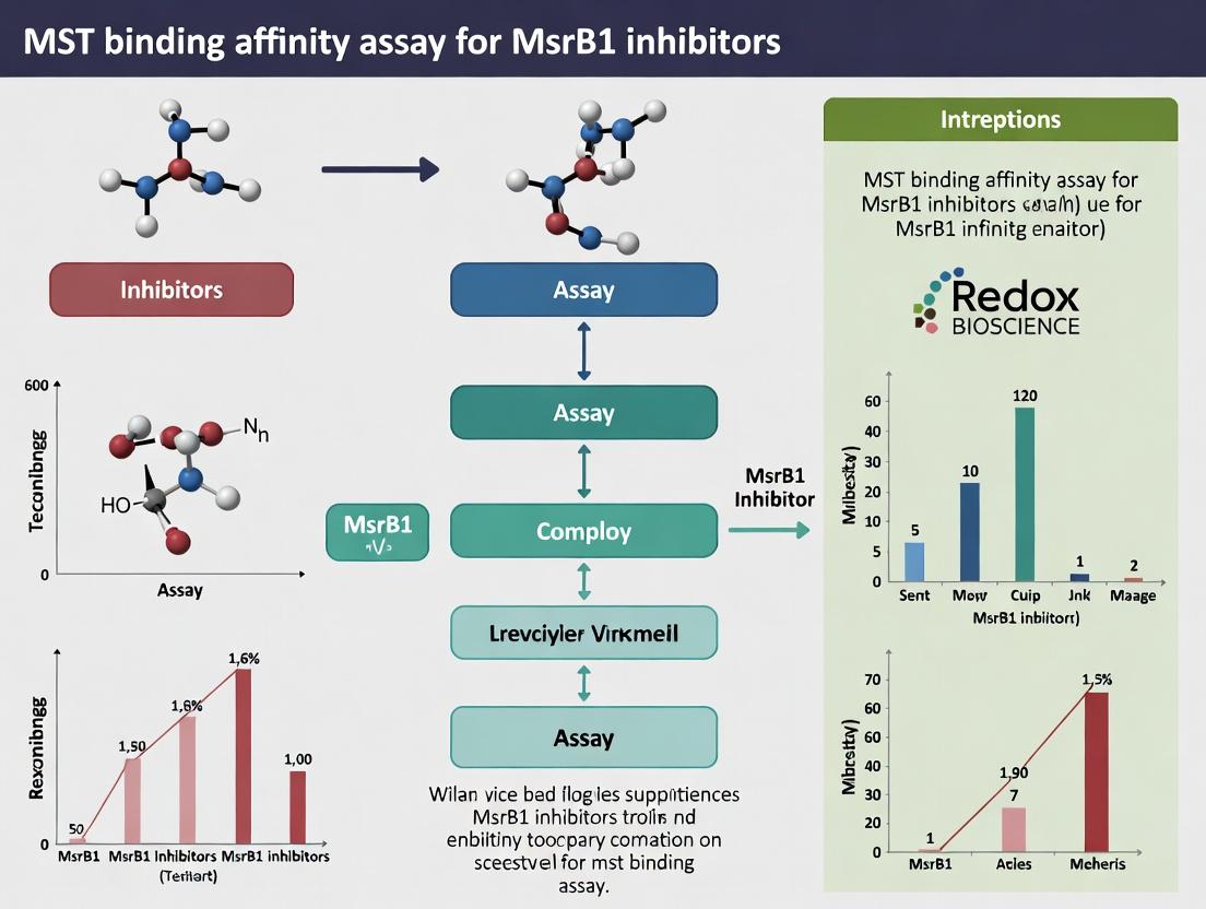
Protocol 3: MST Binding Assay for MsrB1 Inhibitor Screening (Thesis Context)
Purpose: To determine the binding affinity (Kd) of small molecule inhibitors to MsrB1 using Microscale Thermophoresis. Materials:
- Monolith Series instrument (NanoTemper Technologies)
- Purified, fluorescently labeled MsrB1 (e.g., NT-647-NHS dye)
- Candidate inhibitor compounds in DMSO
- Assay buffer: 50 mM Tris, 150 mM NaCl, 10 mM MgCl2, 0.05% Tween-20, pH 7.5 (optimized for MsrB1)
- Premium coated capillaries
Procedure:
- Labeling: Label purified MsrB1 with RED-NHS 2nd Generation dye according to manufacturer's protocol. Remove excess dye using size-exclusion columns.
- Sample Preparation: Prepare a 16-step, 1:1 serial dilution of the inhibitor compound in assay buffer, starting from the highest soluble concentration (e.g., 1 mM). Keep DMSO concentration constant (<2%).
- Complex Formation: Mix a constant concentration of labeled MsrB1 (e.g., 50 nM) with each compound dilution. Incubate for 15-30 min at RT in the dark.
- Loading & Measurement: Load samples into capillaries. Perform MST measurements using appropriate instrument settings (e.g., 20% LED power, 40% MST power).
- Data Analysis: Import thermophoresis + T-Jump data into MO.Affinity Analysis software. Plot normalized fluorescence (Fnorm) vs. compound concentration. Fit curve using the Kd model to determine the dissociation constant.
Diagrams
Title: MsrB1's Role in Redox Homeostasis and Disease
Title: MST Assay Workflow for MsrB1 Inhibitors
The Scientist's Toolkit: Research Reagent Solutions
| Reagent/Material | Supplier Examples | Function in MsrB1 Research |
|---|---|---|
| Recombinant Human MsrB1 Protein | Abcam, Sino Biological, in-house purification | Essential substrate for enzymatic activity assays and binding studies (MST/SPR). |
| Dabsyl-Met-R-O Sulfoxide | Custom synthesis (e.g., Sigma Custom Synthesis) | Stereospecific chromogenic substrate for convenient MsrB1 activity measurement via HPLC. |
| Thioredoxin Reductase (TrxR1) System | Sigma-Aldrich, Cayman Chemical | Provides physiologically relevant reducing equivalents (NADPH/TrxR1/Trx) for MsrB1 activity assays. |
| Anti-MsrB1 Antibody | Santa Cruz Biotechnology, Proteintech | Validation of protein expression in cellular models (knockdown/overexpression) via Western blot, IF. |
| MSRB1 siRNA/shRNA | Dharmacon, Sigma-Aldrich, Origene | For genetic knockdown studies to probe loss-of-function phenotypes in cellular models. |
| NT-647 Fluorescent Dye | NanoTemper Technologies | Covalent labeling of MsrB1 protein for Microscale Thermophoresis (MST) binding assays. |
| MST Instrument & Capillaries | NanoTemper Technologies | Platform for label-free, solution-based measurement of inhibitor binding affinity and kinetics. |
| Cell Viability Assay Kit (MTT) | Thermo Fisher, Abcam, Promega | Quantifying cellular sensitivity to oxidative stress following MsrB1 modulation. |
| ROS Detection Probe (H2DCFDA) | Thermo Fisher, Cayman Chemical | Measuring intracellular reactive oxygen species levels in MsrB1-modulated cells. |
Methionine sulfoxide reductase B1 (MsrB1) is a key selenium-dependent enzyme responsible for the reduction of methionine-R-sulfoxide back to methionine, thereby repairing oxidative damage to proteins. Its role in regulating protein function and cellular redox homeostasis has implicated it in critical pathophysiological processes. Recent research, framed within a broader thesis on MST binding affinity assays for MsrB1 inhibitors, positions MsrB1 as a promising therapeutic target. Inhibition of MsrB1 is hypothesized to modulate pathways involved in cancer progression (e.g., by sensitizing cells to oxidative stress), neurodegeneration (e.g., by affecting tau and α-synuclein pathology), and aging (e.g., by disrupting protein homeostasis and stress resistance).
Table 1: Reported Biological Effects of MsrB1 Modulation
| Condition/Model | Intervention | Key Quantitative Outcome | Reference/Model System |
|---|---|---|---|
| Colorectal Cancer | MsrB1 Knockdown (shRNA) | ~60% reduction in tumor volume in xenograft models; 2.5-fold increase in apoptotic markers. | HCT116 cell xenografts |
| Breast Cancer (Triple Negative) | MsrB1 Inhibition (Small Molecule) | IC₅₀ of 3.2 µM for cell proliferation; synergy with cisplatin reduces cell viability by 85%. | MDA-MB-231 cells |
| Alzheimer's Disease | MsrB1 KO Mouse Model | 40% increase in insoluble tau aggregates; 30% deficit in spatial memory (Morris water maze). | 3xTg-AD mice background |
| Parkinson's Disease | MsrB1 Overexpression | 50% reduction in α-synuclein oligomers; protection against MPTP-induced dopaminergic neuron loss. | A53T α-synuclein model |
| Aging (Lifespan) | MsrB1 C. elegans Knockout | 25% reduction in mean lifespan under oxidative stress; 35% increase in protein carbonylation. | C. elegans strain CL2120 |
| Binding Affinity | Lead Inhibitor (Compound X) | Kd = 180 nM measured by Microscale Thermophoresis (MST); Ki = 220 nM (enzymatic assay). | Recombinant human MsrB1 |
Table 2: Current MsrB1 Inhibitor Candidates
| Compound ID | Chemical Class | Reported Potency (IC₅₀/Ki) | Primary Experimental Evidence |
|---|---|---|---|
| MIPS-213 | Thiourea derivative | 150 nM (Ki) | Reduces glioblastoma cell invasion by 70% in vitro. |
| BRX-017 | Organoselenium mimic | 2.1 µM (IC₅₀) | Sensitizes prostate cancer cells to radiation (Dose enhancement factor: 1.8). |
| Compound X | Peptidomimetic | 220 nM (Ki) | High selectivity (>100x over MsrA); validated by MST and X-ray co-crystallography. |
Experimental Protocols
Protocol 1: Microscale Thermophoresis (MST) Binding Affinity Assay for MsrB1 Inhibitors
(This protocol is central to the thesis context of characterizing inhibitor binding.) Objective: Determine the dissociation constant (Kd) between recombinant human MsrB1 and a novel small-molecule inhibitor. Materials: See "Research Reagent Solutions" below. Procedure:
- Labeling: Reconstitute recombinant MsrB1 protein in PBS-T (0.05% Tween-20). Use the Monolith Protein Labeling Kit RED-NHS 2nd Generation. Mix 100 µL of 50 nM protein with 100 µL of the fluorescent dye. Incubate for 30 min at RT in the dark.
- Purification: Pass the labeling mixture through a pre-equilibrated gravity-flow column (provided in the kit) to remove free dye. Collect the flow-through containing labeled MsrB1. Determine final protein concentration.
- Inhibitor Series Preparation: Prepare a 16-step, 1:1 serial dilution of the inhibitor in assay buffer (PBS-T, 1% DMSO). Use a high starting concentration (typically 100 µM to 1 mM).
- Sample Preparation: Mix 10 µL of labeled MsrB1 (at a final constant concentration of 20 nM) with 10 µL of each inhibitor dilution. Include a control with no inhibitor (0% inhibition) and a reference with unlabeled competitor (100% inhibition). Incubate 15 min at RT.
- MST Measurement: Load samples into premium coated capillaries. Place capillaries in the Monolith NT.115 or NT.Automated. Perform measurements at 25°C, using 20% LED power and 40% MST power.
- Data Analysis: Use MO.Control software to analyze thermophoresis traces. Plot normalized fluorescence (Fnorm) vs. inhibitor concentration. Fit the data using the "Kd model" to obtain the dissociation constant.
Protocol 2: Assessment of MsrB1 Inhibition in a Cellular Oxidative Stress Model
Objective: Evaluate the functional consequence of MsrB1 inhibition on cancer cell viability under oxidative stress. Procedure:
- Seed HeLa or HCT116 cells in a 96-well plate (5,000 cells/well) and incubate overnight.
- Treat cells with a dose range of the MsrB1 inhibitor (e.g., 0.1 nM to 100 µM) or DMSO vehicle for 2 hours.
- Induce oxidative stress by adding 200 µM H₂O₂ to the medium. Incubate for 24 hours.
- Assess cell viability using the CellTiter-Glo 2.0 Luminescent Cell Viability Assay. Record luminescence.
- Calculate % viability relative to untreated control. Determine IC₅₀ values and synergy with H₂O₂ using CompuSyn software.
Signaling Pathways & Logical Workflow Diagrams
Title: MsrB1 Inhibition Disrupts Redox Repair & Drives Disease Phenotypes
Title: MST Binding Affinity Assay Workflow
Research Reagent Solutions Toolkit
Table 3: Essential Materials for MsrB1 Inhibitor Research
| Reagent/Material | Supplier Example | Function in Research |
|---|---|---|
| Recombinant Human MsrB1 Protein | Abcam (ab114288) or in-house expression | Target protein for biochemical assays (MST, enzymatic activity). |
| MONOLITH Protein Labeling Kit RED-NHS 2nd Gen | NanoTemper Technologies | Fluorescently labels lysine residues for highly sensitive MST measurements. |
| Premium Coated Capillaries | NanoTemper Technologies | Minimizes surface interaction for reliable MST/DSF measurements. |
| CellTiter-Glo 2.0 Assay | Promega | Luminescent assay to quantify viable cells based on ATP levels. |
| MIPS-213 / BRX-017 (Reference Inhibitors) | Sigma-Aldrich / Tocris | Pharmacological tools for validating experimental models and assays. |
| Anti-Methionine-R-Sulfoxide Antibody | MilliporeSigma (ABS1000) | Detects the primary substrate of MsrB1 in cells/tissues (Western, IF). |
| Se-deficient Media (e.g., FBS dialyzed) | Thermo Fisher Scientific | Used to manipulate cellular MsrB1 activity, as it is a selenoprotein. |
| H₂O₂ / Menadione | Various | Inducers of oxidative stress to challenge the MsrB1 repair system in vitro. |
This document provides detailed application notes and protocols for the design and evaluation of methionine sulfoxide reductase B1 (MsrB1) inhibitors, framed within a broader research thesis utilizing Microscale Thermophoresis (MST) binding affinity assays. The focus is on the critical interplay between active site chemistry and substrate recognition.
Application Notes: Active Site Chemistry of MsrB1
MsrB1 is a selenocysteine (Sec)-containing enzyme critical for repairing methionine-R-sulfoxide residues in proteins. Its active site features a catalytic triad composed of Sec, a resolving cysteine, and a glutamine. The selenol group of Sec (pKa ~5.2) is highly nucleophilic at physiological pH, making it a prime target for electrophilic inhibitors. Substrate recognition is governed by a hydrophobic pocket that accommodates the methionine side chain and precise geometry for the sulfoxide moiety.
Table 1: Key Active Site Residues and Their Roles in MsrB1 Catalysis
| Residue (Human MsrB1) | Role in Catalysis/Recognition | Chemical Property Target for Inhibition |
|---|---|---|
| Sec95 (U95) | Nucleophilic attack on sulfoxide. | Irreversible electrophiles (e.g., vinyl sulfones, chloroacetamides). |
| Cys99 | Resolving thiol; forms diselenide/selenylsulfide intermediate. | Reversible disulfide/diselenide disruptors. |
| Gln102 | Stabilizes the transition state. | Hydrogen-bond competitors. |
| Trp52, Phe86, Phe110 | Form hydrophobic pocket for methionine side chain. | Bulk group introduction for steric hindrance. |
Experimental Protocols
Protocol 2.1: In Silico Docking for Substrate Recognition Analysis
Objective: To predict binding modes of inhibitor candidates within the MsrB1 active site.
- Preparation:
- Retrieve the crystal structure of human MsrB1 (e.g., PDB ID: 2F3N) from the RCSB PDB.
- Prepare the protein using molecular modeling software: remove water molecules, add hydrogen atoms, assign charges (e.g., AMBER ff14SB force field).
- Define the binding site as a 10 Å sphere centered on the Sec95 residue.
- Ligand Preparation:
- Draw or import inhibitor structures (e.g., in .sdf format).
- Generate 3D conformations and minimize energy using the MMFF94 force field.
- Docking Execution:
- Perform docking using AutoDock Vina or GOLD.
- Set exhaustiveness to 32 (Vina) or search efficiency to 100% (GOLD).
- Analysis:
- Cluster results by root-mean-square deviation (RMSD < 2.0 Å).
- Rank poses by calculated binding affinity (ΔG, kcal/mol).
- Analyze key interactions: hydrogen bonds with Gln102, hydrophobic contacts with Trp52/Phe86, distance to Sec95 nucleophile.
Protocol 2.2: MST Binding Affinity Assay for Inhibitor Screening
Objective: To quantitatively measure the binding affinity (Kd) of designed inhibitors for recombinant MsrB1.
- Sample Preparation:
- Label recombinant MsrB1 with a fluorescent dye (e.g., NT-647-NHS dye, NanoTemper Technologies) according to the manufacturer's protocol. Use a protein concentration of 50 nM in assay buffer (50 mM HEPES, 150 mM NaCl, 10 mM MgCl2, 0.05% Tween-20, pH 7.5).
- Prepare a 16-step, 1:1 serial dilution of the unlabeled inhibitor in the same buffer. Start from a top concentration 20x the expected Kd.
- MST Measurement:
- Mix 10 µL of labeled MsrB1 with 10 µL of each inhibitor dilution in premium coated capillaries. Include a control (protein with buffer only).
- Load capillaries into the Monolith instrument (e.g., NT.Automated).
- Set instrument parameters: LED power to 20%, MST power to 40% at 25°C.
- Perform triplicate measurements.
- Data Analysis:
- Import data into MO.Control or PALMIST software.
- Plot normalized fluorescence (Fnorm) vs. inhibitor concentration.
- Fit the dose-response curve using the "Kd model" to obtain the dissociation constant (Kd).
Table 2: Representative MST Binding Data for Hypothetical MsrB1 Inhibitors
| Inhibitor ID | Core Scaffold | Targeted Residue | MST Kd (nM) ± SD | Comment on Recognition |
|---|---|---|---|---|
| INH-01 | Vinyl sulfone | Sec95 (Irreversible) | N/A (k2/Ki = 1.5 x 10^4 M⁻¹s⁻¹)* | Covalent binder; excellent shape complementarity. |
| INH-02 | Benzothiazole | Hydrophobic pocket | 250 ± 15 | High affinity driven by π-stacking with Phe86. |
| INH-03 | Acetamide derivative | Gln102 / Hydrophobic pocket | 1250 ± 85 | Moderate affinity; hydrogen bond donor confirmed. |
| INH-04 | Peptidomimetic | Full active site | 18 ± 3 | Mimics methionine sulfoxide; best-in-class recognition. |
*Covalent inhibitors are characterized by kinetics (k2/Ki); a MST dose-response yields an apparent IC50.
Visualizations
Title: MST-Based MsrB1 Inhibitor Design Workflow
Title: MsrB1 Substrate and Inhibitor Recognition
The Scientist's Toolkit: Research Reagent Solutions
Table 3: Essential Materials for MsrB1 Inhibitor Design & MST Assays
| Item | Function in Research | Example Product / Specification |
|---|---|---|
| Recombinant Human MsrB1 | Purified target protein for biochemical and biophysical assays. | ≥95% purity (SDS-PAGE), Sec-active site confirmed. |
| NT-647-NHS Fluorescent Dye | Covalently labels lysine residues on MsrB1 for MST detection. | NanoTemper Technologies, 2nd Generation dye. |
| MST-Optimized Buffer | Provides stable pH and ionic strength, minimizes non-specific binding for MST. | 50 mM HEPES, 150 mM NaCl, 10 mM MgCl2, 0.05% Tween-20, pH 7.5. |
| Premium Coated Capillaries | Low-binding capillaries for MST sample loading. | NanoTemper Technologies, 16/pack. |
| DTT (Dithiothreitol) | Maintains reduced state of catalytic Cys99; control for redox activity. | Molecular biology grade, 1M stock solution. |
| Methionine-R-Sulfoxide | Native substrate for competitive inhibition and enzymatic activity assays. | High-purity (≥98%), stereochemically defined. |
| Covalent Inhibitor Warheads | Chemical building blocks for targeting Sec95. | e.g., Vinyl sulfone, Chloroacetamide, Acrylamide derivatives. |
| Molecular Modeling Software Suite | For active site analysis, docking, and binding pose visualization. | e.g., PyMOL, AutoDock Vina, Schrödinger Maestro. |
Within the context of a broader thesis on Methionine Sulfoxide Reductase B1 (MsrB1) inhibitor research, understanding and quantifying binding affinity is paramount. MsrB1, a key enzyme in oxidative stress response, is a promising therapeutic target. The journey from identifying initial hit compounds to optimizing a lead candidate hinges on precise affinity measurements. Microscale Thermophoresis (MST) has emerged as a powerful, label-free technique for this purpose, enabling accurate determination of dissociation constants (Kd) across a wide range of conditions and molecular complexities. This application note details the critical role of binding affinity and provides protocols for its assessment using MST in the MsrB1 drug discovery pipeline.
The Centrality of Binding Affinity in Drug Discovery
Binding affinity, quantified as the dissociation constant (Kd), measures the strength of the interaction between a target protein and a ligand. It is the foundational metric that guides decision-making.
| Discovery Stage | Affinity Role | Typical Kd Range | MST Utility |
|---|---|---|---|
| Hit Identification | Primary screen to identify initial binders from large libraries. | High µM to mM | High-throughput screening of fragments/compounds; low sample consumption. |
| Hit-to-Lead | Prioritize hits based on potency; establish Structure-Activity Relationship (SAR). | µM range | Rapid comparison of analog series; works in complex buffers (e.g., with reductants for MsrB1). |
| Lead Optimization | Fine-tune chemical structure for maximal target engagement and selectivity. | nM to low µM | Precise measurement for tight binders; assess binding in presence of cellular lysates. |
Quantitative Data from Recent MsrB1 Inhibitor Studies (Representative):
| Compound ID | MST-Measured Kd (nM) | Target (MsrB1) | Assay Conditions | Reference Year |
|---|---|---|---|---|
| Inhibitor A | 150 ± 20 | Human Recombinant | PBS, 1 mM DTT, 0.05% Tween-20 | 2023 |
| Inhibitor B | 1,850 ± 300 | Human Recombinant | TRIS Buffer, 5 mM TCEP | 2022 |
| Fragment 1 | 15,000 ± 2,000 | Mouse Recombinant | PBS, 2 mM β-mercaptoethanol | 2024 |
| Lead Candidate X | 8.5 ± 1.2 | Human Recombinant | Cell Lysate Supplement | 2023 |
Detailed MST Protocol for MsrB1-Inhibitor Binding Affinity Determination
Protocol 1: MST Assay for MsrB1 Fragment Screening
Objective: Determine the binding affinity (Kd) of fragment library compounds to recombinant MsrB1.
Materials & Reagents:
- Monolith NT.115 Premium Capillaries
- Labelled MsrB1 Protein: Recombinant human MsrB1, labelled with RED-NHS 2nd Generation dye (NanoTemper Technologies).
- Fragment Library: 500 compounds in 100% DMSO.
- Assay Buffer: 50 mM PBS pH 7.4, 150 mM NaCl, 1 mM TCEP, 0.05% Tween-20.
- Instrument: Monolith X.
Procedure:
- Sample Preparation:
- Dilute RED-labelled MsrB1 to a final concentration of 50 nM in assay buffer.
- Prepare a 16-point, 1:1 serial dilution of the test fragment in assay buffer, starting from a top concentration of 2 mM (final DMSO ≤ 2%).
- Mix equal volumes (10 µL) of diluted protein and each fragment dilution. Include a control with protein + buffer only.
- Incubate for 10 minutes at room temperature protected from light.
MST Measurement:
- Load samples into standard treated capillaries.
- Place capillaries in the Monolith X instrument.
- Set instrument parameters: LED power 20%, MST power "Medium", measurement time 30 s.
- Run the experiment.
Data Analysis:
- Import data into MO.Affinity Analysis software (v3.0+).
- Select the
T-JumporMST-onphase for analysis. - Fit the dose-response curve using the
Kd modelto obtain the Kd value.
Protocol 2: Competitive MST for Selectivity Profiling
Objective: Assess inhibitor selectivity by competing for binding to MsrB1 versus MsrA.
Procedure:
- Label MsrB1 as in Protocol 1.
- Prepare a fixed concentration of a known high-affinity MsrB1 inhibitor (Kd ~100 nM) at 2x its Kd (200 nM).
- Titrate unlabelled MsrA protein into a mixture containing constant concentrations of labelled MsrB1 and the fixed inhibitor.
- Perform MST. A shift in the signal indicates the test inhibitor's displacement by MsrA, suggesting cross-reactivity. Analyze using the competitive binding model in MO.Affinity Analysis.
Visualizing the Workflow and Biological Context
Title: MST-Driven Drug Discovery Pipeline for MsrB1 Inhibitors
Title: MsrB1 Role and Inhibitor Mechanism of Action
The Scientist's Toolkit: Key Research Reagent Solutions
| Item | Function in MsrB1/MST Research |
|---|---|
| Recombinant Human/Mouse MsrB1 | Purified, active target protein for binding assays and enzymatic activity validation. |
| RED-NHS 2nd Generation Dye | Covalent fluorescent label for target protein in MST; minimal interference with binding. |
| Monolith X Instrument | Platform for performing MST measurements, offering high sensitivity and precision. |
| TCEP (Tris(2-carboxyethyl)phosphine) | Reducing agent for maintaining MsrB1's active site cysteine residues in assay buffers. |
| MO.Affinity Analysis Software | Specialized software for fitting MST data to calculate Kd values and statistical confidence. |
| Premium Capillaries | Low-binding capillaries for loading MST samples, ensuring consistent measurement quality. |
| Selectivity Panel Proteins | Related proteins (e.g., MsrA) for competitive MST assays to determine inhibitor selectivity. |
Step-by-Step MST Protocol: Measuring MsrB1-Inhibitor Binding Constants (Kd)
This protocol details the essential preparatory steps for conducting MicroScale Thermophoresis (MST) binding affinity assays to identify and characterize inhibitors of Methionine Sulfoxide Reductase B1 (MsrB1). MsrB1 is a key enzyme in redox regulation, reducing methionine-R-sulfoxide back to methionine. Its dysregulation is implicated in aging, neurodegenerative diseases, and cancer, making it a promising therapeutic target. The accuracy of MST, which measures biomolecular interactions by detecting temperature-induced fluorescence changes, is critically dependent on the quality of the labeled protein and the prepared compound library. These prerequisites ensure high signal-to-noise ratios and reliable determination of dissociation constants (Kd).
Table 1: Critical Parameters for Recombinant MsrB1 Labeling
| Parameter | Optimal Value/Range | Purpose/Rationale |
|---|---|---|
| Protein Purity (HPLC/SDS-PAGE) | >95% | Minimizes non-specific labeling and background. |
| Protein Concentration for Labeling | 20 µM | Ensures efficient dye conjugation while minimizing aggregation. |
| Dye:Protein Molar Ratio | 2:1 to 3:1 | Optimizes labeling efficiency without over-labeling. |
| Labeling Reaction Temperature | 4°C | Preserves protein activity and stability. |
| Labeling Reaction Time | 30 min - 1 hr | Balances complete conjugation with protein integrity. |
| Free Dye Removal (Column) | ≥ 2 passes | Critical for low MST background; target >99% removal. |
| Final Labeled Protein Concentration | ≥ 10 µM | Provides sufficient stock for serial dilution in MST. |
Table 2: Small Molecule Inhibitor Library Preparation
| Parameter | Specification | Rationale |
|---|---|---|
| Compound Purity | >90% (by HPLC) | Ensures observed activity is from the target compound. |
| Stock Solvent | 100% DMSO (anhydrous) | Standard for compound libraries; prevents hydrolysis. |
| Stock Concentration | 10-20 mM | High enough for serial dilution without solvent effects. |
| Final DMSO Concentration in MST | ≤ 1% (v/v) | Prevents protein denaturation and buffer interference. |
| Storage Temperature | -20°C to -80°C (desiccated) | Maintains long-term compound stability. |
| Serial Dilution Scheme | 1:1 or 1:2 in assay buffer | Creates 16 concentrations for full binding curve. |
Experimental Protocols
Protocol 3.1: Expression and Purification of Recombinant Human MsrB1 (Prerequisite)
- Expression: Transform BL21(DE3) E. coli with pET-28a vector containing human MSRB1 gene (with N-terminal 6xHis-tag). Grow in LB + Kanamycin (50 µg/mL) at 37°C to OD600 ~0.6. Induce with 0.5 mM IPTG for 16-18 hours at 18°C.
- Purification: Pellet cells, lyse in Lysis Buffer (50 mM Tris-HCl pH 8.0, 300 mM NaCl, 10 mM imidazole, 1 mg/mL lysozyme, protease inhibitors). Clarify by centrifugation. Purify supernatant using Ni-NTA affinity chromatography. Wash with 20 mM imidazole buffer. Elute with 250 mM imidazole buffer.
- Buffer Exchange & Storage: Dialyze into Labeling Storage Buffer (50 mM Tris-HCl pH 7.4, 150 mM NaCl, 10% glycerol). Concentrate to >1 mg/mL, aliquot, flash-freeze in liquid N₂, and store at -80°C. Confirm purity by SDS-PAGE and concentration by A280.
Protocol 3.2: Fluorescent Labeling of MsrB1 with NT-647-NHS Dye
Materials: Purified MsrB1, NT-647-NHS dye (Monolith Protein Labeling Kit RED), Labeling Buffer (PBS, pH 7.4), Reaction Buffer (provided in kit), Spin Columns (provided in kit).
- Prepare Protein: Thaw MsrB1 on ice. Centrifuge at 4°C, 15,000 x g for 10 min to remove aggregates. Determine exact concentration.
- Dilution: Dilute protein to 20 µM in Labeling Buffer.
- Prepare Dye: Reconstitute lyophilized NT-647-NHS dye in 20 µL of ultrapure DMSO to create the stock solution.
- Conjugation: Mix 100 µL of diluted protein (20 µM) with 10 µL of Reaction Buffer. Add 2-3 molar equivalents of NT-647-NHS dye stock (e.g., for a 2:1 ratio, add ~0.4 µL). Mix gently by pipetting.
- Incubation: Incubate reaction in the dark for 30 minutes at 4°C.
- Remove Free Dye: Equilibrate a provided spin column with 400 µL of Labeling Buffer. Apply the entire labeling reaction to the column center. Centrifuge at 1500 x g for 2 min. CRITICAL: Re-apply the flow-through to the column center and centrifuge again. This double-pass ensures >99% free dye removal.
- Characterization: Measure A280 and A650 to determine degree of labeling (DOL). Ideal DOL is between 0.5 and 1.0. Aliquot labeled protein, flash-freeze, and store at -80°C in the dark.
Protocol 3.3: Preparation of Small Molecule Inhibitor Stocks and Dilution Series
Materials: Small molecule compounds, anhydrous DMSO, assay buffer (50 mM HEPES pH 7.4, 150 mM NaCl, 10 mM MgCl₂, 0.05% Tween-20), low-protein-binding microplates/tubes.
- Stock Solution Preparation: Weigh compounds precisely. Dissolve in 100% anhydrous DMSO to a final concentration of 10 mM. Vortex and sonicate briefly to ensure complete dissolution.
- Quality Control: Verify concentration via absorbance if chromophore present, or by LC-MS.
- Master Stock Plate: Prepare a source plate with all compounds at 10 mM in DMSO. Seal and store desiccated at -20°C.
- Serial Dilution for MST: In a low-binding 96-well plate, perform a 1:1 serial dilution in assay buffer. Start with the highest concentration (typically 100 µM, yielding 1% DMSO). Pipette 20 µL of assay buffer into wells 2-16. Add 20 µL of 100 µM compound (in 2% DMSO/buffer) to well 1. Perform serial transfers from well 1 to 16. The final well contains buffer only (0 µM compound). This creates 16 concentrations with constant 1% DMSO.
Visualizations
Diagram 1: MST Assay Workflow from Prerequisites to Thesis Output
Diagram 2: Chemistry of MsrB1 NT-647-NHS Fluorescent Labeling
The Scientist's Toolkit: Research Reagent Solutions
Table 3: Essential Materials for MsrB1 MST Preparations
| Item | Function & Rationale | Example Supplier/Cat. No. (for reference) |
|---|---|---|
| NT-647-NHS Protein Labeling Kit | Contains site-directed, amine-reactive fluorescent dye optimized for MST. Minimizes perturbation of protein function. | NanoTemper Technologies, MO-L011 |
| HisTrap HP Ni-NTA Column | For high-performance purification of 6xHis-tagged recombinant MsrB1. Ensures high purity for labeling. | Cytiva, 17524801 |
| Size-Exclusion Chromatography Column (e.g., Superdex 75) | Optional but recommended for final polishing step to remove aggregates before labeling. | Cytiva, 28989333 |
| Anhydrous DMSO (≥99.9%) | Solvent for preparing stable, high-concentration small molecule stocks. Prevents water-induced compound degradation. | Sigma-Aldrich, 276855 |
| Low-Protein-Binding 96-Well Plates & Tubes | Prevents loss of protein and compound via adsorption to plastic during serial dilution steps. | Corning, CLS3997 |
| MST-Optimized Assay Buffer | Ready-to-use buffer with surfactant to reduce non-specific binding and stabilize proteins for MST. | NanoTemper Technologies, MO-B002 |
| Desiccator Cabinet | For long-term storage of small molecule stocks at -20°C/-80°C to prevent humidity-induced hydrolysis. | - |
| Nanodrop or equivalent Spectrophotometer | For precise measurement of protein concentration (A280) and dye labeling ratio (A650). | Thermo Fisher Scientific |
This document details the critical experimental setup for MicroScale Thermophoresis (MST) binding affinity assays, framed within a broader thesis investigating novel inhibitors of Methionine Sulfoxide Reductase B1 (MsrB1). MsrB1 is a key enzyme in redox homeostasis and a potential therapeutic target. Reproducible, high-quality MST data for characterizing small-molecule inhibitors relies on three pillars: appropriate capillary selection, rigorous buffer optimization, and precise instrument parameter configuration.
Capillary Choice
The selection of the appropriate capillary type is fundamental to the signal quality and experimental robustness.
Capillary Types and Properties
Table 1: Standard MST Capillary Properties and Recommendations
| Capillary Type | Surface Coating | Recommended Use | Key Advantage | Consideration for MsrB1/Inhibitors |
|---|---|---|---|---|
| Standard Treated | Hydrophilic polymer | Most proteins, standard buffers | Low non-specific binding; cost-effective. | Suitable for initial MsrB1 titrations in optimized buffer. |
| Premium Coated | Next-generation hydrophilic coating | Challenging proteins (hydrophobic, sticky) | Ultra-low protein adsorption. | Recommended for MsrB1 if aggregation or sticking is observed. |
| Untreated (Silicate) | Bare glass | Molecules binding to glass (e.g., DNA, some lipids) | Highest possible MST signal for strong binders. | Not recommended for protein-ligand studies due to high non-specific binding. |
Protocol 2.1: Capillary Selection and Pre-Screening
- Prepare Samples: Prepare a constant concentration of fluorescently labeled MsrB1 (e.g., 20 nM in assay buffer) and a non-binding control (buffer only).
- Capillary Loading: Using the provided tray, load the MsrB1 sample into 2-3 capillaries of each type (Standard Treated, Premium Coated). Load the buffer control into one capillary of each type.
- Initial MST Measurement: Perform a quick MST run (20-30% LED power, medium MST power) to measure the initial fluorescence (F₀) and thermophoresis signal.
- Analysis Criteria: Select the capillary type that yields: a) Stable and uniform F₀ across capillaries, b) High initial fluorescence intensity, c) No visible aggregation or precipitation in the capillary during the scan.
Buffer Optimization
Buffer composition is critical for maintaining protein stability, preventing non-specific interactions, and ensuring the observed thermophoresis is driven solely by the binding event.
Core Buffer Components and Optimization Strategy
Table 2: Key Buffer Components and Optimization Targets for MsrB1-Inhibitor MST
| Component | Typical Range | Purpose | Optimization Goal for MsrB1 |
|---|---|---|---|
| Buffer Agent | 20-50 mM HEPES, Tris, Phosphate | pH stability | Maintain pH 7.0-7.5, physiological for MsrB1 activity. |
| Salt (NaCl/KCl) | 0-300 mM | Modulate ionic strength | Minimize non-specific electrostatic interactions. Start at 150 mM. |
| Non-Ionic Detergent | 0.01-0.1% Tween-20, Pluronic F-127 | Reduce surface adhesion | Prevent MsrB1 sticking to capillary. Use lowest effective concentration. |
| Reducing Agent | 0.5-5 mM TCEP or DTT | Maintain reduced state of Msr active site | Essential for MsrB1 structural integrity. Use TCEP for pH stability. |
| Carrier Protein | 0.1-0.5 mg/mL BSA | Further reduce adsorption | Include if low nM MsrB1 concentrations are used. Use fatty-acid free. |
| Additives | Glycerol (2-5%), Sucrose | Stabilize protein conformation | Test if MsrB1 shows instability during thermophoresis. |
Protocol 3.1: Systematic Buffer Optimization via MST Signal Stability
- Prepare Buffer Variants: Prepare your base assay buffer (e.g., 50 mM HEPES pH 7.4, 150 mM NaCl, 0.5 mM TCEP). Create variants systematically altering ONE component at a time (e.g., ± 50 mM NaCl, ± 0.01% Tween-20, with/without 0.1% BSA).
- Prepare Constant Sample: Dilute fluorescently labeled MsrB1 to 2x the final assay concentration (e.g., 40 nM) in each buffer variant.
- Prepare Ligand Sample: Prepare a high-concentration stock of a known strong inhibitor (positive control) in each buffer variant.
- MST Measurement: Load capillaries with the MsrB1 sample. Perform an MST time trace (2-5 s MST on) in the absence and presence of a saturating concentration of the inhibitor.
- Evaluation Criteria: The optimal buffer yields: a) A stable, flat MST trace for MsrB1 alone (no aggregation/drift), b) A maximal, stable MST response (ΔFnorm) upon inhibitor addition, c) Minimal capillary-to-capillary variation.
Instrument Parameters
Fine-tuning the Monolith series instrument parameters maximizes the signal-to-noise ratio of the binding curve.
Key Parameters and Guidance
Table 3: Critical MST Instrument Parameters and Setup Protocol
| Parameter | Control | Typical Range for MsrB1 (Red Dye) | Optimization Goal |
|---|---|---|---|
| Excitation Power | LED Power | 20-80% | Use the lowest power that gives a stable F₀ > 500-1000 counts. |
| MST Power | IR-Laser Power | 20-100% (Start at 40-60%) | Sufficient to generate a clear thermophoresis signal without causing local heating artifacts or protein damage. |
| Measurement Time | On/Off Times | MST On: 5-30 s; Off: 5 s | Ensure thermophoresis signal reaches a plateau. 20s on/5s off is a common start point. |
| Focus Position | Capillary Scan | Automatic or manual | Must be set precisely in the center of the capillary for each experiment. |
| Temperature | Instrument Control | 22-25°C (ambient) | Keep constant. Ensure all samples and buffer are equilibrated to this temperature. |
Protocol 4.1: Stepwise Parameter Optimization
- Load Sample: Load optimized buffer into a capillary to set the baseline. Then load your constant sample (labeled MsrB1 at target concentration).
- Optimize LED Power: Perform a capillary scan. Adjust LED power until the peak fluorescence (F₀) is between 1,000 and 10,000 counts (ideal range). Record the % setting.
- Set Focus: Use the auto-focus function to precisely position the detection region in the center of the capillary.
- Test MST Power: With the IR-laser off, record initial fluorescence for 5s. Turn on the MST laser at 40% power and record for 20s. Inspect the trace. The
MST Onphase should show a clear, rapid drop (thermophoresis) followed by a stable plateau. If the signal is noisy or decreasing continuously, reduce MST power. If the signal change is very small (< 100 counts), increase MST power incrementally. - Finalize Settings: Once optimal LED and MST power are found, use these exact settings for all capillaries in the binding experiment to ensure consistency.
The Scientist's Toolkit: Essential Research Reagent Solutions
Table 4: Key Reagents for MST-based MsrB1 Inhibitor Screening
| Item | Function & Rationale |
|---|---|
| Monolith His-Tag Labeling Kit (RED-tris-NTA 2nd Gen) | Site-specific, fluorescent labeling of His-tagged recombinant MsrB1. Minimizes perturbation of the active site compared to cysteine labeling. |
| Monolith Premium Coated Capillaries | Minimizes non-specific adsorption of MsrB1 protein to capillary walls, crucial for obtaining clean binding data. |
| Tris(2-carboxyethyl)phosphine (TCEP) | Reducing agent; maintains MsrB1 catalytic cysteine in reduced state without affecting pH, unlike DTT. |
| Fatty-Acid Free Bovine Serum Albumin (BSA) | Carrier protein to stabilize low-concentration MsrB1 solutions and passivate surfaces. Fatty-acid free avoids unintended ligand binding. |
| Pluronic F-127 (10% solution) | Non-ionic surfactant alternative to Tween-20; can be more effective at preventing aggregation for some membrane-associated or hydrophobic proteins. |
| DMSO (Molecular Biology Grade) | High-purity solvent for dissolving small-molecule inhibitors. Must be matched in all samples (typically ≤ 1-2% final concentration). |
| Assay Buffer Concentrate (10X) | Ensures perfect buffer matching between protein, ligand, and serial dilution stocks, a prerequisite for accurate Kd determination. |
Visualized Workflows and Pathways
Title: MST Experimental Setup and Workflow
Title: Buffer Components for MsrB1 Stability
Title: MST Signal Generation and Kd Determination
Within the broader research on identifying and characterizing inhibitors of Methionine Sulfoxide Reductase B1 (MsrB1) using Microscale Thermophoresis (MST), this document details the critical experimental phase of assay execution. Precise titration series design, robust data acquisition, and rigorous initial analysis are fundamental to obtaining reliable binding affinity (Kd) values, directly informing on inhibitor potency and supporting structure-activity relationship (SAR) studies in drug development.
The Scientist's Toolkit: Essential Research Reagents & Materials
| Item | Function in MST Assay |
|---|---|
| Monolith NT.115 Premium Capillaries | High-quality glass capillaries for consistent sample loading and laser path. |
| Monolith NT.LabelFree OR His-Tag Labeling Kits | For fluorescently labeling the target protein (MsrB1) without affecting its active site. |
| Purified Recombinant MsrB1 Protein | The target enzyme, preferably with a purity >95% for specific binding measurements. |
| Small Molecule Inhibitor Compounds | Putative inhibitors, solubilized in DMSO, for titration against labeled MsrB1. |
| Assay Buffer (e.g., PBS, Tris-HCl) | Matched buffer with potential additives (e.g., 0.05% Tween-20) to minimize surface interactions. |
| MO.Control Software | Instrument software for defining experiment parameters, capillary scanning, and data collection. |
| MO.Affinity Analysis Software | Software for fitting thermophoresis data to binding models and calculating Kd values. |
Experimental Protocol: Titration Series Design & MST Measurement
A. Sample Preparation
- Label MsrB1: Using the Monolith His-Tag Labeling Kit RED-tris-NTA, label purified MsrB1 (with a His-tag) at a concentration of 50-100 nM in the chosen assay buffer. Incubate for 30 minutes in the dark.
- Prepare Compound Stock: Dissolve lyophilized inhibitor in 100% DMSO to create a high-concentration stock (e.g., 10 mM). Subsequent dilutions will use the same assay buffer to maintain constant DMSO concentration (<5% final).
- Design Titration Series: Prepare a 1:1 serial dilution of the inhibitor compound in assay buffer. A typical series consists of 16 capillaries, covering a concentration range from ~100 μM down to sub-nanomolar levels, ensuring the expected Kd is centered within the range.
B. MST Experiment Setup & Data Acquisition
- Load Capillaries: Mix a constant concentration of labeled MsrB1 (e.g., 20 nM) with each dilution of the inhibitor compound. Use a final volume of 10-20 μL per capillary.
- Instrument Setup: Place capillaries in the Monolith NT.115 instrument. In MO.Control software, set the following parameters:
- LED Power: 20% (for RED dye)
- MST Power: Medium (40%)
- Measurement Time: 30 sec (5 sec off, 20 sec on, 5 sec off)
- Capillary Scan: Perform before measurement to check for precipitation or bubbles.
- Run Experiment: Initiate the MST run. The instrument records fluorescence (F) before and during the infrared laser-induced temperature gradient.
Data Presentation & Initial Analysis
A. Key Data Output Table The raw MST data is processed to generate normalized responses (ΔFnorm [‰]) for each inhibitor concentration.
| Capillary | [Inhibitor] (nM) | log([Inhibitor]) | Fluorescence F1 (Start) | Fluorescence F2 (Hot) | ΔFnorm (‰) |
|---|---|---|---|---|---|
| 1 | 100000 | 5.0 | 5432 | 5221 | -12.4 |
| 2 | 33333 | 4.52 | 5501 | 5310 | -10.1 |
| ... | ... | ... | ... | ... | ... |
| 8 | 15.24 | 1.18 | 5488 | 5480 | -0.5 |
| 9 (Ctrl) | 0 | - | 5490 | 5485 | -0.4 |
| 10 | 5.08 | 0.71 | 5485 | 5488 | 0.2 |
| ... | ... | ... | ... | ... | ... |
| 16 | 0.017 | -1.77 | 5480 | 5482 | 0.1 |
B. Initial Analysis Workflow
- Data Normalization: MO.Affinity Analysis software automatically calculates ΔFnorm = (Fhot/Fcold)*1000.
- Curve Fitting: The software fits the ΔFnorm vs. log([Inhibitor]) data to a law of mass action (Kd) model.
- Kd Determination: The Kd value is derived from the fit at the point of half-maximal binding.
Visualization of Experimental Workflow & Analysis Logic
Title: MST Binding Assay Workflow for MsrB1 Inhibitors
Title: MST Data Analysis & Quality Control Logic
Within the broader thesis on identifying and characterizing novel inhibitors of Methionine Sulfoxide Reductase B1 (MsrB1) for therapeutic intervention, MicroScale Thermophoresis (MST) has been employed as a core technology for determining binding affinity and stoichiometry. This application note details protocols and data interpretation for deriving dissociation constants (Kd) and binding models from MST data, which is critical for validating hit compounds and guiding structure-activity relationship studies in drug development.
Key Research Reagent Solutions
The following table lists essential materials and reagents used in a standard MST binding assay for MsrB1 inhibitors.
| Research Reagent / Material | Function in Experiment |
|---|---|
| Purified, Labeled MsrB1 Protein | The target protein, fluorescently labeled for detection in the MST instrument. |
| MST-Compatible Buffer (e.g., PBS with 0.05% Tween-20) | Provides a stable, non-interfering chemical environment for binding. |
| Candidate Inhibitor Compounds | Small molecules or peptides screened for binding to the MsrB1 active site. |
| RED-tris-NTA 2nd Generation Dye (for His-tag labeling) | Fluorescent dye used to label His-tagged MsrB1 protein. |
| Capillary Chips (Monolith NT.115) | Hold the sample for analysis in the MST instrument. |
| Reference Control (e.g., DMSO) | Controls for non-specific signal changes from solvent or buffer components. |
Experimental Protocol: MST Binding Assay for MsrB1 Inhibitors
A. Protein Labeling (His-Tag Specific)
- Centrifuge the vial of RED-tris-NTA dye briefly. Prepare a 100 nM stock solution in assay buffer.
- Mix purified His-tagged MsrB1 protein at a concentration of 100-200 nM with the 100 nM dye stock at a 1:1 volume ratio.
- Incubate the mixture for 30 minutes at room temperature in the dark. The labeling is complete and stable.
B. Titration Series Preparation
- Prepare a 16-step, 1:1 serial dilution of the unlabeled ligand (inhibitor compound) in assay buffer. The highest concentration should be well above the expected Kd (typically 10-100x).
- Using a constant concentration of labeled MsrB1 (typically 10-50 nM), mix it with an equal volume of each ligand dilution. This creates a series where the ligand concentration varies, but the protein concentration is constant.
- Include a control sample with labeled MsrB1 and zero ligand (buffer only).
C. MST Measurement
- Load each sample into a premium coated capillary. Insert capillaries into the capillary holder.
- Place the holder into the Monolith instrument.
- Set instrument parameters: 20-40% LED power, medium MST power, 30 sec on-time. Perform the measurement.
D. Data Analysis (Protocol)
- Initial Processing: Import data into analysis software (e.g., MO.Affinity Analysis). The software calculates the normalized fluorescence (Fnorm) from the initial capillary scan and the thermophoresis trace.
- Response Curve: Plot the MST signal (ΔFnorm [‰] or Fluorescence Ratio) against the logarithm of the ligand concentration.
- Model Fitting: Fit the binding isotherm using the "Kd model." The software solves the law of mass action to fit the curve and calculate the Kd.
- Stoichiometry Assessment: Inspect the fitted curve. A single, clear transition suggests a 1:1 binding model. For multiple binding sites, advanced models (e.g., two-site model) must be tested. The ratio of protein concentration to the determined Kd and the shape of the saturation curve inform stoichiometry.
Data Presentation: Representative MST Results for Candidate MsrB1 Inhibitors
The table below summarizes quantitative binding data for three hypothetical inhibitor candidates (A-C) derived from MST assays.
| Compound ID | Kd (nM) [Mean ± SD] | Binding Model (Stoichiometry) | N (replicates) | Comments / Next Step |
|---|---|---|---|---|
| Inhibitor A | 25.3 ± 3.1 | 1:1 (MsrB1:Compound) | 3 | High-affinity hit. Proceed to ITC confirmation. |
| Inhibitor B | 1020 ± 110 | 1:1 (MsrB1:Compound) | 3 | Moderate affinity. SAR optimization required. |
| Inhibitor C | No binding observed | N/A | 2 | Exclude from further studies. |
| *Positive Control | 18.5 ± 1.8 | 1:1 | 2 | Known tight-binding substrate analog. |
*Used for assay validation.
Visualization of Workflows and Relationships
MST Binding Assay Core Workflow
Therapeutic Role of MsrB1 & Inhibitor Binding
Solving Common MST Challenges: Artifacts, Low Signal, and Data Variability in MsrB1 Assays
This document provides essential Application Notes and Protocols for Microscale Thermophoresis (MST) binding affinity assays, framed within the ongoing thesis research on the identification and characterization of inhibitors targeting Methionine Sulfoxide Reductase B1 (MsrB1). MsrB1 is a key enzyme in redox homeostasis and a promising therapeutic target. Accurate determination of binding constants (Kd) is critical for validating hit compounds. However, artifacts from fluorescence quenching, compound aggregation, and non-specific surface binding frequently compromise data integrity. This guide details protocols to identify and mitigate these artifacts, ensuring robust MST data for the MsrB1 inhibitor discovery pipeline.
Table 1: Common MST Artifacts and Diagnostic Signatures
| Artifact Type | Primary Cause | MST Trace Signature | Impact on Calculated Kd |
|---|---|---|---|
| Fluorescence Quenching | Direct interaction of ligand with fluorophore. | Decrease in initial fluorescence (F0) with increasing ligand concentration. Kd curve may still fit but is unreliable. | False positive or significantly distorted affinity. |
| Compound Aggregation | Ligand forms nano-aggregates that sequester protein. | Non-hyperbolic, sigmoidal, or irregular binding curves. High variance in data points. | Can produce false positives with apparent sub-micromolar affinity. |
| Surface Binding | Ligand or protein adsorbs to capillary surface. | Inconsistent MST signals between replicates; drifting baseline over time. | Unreliable, non-reproducible binding constants. |
Detailed Protocols
Protocol 1: Identifying Fluorescence Quenching in MsrB1-Ligand Interactions
Objective: To distinguish true thermophoretic mobility changes from artifactual fluorescence intensity changes. Materials: Labeled MsrB1 protein (e.g., NT-647-red fluorescent dye), test inhibitor compounds, MST buffer (e.g., PBS with 0.05% Tween-20), standard MST capillaries. Procedure:
- Prepare a 16-step, 1:1 serial dilution of the test compound in assay buffer. Do not add protein.
- Prepare a matching dilution series of the compound, but into a constant, low concentration (e.g., 20 nM) of fluorescently labeled MsrB1. Incubate for 15 min.
- Load both series (compound-only and protein-compound mix) into MST capillaries.
- Run the MST instrument using the same settings (e.g., 20% LED power, medium MST power).
- Analysis: Plot the initial fluorescence (F0) for each capillary vs. compound concentration for both series.
- Interpretation: A decrease in F0 in the compound-only series directly indicates fluorescence quenching. Any binding curve generated from the protein mix series is suspect and requires validation via Label-Free or alternative assays.
Protocol 2: Aggregation Detection via Dynamic Light Scattering (DLS)
Objective: To confirm if a putative MsrB1 inhibitor forms aggregates in assay conditions. Materials: Test compound at 10x the highest concentration used in MST, MST assay buffer, DLS instrument. Procedure:
- Centrifuge the compound stock solution at high speed (e.g., 16,000 x g) for 10 minutes to pellet any large aggregates.
- Prepare the compound in assay buffer at the final high concentration (e.g., 500 µM). Do not include protein.
- Incubate the solution at the assay temperature for 30 minutes.
- Load the sample into a clean DLS cuvette.
- Measure the hydrodynamic radius (Rh) distribution.
- Interpretation: A significant population of particles with Rh > 10 nm (besides known buffer components) indicates aggregation. The compound should be considered a potential aggregator. Mitigation includes adding detergent (0.01-0.1% Tween-20) or reducing compound concentration.
Protocol 3: Mitigating Surface Binding Artifacts
Objective: To minimize non-specific adsorption of MsrB1 or inhibitors to capillary walls. Materials: Labeled MsrB1, test compound, standard and surface-treated capillaries, additives (BSA, detergents). Procedure:
- Buffer Optimization: Add a non-interacting carrier protein (e.g., 0.1 mg/mL BSA) or increase non-ionic detergent (e.g., 0.1% Tween-20) to the assay buffer. BSA occupies non-specific binding sites.
- Capillary Selection: Use surface-treated capillaries (e.g., hydrophilic polymer coating) instead of standard plain glass capillaries for problematic compounds.
- Control Experiment: Perform a "reverse" MST experiment where a fixed concentration of a red-fluorescent tracer ligand is titrated with unlabeled MsrB1. A clean binding curve confirms the inhibitor's signal is not driven by surface effects.
- Validation: Compare binding curves and Kd values with and without mitigating agents. A consistent Kd value suggests the artifact has been minimized.
The Scientist's Toolkit: Key Research Reagent Solutions
| Item | Function in MsrB1 MST Assays |
|---|---|
| NT-647-NHS 2nd Generation Dye | Covalent fluorescent label for MsrB1 protein. Provides high brightness and photostability for MST. |
| MST Premium Coated Capillaries | Hydrophilic polymer-coated capillaries to minimize surface adsorption of proteins/lipids. |
| Tween-20 (Molecular Biology Grade) | Non-ionic detergent added to buffers (typically 0.05%) to reduce aggregation and surface binding. |
| BSA (Fatty-Acid Free) | Carrier protein added to assay buffers to block non-specific binding sites. |
| DTT or TCEP Reducing Agents | Maintains MsrB1, a redox enzyme, in its active reduced state during the assay. |
| Label-Free Detection MST Chips | Alternative to labeling; detects binding via intrinsic tryptophan fluorescence, eliminating quenching artifacts. |
Visualizations
Title: MST Artifact Identification Decision Workflow
Title: Artifact Mechanisms and Mitigation Paths
Within the broader research thesis on targeting methionine sulfoxide reductase B1 (MsrB1) for therapeutic intervention, MicroScale Thermophoresis (MST) binding affinity assays are central for identifying and characterizing novel inhibitors. The efficacy of these assays is critically dependent on the signal-to-noise ratio (SNR). This document details optimized protocols and application notes focusing on three pillars of SNR optimization: fluorescent labeling efficiency of the MsrB1 protein, solubility of small-molecule ligands (inhibitors), and strategic use of buffer additives to stabilize the interaction.
Key Research Reagent Solutions
| Reagent/Material | Function in MsrB1 Inhibitor MST Assay |
|---|---|
| Monolith NTT 2nd Generation RED Dye | Amine-reactive fluorescent dye for covalent, site-specific labeling of recombinant MsrB1. High photon yield and photostability are crucial for MST. |
| His-Tagged Recombinant Human MsrB1 | Purified target protein. His-tag facilitates purification and can be used for labeling control experiments. |
| DMSO (High Purity, Sterile) | Universal solvent for dissolving hydrophobic small-molecule inhibitors. Maintaining low final concentration (<5%) is vital to avoid protein denaturation. |
| Hydrophobic/Inert Carrier (e.g., Cyclodextrins) | Molecular carriers that enhance apparent solubility of ligands without interfering with binding, reducing non-specific aggregation. |
| Pluronic F-127 | Non-ionic surfactant used as a buffer additive (0.01-0.05%) to prevent adhesion of proteins and ligands to surfaces (vials, capillaries). |
| BSA (Fatty Acid-Free) | Carrier protein (0.1-0.5 mg/mL) that minimizes surface adsorption, stabilizes dilute MsrB1, and reduces false-positive depletion signals. |
| TCEP (Tris(2-carboxyethyl)phosphine) | Reducing agent to maintain MsrB1 active site cysteines in a reduced state, essential for catalytic function and inhibitor binding. |
Table 1: Impact of Labeling Efficiency on MST Signal Quality
| Labeling Ratio (Dye:Protein) | MST ΔFnorm [%] Amplitude | Baseline Noise | Recommended Use |
|---|---|---|---|
| 0.5:1 | Low (< 100%) | Low | Insufficient signal; not recommended. |
| 0.8 - 1.2:1 (Optimal) | High (> 300%) | Low | Ideal. Provides maximum SNR. |
| 1.5:1 | High | Increased | Acceptable, but may increase non-specific effects. |
| >2:1 | Variable/High | High | Not recommended. Risk of altered protein function & high noise. |
Table 2: Effect of Buffer Additives on Apparent Kd of a Model MsrB1 Inhibitor
| Buffer Condition | Measured Kd (nM) | Std. Error | Notes |
|---|---|---|---|
| Standard Buffer (Tris, NaCl, TCEP) | 150 | ± 25 | High variability, ligand depletion at high concentrations. |
| + 0.1 mg/mL BSA | 155 | ± 15 | Reduced variability, stabilized protein. |
| + 0.01% Pluronic F-127 | 148 | ± 10 | Minimized surface adhesion, cleanest titration curves. |
| + 0.5% DMSO (constant) | 160 | ± 20 | Necessary for ligand solubility; control for solvent effects. |
Detailed Experimental Protocols
Protocol A: Optimal Labeling of MsrB1 with RED-NHS 2nd Generation Dye
Objective: Achieve a 1:1 dye-to-protein ratio for maximum MST SNR.
- Prepare Protein: Buffer-exchange recombinant MsrB1 into labeling buffer (100 mM NaHCO₃, 100 mM NaCl, pH 8.5) using a desalting column. Final concentration should be 5-10 µM.
- Prepare Dye: Centrifuge vial of RED-NHS 2nd Generation dye (lyophilized) and reconstitute in anhydrous DMSO to a final concentration of 100 µM.
- Labeling Reaction: Add a 1.2x molar excess of dye solution to the protein solution. Mix gently and incubate in the dark at 25°C for 30 minutes.
- Purification: Remove free dye using the provided dye-removal columns equilibrated with MST assay buffer (e.g., 50 mM Tris-HCl, 150 mM NaCl, 10 mM TCEP, 0.05% Pluronic F-127, pH 7.5). Collect the labeled protein (eluent).
- Quantification: Measure absorbance at 280 nm (protein) and 650 nm (dye). Calculate degree of labeling (DOL) using the formula: DOL = (A650 * εprotein) / (A280 - (A650 * CF280)) * εdye⁻¹, where CF280 is the dye's correction factor at 280 nm (provided by manufacturer). Aim for DOL = 0.8 - 1.2.
Protocol B: Ligand Solubilization & MST Titration Setup
Objective: Prepare a soluble inhibitor stock and perform a 16-point serial dilution for MST.
- Primary Stock: Dissolve lyophilized inhibitor in 100% DMSO to a high concentration (e.g., 10-50 mM). Sonicate if necessary.
- Intermediate Dilution: Dilute the primary stock in MST assay buffer containing 1% DMSO (and 0.1 mg/mL BSA if needed) to create a 100x working stock at 100 µM. Ensure no precipitation.
- Serial Dilution: Using a transparent-bottom 96-well plate, perform a 1:1 serial dilution in assay buffer. Place buffer in all wells. Add the 100x ligand working stock to the first well (high concentration), mix, and transfer serially. The final constant [DMSO] should be ≤1%.
- Sample Preparation: Add a constant volume of labeled MsrB1 (at 2x final desired concentration, typically 20-50 nM) to each ligand dilution. Mix thoroughly by pipetting. Final volume per capillary: 10 µL.
- Measurement: Load samples into Monolith Premium Capillaries. Run MST on a Monolith instrument (e.g., Pico) using 40-80% LED power and medium/high MST power. Analyze data with MO.Affinity Analysis software using the Kd model.
Visualization of Key Concepts
MST Assay Workflow for MsrB1 Inhibitors
Key Factors Influencing MST Signal-to-Noise
Application Notes & Protocols Thesis Context: Methodological rigor in MicroScale Thermophoresis (MST) is paramount for generating reliable binding affinity (Kd) data in the ongoing investigation of MsrB1 methionine sulfoxide reductase inhibitors, a promising target for age-related and oxidative stress-related pathologies. This document outlines standardized protocols for critical pre-analytical and analytical variables to ensure cross-experiment and cross-laboratory reproducibility.
I. Sample Handling & Preparation Protocols
A. Protein (MsrB1) Purification & Labeling
- Objective: To produce homogenous, functionally active, fluorescently labeled MsrB1.
- Protocol:
- Expression & Purification: Express recombinant His-tagged MsrB1 in E. coli BL21(DE3). Purify via immobilized metal affinity chromatography (IMAC) using a step-wise imidazole elution (20mM, 250mM) in storage buffer (50mM Tris-HCl, 150mM NaCl, 5% Glycerol, pH 7.4).
- Buffer Exchange: Immediately exchange into MST-compatible labeling buffer (e.g., 50mM HEPES, 150mM NaCl, 10mM MgCl2, pH 7.5) using a desalting column. Confirm concentration via absorbance at 280 nm.
- Fluorescent Labeling: Use a RED-tris-NTA 2nd generation dye for His-tag labeling. Incubate 100 nM MsrB1 with 200 nM dye for 30 minutes in the dark at RT. Critical: The protein must be in a buffer without EDTA or other chelators.
- Cleaning: Remove excess dye using the provided dye removal columns. Aliquot and flash-freeze labeled protein for single-use to avoid freeze-thaw cycles.
B. Compound (Inhibitor) Stock Management
- Objective: To maintain inhibitor integrity and ensure accurate serial dilution.
- Protocol:
- Solubilization: Dissolve lyophilized inhibitors in 100% molecular biology grade DMSO to create a 10 mM primary stock. Vortex thoroughly for 1 min and sonicate if necessary.
- Storage: Store single-use aliquots at -80°C under desiccated, inert atmosphere (argon).
- Dilution Series: Prepare a 16-step, 1:1 serial dilution in MST assay buffer, maintaining a constant final DMSO concentration (typically ≤1%). Use low-protein-binding tubes and fresh tips for each transfer.
II. Temperature Control Protocol
A. Instrument & Sample Equilibration
- Objective: Eliminate thermal gradients that cause unwanted capillary convection.
- Protocol:
- Instrument: Power on the MST instrument (e.g., Monolith series) at least 1 hour before measurement. Use the integrated temperature control to set to 25°C (or desired assay temp).
- Sample Plate: After preparing the dilution series in the microplate, centrifuge briefly (1000 x g, 1 min) to collect liquid. Seal and incubate in the instrument or a thermally controlled block at the assay temperature for 15-20 minutes.
- Capillary Pre-incubation: Load capillaries and allow them to sit in the instrument for 5 minutes prior to the first measurement for thermal equilibration.
III. Instrument Calibration & QC Protocol
A. Daily Performance Check
- Objective: Verify laser and detector stability.
- Protocol:
- Perform the instrument's built-in "Power & Focus" calibration using the provided reference dye.
- Record the LED power and MST power values. Deviations >5% from the established baseline for your lab require investigation and potential service.
B. Capillary Lot QC
- Objective: Control for variability in capillary coating and dimensions.
- Protocol:
- Select a reference protein-ligand pair with a well-characterized Kd (e.g., carbonic anhydrase – acetazolamide).
- Perform a full MST titration in triplicate using a new capillary lot alongside the current lot.
- Compare the derived Kd values and the initial fluorescence (Fnorm) signals. Reject lots that cause a >20% shift in reference Kd or a significant change in sample meniscus shape.
Table 1: Impact of Sample Handling Variables on MsrB1-Inhibitor Kd Reproducibility
| Variable Tested | Protocol Deviation | Resulting CV of Kd* | Recommended Practice |
|---|---|---|---|
| Protein Storage | 3 Freeze-Thaw Cycles | 35% | Single-use aliquots at -80°C |
| Labeling Buffer | Presence of 1mM EDTA | Labeling Failed | Use chelator-free buffer |
| DMSO Consistency | Variable (0.5%-2%) across dilution | 28% | Fix at 1% for all samples |
| Compound Age | 6-month old DMSO stock at RT | 50% (Potency Loss) | Aliquot, store at -80°C, use within 1 month |
*CV = Coefficient of Variation from n=5 independent titrations.
Table 2: Temperature Equilibration Effects on MST Signal Quality
| Condition | Initial Fluorescence (Fnorm) Noise | MST Amplitude (ΔFnorm) | Resulting Kd Confidence Interval (95%) |
|---|---|---|---|
| No Equilibration (Samples at RT) | High (≈ 5-8%) | Low, Variable | 112 nM ± 85 nM |
| Full Equilibration (Samples at 25°C) | Low (≈ 1-2%) | High, Consistent | 105 nM ± 12 nM |
V. The Scientist's Toolkit: Key Research Reagent Solutions
| Item | Function in MsrB1 MST Assay |
|---|---|
| RED-tris-NTA 2nd Generation Dye (MO-L018) | High-affinity, low-stoichiometry fluorescent label for His-tagged MsrB1. Minimizes perturbation. |
| Premium Coated Capillaries (MO-K022) | Standardized, hydrophilic-coated capillaries for consistent sample loading and meniscus shape. |
| MST Assay Buffer Kit | Optimized, lyophilized buffer for consistent pH, salt, and additive background. |
| His-Tagged Reference Protein (e.g., CA II) | Essential positive control for instrument and capillary lot quality control. |
| DMSO, Molecular Biology Grade | High-purity solvent for inhibitor stocks; minimizes oxidative degradation. |
VI. Visualizations
Diagram 1: MST Assay Workflow for MsrB1 Inhibitors
Diagram 2: Key Variables in Reproducibility
Within the context of investigating inhibitors for the methionine sulfoxide reductase B1 (MsrB1) enzyme using MicroScale Thermophoresis (MST), managing low-affinity interactions and competitive binding scenarios is paramount. These challenges are central to hit validation and lead optimization in drug discovery. This document provides advanced protocols and application notes for robust experimental design and data analysis in such contexts, specifically framed within ongoing MsrB1 inhibitor research.
Challenges of Low-Affinity Binders in MST
Low-affinity binders (typically with Kd values in the high µM to mM range) present specific challenges in MST assays. The shallow binding isotherms are sensitive to experimental noise, and the low signal amplitude can approach the detection limit of the instrument.
Key Considerations:
- Signal-to-Noise Ratio (SNR): Maximizing the amplitude of the thermophoresis signal is critical.
- Protein and Ligand Stability: High concentrations required can lead to aggregation or precipitation.
- Buffer Optimization: Buffer composition significantly impacts weak interactions.
Protocol: Optimizing MST for Low-Affinity MsrB1 Binders
1. Target (MsrB1) Labeling and Preparation:
- Use RED-tris-NTA 2nd generation dye for His-tagged MsrB1. It offers superior performance over 1st generation dyes.
- Keep labeling stoichiometry low (recommended dye:protein ratio of 0.5-2:1) to minimize dye-induced artifacts.
- Perform a labeling test via a mobility shift assay in the MST instrument to confirm successful labeling without aggregation.
2. Assay Buffer Optimization:
- Baseline Buffer: 50 mM HEPES, 150 mM NaCl, 10 mM MgCl₂, 0.05% Tween-20, pH 7.5.
- Additives: Include 1-5% DMSO if compounds are dissolved in DMSO to match final conditions precisely. Consider adding 0.1-1 mg/mL BSA to prevent non-specific sticking.
- Critical: Use the exact same buffer for compound serial dilution as for the protein-dye mix. Perform a "Capillary Scan" and "Temperature Scan" prior to the experiment to establish optimal measurement parameters.
3. Experimental Design for Low-Affinity Range:
- Prepare a 16-step 1:1 serial dilution of the unlabeled low-affinity binder, starting from a concentration 20-50 times above the estimated Kd (e.g., 10-20 mM for a ~500 µM binder).
- Keep the concentration of labeled MsrB1 constant at a value below the expected Kd (e.g., 50-100 nM for a 500 µM Kd). This ensures the "Cold Target" regime, simplifying data analysis.
- Incubate the dilution series with the labeled protein for 10-15 minutes at room temperature before loading into Monolith premium capillaries.
4. MST Measurement & Data Analysis:
- Use a LED power and MST power that provide a strong initial fluorescence (≥ 500-1000 counts) and a clear thermophoresis signal.
- For low-affinity binders, analyze the "Thermophoresis + T-Jump" signal (often provides better SNR for shallow curves).
- Fit data using the "Kd model" in MO.Affinity Analysis software. Ensure the concentration of the fluorescent molecule (MsrB1) is correctly set in the analysis model.
Competitive Binding Experiments for Inhibitor Characterization
Competitive MST experiments are used to rank inhibitor potency, determine binding modes, and measure IC50 values for compounds that compete with a known, high-affinity fluorescent ligand.
Principle:
A fluorescent tracer (e.g., a high-affinity inhibitor labeled directly, or a labeled antibody) binds to MsrB1. An unlabeled competitor inhibitor displaces the tracer, causing a change in the MST signal. This allows determination of the competitor's affinity (Ki).
Protocol: Competitive MST for MsrB1 Inhibitors
1. Tracer Selection & Characterization:
- Identify a high-affinity binder (Kd < 100 nM) to MsrB1. This could be a known potent inhibitor conjugated to a dye (e.g., ATTO-647N via amine coupling) or a labeled antibody/ nanobody.
- First, perform a direct MST titration to determine the precise Kd of the tracer to labeled MsrB1 under your assay conditions.
2. Competitive Binding Experiment Setup:
- Prepare a constant, pre-mixed complex of MsrB1 and tracer at a concentration near the Kd of the tracer (e.g., [MsrB1] = 2 x Kdtracer, [Tracer] = 1 x Kdtracer).
- Prepare a 16-step 1:1 serial dilution of the unlabeled competitor inhibitor.
- Mix each competitor dilution with the pre-formed MsrB1-tracer complex. Final DMSO concentration must be constant across all points.
- Incubate for 30-60 minutes to reach equilibrium (longer for very high-affinity competitors).
- Load into capillaries and run the MST experiment.
3. Data Analysis for Ki Determination:
- Plot the normalized fluorescence or thermophoresis signal against the competitor concentration.
- Fit the data using the "Inhibition model (Ki)" in MO.Affinity Analysis.
- The software requires the known Kd of the tracer and the concentrations of MsrB1 and tracer as input parameters to calculate the competitor's Ki.
Table 1: Comparison of Direct vs. Competitive MST for MsrB1 Inhibitor Screening
| Parameter | Direct Binding MST (Low-Affinity) | Competitive Binding MST |
|---|---|---|
| Optimal Kd Range | 1 µM - 10 mM | 1 nM - 10 µM (for competitor) |
| Protein Consumption | Low to Moderate (nM concentrations) | Low (nM concentrations) |
| Ligand/Dye Requirement | Target (MsrB1) must be labeled | Requires a high-affinity fluorescent tracer |
| Information Gained | Direct affinity measurement, stoichiometry | Relative affinity (Ki), binding site competition |
| Key Challenge | Low signal amplitude, buffer sensitivity | Tracer development, longer equilibration times |
| Typical Incubation Time | 10-15 minutes | 30-60 minutes |
Table 2: Essential Buffer Components for MsrB1 MST Assays
| Component | Typical Concentration | Function & Rationale |
|---|---|---|
| HEPES | 50 mM | Maintains physiological pH with minimal metal chelation. |
| NaCl | 150 mM | Provides ionic strength to mimic physiological conditions. |
| MgCl₂ | 10 mM | Cofactor for MsrB1 enzymatic activity; may influence binding. |
| Tween-20 | 0.05% (v/v) | Reduces non-specific hydrophobic interactions and surface adsorption. |
| BSA | 0.1 mg/mL | Blocks non-specific binding to capillaries and protein surfaces. |
| DTT/TCEP | 0.5-1 mM | Keeps MsrB1 catalytic cysteine reduced; essential for activity. |
| DMSO | ≤5% (v/v) | Matches compound solvent; critical for signal stability. |
The Scientist's Toolkit: Research Reagent Solutions
Table 3: Essential Materials for MsrB1 Inhibitor MST Studies
| Item | Function/Application | Example Product (Vendor) |
|---|---|---|
| Monolith His-Tag Labeling Kit | For fluorescent labeling of His-tagged MsrB1 with minimal perturbation. | RED-tris-NTA 2nd Gen (NanoTemper) |
| Premium Coated Capillaries | Low-binding capillaries for consistent sample loading and measurement. | MONOLITH NT.115 Premium Capillaries (NanoTemper) |
| Recombinant Human MsrB1 | Purified, active target protein, preferably with a His-tag for labeling. | Recombinant Human MSRB1 Protein, His-tagged (Sino Biological) |
| High-Affinity Tracer Molecule | A known potent inhibitor for MsrB1, suitable for dye conjugation. | (Compound-specific; e.g., a published lead inhibitor) |
| Amine-Reactive Dye | For custom conjugation of tracer molecules. | ATTO 647N NHS ester (ATTO-TEC) |
| Assay-Optimized Buffer Kit | Pre-formulated buffers for stability and reduced artifacts. | MST Optimized Buffer (NanoTemper) |
| Positive Control Inhibitor | A well-characterized low/medium affinity binder for assay validation. | e.g., a substrate analog or early-hit compound |
Visualization Diagrams
Low-Affinity MST Workflow
Competitive Binding Principle
MsrB1 Pathway & Inhibition
Benchmarking MST Data: Validation Strategies and Comparative Analysis with ITC, SPR, and DSF
1. Introduction and Application Notes
Within the thesis research on targeting the methionine sulfoxide reductase B1 (MsrB1) for therapeutic intervention, establishing a direct quantitative link between binding affinity and functional inhibition is paramount. MicroScale Thermophoresis (MST) provides a precise method for determining the dissociation constant (Kd) of small-molecule inhibitors binding to purified MsrB1 protein. However, Kd alone does not predict the potency of enzymatic inhibition. The half-maximal inhibitory concentration (IC50), derived from a functional enzymatic assay, measures the compound's efficacy in blocking MsrB1 activity. Correlating Kd with IC50 is critical for hit validation and lead optimization, as it confirms that observed binding directly translates to the desired pharmacological effect. A strong correlation validates the binding site's relevance, while discrepancies may indicate allosteric binding or assay interference.
2. Quantitative Data Summary
Table 1: Representative Data for MsrB1 Inhibitors from Thesis Research
| Compound ID | MST Kd (nM) | Enzymatic IC50 (nM) | Ratio (IC50/Kd) | Correlation Notes |
|---|---|---|---|---|
| M-107 | 15 ± 3 | 42 ± 8 | 2.8 | Strong correlation, competitive inhibitor. |
| M-112 | 120 ± 20 | 35 ± 5 | 0.29 | IC50 < Kd suggests non-competitive or irreversible mechanism. |
| M-099 | 5.2 ± 0.9 | 220 ± 40 | 42.3 | Poor correlation, may bind outside active site (allosteric). |
| M-121 | 850 ± 150 | 1100 ± 200 | 1.3 | Good correlation, but weak affinity/potency. |
3. Experimental Protocols
Protocol 3.1: MST Binding Assay for MsrB1 Inhibitors (Kd Determination) Objective: Determine the binding affinity (Kd) of small molecules to purified, fluorescently labeled MsrB1 protein. Materials: Monolith X-series instrument, RED-NHS 2nd Generation dye (NanoTemper), recombinant human MsrB1, assay buffer (50 mM Tris, 150 mM NaCl, 10 mM MgCl2, 0.05% Tween-20, pH 7.5). Procedure:
- Labeling: Label purified MsrB1 with RED-NHS dye per manufacturer's protocol. Remove excess dye via size-exclusion chromatography.
- Sample Preparation: Prepare a 16-step, 1:1 serial dilution of the unlabeled inhibitor compound in assay buffer. Use a top concentration ~100x the expected Kd.
- Mixing: Mix a constant concentration of labeled MsrB1 (~20 nM) with an equal volume of each compound dilution. Include a no-inhibitor control (buffer only).
- Loading: Load samples into premium-coated capillaries.
- Measurement: Run MST using 40% LED power and 40% MST power. Thermophoresis is measured.
- Analysis: Fit the dose-response curve (ΔFnorm vs. inhibitor concentration) using the "Kd model" in MO.Affinity Analysis software to extract the Kd value.
Protocol 3.2: MsrB1 Enzymatic Activity Assay (IC50 Determination) Objective: Determine the functional inhibition potency (IC50) of compounds against MsrB1 reductase activity. Materials: Purified MsrB1, dithiothreitol (DTT), methionine-R-sulfoxide (Met-R-SO) substrate, DTNB [5,5'-dithio-bis-(2-nitrobenzoic acid)], reaction buffer (50 mM HEPES, pH 7.5). Procedure:
- Reaction Principle: MsrB1 reduces Met-R-SO to methionine using DTT. The reaction is coupled with DTNB, which reacts with the consumed DTT to produce the yellow-colored TNB, measurable at 412 nm.
- Inhibitor Dilution: Prepare a 10-point, 1:3 serial dilution of the inhibitor in reaction buffer.
- Assay Setup: In a 96-well plate, mix inhibitor (or buffer control), MsrB1 (final 50 nM), and DTT (final 1 mM) in reaction buffer. Pre-incubate for 15 min at 25°C.
- Reaction Initiation: Simultaneously add Met-R-SO (final 200 µM) and DTNB (final 0.2 mM) to start the reaction.
- Kinetic Readout: Immediately monitor absorbance at 412 nm for 10-20 minutes using a plate reader.
- Analysis: Calculate initial reaction rates (Vo). Normalize rates to the no-inhibitor control. Fit normalized activity (%) vs. log[Inhibitor] to a four-parameter logistic equation to determine the IC50.
4. Visualizations
Title: MST Binding Assay Workflow
Title: Kd-IC50 Correlation Interpretation
5. The Scientist's Toolkit: Essential Research Reagents & Materials
Table 2: Key Reagent Solutions for MsrB1 Binding & Activity Studies
| Item | Function in Research | Example/Note |
|---|---|---|
| Recombinant Human MsrB1 | Purified protein target for both MST binding and enzymatic assays. | Must be catalytically active and >95% pure. |
| RED-NHS 2nd Gen Dye (NanoTemper) | Covalent fluorescent label for MST; minimal perturbation of protein function. | Optimal for labeling lysines on MsrB1. |
| Monolith X-series Instrument | Performs MST measurements for label-free or fluorescence-based binding. | Enables Kd determination at low sample consumption. |
| Methionine-R-Sulfoxide (Met-R-SO) | Specific physiological substrate for the MsrB1 enzyme. | Critical for functional activity assay. |
| DTNB (Ellman's Reagent) | Colorimetric thiol detection reagent; couples DTT consumption to A412. | Enables continuous kinetic readout of MsrB1 activity. |
| Premium-Coated Capillaries | Surface-treated capillaries to prevent protein adsorption during MST. | Essential for robust, reproducible MST data. |
| DTT (Dithiothreitol) | Reducing agent serving as the electron donor in the MsrB1 catalytic cycle. | Required for enzymatic turnover in activity assay. |
In the broader thesis investigating Methionine Sulfoxide Reductase B1 (MsrB1) inhibitors using Microscale Thermophoresis (MST) for primary binding affinity screening, cross-validation with Isothermal Titration Calorimetry (ITC) is a critical step. MST provides high-sensitivity dissociation constant (Kd) values from fluorescence-based thermophoresis, but ITC delivers a full thermodynamic profile—enthalpy change (ΔH), entropy change (ΔS), and binding stoichiometry (n)—in a label-free manner. This protocol details the use of ITC to validate and deepen the thermodynamic understanding of lead compound interactions with recombinant MsrB1, ensuring robust data for drug development.
Application Notes: The Role of ITC in Validating MST Findings for MsrB1
- Primary Goal: To confirm the binding affinity (Kd) of putative MsrB1 inhibitors identified via MST and to elucidate the driving forces (enthalpy/entropy) of the interaction.
- Rationale: Agreement between Kd values from MST (kinetic/affinity) and ITC (thermodynamic equilibrium) strongly corroborates the binding event. Discrepancies may indicate artifacts from the fluorescent labeling in MST or buffer/sample condition issues.
- Key Insight: For drug development, the thermodynamic signature (ΔH, ΔS) provided by ITC helps in lead optimization. A binding driven by favorable enthalpy is often associated with specific hydrogen bonding and van der Waals interactions, which can be optimized for selectivity.
Detailed Experimental Protocol
Protocol 1: Sample Preparation for ITC
Objective: Prepare pure, active recombinant MsrB1 and inhibitor compounds in matched, degassed buffers.
- Protein (MsrB1): Dialyze purified, recombinant MsrB1 (>95% purity) extensively into ITC assay buffer (e.g., 25 mM HEPES, 150 mM NaCl, 1 mM TCEP, pH 7.4). Centrifuge at 15,000 x g for 10 minutes at 4°C to remove aggregates. Determine concentration using A280 absorbance.
- Ligand (Inhibitor): Dissolve lyophilized inhibitor in the same batch of dialysis buffer used for the protein. For DMSO-solubilized compounds, ensure the final DMSO concentration is ≤2% v/v and match this concentration in the protein sample via buffer addition.
- Degassing: Degas both protein and ligand solutions for 10-15 minutes under vacuum with gentle stirring to prevent bubbles in the ITC cell during titration.
Protocol 2: ITC Titration Experiment
Objective: Perform the titration to obtain raw heat data for analysis.
- Instrument Setup: Equilibrate the ITC instrument (e.g., Malvern MicroCal PEAQ-ITC) at 25°C. Rinse the sample cell and syringe with water and buffer.
- Loading: Load the syringe with the inhibitor solution (typically 10-20 times the concentration of the protein in the cell). Fill the sample cell (~200 µL) with the MsrB1 protein solution.
- Titration Parameters (Typical):
- Reference Power: 10 µCal/s
- Cell Temperature: 25°C
- Stirring Speed: 750 rpm
- Initial Delay: 60 s
- Number of Injections: 19
- Injection Volume: 2 µL (first injection of 0.4 µL discarded from analysis)
- Duration per Injection: 4 s
- Spacing between Injections: 150 s
- Control Experiment: Perform an identical titration of the inhibitor into buffer alone to measure heats of dilution. This data is subtracted from the main experiment.
Protocol 3: Data Analysis and Cross-Validation
Objective: Fit data to a binding model and compare Kd to MST results.
- Integration & Subtraction: Integrate raw heat peaks to obtain injection heats. Subtract the control titration data.
- Nonlinear Regression: Fit the corrected isotherm to a "One Set of Sites" model using the instrument's software (e.g., MicroCal PEAQ-ITC Analysis Software).
- Derived Parameters: The fit yields n (stoichiometry), Kd (dissociation constant = 1/Ka), ΔH (enthalpy change), and ΔS (entropy change, calculated via ΔG = ΔH - TΔS = RT ln Kd).
- Cross-Validation: Compare the ITC-derived Kd with the MST-derived Kd for the same MsrB1-inhibitor pair under identical buffer and temperature conditions.
Data Presentation
Table 1: Cross-Validation of MST and ITC Data for Candidate MsrB1 Inhibitors
| Compound ID | MST Kd (µM) [95% CI] | ITC Kd (µM) | ITC n | ITC ΔH (kcal/mol) | ITC -TΔS (kcal/mol) | Agreement (Within 3-fold?) |
|---|---|---|---|---|---|---|
| INH-02 | 1.54 [1.2 - 2.0] | 1.21 | 0.98 | -8.45 | 1.23 | Yes |
| INH-07 | 0.23 [0.18 - 0.29] | 0.19 | 1.02 | -12.10 | -0.05 | Yes |
| INH-15 | 15.7 [12.3 - 20.1] | 5.80 | 0.95 | -4.20 | -2.95 | Yes |
| INH-19 | 0.95 [0.7 - 1.3] | 5.21 | 0.87 | +2.15 | -7.10 | No (Further investigation) |
Table 2: The Scientist's Toolkit: Key Research Reagent Solutions
| Item | Function in ITC/MSrB1 Research |
|---|---|
| Recombinant Human MsrB1 | Purified target protein. Must be homogeneous and active for reliable binding studies. |
| HEPES Buffer (25 mM, pH 7.4) | Provides physiologically relevant, non-interfering pH buffering capacity. |
| Tris(2-carboxyethyl)phosphine (TCEP) | Reducing agent to keep MsrB1 active site cysteines reduced, preventing disulfide formation. |
| DMSO (Molecular Biology Grade) | High-purity solvent for dissolving hydrophobic small-molecule inhibitors. |
| ITC Degassing Station | Removes dissolved gases from samples to prevent bubble formation during titration. |
| Dialysis Cassettes (10k MWCO) | For exhaustive buffer exchange of the protein into the exact assay buffer. |
Diagrams
Workflow for Cross-Validating MST and ITC Data
ITC Measures Binding Enthalpy Directly
Within the broader thesis on characterizing MsrB1 inhibitors for therapeutic development, understanding the binding interaction is paramount. Two principal methodologies are employed: Surface Plasmon Resonance (SPR) and MicroScale Thermophoresis (MST). While SPR excels at resolving real-time kinetics (association/dissociation rates, ka and kd), MST directly measures the equilibrium dissociation constant (KD) in solution. This Application Note details the complementary use of these techniques to provide a complete binding profile of candidate MsrB1 inhibitors, contrasting kinetic and equilibrium affinity data.
Quantitative Data Comparison
The following table summarizes key parameters obtained from SPR and MST for a representative MsrB1 inhibitor (Compound X).
Table 1: Comparative Binding Data for MsrB1 and Compound X
| Parameter | SPR (Kinetics) | MST (Equilibrium) | Significance |
|---|---|---|---|
| KD | 15.2 ± 2.1 nM (Calculated from kd/ka) | 18.7 ± 3.5 nM (Direct measurement) | Confirms binding affinity; minor differences may arise from immobilized vs. free-state protein. |
| ka (M-1s-1) | (2.8 ± 0.3) x 105 | Not directly measured | Informs on the rate of complex formation, relevant for target engagement speed. |
| kd (s-1) | (4.3 ± 0.5) x 10-3 | Not directly measured | Informs on complex stability and inhibitor residence time (τ = 1/kd ≈ 233 s). |
| Sample Consumption | ~100-200 µg (immobilization + runs) | ~5-10 µg (single capillary) | MST is advantageous for scarce or precious samples like MsrB1. |
| Assay Time | ~1-2 hours per cycle (including regeneration) | ~30 minutes (16 capillaries in parallel) | MST offers higher throughput for equilibrium KD screening. |
| Buffer Flexibility | Restricted (must minimize non-specific surface binding) | High (can use physiological buffers, detergents) | MST allows measurement in conditions closer to the native cellular environment for MsrB1. |
Experimental Protocols
Protocol 1: SPR Kinetic Analysis of MsrB1-Inhibitor Binding
Objective: Determine the association (ka) and dissociation (kd) rate constants for Compound X binding to immobilized MsrB1.
Materials: See "The Scientist's Toolkit" below. Method:
- Sensor Chip Preparation: A CMS Series S chip is activated using a 1:1 mixture of 0.4 M EDC and 0.1 M NHS for 7 minutes.
- MsrB1 Immobilization: Recombinant human MsrB1 (in 10 mM sodium acetate, pH 5.0) is diluted to 20 µg/mL and injected over the activated surface to achieve a target immobilization level of 8000-12000 Response Units (RUs). Remaining active esters are deactivated with 1 M ethanolamine-HCl (pH 8.5).
- Kinetic Binding Experiment:
- HBS-EP+ (10 mM HEPES, 150 mM NaCl, 3 mM EDTA, 0.05% v/v Surfactant P20, pH 7.4) is used as running and dilution buffer.
- A 2-fold serial dilution of Compound X (e.g., 0.78 nM to 100 nM) is prepared in running buffer.
- Each sample is injected over the MsrB1 and reference surfaces at a flow rate of 30 µL/min for 180s (association phase), followed by dissociation in running buffer for 300s.
- The sensor surface is regenerated with a 30s pulse of 10 mM glycine-HCl, pH 2.0.
- Data Analysis: Reference-subtracted sensorgrams are fit to a 1:1 Langmuir binding model using the SPR evaluation software to extract ka and kd. The KD is calculated as kd/ka.
Protocol 2: MST Equilibrium Affinity Measurement
Objective: Directly determine the equilibrium KD for the MsrB1-Compound X interaction in solution.
Materials: See "The Scientist's Toolkit" below. Method:
- Labeling of MsrB1: Recombinant MsrB1 is labeled using the Monolith Protein Labeling Kit RED-NHS 2nd Generation. 100 µL of 20 nM MsrB1 in labeling buffer is mixed with the dye and incubated for 30 minutes at room temperature in the dark. Free dye is removed using the supplied dye removal columns.
- Compound Titration: A 16-step, 1:1 serial dilution of Compound X is prepared in assay buffer (e.g., PBS + 0.05% Tween-20).
- Sample Preparation: Constant concentration of labeled MsrB1 (e.g., 20 nM) is mixed 1:1 with each dilution of Compound X, resulting in a final MsrB1 concentration of 10 nM. One capillary is filled with MsrB1 alone (control).
- MST Measurement: Load capillaries into the Monolith instrument. Measure Thermophoresis + T-Jump at an LED excitation power and MST power optimized for the labeled protein (typically 20-40% MST power). Use medium or premium coated capillaries to prevent surface adsorption.
- Data Analysis: Plot the normalized fluorescence (Fnorm) against the logarithm of Compound X concentration. Fit the dose-response curve using the KD model in the MO.Affinity Analysis software to obtain the KD value.
Visualizations
Title: SPR and MST Workflow for MsrB1 Inhibitor Profiling
Title: Complementary Data from SPR Kinetics and MST Equilibrium Assays
The Scientist's Toolkit: Key Research Reagent Solutions
Table 2: Essential Materials for MsrB1 Binding Assays
| Item | Function in Experiment | Example/Supplier |
|---|---|---|
| Recombinant Human MsrB1 | The target protein; must be highly pure and active for reliable binding data. | Produced in-house or from commercial vendors (e.g., Abcam, R&D Systems). |
| SPR Instrument & Chips | Platform for immobilization and real-time kinetic measurement. | Biacore series (Cytiva) or Sierra SPR (Bruker); CMS Sensor Chips. |
| MST Instrument & Capillaries | Platform for label-based thermophoresis and equilibrium KD measurement. | Monolith series (NanoTemper); Premium Coated Capillaries. |
| NHS/EDC Coupling Kit | For covalent immobilization of MsrB1 on SPR chip surfaces. | Amine Coupling Kit (Cytiva). |
| Protein Labeling Dye (RED-NHS) | Covalent fluorescent label for MsrB1 for MST detection. | Monolith Protein Labeling Kit RED-NHS 2nd Gen (NanoTemper). |
| HBS-EP+ Buffer | Standard low-nonspecific-binding running buffer for SPR. | Cytiva or prepare in-house. |
| Assay Buffer with Additives | Flexible buffer for MST, can include reducing agents or detergents matching MsrB1's native environment. | PBS + 0.05% Tween-20 + 1 mM TCEP. |
| Regeneration Solution (Glycine-HCl) | Removes bound inhibitor from SPR chip without damaging immobilized MsrB1. | 10 mM Glycine-HCl, pH 2.0. |
| Candidate Inhibitors (e.g., Compound X) | Small molecule compounds targeting the MsrB1 active site. | Synthesized in-house or obtained from compound libraries. |
Application Notes
Within the context of a broader thesis on MST binding affinity assay for MsrB1 inhibitors, orthogonal validation is critical. Microscale Thermophoresis (MST) provides precise quantitative affinity data (Kd) but can be susceptible to artifacts from fluorescent labeling, compound autofluorescence, or buffer effects. Differential Scanning Fluorimetry (DSF), also known as Thermal Shift Assay (TSA), serves as an excellent complementary, label-free primary screen. DSF monitors protein thermal stability changes upon ligand binding, typically indicated by a shift in the protein's melting temperature (ΔTm). A positive ΔTm suggests potential binding and stabilizes the protein, aiding in the rapid triage of candidate MsrB1 inhibitors before committing to more resource-intensive MST assays.
Quantitative data from a representative screen of 120 compounds against recombinant human MsrB1 is summarized below. This combined approach increases confidence in hit identification.
Table 1: Summary of Combined DSF and MST Data for MsrB1 Inhibitor Screen
| Compound ID | DSF ΔTm (°C) | DSF Result (Hit: ΔTm ≥ 1.0°C) | MST Kd (µM) | MST Result (Hit: Kd < 10 µM) | Orthogonal Validation (DSF+/MST+) |
|---|---|---|---|---|---|
| CP-001 | +2.4 | Yes | 5.2 | Yes | Yes |
| CP-002 | -0.2 | No | N/D | No | No |
| CP-003 | +1.8 | Yes | 8.7 | Yes | Yes |
| CP-004 | +0.5 | No | 125.0 | No | No |
| CP-005 | +3.1 | Yes | 0.9 | Yes | Yes |
| ... | ... | ... | ... | ... | ... |
| CP-120 | +0.1 | No | N/D | No | No |
| Totals | 15 Hits | 7 Hits | 6 Confirmed Hits |
N/D: Not Determined. Correlation between a stabilizing ΔTm (>1°C) and sub-10 µM Kd is strong, though not absolute (e.g., CP-004). DSF efficiently filtered out 105 compounds, enabling focused MST validation.
Experimental Protocols
Protocol 1: Differential Scanning Fluorimetry (DSF) Primary Screen for MsrB1 Ligands
Objective: To identify potential MsrB1 binders by detecting ligand-induced thermal stabilization.
Materials:
- Recombinant human MsrB1 protein (purified, >95% purity).
- Test compounds (e.g., 10 mM stock in DMSO).
- SYPRO Orange protein dye (5000X concentrate in DMSO).
- Suitable assay buffer (e.g., 50 mM HEPES, 150 mM NaCl, pH 7.5).
- Real-time PCR instrument with FRET or HRM capability (e.g., Applied Biosystems StepOnePlus, Bio-Rad CFX).
Procedure:
- Sample Preparation: Dilute MsrB1 protein to 2 µM in assay buffer. Prepare a 10X SYPRO Orange solution by diluting the stock to 50X in buffer, then to 10X.
- Plate Setup: In a 96-well PCR plate, mix:
- 18 µL of 2 µM MsrB1 solution.
- 2 µL of test compound (final compound concentration typically 50-100 µM, final DMSO ≤1%).
- For controls, include MsrB1 with 2 µL of DMSO only (negative control) and a known binder if available (positive control).
- Dye Addition: Add 2 µL of 10X SYPRO Orange to each well (final dye concentration 1X). Mix gently by pipetting. Seal the plate with optical film.
- Run DSF Program: Centrifuge the plate briefly. Load into the RT-PCR instrument. Run a temperature ramp from 25°C to 95°C with a slow ramp rate (e.g., 1°C/min). Monitor fluorescence continuously using the ROX/FAM filters (excitation ~470-490 nm, emission ~560-580 nm).
- Data Analysis: Export raw fluorescence (F) vs. temperature (T) data. Fit data to a Boltzmann sigmoidal curve to determine the melting temperature (Tm) for each well. Calculate ΔTm = Tm(compound) - Tm(DMSO control). Compounds with ΔTm ≥ +1.0°C are typically considered primary hits for MST validation.
Protocol 2: Microscale Thermophoresis (MST) Affinity Assay for Confirmed Hits
Objective: To determine the precise binding affinity (Kd) of DSF-hit compounds for MsrB1.
Materials:
- Monolith Series instrument (NanoTemper Technologies).
- Recombinant human MsrB1 protein, labeled with a suitable fluorescent dye (e.g., NT-647-NHS).
- DSF-confirmed hit compounds.
- Premium coated capillaries.
- Assay buffer (as above, plus 0.05% Tween-20 recommended).
Procedure:
- Protein Labeling: Label purified MsrB1 according to the NT-647 protein labeling kit protocol. Remove excess dye via size-exclusion chromatography. Determine degree of labeling (DOL, aim for 0.5-1.5) and protein concentration.
- Ligand Serial Dilution: Prepare a 16-step 1:1 serial dilution of the test compound in assay buffer, typically starting from 2X the expected Kd or a maximum of 1 mM.
- Sample Preparation: Dilute labeled MsrB1 to a final concentration of 10-50 nM in assay buffer. Mix this constant protein solution 1:1 with each concentration of the ligand dilution series, creating a series where protein concentration is constant and ligand concentration varies. Include a "no ligand" control (protein mixed with buffer).
- MST Measurement: Load samples into premium capillaries. Place capillaries in the instrument. Set instrument parameters: 20-40% LED power, medium MST power, on-time of 30 s, off-time of 5 s. Perform the measurement.
- Data Analysis: Using the MO.Affinity Analysis software, plot the normalized fluorescence (Fnorm) or the thermophoretic movement (ΔFnorm) against the ligand concentration. Fit the data to the law of mass action (Kd model) to obtain the dissociation constant (Kd).
Diagrams
DSF as a Primary Filter for MST Validation
DSF Workflow: From Ramp to ΔTm
The Scientist's Toolkit: Key Reagent Solutions for DSF & MST
Table 2: Essential Materials for Orthogonal Screening
| Item | Function in Assay | Key Considerations for MsrB1 Studies |
|---|---|---|
| Recombinant MsrB1 | The target protein for binding studies. | High purity (>95%), monodisperse, functional activity verified. Ensure reducing agents are compatible with labeling (MST) and dye (DSF). |
| SYPRO Orange Dye | Environment-sensitive fluorophore for DSF. Binds hydrophobic regions exposed upon protein unfolding. | 5000X stock in DMSO. Light sensitive. Optimal final concentration must be determined empirically (typically 1-10X). |
| NT-647-NHS Dye | Covalent fluorescent label for MST. | Used to label lysines on MsrB1. Degree of Labeling (DOL) must be optimized to avoid affecting binding sites. |
| Assay Buffer | Provides physiological-like conditions. | Must be optimized for both protein stability and MST/DSF performance. Low fluorescence background. Tween-20 (0.05%) often added for MST to reduce surface interactions. |
| Premium Capillaries | Sample holders for the MST instrument. | Coated to minimize protein adhesion. Essential for consistent, high-quality MST traces. |
| Real-time PCR System | Instrument for DSF. Precisely controls temperature ramp and measures fluorescence. | Requires filters compatible with SYPRO Orange (e.g., ROX, HRM). |
Conclusion
MicroScale Thermophoresis (MST) is a powerful, label-free technique uniquely suited for the rapid and precise determination of binding affinities between MsrB1 and potential inhibitors. By understanding the foundational biology, meticulously applying the protocol, proactively troubleshooting issues, and validating findings with orthogonal methods, researchers can generate reliable data crucial for drug discovery pipelines. The successful implementation of MST assays accelerates the identification and optimization of high-affinity MsrB1 inhibitors, paving the way for novel therapeutics targeting oxidative stress-related diseases. Future directions include applying MST to fragment-based screening and in-cell binding studies to further bridge the gap between biochemical assays and cellular efficacy.