NAD(P)H-Glo Detection System: A Comprehensive Protocol for High-Throughput Screening in Drug Discovery
This article provides a complete guide to the NAD(P)H-Glo™ Detection System for high-throughput screening (HTS) assays.
NAD(P)H-Glo Detection System: A Comprehensive Protocol for High-Throughput Screening in Drug Discovery
Abstract
This article provides a complete guide to the NAD(P)H-Glo™ Detection System for high-throughput screening (HTS) assays. It begins by explaining the fundamental role of NAD(P)H as a key metabolic cofactor in cellular pathways like glycolysis and oxidative phosphorylation, and details the bioluminescent detection principle. We then present a step-by-step, optimized protocol for 96/384/1536-well plate HTS, covering reagent preparation, cell plating, compound addition, and luminescence reading. Critical troubleshooting and optimization strategies are discussed to address common issues such as signal variability, compound interference, and Z'-factor enhancement. Finally, we validate the system's performance by comparing it to alternative NAD(P)H detection methods (e.g., colorimetric, fluorescent) and present real-world case studies in oncology, metabolic disease, and antimicrobial drug discovery. This guide empowers researchers to robustly implement this sensitive, homogeneous assay for identifying modulators of dehydrogenase and oxidase enzymes.
NAD(P)H 101: Understanding the Core Biochemistry and Bioluminescent Detection Principle
Within the context of high-throughput screening (HTS) research using the NAD(P)H-Glo detection system, understanding the central role of NAD(P)H is paramount. These cofactors are not merely electron carriers; they are critical nodes linking metabolic flux, energy production (ATP), and biosynthetic pathways. Their relative ratios (NADH/NAD⁺, NADPH/NADP⁺) serve as real-time indicators of cellular redox state, metabolic preferences, and overall health. This application note details protocols for investigating NAD(P)H dynamics, framed specifically for researchers utilizing luminescent detection systems in drug discovery and metabolic research.
The quantitative distribution and flux of NAD(P)H pools are key to interpreting HTS data. The following tables summarize critical data points.
Table 1: Key Metabolic Pathways and Their NAD(P)H Dependence
| Pathway | Primary Function | NAD(P)H Role | Approximate Yield/Consumption* |
|---|---|---|---|
| Glycolysis | Glucose to Pyruvate | Produces 2 NADH | +2 NADH / glucose |
| TCA Cycle | ATP & Precursor Generation | Produces 6 NADH, 2 FADH₂ | +6 NADH / acetyl-CoA |
| Oxidative Phosphorylation | ATP Synthesis | Consumes NADH (via ETC) | -10 NADH → ~25-30 ATP |
| Pentose Phosphate Pathway | Ribose & NADPH Production | Produces 2 NADPH (oxidative phase) | +2 NADPH / glucose-6-P |
| Fatty Acid Synthesis | Palmitate Production | Consumes 14 NADPH | -14 NADPH / palmitate |
| Glutathione Reduction | Antioxidant Defense | Consumes NADPH | Continuous consumption |
Note: Yields are per molecule of primary input (e.g., glucose).
Table 2: Representative Cellular NAD(P)H Concentrations
| Cofactor Pool | Typical Concentration Range (μM) | Compartment | Notes for HTS |
|---|---|---|---|
| NAD⁺ (total) | 100 - 400 | Cytosol/Nucleus | High turnover. |
| NADH (total) | 10 - 50 | Cytosol/Nucleus | Low relative to NAD⁺. |
| NADH/NAD⁺ Ratio | ~0.01 - 0.1 | Cytosol | Key indicator of glycolytic state. |
| NADP⁺ (total) | 10 - 50 | Cytosol | Maintained low by reductases. |
| NADPH (total) | 50 - 150 | Cytosol | High relative to NADP⁺. |
| NADPH/NADP⁺ Ratio | ~50 - 200 | Cytosol | Key indicator of reductive capacity. |
Source: Aggregated from recent metabolomic studies. Absolute values vary by cell type and condition.
Application Notes & Protocols
Protocol 1: Cell-Based High-Throughput Screening for Modulators of NAD(P)H Homeostasis
Objective: To identify small molecules or genetic perturbations that alter global or phosphate-specific NAD(P)H levels in living cells using the NAD(P)H-Glo Detection System. Background: This luminescent assay measures the total combined pool of NADH and NADPH. Results must be interpreted in the context of follow-up assays (Protocols 2 & 3).
Materials:
- Cells of interest (e.g., HepG2, primary hepatocytes, cancer cell lines)
- White-walled, clear-bottom 96- or 384-well plates
- NAD(P)H-Glo Detection Reagent (Promega, or equivalent)
- Compound library or test reagents
- Appropriate cell culture medium and serum
- CellTiter-Glo 2.0 Reagent (for parallel viability normalization)
- Plate reader capable of luminescence detection
Procedure:
- Cell Seeding: Seed cells in assay plates at an optimized density (e.g., 5,000-10,000 cells/well for 96-well) in full growth medium. Incubate overnight (37°C, 5% CO₂).
- Compound Treatment: Prepare serial dilutions of test compounds in DMSO or medium. Treat cells for a desired duration (e.g., 2-24h). Include DMSO-only vehicle controls and relevant positive controls (e.g., 1 μM Rotenone for NADH increase, 1 μM Tert-Butyl Hydroperoxide for NADPH depletion).
- Luminescent Detection: a. Equilibrate the NAD(P)H-Glo Detection Reagent and assay plates to room temperature. b. For each well, add a volume of detection reagent equal to the volume of medium present (e.g., 50μL to 50μL). c. Mix briefly on an orbital shaker and incubate at room temperature for 30-60 minutes (optimize for cell type) to lyse cells and allow the luminescent reaction to proceed. d. Record luminescence on a plate reader.
- Viability Normalization (Parallel Assay): On a replicate plate treated identically, perform a CellTiter-Glo 2.0 assay according to manufacturer instructions to generate a concurrent viability readout.
- Data Analysis: Normalize raw NAD(P)H luminescence values to the viability luminescence values from the same treatment to correct for cell number/cytotoxicity. Express data as fold-change relative to vehicle control.
Protocol 2: Enzymatic Cycling Assay for Discrimination of NADH vs. NADPH
Objective: To specifically quantify NADH and NADPH levels separately from cell or tissue lysates. Background: This colorimetric/fluorimetric protocol uses enzyme-specific cycling reactions to amplify the signal from each cofactor.
Materials:
- Cell/tissue lysates in PBS or specialized extraction buffer (acid/base extraction recommended for stability)
- Reagent A (NADH Detection): 100 mM Tris-HCl (pH 8.0), 0.5 mM MTT, 2.0 mM PMS, 10 U/mL Alcohol Dehydrogenase, 2% Ethanol.
- Reagent B (NADPH Detection): 100 mM Tris-HCl (pH 8.0), 0.5 mM MTT, 2.0 mM PMS, 1 U/mL Glucose-6-Phosphate Dehydrogenase, 2 mM Glucose-6-Phosphate.
- Clear 96-well plates
- Plate reader capable of absorbance measurement at 565-570 nm.
Procedure:
- Sample Preparation: Lyse cells rapidly in 0.1N HCl (for NADH/NAD⁺) or 0.1N NaOH (for NADPH/NADP⁺) with thermal shock. Neutralize immediately. Clarify by centrifugation (13,000 x g, 5 min, 4°C). Keep samples on ice.
- Reaction Setup:
- For NADH: Combine 50 μL sample with 100 μL Reagent A in a well.
- For NADPH: Combine 50 μL sample with 100 μL Reagent B in a well.
- Prepare standard curves for NADH and NADPH (0-10 μM range) in the appropriate extraction/neutralization buffer.
- Include blank (buffer only) controls.
- Detection: Incubate the plate at 37°C for 5-30 minutes (kinetic mode recommended). Monitor the increase in absorbance at 565-570 nm (formation of formazan dye).
- Calculation: Calculate the rate of absorbance change (ΔA/min) for each sample. Determine concentration from the linear standard curve. Express as pmol/μg protein or nmol/million cells.
Protocol 3: Monitoring Real-Time NAD(P)H Dynamics via Fluorescence (Plate Reader)
Objective: To track rapid, stimulus-induced changes in the autofluorescence of reduced cofactors. Background: NADH and NADPH are fluorescent (excitation ~340 nm, emission ~460 nm), while oxidized forms are not. This protocol captures redox shifts.
Materials:
- Cells seeded in black-walled, clear-bottom 96- or 384-well plates
- Phenol-red free culture medium
- Test compounds/inhibitors in buffer
- Plate reader with temperature control and kinetic fluorescence capability (ex 340-360 nm, em 440-460 nm).
Procedure:
- Plate Preparation: Seed cells and incubate as in Protocol 1. Prior to assay, replace medium with phenol-red free imaging medium.
- Baseline Recording: Place plate in pre-warmed reader (37°C). Record baseline fluorescence for 5-10 cycles (1-2 minute intervals).
- Compound Addition: Pause the reader, automatically inject a concentrated solution of test compound (e.g., glucose, oligomycin, antimycin A, or a pro-oxidant), and resume kinetic reading for 30-60 minutes.
- Data Analysis: Normalize fluorescence to the average baseline value (F/F₀). A sharp increase indicates reduction (accumulation of NADH or NADPH); a decrease indicates oxidation.
Signaling and Metabolic Pathways
NAD(P)H in Central Metabolism Diagram
HTS Workflow for NAD(P)H Modulators
The Scientist's Toolkit: Key Research Reagent Solutions
Table 3: Essential Materials for NAD(P)H Research
| Reagent / Kit Name | Primary Function | Key Application in This Context |
|---|---|---|
| NAD(P)H-Glo Detection System (Promega) | Luminescent detection of total NADH+NADPH. | Primary HTS assay. Measures global redox cofactor pool in lysed cells. |
| CellTiter-Glo Luminescent Viability Assay (Promega) | Quantifies ATP as a proxy for viable cell number. | Critical normalization assay. Corrects NAD(P)H signals for cytotoxicity. |
| NAD/NADH-Glo & NADP/NADPH-Glo Assays (Promega) | Selective, bioluminescent detection of each specific cofactor and its ratio. | Follow-up specificity. Distinguishes NADH from NADPH and measures redox ratios. |
| PicoProbe NADH Fluorometric Assay (BioVision) & NADPH Assay Kits (Colorimetric/Fluorimetric) | Enzyme-based, specific quantification. | Biochemical validation. Accurate concentration measurement in cell/tissue extracts. |
| Seahorse XFp/XFe Analyzer Kits (Agilent) | Real-time measurement of OCR (linked to NADH oxidation) and ECAR. | Functional phenotyping. Links NAD(P)H changes to mitochondrial function & glycolysis. |
| MitoTracker Probes & roGFP-based Redox Sensors (Thermo Fisher) | Live-cell imaging of mitochondrial mass/potential and glutathione redox state. | Subcellular context. Correlates NAD(P)H changes with organelle health and H₂O₂ levels. |
| Acid/Base Extraction Buffers (e.g., 0.1N HCl / 0.1N NaOH) | Stabilizes labile reduced cofactors during lysis. | Sample preparation. Essential for accurate enzymatic cycling assays. |
Within the framework of a thesis on NAD(P)H detection systems for HTS, the NAD(P)H-Glo Assay represents a cornerstone technology for quantifying total oxidized and reduced nicotinamide adenine dinucleotide (phosphate) pools. This homogeneous, bioluminescent assay enables the rapid, sensitive, and robust detection of NAD(P)H in cell-based and biochemical assays, directly supporting drug discovery efforts targeting metabolic pathways, oxidoreductases, and NAD(P)H-dependent enzymes.
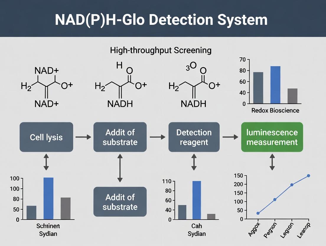
Core Bioluminescent Reaction Mechanism
The assay employs a proprietary thermostable luciferase (Ultra-Glo Recombinant Luciferase) in a coupled enzymatic reaction that converts NAD(P)H into a luminescent signal.
Key Reaction Steps:
- Prolyl Substrate Reduction: A proluciferin substrate is reduced by NAD(P)H in a reaction catalyzed by a reductase enzyme.
- Luciferin Formation: The reduced proluciferin converts to luciferin.
- Light Production: The thermostable luciferase utilizes the newly formed luciferin, ATP, and oxygen to produce light (λmax ~560 nm).
- Signal Stabilization: The proprietary luciferase and buffer system generate a stable, "glow-type" signal lasting over hours, compatible with automated screening platforms.
Quantitative Performance Data
Table 1: Key Performance Metrics of the NAD(P)H-Glo Assay
| Parameter | Value/Range | Notes |
|---|---|---|
| Detection Limit | < 0.1 pmol of NADH in 384-well format | Enables detection in small sample volumes. |
| Dynamic Range | 3-4 orders of magnitude (e.g., 0.1 nM to 1 µM NADH) | Suitable for both high and low abundance samples. |
| Signal Half-Life | > 5 hours | "Glow-type" kinetics ideal for batch processing in HTS. |
| Z'-Factor (HTS suitability) | Typically >0.7 | Excellent for robust high-throughput screening. |
| Sample Compatibility | Cell lysates, purified enzymes, serum | Homogeneous "add-mix-measure" format. |
| Assay Time | < 1 hour post-reagent addition | Fast turnaround for screening campaigns. |
Table 2: Example Data from an Inhibitor Screen (Dehydrogenase Target)
| Condition | Luminescence (RLU) | % Control | Standard Deviation (n=4) |
|---|---|---|---|
| No Enzyme Control (Background) | 1,250 | 0.5% | 45 |
| DMSO Vehicle Control (100% Activity) | 250,000 | 100% | 8,200 |
| Reference Inhibitor A (10 µM) | 25,500 | 10.2% | 950 |
| Test Compound X (10 µM) | 150,000 | 60% | 6,500 |
Detailed Experimental Protocols
Protocol 1: Determining NAD(P)H Concentration in Cultured Cells (384-well format)
A. Materials & Reagents
- NAD(P)H-Glo Detection Reagent (lyophilized or ready-to-use)
- Cultured cells in white-walled, clear-bottom 384-well plates
- Appropriate cell culture medium and lysis buffer (often included in or compatible with the detection reagent)
- NADH standard curve dilutions (0-10 µM in assay buffer)
- Plate-reading luminometer compatible with 384-well plates
B. Procedure
- Cell Preparation: Seed cells at optimal density (e.g., 5,000 cells/well) in 25 µL medium. Incubate overnight.
- Treatment: Add test compounds in 5 µL volume. Incubate for desired duration (e.g., 2-48h).
- Assay Initiation: Equilibrate NAD(P)H-Glo Detection Reagent to room temperature. Add 30 µL directly to all wells (final volume ~60 µL). Mix briefly on an orbital shaker.
- Incubation: Incubate plate at room temperature for 30-60 minutes to allow cell lysis and signal stabilization.
- Detection: Measure luminescence on a plate-reading luminometer with an integration time of 0.5-1 second/well.
- Analysis: Generate a standard curve from NADH standards and interpolate unknown sample values. Report as pmol/well or normalize to cell count/protein.
Protocol 2: Biochemical Assay for Dehydrogenase/Reductase Activity
A. Materials & Reagents
- Purified recombinant enzyme
- Enzyme-specific substrate and cofactors (e.g., NAD⁺ or NADP⁺)
- Assay buffer optimized for enzyme activity
- Test inhibitors in DMSO (final DMSO ≤1%)
- NAD(P)H-Glo Detection Reagent
B. Procedure
- Reaction Setup: In a white 384-well plate, combine in order:
- 10 µL enzyme in assay buffer.
- 5 µL test compound or DMSO control.
- 10 µL substrate/cofactor mix. Pre-incubate 10-15 minutes.
- Reaction Initiation: Start the enzymatic reaction by adding the limiting component (often substrate). Incubate at desired temperature (e.g., 25-37°C) for 30 minutes.
- Detection: Add 25 µL NAD(P)H-Glo Detection Reagent to stop the reaction and initiate the luminescent detection. Incubate 30 min at RT.
- Measurement: Read luminescence.
- Analysis: Calculate enzyme activity as the rate of NAD(P)H production. Plot inhibitor dose-response curves to determine IC₅₀ values.
Signaling Pathways and Workflow Diagrams
Title: NAD(P)H-Glo Core Reaction Pathway
Title: Cell-Based NAD(P)H Detection Workflow
Title: Biochemical Enzyme Activity Assay Principle
The Scientist's Toolkit: Key Research Reagent Solutions
Table 3: Essential Materials for NAD(P)H-Glo Assay Implementation
| Item | Function & Role in Assay | Key Considerations for HTS |
|---|---|---|
| NAD(P)H-Glo Detection Reagent (Core) | Proprietary, lyophilized or liquid formulation containing the reductase, proluciferin substrate, and Ultra-Glo Luciferase in an optimized buffer. Generates the bioluminescent signal. | Single-addition, "just-add" reagent enables homogeneous assay format critical for automation. Stable glow signal minimizes timing constraints. |
| White Multiwell Plates (384/1536) | Maximizes light signal collection and minimizes well-to-well crosstalk. | Low-volume, small-footprint plates are standard for HTS. Solid white walls are essential; clear bottoms optional for microscopy. |
| NADH / NADPH Standards | Used to generate a standard curve for quantifying absolute amounts of NAD(P)H in unknown samples. | High-purity, lyophilized stocks required. Must be prepared fresh in relevant assay buffer. |
| Cell Lysis Reagent (if not in kit) | Efficiently and rapidly lyses cells to release intracellular NAD(P)H for detection. Must be compatible with the luciferase reaction. | Non-lytic protocols also exist for real-time monitoring. For endpoint HTS, a rapid, complete lysis is key. |
| DMSO-Tolerant Assay Buffer | Maintains enzyme activity (for biochemical assays) and compound solubility. | Final DMSO concentration must be normalized and kept low (typically ≤1%) to avoid assay interference. |
| Liquid Handling Robotics | For automated reagent dispensing, compound addition, and plate stacking in large-scale screens. | The assay's homogeneous, single-addition format is highly amenable to automation on standard platforms. |
The NAD(P)H-Glo Detection System is a bioluminescent, homogeneous assay that quantifies NAD(P)H and NAD(P)+ by coupling reductase activity to a luminescent signal. Within high-throughput screening (HTS) research for drug discovery, this system provides a critical tool for directly targeting a vast array of dehydrogenases, reductases, and oxidoreductases (collectively, Oxidoreductases). These enzyme families are central to disease pathways such as cancer metabolism, microbial pathogenesis, and neurodegenerative disorders. By enabling rapid, sensitive, and selective quantification of cofactor turnover, the NAD(P)H-Glo system facilitates the identification of modulators (inhibitors/activators) of these target enzymes in a 96- or 384-well plate format, supporting lead compound discovery in phenotypic and target-based screens.
Application Notes: Targeting Disease-Relevant Oxidoreductases
2.1 Cancer: Targeting Metabolic Reprogramming via Dehydrogenases Cancer cells undergo metabolic reprogramming (Warburg effect and beyond), increasing dependence on specific dehydrogenases. Lactate dehydrogenase (LDH-A) and isocitrate dehydrogenase (IDH1/2) are prominent oncology targets.
- LDH-A Inhibition: Drives glycolysis, converting pyruvate to lactate while regenerating NAD+. Inhibition suppresses proliferation and induces oxidative stress.
- IDH1/2 Inhibition/Mutant Targeting: Gain-of-function mutations in IDH1/2 produce the oncometabolite 2-hydroxyglutarate (2-HG). Inhibitors (e.g., Ivosidenib) normalize cellular differentiation.
- Application: The NAD(P)H-Glo assay is configured to screen for LDH-A inhibitors by monitoring NADH depletion or for mutant IDH1 inhibitors by monitoring NADPH production.
2.2 Infectious Disease: Targeting Microbial Reductases Bacterial and parasitic pathogens often possess essential, divergent oxidoreductases absent in humans, offering selective drug targets.
- Dihydrofolate Reductase (DHFR) in Plasmodium: Critical for folate synthesis in malaria parasites. Pyrimethamine targets parasitic DHFR.
- Enoyl-Acyl Carrier Protein Reductase (FabI) in Mycobacterium tuberculosis: Essential for mycolic acid synthesis in tuberculosis. Isoniazid is a prodrug activated by bacterial catalase-peroxidase.
- Application: Screening for novel antimicrobials by quantifying NAD(P)H consumption in pathogen-specific reductase assays.
2.3 Neurodegenerative Disease: Oxidative Stress and Reductase Defense Alzheimer's and Parkinson's diseases involve oxidative stress. Protective cellular systems like aldose reductase (in polyol pathway) and thioredoxin reductase (TrxR) are potential targets for neuroprotection.
- Aldose Reductase (AR) Inhibition: Reduces glucose to sorbitol, consuming NADPH. Hyperactivity in diabetes contributes to neuropathic complications.
- Thioredoxin Reductase (TrxR) Modulation: Maintains redox balance via NADPH. Its dysfunction exacerbates neuronal oxidative damage.
- Application: HTS for AR inhibitors by measuring NADPH oxidation, or for TrxR activators to bolster cellular antioxidant capacity.
Table 1: Key Oxidoreductase Targets in Disease Pathways
| Disease Area | Target Enzyme (EC Class) | Cofactor Link | Role in Disease Pathway | Example Drug/Modulator |
|---|---|---|---|---|
| Oncology | Lactate Dehydrogenase A (LDH-A, EC 1.1.1.27) | NADH ➔ NAD+ | Glycolytic flux, tumor growth & metastasis | FX-11 (experimental) |
| Oncology | Isocitrate Dehydrogenase 1 (IDH1 mutant, EC 1.1.1.42) | NADP+ ➔ NADPH | Production of oncometabolite 2-HG | Ivosidenib (FDA-approved) |
| Infectious Disease | Plasmodium Dihydrofolate Reductase (DHFR, EC 1.5.1.3) | NADPH ➔ NADP+ | Pyrimidine synthesis for DNA replication | Pyrimethamine |
| Infectious Disease | M. tuberculosis InhA (Enoyl-ACP reductase, EC 1.3.1.9) | NADH ➔ NAD+ | Mycolic acid cell wall biosynthesis | Isoniazid (activated form) |
| Neurodegeneration | Aldose Reductase (AR, EC 1.1.1.21) | NADPH ➔ NADP+ | Polyol pathway, osmotic & oxidative stress | Epalrestat |
| Neurodegeneration | Thioredoxin Reductase (TrxR, EC 1.8.1.9) | NADPH ➔ NADP+ | Antioxidant defense, redox homeostasis | Auranofin (inhibitor) |
Detailed Experimental Protocols
Protocol 1: HTS for Lactate Dehydrogenase A (LDH-A) Inhibitors Using NAD(P)H-Glo
Objective: Identify small molecule inhibitors of human LDH-A in a 384-well plate format.
Materials:
- Recombinant human LDH-A protein
- NAD(P)H-Glo Detection Reagent (lyophilized)
- NADH (substrate)
- Sodium Pyruvate (substrate)
- LDH Assay Buffer (e.g., Tris-HCl, pH 7.5)
- Test compound library (in DMSO)
- Known inhibitor (e.g., GSK2837808A, for controls)
- 384-well white, solid-bottom assay plates
- Plate reader capable of measuring luminescence
Workflow:
- Plate Preparation: Dispense 2.5 µL of test compound or control (DMSO for 100% activity, reference inhibitor for 0% activity) into assay plates using an acoustic liquid handler. Include a no-enzyme control for background.
- Enzyme/Substrate Addition: Prepare a master mix containing LDH-A (final 5 nM) and NADH (final 50 µM) in LDH Assay Buffer. Add 22.5 µL of this master mix to all wells. Pre-incubate for 15 minutes at room temperature.
- Reaction Initiation: Initiate the enzymatic reaction by adding 5 µL of sodium pyruvate solution (final concentration 1 mM) to all wells. Incubate for 30-60 minutes at room temperature.
- Detection: Prepare the NAD(P)H-Glo Detection Reagent according to the manufacturer's instructions. Add 25 µL of the detection reagent to each well to stop the enzymatic reaction and initiate the luminescent detection reaction.
- Signal Measurement: Incubate for 30-60 minutes at room temperature (signal stabilizes). Measure luminescence on a plate reader.
- Data Analysis: Normalize signals: % Inhibition = [(Avg. High Ctrl (DMSO) – Sample) / (Avg. High Ctrl – Avg. Low Ctrl (Ref. Inhibitor))] x 100. Calculate Z'-factor for quality control.
Protocol 2: Screening for Plasmodium falciparum DHFR Inhibitors
Objective: Screen compound libraries for inhibitors of P. falciparum DHFR to discover novel antimalarials.
Materials:
- Recombinant P. falciparum DHFR enzyme
- NAD(P)H-Glo Detection Reagent
- NADPH (cofactor)
- Dihydrofolate (DHF, substrate)
- DHFR Assay Buffer (Potassium Phosphate, pH 7.4, with 1 mM DTT)
- Test compounds and reference inhibitor (Pyrimethamine)
- 384-well white plates
Workflow:
- Plate Preparation: Dispense 5 nL of compound/DMSO into plates.
- Enzyme Addition: Add 20 µL of PfDHFR (final 2 nM) in assay buffer. Pre-incubate 15 min.
- Reaction Initiation: Add 5 µL of a substrate/cofactor mix containing DHF (final 10 µM) and NADPH (final 50 µM).
- Incubation & Detection: Incubate for 60 min at 25°C. Add 25 µL of NAD(P)H-Glo Detection Reagent, incubate 40 min, and read luminescence.
- Analysis: The signal is proportional to remaining NADPH. Inhibitors increase luminescence. Calculate % inhibition relative to DMSO and pyrimethamine controls.
Visualizations: Pathways & Workflows
LDH-A HTS Screening Protocol Flow
Oxidoreductase Targets in Cancer Metabolism
The Scientist's Toolkit: Research Reagent Solutions
Table 2: Essential Materials for Oxidoreductase HTS using NAD(P)H-Glo
| Item / Reagent Solution | Function in Assay | Key Consideration |
|---|---|---|
| NAD(P)H-Glo Detection System | Core bioluminescent detection. Converts remaining NAD(P)H into a stable luminescent signal via reductase/enhancer coupling. | Homogeneous, "add-and-read". Sensitive to femtomole levels of cofactor. |
| Recombinant Target Enzyme | The disease-relevant oxidoreductase (e.g., LDH-A, PfDHFR). Source (human, parasitic) and purity are critical for specificity. | Requires optimization of enzyme concentration (signal-to-background, linear range). |
| NADH or NADPH Cofactor | Primary substrate for the detection system and often a co-substrate for the target enzyme. | Choice depends on enzyme specificity (NAD vs NADP). Stability in buffer must be considered. |
| Chemical Substrate | The enzyme-specific substrate (e.g., pyruvate for LDH, DHF for DHFR). | Km should be determined; use near-Km concentration for robust assay window. |
| HTS-Compatible Assay Plates | White, solid-bottom plates for optimal luminescence signal collection. | 384-well or 1536-well format for screening. Low protein binding is beneficial. |
| Reference Inhibitors/Activators | Pharmacological controls for validation and data normalization (e.g., GSK2837808A for LDH-A). | Validates target engagement and establishes assay performance (Z' > 0.5). |
| Automated Liquid Handlers | For precise, high-throughput dispensing of compounds, enzymes, and reagents. | Essential for miniaturization, reproducibility, and screening 10,000+ compounds. |
| Luminescence Plate Reader | Quantifies the endpoint luminescent signal from the detection reaction. | Requires sensitivity, dynamic range, and fast read times for HTS. |
Within high-throughput screening (HTS) research, particularly for assays utilizing the NAD(P)H-Glo Detection System, three interconnected technical advantages are paramount: the homogeneous "add-and-read" format, exceptional sensitivity, and a broad dynamic range. These features collectively enable the rapid, robust, and reliable identification of modulators of enzymes that utilize or produce NAD(P)H, a critical cofactor in numerous metabolic and signaling pathways. This application note details these advantages within the context of a protocol for screening NAD(P)H-dependent enzymes, providing researchers with a framework for optimal assay development.
Core Advantages: A Comparative Analysis
Homogeneous "Add-and-Read" Format
A homogeneous assay format requires no separation steps (e.g., washing, filtration) between reagent addition and signal measurement. The NAD(P)H-Glo System operates on this principle, where a single reagent addition simultaneously stops the enzymatic reaction and initiates the luminescent detection. This format is ideal for HTS.
Key Benefits:
- Minimized Hands-on Time & Complexity: Enables automation-friendly workflows.
- Reduced Variability: Eliminates errors associated with multiple liquid handling steps.
- Increased Throughput: Allows for rapid processing of 384- or 1536-well plates.
Sensitivity and Dynamic Range
Sensitivity defines the lowest detectable concentration of an analyte. Dynamic range is the span over which a change in analyte concentration produces a proportional change in signal. The NAD(P)H-Glo System converts NAD(P)H into a stabilized luminescent signal via a coupled enzymatic reaction, offering superior performance over traditional absorbance (UV/Vis) methods.
Quantitative Performance Data:
Table 1: Comparison of NAD(P)H Detection Methods
| Detection Method | Format | Assay Time | Sensitivity (NADH) | Dynamic Range | Suitable for HTS? |
|---|---|---|---|---|---|
| Absorbance (340 nm) | Homogeneous | Fast (seconds) | ~1-10 µM | ~1-2 logs | Limited (low S/B, interference) |
| Fluorescence | Homogeneous | Fast (seconds) | ~10-100 nM | ~2-3 logs | Yes (but prone to compound interference) |
| NAD(P)H-Glo Luminescence | Homogeneous | Moderate (30-60 min) | < 1 nM | > 4-5 logs | Excellent (robust, minimal interference) |
Table 2: Exemplar HTS Performance Metrics for a Kinase Dehydrogenase Screen
| Performance Parameter | Result (Mean ± SD, n=4 plates) |
|---|---|
| Z'-Factor | 0.78 ± 0.04 |
| Signal-to-Background (S/B) Ratio | 12.5 ± 1.2 |
| Coefficient of Variation (CV) of High Control | 4.2% ± 0.8% |
| Coefficient of Variation (CV) of Low Control | 5.1% ± 1.1% |
Detailed Protocol: HTS for an NADH-Producing Dehydrogenase Inhibitor
Objective: To identify small-molecule inhibitors of a target dehydrogenase enzyme in a 384-well plate format using the NAD(P)H-Glo Detection System.
Research Reagent Solutions Toolkit
Table 3: Essential Materials and Reagents
| Item | Function/Description |
|---|---|
| NAD(P)H-Glo Detection Reagent | Single lyophilized or liquid reagent containing substrates for the coupled luminescent reaction. Reconstituted in specified buffer. |
| Recombinant Target Dehydrogenase | Purified enzyme. Store and dilute in assay buffer per stability guidelines. |
| Enzyme Substrate | Specific substrate for the target dehydrogenase. Prepared in assay buffer. |
| NAD⁺ Cofactor | Essential reaction cofactor for dehydrogenase activity. |
| Positive Control Inhibitor | A known potent inhibitor (e.g., reaction product or specific chemical inhibitor) for assay validation. |
| DMSO | Universal solvent for compound libraries. Final concentration in assay must be normalized (typically ≤1%). |
| White, Solid-Bottom 384-Well Plates | Optically optimal plates for luminescence detection, minimizing cross-talk. |
| Assay Buffer | Typically a physiologically-relevant buffer (e.g., Tris or PBS at optimal pH). May contain stabilizing agents like BSA. |
| HTS-Compatible Liquid Handler | For precise, nanoliter-scale dispensing of compounds and reagents. |
| Plate Reader with Luminescence Detector | Capable of reading 384-well plates with integration times of 0.1-1 second/well. |
Experimental Workflow
Protocol Steps:
- Plate Preparation: Dispense 20 nL of compound (in DMSO) or control into assigned wells of a 384-well plate using a pintool or acoustic dispenser.
- Enzyme/Substrate Mixture Preparation: Prepare a master mix containing the target dehydrogenase (at the predetermined Km or EC₅₀ concentration) and its substrate in assay buffer. Keep on ice.
- Reaction Initiation: Using a bulk dispenser, add 5 µL of the enzyme/substrate master mix to all wells of the assay plate. Centrifuge briefly (500 rpm, 30 sec) to ensure mixing and no bubbles.
- Incubation: Incubate the plate at room temperature for the predetermined optimal time (e.g., 30 minutes) to allow the enzymatic conversion and NADH production.
- Detection Reagent Addition: Add 5 µL of the room-temperature, reconstituted NAD(P)H-Glo Detection Reagent to each well. This addition simultaneously quenches the dehydrogenase reaction and initiates the luminescent detection reaction.
- Signal Development: Incubate the plate at room temperature for 30-60 minutes to allow the luminescent signal to stabilize.
- Signal Measurement: Read plate luminescence on a compatible plate reader with an integration time of 0.5 seconds/well.
- Data Analysis: Normalize data: % Inhibition = [(Median Low Control - Test Compound) / (Median Low Control - Median High Control)] * 100. Calculate statistical robustness parameters (Z'-factor, S/B).
Biochemical Principle of Detection
The core mechanism involves a coupled enzymatic reaction that converts NAD(P)H into a quantifiable luminescent signal.
Step-by-Step HTS Protocol: From Plate Setup to Data Acquisition
Within the context of a high-throughput screening (HTS) research program utilizing the NAD(P)H-Glo Detection System, the reliability and signal-to-noise ratio of assays are fundamentally dependent on meticulous materials and reagent preparation. This protocol details the optimization of buffer conditions and substrate stability to ensure robust, reproducible detection of NAD(P)H-consuming or -producing enzymes in drug discovery campaigns. Proper execution mitigates common pitfalls such as high background luminescence, enzymatic instability, and substrate hydrolysis.
Key Research Reagent Solutions
The following table catalogs essential materials for establishing an optimized NAD(P)H-Glo assay workflow.
| Reagent / Material | Function & Rationale |
|---|---|
| NAD(P)H-Glo Detection Reagent (Lyophilized) | Core luciferase-based detection system. Reconstituted reagent stability is paramount. |
| Recombinant Target Enzyme (e.g., Kinase, Dehydrogenase) | The enzymatic activity being modulated in the HTS campaign. Buffer compatibility is critical. |
| NAD⁺ or NADP⁺ Cofactor | Enzyme substrate. Solution pH affects stability; prepare fresh or aliquot and store at -80°C. |
| Assay Buffer (Optimized Tris or HEPES) | Maintains enzyme activity and compatibility with detection chemistry. Ionic strength and pH are key variables. |
| Test Inhibitors / Compound Library | Small molecules screened for modulation of target enzyme activity. Dissolved in DMSO; final concentration ≤1%. |
| White, Solid-Bottom 384-Well Plates | Minimizes light crosstalk and maximizes luminescent signal capture for HTS. |
| Non-ionic Surfactant (e.g., 0.01% Tween-20) | Reduces compound and protein adsorption to plasticware, improving well-to-well reproducibility. |
| Dithiothreitol (DTT) or Tris(2-carboxyethyl)phosphine (TCEP) | Maintains reduction state of cysteine residues in enzymes. Can interfere with luciferase if carried over. |
Optimized Buffer Composition and Stability Data
Systematic testing of buffer components identifies conditions that maximize enzymatic activity while maintaining low background and detection reagent integrity. The following table summarizes quantitative findings.
| Buffer Parameter | Tested Range | Optimal Condition for Kinase X Assay | Impact on NAD(P)H-Glo Signal (vs. Standard Buffer) |
|---|---|---|---|
| pH (HEPES Buffer) | 6.8 - 8.2 | pH 7.5 | 45% increase in Z'-factor due to lower background and maintained enzyme velocity. |
| Mg²⁺ Concentration | 0 - 10 mM | 5 mM | Essential for kinase activity; >8 mM increases precipitate with detection reagent. |
| DTT Concentration | 0 - 2 mM | 0.5 mM (in enzyme pre-dilution only) | >1 mM in final assay quenches luminescence by ~30%. Use TCEP as alternative. |
| Bovine Serum Albumin (BSA) | 0 - 0.1% | 0.01% | Stabilizes dilute enzyme, reduces plate adsorption. Higher % increases background. |
| Reconstituted Detection Reagent Stability (4°C) | 0 - 8 hours | < 4 hours | Background increases linearly (~15% per hour) after 4 hours; use immediately. |
| NAD⁺ Stock Solution Stability (-80°C) | 1 month | Aliquoted, single-use | No activity loss after 30 days. Three freeze-thaw cycles degrade activity by 25%. |
Detailed Protocol: Optimized Reagent Preparation and Assay Setup
Part A: Buffer and Substrate Preparation
- 5X Assay Buffer (500 mL): 250 mM HEPES (pH 7.5 @ 25°C), 25 mM MgCl₂, 0.05% BSA (protease-free), 0.05% Tween-20. Filter sterilize (0.22 µm), store at 4°C for up to 2 weeks.
- 10 mM NAD⁺ Stock: Reconstitute lyophilized NAD⁺ in nuclease-free water. Aliquot 20 µL into single-use PCR tubes. Flash-freeze in liquid nitrogen and store at -80°C. Avoid thaw cycles.
- Enzyme Dilution Buffer: 1X Assay Buffer supplemented with 0.5 mM DTT (or 0.1 mM TCEP). Prepare fresh and keep on ice.
- Detection Reagent: Reconstitute lyophilized NAD(P)H-Glo Detection Substrate with the specified volume of Buffer. Vortex for 60 seconds until fully dissolved. Use within 4 hours. Keep protected from light at room temperature until use.
Part B: HTS-Compatible Assay Workflow
- Day 1 - Plate Compound: Using an acoustic or pintool dispenser, transfer 50 nL of compound library (in DMSO) to white 384-well assay plates. Include control wells (100% activity, 0% activity with inhibitor).
- Prepare Reaction Mix (2X Concentration): In order, combine:
- 1X Assay Buffer
- Target Enzyme (2X final desired concentration in Enzyme Dilution Buffer)
- NAD⁺ (2X final concentration, typically 10-100 µM).
- Keep on ice.
- Initiate Reaction: Add 5 µL of the 2X Reaction Mix to each well of the compound plate using a multidispenser. Centrifuge plates briefly (500 rpm, 30 sec).
- Incubate: Seal plates and incubate at room temperature (or desired assay temperature) for the predetermined enzymatic linear range (e.g., 30-60 minutes).
- Stop and Detect: Add 10 µL of reconstituted NAD(P)H-Glo Detection Reagent to each well. Seal, shake plates on an orbital shaker for 2 minutes, and incubate at room temperature for 30 minutes to allow signal development.
- Read Luminescence: Read plates on a compatible plate reader (e.g., PerkinElmer EnVision, BMG CLARIOstar) using an integration time of 0.5-1 second per well.
Visualization of Protocols and Pathways
Titles:
- HTS Assay Workflow with NAD(P)H-Glo
- NAD(P)H-Glo Detection Principle
This application note details the comparative timelines for cell-based and biochemical assays conducted in microplates, specifically within the framework of high-throughput screening (HTS) research utilizing NAD(P)H-Glo Detection Systems. This detection platform provides a bioluminescent method for quantifying NAD(P)H, serving as a universal indicator of enzymatic activity in biochemical assays or cellular metabolic health in cell-based assays. Understanding the distinct workflows and time investments for each assay type is critical for efficient screening campaign planning in drug discovery.
The following table outlines the major phases and estimated time commitments for typical microplate-based assays using NAD(P)H detection. Times are estimates for a 384-well plate format.
Table 1: Detailed Timeline Comparison: Cell-Based vs. Biochemical NAD(P)H Assays
| Phase | Step | Cell-Based Assay (Adherent Cells) | Biochemical Assay (Purified Enzyme) | Notes |
|---|---|---|---|---|
| Phase 1: Preparation | Plate Coating (if needed) | 1-24 hours (overnight common) | Not Applicable | For extracellular matrix components. |
| Cell Seeding & Incubation | 6-24 hours (overnight) | Not Applicable | Time for cells to adhere and resume normal growth. | |
| Compound/Reagent Preparation | 1-2 hours | 1-2 hours | Includes compound dilution series, control prep. | |
| Phase 2: Assay Execution | Compound Addition & Incubation | Variable: 5 min - 72+ hours | 5-30 minutes | Treatment time is experiment-dependent. |
| Assay Reagent Addition (NAD(P)H-Glo) | 15-30 minutes | 15-30 minutes | Homogeneous "add-mix-measure" protocol. | |
| Signal Development Incubation | 30-60 minutes | 30-60 minutes | Time for luminescent signal to stabilize. | |
| Phase 3: Readout & Analysis | Microplate Reading | 5-10 minutes | 5-10 minutes | Luminescence read on a compatible reader. |
| Data Analysis (Primary) | 1-2 hours | 1-2 hours | Normalization, hit identification (Z' calculation). | |
| Total Hands-On Time | ~3-5 hours | ~2-3 hours | Excludes long incubation periods. | |
| Total Elapsed Time | 24 - 96+ hours | ~1.5 - 3 hours | Critical for scheduling. |
Detailed Experimental Protocols
Protocol 3.1: Biochemical Enzyme Activity Assay Using NAD(P)H-Glo
Objective: To screen for inhibitors of a dehydrogenase enzyme using purified protein in a 384-well microplate format.
Materials:
- White, solid-bottom 384-well assay plates.
- Purified target dehydrogenase enzyme.
- Enzyme substrate and cofactor (e.g., NAD⁺ or NADP⁺).
- Test compounds in DMSO.
- NAD(P)H-Glo Detection Reagent.
- Appropriate assay buffer.
- Multichannel pipettes, plate shaker, microplate luminometer.
Method:
- Plate Preparation: Dispense 2.5 µL of compound in DMSO or control (DMSO only for 100% activity, known inhibitor for background) to the assay plate using an acoustic dispenser or pin tool.
- Enzyme/Substrate Mix: Prepare a master mix containing the dehydrogenase enzyme and its required substrate in assay buffer. Keep cofactor (NAD⁺) separate if the enzyme reaction is to be initiated simultaneously.
- Reaction Initiation: Add 22.5 µL of the enzyme/substrate master mix to all wells, initiating the biochemical reaction. Final DMSO concentration should be ≤1%.
- Incubation: Incubate plate at room temperature for 30 minutes.
- Detection: Add 25 µL of NAD(P)H-Glo Detection Reagent to all wells. Mix thoroughly on a plate shaker for 30 seconds.
- Signal Development: Incubate plate at room temperature for 30-60 minutes to allow luminescent signal to develop and stabilize.
- Measurement: Read luminescence on a compatible microplate luminometer.
- Data Analysis: Calculate % inhibition relative to controls (100% activity = DMSO control; 0% activity = background/no-enzyme or full-inhibition control). Calculate Z'-factor to validate assay quality.
Protocol 3.2: Cell Viability/Proliferation Assay Using NAD(P)H-Glo
Objective: To assess compound cytotoxicity or effects on cellular metabolism in adherent cells.
Materials:
- White, clear-bottom 384-well cell culture plates.
- Adherent cell line (e.g., HepG2, HeLa).
- Complete cell culture medium.
- Trypsin-EDTA, PBS.
- Test compounds in DMSO.
- NAD(P)H-Glo Detection Reagent.
- Cell culture incubator (37°C, 5% CO₂).
- Multichannel pipettes, plate shaker, microplate luminometer.
Method:
- Cell Seeding: Harvest and count cells. Seed 50 µL of cell suspension (e.g., 2,000-5,000 cells/well) in complete medium into each well of the microplate.
- Cell Attachment: Incubate plates overnight (16-24 hours) in a humidified 37°C, 5% CO₂ incubator.
- Compound Treatment: Prepare compound dilutions in medium. Remove plate from incubator and carefully add 10 µL of compound dilution to each well, yielding final desired concentration. Include vehicle (DMSO) control and a cytotoxicity control (e.g., digitonin).
- Treatment Incubation: Return plate to incubator for the desired treatment period (e.g., 48 hours).
- Equilibration: Remove plate from incubator and equilibrate to room temperature for 15-30 minutes.
- Detection: Add 60 µL of NAD(P)H-Glo Detection Reagent directly to each well. Mix gently on an orbital shaker for 2 minutes to induce cell lysis.
- Signal Development: Incubate at room temperature for 30 minutes to stabilize luminescence.
- Measurement: Read luminescence on a microplate luminometer.
- Data Analysis: Normalize data to vehicle control cells (100% viability) and cytotoxicity control (0% viability). Calculate IC₅₀ values as applicable.
Visualization of Workflows and Pathways
Title: NAD(P)H-Glo Biochemical Detection Pathway
Title: Cell-Based vs. Biochemical Assay Workflow Timeline
The Scientist's Toolkit: Essential Research Reagent Solutions
Table 2: Key Reagents and Materials for NAD(P)H-Glo Microplate Assays
| Item | Function in Assay | Key Considerations |
|---|---|---|
| NAD(P)H-Glo Detection Reagent | Core detection solution. Contains a proprietary proluciferin substrate and a reductase-luciferase enzyme. Converts NAD(P)H to a luminescent signal proportional to its concentration. | Homogeneous, "add-mix-measure" format. Compatible with 96-, 384-, and 1536-well formats. Stable signal. |
| White, Solid-Bottom Microplates | Optimal for luminescence detection. White walls reflect light to the detector, maximizing signal and sensitivity. | Essential for biochemical and endpoint cell assays. Low luminescence background is critical. |
| Cell Culture Microplates | For cell-based assays. Clear-bottom plates allow for microscopic inspection. Opaque/white walls are best for final read. | Tissue-culture treated for cell adherence. May opt for white walls with clear bottom. |
| Recombinant Dehydrogenase Enzyme | Target protein for biochemical screening. Catalyzes the conversion of substrate and NAD(P)⁺ to product and NAD(P)H. | Requires purity, stability, and known kinetic parameters (Km for substrate/cofactor). |
| NAD⁺ or NADP⁺ Cofactor | Essential reactant for dehydrogenase enzymes. Its reduction to NAD(P)H is the measured event. | Concentration must be optimized to be non-limiting in the reaction for robust window. |
| Cell Lines (e.g., HepG2, HEK293) | Model systems for cell-based assays. Intracellular NAD(P)H pools reflect metabolic activity/viability. | Choose relevant disease/pathway model. Seeding density and health are critical for reproducibility. |
| DMSO (Cell Culture Grade) | Universal solvent for small-molecule compound libraries. | Final concentration in assay (typically ≤0.5-1%) must be non-toxic and not interfere with detection. |
| Lysis Buffer (for cell assays) | Optional component to ensure complete cell lysis upon reagent addition, releasing all NAD(P)H. | The NAD(P)H-Glo reagent itself has lysing properties for many cell types. Optimization may be needed. |
Within high-throughput screening (HTS) research utilizing the NAD(P)H-Glo Detection System, rigorous plate layout design is critical for robust data interpretation. This protocol details the establishment of controls and compound dilution series for assays measuring NAD(P)H-dependent dehydrogenase activity or the generation of NAD(P)H in coupled systems. Proper controls normalize for systematic variability, while well-designed dilution series enable accurate concentration-response analyses for hit identification and validation in drug discovery pipelines.
Key Control Definitions and Functions
Positive Controls
Positive controls are wells that produce a maximum assay signal. They are used to:
- Normalize compound activity data (e.g., % inhibition or activation).
- Monitor assay performance and robustness across plates and days.
- Calculate the Z'-factor, a statistical measure of assay quality.
Typical Positive Controls for NAD(P)H-Glo Assays:
- For Dehydrogenase Inhibition Screens: A well-characterized, potent inhibitor of the target dehydrogenase.
- For Enzyme Activation Screens: The known substrate at saturating concentration or a known allosteric activator.
- For Coupled Systems: A reaction component known to generate maximal NAD(P)H.
Negative Controls
Negative controls are wells that produce a minimum assay signal (background). They are used to:
- Define the baseline signal.
- Assess non-specific compound interference or assay artifacts.
- Calculate signal-to-background (S/B) and signal-to-noise (S/N) ratios.
Typical Negative Controls for NAD(P)H-Glo Assays:
- For Inhibition Screens: Vehicle-only control (DMSO, buffer) with active enzyme, representing 0% inhibition.
- For Activation Screens: Vehicle-only control with enzyme, representing baseline activity.
- No-Enzyme Control: Contains all reagents except the target enzyme, defining the background luminescence of the detection system.
Compound Dilution Series
A systematic concentration range of test compounds, typically prepared via serial dilution, is essential for determining potency (e.g., IC₅₀, EC₅₀). In HTS, this is often applied during secondary confirmation and dose-response experiments.
Standard Plate Layout for a 384-Well HTS Campaign
The following layout incorporates controls for a primary screen and a dose-response confirmation plate.
Table 1: 384-Well Plate Layout for Primary Screening
| 1 | 2-23 (Test Compounds) | 24 | |
|---|---|---|---|
| A-P | Column 1: Negative Control (n=16) | Columns 2-23: Single-Concentration Test Compounds (n=352) | Column 24: Positive Control (n=16) |
Table 2: 384-Well Plate Layout for Dose-Response Confirmation
| 1-2 | 3-22 | 23-24 | |
|---|---|---|---|
| A-P | Columns 1-2: Negative Control (n=32) | Columns 3-22: 10-Point, 1:3 Serial Dilution of Hit Compounds (n=320) | Columns 23-24: Positive Control (n=32) |
Table 3: Example 10-Point, 1:3 Serial Dilution Series (Top Conc. = 10 µM)
| Well Column | 3 | 4 | 5 | 6 | 7 | 8 | 9 | 10 | 11 | 12 |
|---|---|---|---|---|---|---|---|---|---|---|
| Dilution Factor | 1 | 1:3 | 1:9 | 1:27 | 1:81 | 1:243 | 1:729 | 1:2187 | 1:6561 | 1:19683 |
| Final Conc. (µM) | 10.0 | 3.33 | 1.11 | 0.37 | 0.123 | 0.041 | 0.014 | 0.0046 | 0.0015 | 0.0005 |
Detailed Experimental Protocols
Protocol 1: Preparation of Control and Compound Plates for Primary Screening
Objective: To dispense controls and test compounds into assay-ready microplates. Materials: 384-well white solid-bottom plate, DMSO, positive control inhibitor/activator stock, test compound libraries (e.g., 10 mM in DMSO), liquid handler. Procedure:
- Pre-dispense Vehicle: Using a liquid handler, dispense 20 nL of DMSO into all wells of columns 2-23 (test compound area).
- Dispense Test Compounds: Transfer 20 nL of each test compound from the source library plate to the corresponding destination well in columns 2-23. Final DMSO concentration is typically 0.5%.
- Dispense Controls: Dispense 20 nL of DMSO into all wells of column 1 (Negative Control). Dispense 20 nL of positive control stock solution into all wells of column 24 (Positive Control).
- Seal and Store: Seal plates with a lid or adhesive foil. Store desiccated at room temperature or -20°C until assay.
Protocol 2: Performing the NAD(P)H-Glo Assay on a Prepared Compound Plate
Objective: To measure compound effects on NAD(P)H levels in a high-throughput format. Materials: Pre-dispensed compound plate, assay buffer, recombinant dehydrogenase enzyme, substrate, NAD(P)H-Glo Detection Reagent, multi-channel pipettes or dispensers. Procedure:
- Prepare Assay Components: Thaw and prepare all reagents per manufacturer's instructions. Pre-mix enzyme and substrate in assay buffer to 2X final concentration.
- Initiate Biochemical Reaction: Using a dispenser, add 10 µL of the 2X enzyme/substrate mix to all wells of the assay plate. Centrifuge briefly (500 rpm, 30 sec).
- Incubate: Incubate plate at room temperature for the predetermined reaction time (e.g., 30-60 min).
- Add Detection Reagent: Using a dispenser, add 10 µL of the NAD(P)H-Glo Detection Reagent to all wells. The reagent contains luciferase and a pro-luciferin substrate, generating light proportional to NAD(P)H.
- Incubate and Read: Incubate plate at room temperature for 30-60 minutes to allow signal stabilization. Read luminescence on a compatible plate reader.
Protocol 3: Preparing a 10-Point, 1:3 Serial Dilution Series
Objective: To generate a concentration-response curve for confirmed hit compounds. Materials: Hit compound stock (e.g., 10 mM in DMSO), DMSO, 384-well polypropylene dilution plate, liquid handler. Procedure:
- Load Top Concentration: Add 5 µL of DMSO to wells in columns 4-12 of the dilution plate. Add 5 µL of hit compound stock (10 mM) to triplicate wells in column 3. This is the 10 mM source.
- Perform Serial Dilution: Using a liquid handler, perform a 1:3 serial dilution:
- Mix the contents of column 3.
- Transfer 2.5 µL from column 3 to column 4 and mix thoroughly.
- Continue this transfer and mix process from column 4 to 5, and so on, through column 12.
- Discard 2.5 µL from column 12 after mixing.
- Pin Transfer to Assay Plate: Using a pintool, transfer 20 nL from each well of the dilution plate to the corresponding well of a 384-well assay plate, creating the final concentration series as shown in Table 3. Include positive and negative control columns as in Table 2.
The Scientist's Toolkit: Key Reagent Solutions
Table 4: Essential Materials for NAD(P)H-Glo HTS
| Item | Function |
|---|---|
| NAD(P)H-Glo Detection Reagent | Single-reagent, luciferase-based system that quantifies NAD(P)H and NAD(P)+. Generates a luminescent signal proportional to NAD(P)H concentration. |
| White, Solid-Bottom 384-Well Plates | Maximizes luminescent signal collection and minimizes crosstalk between wells during plate reading. |
| Recombinant Dehydrogenase Enzyme | The purified target enzyme of interest. Must be titrated for optimal signal window in the assay. |
| Enzyme-specific Substrate | The physiological or synthetic molecule oxidized/reduced by the target dehydrogenase, coupled to NAD(P)+/NAD(P)H conversion. |
| Potent Inhibitor/Activator (Control Compound) | A well-characterized pharmacological tool for defining the maximum and minimum assay signal range (positive control). |
| Anhydrous DMSO | Universal solvent for small molecule compound libraries. Must be of high purity to prevent enzyme inhibition or degradation. |
| Assay Buffer | Typically a physiologically-relevant buffer (e.g., PBS, Tris-HCl) at optimal pH, containing necessary cofactors (e.g., Mg2+). |
| Automated Liquid Handler / Dispenser | Essential for precision and reproducibility when dispensing nanoliter volumes of compounds and microliter volumes of reagents in 384/1536-well formats. |
Diagrams
Title: Role of Controls in Data Normalization
Title: NAD(P)H-Glo HTS Experimental Workflow
Title: NAD(P)H Detection Principle in Dehydrogenase Assay
Application Notes
Within high-throughput screening (HTS) research utilizing the NAD(P)H-Glo Detection System, precise instrument configuration is paramount. This assay quantifies NAD(P)H by generating a luminescent signal proportional to its concentration. Optimal plate reader configuration minimizes background, maximizes signal-to-noise ratio (S/N), and ensures robust Z'-factor calculations for screening campaigns. Key parameters include integration time, gain, wavelength (if applicable), and temperature control. The following data, compiled from current manufacturer specifications and literature, provides a benchmark for configuration.
Table 1: Recommended Plate Reader Configuration for NAD(P)H-Glo Assay Luminescence Detection
| Parameter | Recommended Setting | Rationale | Impact on Assay Performance |
|---|---|---|---|
| Detection Mode | Luminescence (Glow-type) | NAD(P)H-Glo is a stable "glow" reaction, not requiring injectors. | Eliminates variability from injection timing. |
| Integration Time | 100 - 1000 ms per well | Longer integration collects more photons, improving S/N. Must balance with total read time. | S/N typically increases with √(integration time). Optimize for >10:1 S/N. |
| Gain/PMT Voltage | Medium or Default (Optimized) | Avoid maximum gain to prevent PMT saturation and increased background noise. | Prevents signal saturation; maintains linear dynamic range. |
| Read Height/Position | 1-2 mm above well bottom (or as per plate type) | Minimizes cross-talk between adjacent wells in microplate assays. | Reduces inter-well contamination of signal. |
| Filters | Open or clear luminescence filter | No wavelength selection is required for this broad-spectrum luminescence. | Maximizes signal capture. |
| Plate Temperature | 22-25°C (ambient) or controlled to assay spec. | Luciferase activity is temperature-sensitive. Consistency is critical. | Reduces well-to-well variability; ensures kinetic stability. |
| Automation | Stacker compatibility for HTS | Enables unattended reading of multiple plates. | Essential for throughput; ensures timing consistency. |
Table 2: Typical Performance Metrics for a 384-well NAD(P)H-Glo Assay
| Metric | Value Range | Calculation |
|---|---|---|
| Background Luminescence | 100 - 500 RLU | Mean signal from no-NAD(P)H control wells. |
| Signal (High NAD(P)H) | 10,000 - 50,000 RLU | Mean signal from high-NAD(P)H control wells. |
| Signal-to-Noise Ratio (S/N) | 20:1 - 100:1 | (Mean Signal - Mean Background) / SD of Background |
| Signal-to-Background (S/B) | 10 - 100 | Mean Signal / Mean Background |
| Z'-Factor | 0.5 - 0.8 | 1 - [ (3*(SDhigh + SDlow)) / |Meanhigh - Meanlow| ] |
Experimental Protocols
Protocol 1: Initial Plate Reader Calibration and Validation for Luminescence
Objective: To verify the baseline performance and linear dynamic range of the luminescence detector.
- Materials: Recombinant luciferase (steady light source), NAD(P)H-Glo Assay components, black/white assay plates, plate reader.
- Procedure: a. Prepare a serial dilution of a known luciferase control (or a stable luminescent standard) across a plate. b. Configure the reader per Table 1, starting with manufacturer defaults. c. Read the plate using a range of integration times (e.g., 50 ms, 100 ms, 500 ms, 1000 ms). d. Plot RLU vs. concentration for each integration time. Select the shortest integration time that maintains a linear response (R² > 0.99) over the expected assay range.
- Analysis: Calculate the coefficient of variation (CV) for replicate wells. The optimal configuration yields the lowest CV and highest linearity.
Protocol 2: Configuration Optimization for NAD(P)H-Glo Assay
Objective: To empirically determine the optimal integration time and gain for a specific NAD(P)H-Glo assay protocol.
- Materials: NAD(P)H-Glo Assay Kit, NADH standard, assay buffer, low-volume 384-well white plates, multichannel pipettes, calibrated plate reader.
- Procedure: a. Prepare two sets of controls: "High Signal" (e.g., 1 µM NADH in assay buffer) and "Low Signal" (assay buffer only) in 40 µL volume, 16 replicates each. b. Add 40 µL of NAD(P)H-Glo Detection Reagent to all wells. Incubate at room temperature for 30 min (or as per kit instructions). c. Create a reader method that iterates through a matrix of settings: Gain (Low, Medium, High) and Integration Time (50, 100, 250, 500 ms). d. Read the plate.
- Analysis: For each combination, calculate the S/N and Z'-factor (using the two control groups). The optimal configuration is the one that maximizes Z'-factor while keeping total read time practical for HTS.
Protocol 3: HTS Read Method Setup for a 10-Plate Screen
Objective: To establish an automated, reliable method for screening compound libraries.
- Materials: Configured plate reader with stacker, 10x assay plates (pre-dispensed with compounds, enzymes, and substrate), NAD(P)H-Glo Detection Reagent in bulk dispenser.
- Protocol: a. Pre-read: Initiate kinetic reactions in batches of 5 plates by adding detection reagent via bulk dispenser. Start a 30-minute incubation timer. b. Reader Method: Configure method with optimal settings from Protocol 2. Set read order (e.g., column-wise). Enable data autosave after each plate. c. Automation: Program stacker to feed plates to the reader such that each plate is read at precisely 30 minutes (± 1 min) post-reagent addition. d. Execute run. Include a control plate at the start and end of the stack to monitor assay stability.
- Validation: Monitor the Z'-factor of the control plates. A run is valid if beginning and end control plate Z' > 0.5.
Visualizations
Title: NAD(P)H-Glo Detection Principle
Title: HTS Workflow for NAD(P)H-Glo Assay
The Scientist's Toolkit
Table 3: Essential Research Reagent Solutions for NAD(P)H-Glo HTS
| Item | Function in Assay | Key Consideration |
|---|---|---|
| NAD(P)H-Glo Detection Kit | Provides the optimized, lyophilized substrate and enzyme mixture for selective NAD(P)H detection. | Use bulk kits for HTS; reconstitute fresh or in stable aliquots. |
| White Solid-Bottom Microplates | Maximizes reflection of luminescent signal to the detector; minimizes cross-talk. | Low-volume 384- or 1536-well plates preferred for HTS to reduce reagent costs. |
| Recombinant Target Enzyme | The enzyme consuming NAD(P)H in the primary biochemical reaction. | Must be highly active, pure, and compatible with assay buffer. |
| NAD(P)H Cofactor | The key metabolite being quantified in the reaction. | Use as a standard for calibration; prepare fresh stock solutions. |
| DMSO-Tolerant Assay Buffer | Provides optimal pH and ionic strength for both the enzymatic reaction and luciferase detection. | Must maintain solubility of library compounds (typically in DMSO). |
| Bulk Reagent Dispenser | Enables rapid, uniform addition of Detection Reagent to all assay wells for consistent timing. | Critical for assay precision in multi-plate screens. |
| Luminescence Plate Reader | Precisely measures the photon output from each well. | Requires configurable integration time, PMT gain, and stacker automation. |
Maximizing Assay Performance: Troubleshooting Signal Issues and Enhancing Robustness
Application Notes for NAD(P)H-Glo Detection in HTS Research
The NAD(P)H-Glo Detection System is a bioluminescent assay widely used in high-throughput screening (HTS) to monitor dehydrogenase activity, detect NAD(P)H consumption/production, and identify modulators of NAD(P)-dependent enzymes. Despite its sensitivity, researchers commonly encounter challenges with low signal-to-background (S/B) ratios, high background luminescence, and signal instability, which can compromise data quality and lead to false positives/negatives.
Table 1: Common Pitfalls, Causes, and Impact on HTS Data
| Pitfall | Primary Cause | Typical Impact on Z'-factor | Effect on HTS Outcome |
|---|---|---|---|
| Low Signal | Low enzyme concentration or activity; Sub-optimal substrate Km; Quenching compounds. | Z' < 0.5 | Reduced assay window; increased false negative rate. |
| High Background | Auto-luminescent compounds; Non-enzymatic NAD(P)H generation; Serum or media components. | Z' < 0 | High false positive rate; unusable assay. |
| Signal Instability | Enzyme instability; Temperature fluctuations; Incomplete reagent mixing; Kinetics not at endpoint. | Z' highly variable | Poor inter-plate reproducibility; unreliable hits. |
Table 2: Optimization Parameters and Recommended Ranges
| Parameter | Low Signal Solution | High Background Solution | Signal Instability Solution |
|---|---|---|---|
| Enzyme Concentration | Titrate (typically 1-10 nM) | Use minimal required amount | Pre-incubate at assay temperature |
| Incubation Time | Extend (30-90 min) | Optimize to maximize S/B | Validate endpoint stability window |
| Substrate Concentration | Use at or above Km | Ensure saturating conditions | Use consistent, fresh preparation |
| Detection Reagent Volume | 1:1 sample volume (standard) | Ensure homogeneous mixing | Add using automated, consistent dispenser |
| Plate Type | White, solid-bottom | Avoid transparent bottoms | Use plates with low well-to-well crosstalk |
| Read Time Post-Reagent Addition | 10-30 minutes (standard) | Read immediately if background rises | Fix and validate a strict time window |
Detailed Experimental Protocols
Protocol 1: Initial Assay Setup and Validation for HTS
Objective: Establish baseline parameters and calculate Z'-factor.
- Reagent Preparation: Thaw NAD(P)H-Glo Detection Buffer and equilibrate to room temperature. Reconstitute lyophilized NAD(P)H-Glo Substrate with Buffer to create the Detection Reagent. Protect from light.
- Enzyme Reaction Setup: In a white, 384-well assay plate, add 10 µL of assay buffer containing your target dehydrogenase enzyme. Add 5 µL of substrate/cofactor solution (containing NAD(P)⁺ or NAD(P)H as required).
- Compound/Control Addition: Add 5 µL of buffer (positive control), 5 µL of a known inhibitor (negative control), or DMSO/library compound.
- Initiate Reaction: Incubate plate at desired temperature (e.g., 25°C) for the predetermined enzymatic reaction period (e.g., 60 min).
- Detection: Add 20 µL of Detection Reagent to all wells. Mix thoroughly on an orbital plate shaker for 30-60 seconds.
- Luminescence Measurement: Incubate at room temperature for 10 minutes to allow signal stabilization. Read luminescence on a compatible plate reader (integration time 0.5-1 s/well).
- Data Analysis: Calculate the Z'-factor: Z' = 1 - [3*(σp + σn) / |μp - μn|], where σ=SD, μ=mean, p=positive control, n=negative control. A Z' > 0.5 is required for robust HTS.
Protocol 2: Troubleshooting Low Signal
Objective: Increase the assay window without increasing background.
- Enzyme Titration: Perform the assay from Protocol 1 with a serial dilution of the target enzyme (e.g., 0.1 to 100 nM). Plot signal vs. enzyme concentration to identify the linear range. Select a concentration at the midpoint of the linear range for HTS.
- Substrate Km Determination: Perform the reaction with varying substrate concentrations. Fit data to the Michaelis-Menten equation to determine Km. Use a substrate concentration ≥ 2x Km for HTS to ensure Vmax conditions.
- Cofactor Optimization: If the reaction uses NAD⁺/NADP⁺, titrate cofactor (e.g., 1-500 µM) to find the optimal concentration for maximum signal generation.
Protocol 3: Mitigating High Background
Objective: Identify and minimize sources of non-specific luminescence.
- Compound Interference Test: Pre-dispense test compounds/DMSO into plates. Add Detection Reagent alone (no enzyme system). Read luminescence immediately and after 30 min. Identify auto-luminescent compounds (signal > 3x median background).
- Media/Additive Screening: Repeat Protocol 1, but replace the enzyme reaction buffer with the cell culture media, serum, or other additives planned for use. Quantify background luminescence contribution.
- Quenching Assessment: Spike a known amount of NAD(P)H (e.g., 1 µM) into sample wells containing test compounds. Measure recovery of signal compared to control wells. Signal <70% indicates quenching.
Protocol 4: Stabilizing Unstable Signals
Objective: Ensure consistent signal output across an HTS campaign.
- Kinetic Endpoint Determination: After adding Detection Reagent, read the plate kinetically every 2 minutes for 60 minutes. Graph mean signal of controls vs. time. Identify the time window where the signal varies by <10%.
- Temperature Equilibrium: Pre-warm all reagents and plates to assay temperature (e.g., 25°C) in an incubator for at least 30 minutes prior to starting the assay.
- Automated Dispensing Validation: Using a liquid handler, measure the dispense precision (%CV) of the Detection Reagent across a plate. Optimize dispense height, speed, and tip washing to achieve a CV < 5%.
Diagrams
Title: Low Signal Diagnostic and Solution Pathway
Title: NAD(P)H-Glo HTS Experimental Workflow
Title: Signal Instability Root Cause and Fix
The Scientist's Toolkit: Research Reagent Solutions
Table 3: Essential Materials for Robust NAD(P)H-Glo HTS
| Item | Function & Rationale | Example/Product Note |
|---|---|---|
| NAD(P)H-Glo Detection Kit | Core bioluminescent reagents. Contains lyophilized substrate and detection buffer. Provides consistent, sensitive detection of NAD(P)H. | Promega J9080/J9081. Critical to prepare fresh Detection Reagent daily. |
| White Solid-Bottom Assay Plates | Maximizes luminescence signal capture by reflecting light and minimizing well-to-well crosstalk. Essential for low signal assays. | Corning 3570, Greiner 781074. Avoid clear or black plates. |
| Recombinant Target Dehydrogenase | Enzyme source. High-purity, recombinant enzyme ensures consistent activity and reduces variability in HTS. | Use vendor QC data to verify specific activity and lot consistency. |
| NAD⁺ or NADP⁺ Cofactor | Enzymatic reaction substrate. Required for dehydrogenase reactions producing NAD(P)H. Quality affects background. | Use ultrapure grade (e.g., Sigma N7004, N0505) to minimize contaminant-driven background. |
| Known Potent Inhibitor | Negative control for Z'-factor calculation. Validates assay sensitivity to inhibition. | Essential for daily assay validation. e.g., Oxamate for LDHA. |
| DMSO-Tolerant Liquid Handler | For precise, automated dispensing of compounds and reagents. Ensures mixing consistency and HTS scalability. | Tips with anti-droplet features reduce DMSO carryover. |
| Temperature-Controlled Incubator | Maintains consistent reaction temperature for enzymatic kinetics, critical for signal stability. | In-plate reader incubators ideal for kinetic reads. |
| Luminescence Plate Reader | Detects low-light signals. High sensitivity and dynamic range are required. | Instruments like PerkinElmer EnVision or BMG CLARIOstar. |
Context: These protocols are integral to a thesis focused on establishing robust, high-throughput screening (HTS) platforms using the NAD(P)H-Glo Detection System. The assay quantifies NAD(P)H, a central redox cofactor, as a readout for metabolic activity, enzyme function, and cell viability. Consistent and optimized cell seeding and lysis are critical for assay performance, signal-to-noise ratio, and data reproducibility in HTS.
1. Protocol: Determination of Optimal Cell Seeding Density for NAD(P)H-Glo Assay
Objective: To identify the linear range of the NAD(P)H-Glo signal with respect to cell number, ensuring the assay is neither signal-saturated nor below detection limits.
Materials:
- Cell line of interest (e.g., HepG2, HEK293)
- Appropriate complete growth medium
- NAD(P)H-Glo Detection System (Promega, Cat. #G9061)
- White, clear-bottom 96-well or 384-well tissue culture-treated microplates
- Phosphate-Buffered Saline (PBS)
- Hemocytometer or automated cell counter
- Multichannel pipettes
- Plate reader capable of measuring luminescence
Procedure:
- Cell Preparation: Harvest cells during log-phase growth. Perform a viable cell count.
- Cell Seeding: Prepare a serial dilution of cells in complete medium. Seed triplicate wells across a plate.
- Suggested Range: 100 to 50,000 cells/well for a 96-well plate; 25 to 12,500 cells/well for a 384-well plate.
- Incubation: Allow cells to adhere and recover for 6-24 hours in a 37°C, 5% CO₂ incubator.
- Assay Reagent Preparation: Thaw and equilibrate the NAD(P)H-Glo Detection Buffer and Substrate to room temperature. Prepare the "One-Step" Detection Reagent by adding the lyophilized Substrate to the Buffer. Mix thoroughly.
- Luminescence Measurement: Add a volume of the One-Step Detection Reagent equal to the volume of medium in each well (e.g., add 100µL to 100µL medium in a 96-well plate). Mix gently on an orbital shaker for 30 seconds. Incubate at room temperature for 30-60 minutes to allow for cell lysis and signal stabilization.
- Data Acquisition: Measure luminescence on a plate reader.
Data Analysis: Plot the average luminescent signal (RLU) against the seeded cell number. The optimal range is within the linear portion of the curve. The inflection point where the curve plateaus indicates signal saturation.
Table 1: Example Data for HepG2 Cell Seeding Optimization in 96-Well Plate
| Cell Number Seeded/Well | Mean Luminescence (RLU) ± SD | Coefficient of Variation (CV) |
|---|---|---|
| 100 | 1,250 ± 180 | 14.4% |
| 500 | 5,980 ± 420 | 7.0% |
| 1,000 | 12,100 ± 605 | 5.0% |
| 5,000 | 58,300 ± 2,330 | 4.0% |
| 10,000 | 115,000 ± 4,600 | 4.0% |
| 20,000 | 205,000 ± 12,300 | 6.0% |
| 30,000 | 240,000 ± 16,800 | 7.0% |
| 50,000 | 255,000 ± 20,400 | 8.0% |
Conclusion: For HepG2 cells, the linear range is 500-20,000 cells/well, with optimal signal-to-CV between 5,000-10,000 cells/well.
2. Protocol: Evaluation of Lysis Conditions for Intracellular NAD(P)H Detection
Objective: To compare the efficiency of the detergent-based lysis in the NAD(P)H-Glo Reagent against alternative physical and chemical methods for complete NAD(P)H extraction.
Materials:
- Cells seeded at optimal density (from Protocol 1)
- NAD(P)H-Glo Detection System
- Alternative lysis buffers (e.g., RIPA, 0.1% Triton X-100 in PBS, 70% Ethanol/30% 50mM Tris base)
- Freeze-thaw apparatus (e.g., dry ice or -80°C freezer)
- Sonicator (with microplate horn)
Procedure:
- Plate Preparation: Seed cells at the optimal density (e.g., 5,000 cells/well) in a white 96-well plate. Incubate for 24 hours.
- Lysis Application: Apply different lysis methods to separate plate sections (n=6 wells/method).
- A. Standard Reagent: Add equal volume of NAD(P)H-Glo One-Step Reagent.
- B. Detergent-only Control: Add equal volume of 0.1% Triton X-100 in PBS.
- C. Freeze-Thaw: Place plate on dry ice for 10 min, then thaw at 37°C. Repeat twice.
- D. Solvent Extraction: Aspirate medium, add 100µL of 70% Ethanol/30% 50mM Tris. Incubate 10 min at 4°C.
- Post-Lysis Processing:
- For conditions B, C, D: After lysis, add an equal volume of the NAD(P)H-Glo One-Step Reagent to develop the luminescent signal.
- For condition A: The reagent both lyses and develops the signal.
- Incubation & Reading: Incubate all plates at room temperature for 45 minutes. Measure luminescence.
Data Analysis: Compare the mean RLU and CV for each lysis method. The most effective method yields the highest, most reproducible signal (high RLU, low CV).
Table 2: Comparison of Lysis Methods for NAD(P)H Extraction (HepG2, 5,000 cells/well)
| Lysis Method | Mean Luminescence (RLU) ± SD | % Signal vs. Standard | CV |
|---|---|---|---|
| NAD(P)H-Glo 1-Step Reagent (Standard) | 58,300 ± 2,330 | 100% | 4.0% |
| 0.1% Triton X-100 | 52,500 ± 3,150 | 90% | 6.0% |
| Freeze-Thaw (3 cycles) | 45,800 ± 4,580 | 79% | 10.0% |
| 70% Ethanol / Tris | 61,200 ± 4,284 | 105% | 7.0% |
Conclusion: While ethanol extraction yielded the highest raw signal, the integrated NAD(P)H-Glo 1-Step Reagent provided the optimal balance of high signal and superior reproducibility (lowest CV), critical for HTS.
Visualizations
Title: Workflow for HTS Assay Development with NAD(P)H-Glo
Title: NAD(P)H-Glo Detection Mechanism
The Scientist's Toolkit: Research Reagent Solutions
| Item | Function in NAD(P)H-Glo Assays |
|---|---|
| NAD(P)H-Glo Detection System (Promega) | Integrated, "add-mix-read" reagent providing both cell lysis and luminescent detection chemistry. Essential for homogeneous HTS. |
| White, Clear-Bottom Microplates (e.g., Corning, Greiner) | White walls maximize light reflection for luminescence signal; clear bottom allows for microscopic confirmation of cell seeding. |
| Tissue Culture-Treated Plates | Surface treatment promotes consistent cell attachment and spreading, reducing well-to-well variability. |
| Dimethyl Sulfoxide (DMSO), Cell Culture Grade | Standard solvent for small molecule libraries. Final concentration (<0.5%) must be controlled in assays to avoid cytotoxicity. |
| RIPA Buffer | A robust detergent-based lysis buffer used as a comparator for validating lysis efficiency of the proprietary reagent. |
| Triton X-100 Detergent | Mild non-ionic detergent used to create custom lysis buffers for method optimization studies. |
| CellTiter-Glo 2.0 Assay (Promega) | Parallel ATP-based viability assay used to normalize NAD(P)H signals to cell number or assess compound cytotoxicity. |
| Bovine Serum Albumin (BSA), Fatty-Acid Free | Often added to lysis or assay buffers to stabilize enzymes and reduce non-specific adsorption. |
Within the framework of high-throughput screening (HTS) research utilizing the NAD(P)H-Glo Detection System, compound interference represents a critical source of false-positive and false-negative results. This application note details protocols for identifying and mitigating three primary interference mechanisms: luminescence quenching, compound auto-fluorescence, and compound redox activity. Effective management of these interferences is essential for ensuring the integrity of data generated in assays targeting NAD(P)H-dependent enzymes, such as dehydrogenases, reductases, and oxidoreductases.
Mechanisms of Interference & Quantitative Impact
The following table summarizes the primary interference mechanisms, their effect on the NAD(P)H-Glo signal, and typical incidence in screening libraries.
Table 1: Summary of Compound Interference Mechanisms in NAD(P)H-Glo Assays
| Interference Type | Mechanism | Effect on NAD(P)H-Glo Luminescence | Estimated Prevalence in Screening Libraries* |
|---|---|---|---|
| Quenching | Non-specific absorption of emitted light or interaction with luciferase. | Decrease in signal (Signal Loss) | 1-5% |
| Auto-fluorescence | Compound emits light at wavelengths detected by the luminescence reader. | Increase in background (False Increase) | 2-8% |
| Redox Activity | Compound directly reacts with assay components (e.g., reduces proluciferin). | Increase in signal (False Positive) | 0.5-3% |
*Prevalence data aggregated from recent literature and internal screening campaigns.
Research Reagent Solutions & Essential Materials
Table 2: Key Reagents and Materials for Interference Mitigation
| Item | Function & Rationale |
|---|---|
| NAD(P)H-Glo Detection Reagent | Core detection system. Contains a reductase, substrate, and Ultra-Glo Luciferase to generate luminescence proportional to NAD(P)H. |
| Recombinant Target Enzyme | Enzyme of interest for primary screening. Source and purity are critical for assay robustness. |
| NAD(P)H Cofactor | Reaction co-substrate. Stability is key; prepare fresh solutions or aliquot and store at -80°C. |
| Test Compound Library | Compounds in DMSO. Final DMSO concentration must be normalized and kept low (typically ≤1%). |
| Control Compounds | Known inhibitors (positive controls) and inactive analogs (negative controls) for assay validation. |
| Signal Quenching Control (e.g., Charcoal) | A non-specific quencher used in control wells to establish the maximum quenching level. |
| Redox-Sensitive Dye (e.g., Resazurin) | Used in counter-screen assays to identify redox-active compounds. |
| 384-well or 1536-well White, Solid-Bottom Plates | Optimum for luminescence signal detection and minimization of cross-talk. |
| Multimode Plate Reader | Capable of detecting luminescence and fluorescence (for counter-screens). |
Experimental Protocols
Protocol 1: Primary HTS with NAD(P)H-Glo Detection
Objective: To screen compounds for modulators of an NAD(P)H-dependent enzyme activity while flagging potential interferers.
- Reaction Setup: In a white assay plate, combine in order:
- 2.5 µL of compound in DMSO or buffer (final [DMSO] = 1%).
- 10 µL of enzyme/substrate mixture in reaction buffer.
- 10 µL of NAD(P)H cofactor solution.
- Incubation: Incubate at room temperature for the enzymatic reaction period (e.g., 30-60 min).
- Detection: Add 22.5 µL of NAD(P)H-Glo Detection Reagent. Mix briefly on a plate shaker.
- Signal Measurement: Incubate at room temperature for 30-60 minutes to allow signal stabilization. Measure luminescence on a plate reader.
Protocol 2: Quenching Control (LCV: Luminescence Control Plate)
Objective: To identify compounds that non-specifically quench the luminescent signal.
- Plate Preparation: Dispense compounds and controls into a new plate as in Protocol 1, omitting the enzyme and substrate.
- Spike-in: Add a fixed, known concentration of NAD(P)H (e.g., 1 µM final) to all wells.
- Detection & Analysis: Add NAD(P)H-Glo Detection Reagent and measure luminescence as in Protocol 1. Calculate the percentage of signal loss relative to DMSO-only control wells. Compounds causing >30% signal loss are flagged as quenchers.
Protocol 3: Auto-fluorescence Counter-Screen
Objective: To identify compounds that emit light in the luminescence detection window.
- Plate Preparation: Dispense compounds into a plate containing buffer only (no enzyme, no substrate, no NAD(P)H).
- Direct Measurement: Immediately read the plate using the same luminescence reader settings as the primary assay. A significant signal (>3 standard deviations above buffer background) indicates auto-fluorescence.
Protocol 4: Redox Activity Counter-Screen (Resazurin Reduction Assay)
Objective: To identify compounds that are redox-active and may directly reduce the proluciferin in the detection reagent.
- Reaction Setup: In a black, clear-bottom plate, combine:
- 5 µL of compound.
- 40 µL of resazurin solution (e.g., 10 µM in assay buffer).
- Incubation & Measurement: Incubate at room temperature for 60-120 min. Measure fluorescence (Ex ~560 nm, Em ~590 nm). An increase in fluorescence indicates reduction of resazurin to resorufin, signaling general redox activity.
Data Analysis & Triage Workflow
Primary hits from Protocol 1 are triaged against data from Protocols 2-4. Hits that are positive in the primary screen but are also flagged as quenchers, fluorescent, or redox-active require orthogonal, non-luminescent assays (e.g., coupled UV/Vis) for confirmation.
HTS Hit Triage Workflow for NAD(P)H-Glo Assays
Implementing the described quenching, auto-fluorescence, and redox counter-screens is mandatory for rigorous HTS campaigns using the NAD(P)H-Glo Detection System. The structured protocols and triage workflow enable researchers to efficiently identify and eliminate false hits arising from compound interference, thereby increasing the probability of discovering true modulators of NAD(P)H-dependent enzymatic activity.
Within the context of NAD(P)H-Glo detection system protocol for high-throughput screening (HTS) research, robust statistical assay validation is paramount. This application note details the calculation, interpretation, and optimization of three critical statistical parameters: the Z'-factor (Z'), Signal-to-Background ratio (S/B), and Coefficient of Variation (CV). These metrics are essential for evaluating the quality, reliability, and suitability of an assay for high-throughput drug discovery campaigns targeting cellular NAD(P)H metabolism.
Key Statistical Parameters: Definitions and Calculations
The performance of an HTS assay, such as one utilizing the NAD(P)H-Glo detection system, is quantitatively assessed using the following parameters derived from positive (e.g., treated with a known inhibitor) and negative (e.g., untreated or vehicle-treated) control wells.
| Parameter | Formula | Ideal Value | Interpretation in NAD(P)H Assay Context | ||
|---|---|---|---|---|---|
| Signal-to-Background (S/B) | ( S/B = \frac{\mu{positive}}{\mu{negative}} ) | >3 | Ratio of luminescence signal in inhibited (low NAD(P)H) controls to untreated (high NAD(P)H) controls. Indicates assay window magnitude. | ||
| Coefficient of Variation (CV) | ( CV = \frac{\sigma}{\mu} \times 100\% ) | <10% for each control | Measures data dispersion. Low CV for both controls indicates precision and robustness of the detection system and cell handling. | ||
| Z'-Factor (Z') | ( Z' = 1 - \frac{3(\sigma{p} + \sigma{n})}{ | \mu{p} - \mu{n} | } ) | ≥0.5 | A dimensionless metric integrating both the assay dynamic range (S/B) and the data variation (CV). An excellent assay for screening. |
Where:
- (\mu{p}), (\mu{n}): Mean signal of positive and negative controls.
- (\sigma{p}), (\sigma{n}): Standard deviation of positive and negative controls.
Experimental Protocol: Assessing Assay Performance with NAD(P)H-Glo
This protocol outlines the steps to generate control data and calculate Z', S/B, and CV for a cell-based HTS assay using the NAD(P)H-Glo detection system.
Objective: To validate a 384-well format assay for screening compounds that modulate intracellular NAD(P)H levels.
Materials & Reagents: See "The Scientist's Toolkit" below.
Procedure:
- Plate Preparation:
- Seed cells (e.g., HepG2) in white, solid-bottom 384-well plates at an optimized density (e.g., 5,000 cells/well in 25 µL culture medium). Incubate overnight.
- Control Treatment:
- Negative Control (High Signal): Treat 32 wells with vehicle (e.g., 0.1% DMSO).
- Positive Control (Low Signal): Treat 32 wells with a potent inhibitor of NAD(P)H production (e.g., 100 µM of a known metabolic inhibitor in 0.1% DMSO).
- Incubate plates for the desired treatment period (e.g., 4-6 hours).
- NAD(P)H Detection:
- Equilibrate the NAD(P)H-Glo Detection Reagent to room temperature.
- Add an equal volume of detection reagent to each well (e.g., 25 µL). The reagent lyses the cells and initiates the luminescent reaction proportional to NAD(P)H.
- Mix plates on an orbital shaker for 30 seconds.
- Incubate at room temperature for 30-60 minutes to allow signal stabilization.
- Signal Measurement:
- Read luminescence on a compatible plate reader (e.g., PerkinElmer EnVision, BioTek Synergy) with an integration time of 0.1-1 second/well.
- Data Analysis:
- Calculate the mean ((\mu)) and standard deviation ((\sigma)) of the raw luminescence values for the 32 positive and 32 negative control wells.
- Apply the formulas in the table above to compute S/B, CV for each control set, and Z'-factor.
Strategies for Parameter Optimization
If initial results are suboptimal (e.g., Z' < 0.5, high CV), consider the following troubleshooting approaches.
| Parameter to Improve | Potential Causes in NAD(P)H Assay | Optimization Strategies |
|---|---|---|
| Low S/B (Weak Window) | Ineffective positive control; incorrect cell number; short reagent incubation. | Titrate positive control (inhibitor) concentration. Optimize cell seeding density. Extend incubation time with detection reagent. |
| High CV (Poor Precision) | Inconsistent cell seeding/viability; poor liquid handling; edge effects; reagent instability. | Use automated, calibrated liquid handlers. Include plate acclimatization step. Use plate seals to minimize evaporation. Use fresh, equilibrated detection reagent. |
| Low Z'-Factor | Combination of the above: either low S/B, high CV, or both. | Systematically apply S/B and CV optimization steps. Validate each reagent lot. Ensure environmental control (temperature, CO₂). |
Visualizing the Workflow and Pathway
Diagram 1: NAD(P)H-Glo Assay Workflow
Diagram 2: NAD(P)H Detection Signaling Pathway
The Scientist's Toolkit: Key Research Reagent Solutions
| Item | Function in NAD(P)H-Glo Assay |
|---|---|
| NAD(P)H-Glo Detection Assay | A single-reagent, luminescent system for quantifying total NAD(P)H in cells. It lyses cells and generates a signal proportional to NAD(P)H concentration. |
| White, Solid-Bottom 384-Well Plates | Optimized for luminescence signal output and minimal cross-talk between wells in HTS formats. |
| Validated Positive Control Inhibitor | A compound (e.g., a metabolic poison like 2-Deoxy-D-glucose) that reliably lowers cellular NAD(P)H, defining the low-signal control. |
| DMSO (Cell Culture Grade) | Standard vehicle for compound solubilization. Concentration must be kept low (typically ≤0.5%) to avoid cytotoxicity. |
| Automated Liquid Handler | Critical for precision and reproducibility when dispensing cells, compounds, and reagents in 384/1536-well formats to minimize CV. |
| Luminescence Plate Reader | Instrument capable of detecting the specific luminescent output from the assay with high sensitivity and a wide dynamic range. |
Benchmarking the NAD(P)H-Glo™ System: Validation Data and Comparative Analysis
1. Introduction
This application note details the performance validation of the NAD(P)H-Glo Detection System within the context of a high-throughput screening (HTS) research thesis. The assay measures the conversion of a proluciferin substrate to luciferin by a reductase enzyme coupled to NAD(P)H levels, generating a luminescent signal proportional to NAD(P)H concentration. Robust validation of linearity, limit of detection (LOD), and precision is critical for reliable screening of compounds that modulate cellular metabolic pathways.
2. Research Reagent Solutions Toolkit
| Item | Function in NAD(P)H-Glo Assay |
|---|---|
| NAD(P)H-Glo Detection Reagent | Contains proluciferin substrate and reductase enzyme. Generates luminescent signal upon reaction with NAD(P)H. |
| Lyophilized NADH/NADPH Standards | Used to prepare calibration curves for quantifying linear range and sensitivity. |
| Cell Lysis Buffer (Compatible) | Optional component for cell-based assays to ensure reagent access to intracellular NAD(P)H pools. |
| White, Solid-Bottom Microplates | Optimized for luminescence detection, minimizing signal cross-talk between wells. |
| Recombinant Dehydrogenase Enzyme (e.g., G6PDH) | Positive control enzyme to generate NAD(P)H in cell-free validation experiments. |
| Known Modulator (e.g., Rotenone, FK866) | Pharmacological inhibitors of mitochondrial Complex I or NAMPT, used as control compounds to decrease or increase cellular NADH/NADPH levels. |
3. Experimental Protocols
3.1. Protocol for Linearity and LOD Determination (Cell-Free) Objective: To establish the quantitative relationship between luminescent signal and NAD(P)H concentration and determine the assay's sensitivity.
- Prepare a 2X serial dilution of NADH (or NADPH) in assay buffer (e.g., PBS) across a 10,000-fold range (e.g., from 10 µM to ~1 nM) in a low-volume microtube.
- In a white 384-well plate, dispense 5 µL of each NADH standard or blank (buffer only) per well (n=8 replicates).
- Equilibrate the NAD(P)H-Glo Detection Reagent to room temperature and protect from light. Add 5 µL of reagent to each well using a multidispenser.
- Incubate plate at room temperature for 30-60 minutes (as per manufacturer's latest guidelines) to allow signal development.
- Measure luminescence on a compatible plate reader (integration time: 0.5-1 second/well).
3.2. Protocol for Intra-Assay Precision (Within-Run) Objective: To assess repeatability within a single assay run.
- Prepare three quality control (QC) samples in assay buffer: Low QC (~2x LOD), Mid QC (mid-linear range), High QC (upper linear range).
- Following Section 3.1, dispense each QC sample into 24 replicate wells within the same microplate.
- Proceed with reagent addition, incubation, and luminescence measurement.
3.3. Protocol for Inter-Assay Precision (Between-Run) Objective: To assess reproducibility across multiple independent assay runs.
- Prepare fresh aliquots of the three QC samples (Low, Mid, High) from independent stock solutions for each run.
- Perform the complete assay (Section 3.1) on three separate days, using fresh reagent lots and different analysts if possible.
- On each day, dispense each QC sample into 8 replicate wells per plate.
4. Data Presentation
Table 1: Linearity and LOD Data for NADH Calibration
| Parameter | Value for NADH |
|---|---|
| Linear Range | 1.56 nM to 400 nM |
| R² of Calibration Curve | 0.998 |
| LOD (Mean Blank + 3SD) | 0.8 nM |
| LOQ (Mean Blank + 10SD) | 2.5 nM |
Table 2: Precision Profile of NAD(P)H-Glo Assay
| Precision Type | QC Level | Mean RLU | %CV |
|---|---|---|---|
| Intra-Assay (n=24) | Low (5 nM) | 12,540 | 4.2% |
| Mid (50 nM) | 98,750 | 3.1% | |
| High (200 nM) | 385,200 | 2.7% | |
| Inter-Assay (n=3 runs) | Low (5 nM) | 11,980 | 7.8% |
| Mid (50 nM) | 96,300 | 6.5% | |
| High (200 nM) | 379,500 | 5.9% |
5. Diagrams
Title: NAD(P)H-Glo Detection Principle
Title: Performance Validation Workflow
Title: Linking NADPH Production to Assay Signal
This application note provides a critical comparison of three dominant methodologies for assessing cell viability, proliferation, and metabolic activity in high-throughput screening (HTS): the bioluminescent NAD(P)H-Glo Detection System, traditional colorimetric assays (MTT, WST), and fluorescent redox probes (e.g., resazurin). Framed within a thesis on optimizing HTS protocols, this analysis focuses on sensitivity, dynamic range, workflow compatibility, and data quality to guide researchers in selecting the optimal assay for their drug discovery campaigns.
Table 1: Key Assay Characteristics for HTS
| Feature | NAD(P)H-Glo Assay | Colorimetric (MTT/WST) | Fluorescent Probes (Resazurin) |
|---|---|---|---|
| Detection Principle | Bioluminescent detection of NAD(P)H | Colorimetric; Formazan reduction | Fluorescent; Resorufin reduction |
| Signal Type | Luminescence | Absorbance | Fluorescence |
| Assay Time (post-incubation) | ~30-60 minutes | 1-4 hours | 1-4 hours |
| Homogeneous (Add-and-read) | Yes | No (MTT requires solubilization); Yes (WST-1/8) | Typically Yes |
| HTS Compatibility | Excellent | Moderate to Good | Good |
| Dynamic Range | >4-5 logs | 2-3 logs | 3-4 logs |
| Sensitivity (Cells/well) | Very High (<100 cells) | Moderate (1000+ cells) | High (500+ cells) |
| Interference from Colored Compounds | No | High | Low to Moderate |
| Instrument Required | Luminescence plate reader | Absorbance plate reader | Fluorescence plate reader |
Table 2: Quantitative Performance in a Model HTS Experiment (Cytotoxicity Screen)*
| Parameter | NAD(P)H-Glo | WST-8 | Resazurin |
|---|---|---|---|
| Z'-Factor | 0.78 | 0.65 | 0.71 |
| Signal-to-Background Ratio | 125 | 18 | 45 |
| Coefficient of Variation (CV) | 5.2% | 8.7% | 7.1% |
| Assay Vol. (μL in 384-well) | 25 | 50 | 50 |
| *Data is representative of published comparisons using HeLa cells treated with a serial dilution of staurosporine over 48 hours. |
Experimental Protocols
Protocol 1: NAD(P)H-Glo Assay for HTS Cytotoxicity Screening
Principle: A single-reagent, bioluminescent method that quantifies the reductase capacity of cells by measuring NAD(P)H. The reagent lyses cells and uses a proprietary pro-luciferin substrate that requires NAD(P)H for conversion to luciferin, which is then detected by Ultra-Glo Recombinant Luciferase.
Materials:
- NAD(P)H-Glo Detection Reagent (Promega, cat. #G9061)
- White, opaque-walled, tissue-culture-treated assay plates (384- or 1536-well)
- Compound/library plates
- Cell suspension (e.g., HeLa, HepG2)
- Cell culture medium
- Luminescence plate reader
Procedure:
- Cell Seeding: Seed cells in 25 μL culture medium per well in a 384-well plate. Incubate overnight (37°C, 5% CO₂).
- Compound Addition: Using a pintool or liquid handler, transfer 100 nL of test compounds from library plates. Incubate for desired period (e.g., 48h).
- Assay Reagent Addition: Equilibrate NAD(P)H-Glo Detection Reagent to room temperature. Add 25 μL of reagent directly to each well (1:1 ratio).
- Signal Development: Incubate plate at room temperature for 30 minutes to stabilize the luminescent signal.
- Detection: Read luminescence on a compatible plate reader with an integration time of 0.5-1 second/well.
Protocol 2: Colorimetric WST-8 Assay
Principle: The water-soluble tetrazolium salt WST-8 is reduced by cellular dehydrogenases to a yellow-colored formazan product, soluble in culture medium.
Materials:
- Cell Counting Kit-8 (CCK-8, Dojindo) or equivalent WST-8 reagent
- Clear, flat-bottom, tissue-culture-treated 96- or 384-well plates
- Microplate absorbance reader (450 nm filter)
Procedure:
- Cell Treatment: Seed and treat cells as described in Protocol 1, step 1 & 2, using appropriate volumes (e.g., 50 μL/well for 384-well).
- Reagent Addition: Add 5-10 μL of WST-8 reagent directly to each well.
- Incubation: Incubate plate at 37°C for 1-4 hours, protected from light.
- Detection: Measure the absorbance at 450 nm using a plate reader. A reference wavelength of 600-650 nm may be used.
Protocol 3: Fluorescent Resazurin Reduction Assay
Principle: Viable cells reduce the blue, non-fluorescent dye resazurin to pink, highly fluorescent resorufin.
Materials:
- Resazurin sodium salt (e.g., AlamarBlue, Thermo Fisher)
- Black-walled, clear-bottom, tissue-culture-treated plates
- Fluorescence plate reader (Ex ~560 nm, Em ~590 nm)
Procedure:
- Cell Treatment: Seed and treat cells in black-walled plates.
- Reagent Preparation: Prepare a 0.15 mg/mL stock of resazurin in PBS. Filter sterilize.
- Reagent Addition: Add resazurin solution to each well at 1/10th of the total culture volume.
- Incubation: Incubate at 37°C for 1-4 hours.
- Detection: Read fluorescence with appropriate Ex/Em settings.
Signaling Pathways and Workflows
Title: Metabolic Detection Pathway Comparison
Title: HTS Workflow for Viability Assays
The Scientist's Toolkit: Research Reagent Solutions
Table 3: Essential Materials for HTS Metabolic Assays
| Reagent/Material | Function in Assay | Key Consideration for HTS |
|---|---|---|
| NAD(P)H-Glo Detection Reagent | Single-reagent system for bioluminescent quantification of NAD(P)H. | Homogeneous, "add-and-read". Minimizes pipetting steps, ideal for automation. |
| WST-8 (CCK-8) Reagent | Water-soluble tetrazolium salt for colorimetric detection of dehydrogenase activity. | More stable and less toxic than MTT. No solubilization step required. |
| Resazurin Sodium Salt | Cell-permeable redox dye for fluorescent detection of metabolic activity. | Can be used for real-time kinetic readings. Potential photo-sensitivity. |
| White Opaque Microplates (384/1536-well) | Prevents signal crosstalk for luminescence/fluorescence assays. | Essential for maximizing signal-to-noise in NAD(P)H-Glo assays. |
| Automated Liquid Handler | For precise, high-speed dispensing of cells, compounds, and reagents. | Critical for reproducibility and efficiency in large-scale screens. |
| Multimode Plate Reader | Detects luminescence, absorbance, and fluorescence. | Enables comparison of different assay formats on the same instrument platform. |
| DMSO-Tolerant Tips/Systems | For transferring compound libraries dissolved in DMSO. | Prevents compound loss and ensures accurate dosing, especially in nanoliter transfers. |
| Cell Dispenser | For rapid, uniform seeding of cells into microplates. | Ensures consistent cell density, a major factor in assay variability. |
For HTS research where sensitivity, dynamic range, and workflow simplicity are paramount, the NAD(P)H-Glo Detection System offers significant advantages. Its homogeneous, add-and-read format, combined with a robust luminescent signal, results in superior Z'-factors and lower CVs compared to colorimetric and fluorescent alternatives. While WST-8 and resazurin assays remain valuable tools, particularly in lower-throughput settings or with constrained budgets, the NAD(P)H-Glo protocol is the recommended method for rigorous, high-quality HTS campaigns in drug discovery, as emphasized in the broader thesis on optimizing detection systems for screening.
This application note details a high-throughput screening (HTS) protocol for identifying inhibitors of Lactate Dehydrogenase A (LDHA), a critical enzyme in the Warburg effect of cancer metabolism. The assay is framed within the broader thesis of utilizing the NAD(P)H-Glo Detection System for HTS campaigns targeting oxidoreductases. The bioluminescent assay monitors the consumption of NADH cofactor by recombinant LDHA, providing a robust, homogeneous, and miniaturizable screening platform for drug discovery professionals.
Key Research Reagent Solutions
Table 1: Essential Materials and Reagents
| Reagent/Material | Function in Assay |
|---|---|
| Recombinant Human LDHA Protein | Catalyzes the conversion of pyruvate to lactate while oxidizing NADH to NAD⁺. |
| NAD(P)H-Glo Detection Reagent (Promega) | Provides a luminescent signal proportional to the remaining NADH after the LDHA reaction. |
| NADH (β-Nicotinamide adenine dinucleotide) | Enzyme co-substrate; its consumption is measured. |
| Sodium Pyruvate | Primary substrate for the LDHA enzymatic reaction. |
| Test Compound Library (e.g., 100,000 small molecules) | Source of potential LDHA inhibitory chemotypes. |
| White, Solid-Bottom 1536-Well Microplates | Optimal for luminescent signal detection in HTS format. |
| Tris-HCl Buffer (pH 7.5) | Provides optimal pH environment for LDHA activity. |
| DMSO (Dimethyl Sulfoxide) | Standard solvent for compound library storage and dilution. |
Quantitative Assay Performance Data
Table 2: Validated HTS Assay Parameters
| Parameter | Value / Result |
|---|---|
| Assay Format | Homogeneous, "add-and-read" luminescence |
| Assay Volume | 5 µL (1536-well plate) |
| Final [LDHA] | 2 nM |
| Final [NADH] | 50 µM |
| Final [Pyruvate] | 500 µM |
| Incubation Time (LDHA reaction) | 30 min at 23°C |
| Incubation Time (Detection) | 30 min at 23°C |
| Z'-Factor (Mean ± SD, n=4 plates) | 0.82 ± 0.04 |
| Signal-to-Background Ratio | 12:1 |
| Coefficient of Variation (CV) | < 8% |
| Reference Inhibitor (Oxamate) IC₅₀ | 450 ± 35 µM |
Detailed HTS Protocol
Primary Screening Protocol
Objective: Identify initial hits from a compound library that inhibit >70% of LDHA activity.
- Plate Preparation: Dispense 20 nL of each test compound (10 mM in DMSO) or DMSO control (0.4% final) into white 1536-well plates using a non-contact acoustic dispenser.
- Enzyme/Substrate Mixture Preparation: Prepare a master mix in Tris-HCl buffer (pH 7.5) containing:
- Recombinant LDHA (final well concentration: 2 nM)
- NADH (final well concentration: 50 µM)
- Reaction Initiation: Dispense 2.5 µL of the enzyme/NADH master mix into all wells. Pre-incubate for 10 minutes at 23°C.
- Reaction Start: Dispense 2.5 µL of sodium pyruvate solution (final concentration: 500 µM) to all wells to initiate the reaction.
- Incubation: Incubate plate for 30 minutes at 23°C to allow the LDHA-catalyzed reaction to proceed.
- NADH Detection: Add 5 µL of NAD(P)H-Glo Detection Reagent to each well. Incubate for 30 minutes at 23°C to allow luminescence development.
- Signal Measurement: Read luminescence on a compatible plate reader (e.g., PerkinElmer EnVision) with a 0.5 s integration time.
- Data Analysis: Calculate percent inhibition:
% Inhibition = (1 - (Lum_sample - Lum_no enzyme)/(Lum_DMSO - Lum_no enzyme)) * 100. Hits are defined as compounds showing >70% inhibition and luminescence within 3 median absolute deviations of the sample median.
Confirmatory Dose-Response Protocol
Objective: Determine potency (IC₅₀) of primary hits.
- Compound Titration: Prepare 10-point, 1:3 serial dilutions of hit compounds in DMSO, starting from 10 mM.
- Plate Reformating: Transfer diluted compounds to assay plates as in Step 4.1.1., including oxamate as a reference control.
- Assay Execution: Follow Steps 4.1.2. through 4.1.7. identically.
- Curve Fitting: Fit the normalized dose-response data to a four-parameter logistic model using software (e.g., GraphPad Prism) to calculate IC₅₀ values.
Diagrams of Pathways and Workflows
Diagram 1: LDHA HTS Assay Workflow
Diagram 2: Warburg Effect & LDHA Role
Diagram 3: NAD(P)H-Glo Detection Principle
1. Introduction & Application Notes Within the framework of a thesis investigating the NAD(P)H-Glo Detection System for high-throughput screening (HTS), this protocol details its application for profiling antimicrobial compounds that disrupt bacterial metabolic pathways. The assay bioluminescently quantifies intracellular NAD(P)H pools, serving as a direct indicator of metabolic perturbation. Compounds inhibiting key metabolic enzymes (e.g., dehydrogenases in glycolysis, TCA cycle, or fatty acid synthesis) will alter NAD(P)H levels, enabling rapid identification and mechanistic categorization of novel antimicrobials in a 96- or 384-well microplate format. This method is superior to traditional growth assays as it provides an early, direct readout of metabolic inhibition, often prior to observable changes in bacterial growth.
2. Protocol: High-Throughput Screening Using the NAD(P)H-Glo Assay
2.1. Primary Screening for Metabolic Inhibitors Objective: Identify hits that significantly deplete or elevate bacterial NAD(P)H levels. Materials:
- Bacterial culture (e.g., Staphylococcus aureus, Escherichia coli) in mid-log phase.
- Compound library (e.g., 10 mM in DMSO).
- NAD(P)H-Glo Detection Reagent (Promega, Cat# G9061).
- White, clear-bottom 384-well assay plates.
- Suitable broth medium.
- Microplate luminometer.
- Protocol:
- Day 1 - Inoculation: Dilute bacterial culture to ~5 x 10^5 CFU/mL in broth. Dispense 45 µL per well into the assay plate.
- Compound Addition: Using a pin tool, transfer 100 nL of compound (or DMSO control) to respective wells. Final compound concentration is typically ~20 µM. Include wells for background (medium only) and untreated control (bacteria + DMSO).
- Incubation: Incubate plate at 37°C for 2-4 hours (optimized to maintain mid-log phase).
- Detection: Equilibrate plate and NAD(P)H-Glo Reagent to room temperature. Add 25 µL of reconstituted detection reagent to each well. Mix briefly on an orbital shaker.
- Incubation & Read: Incubate for 30-60 minutes at room temperature (to allow bacterial lysis and signal stabilization). Measure luminescence using an integration time of 0.5-1 second per well.
2.2. Secondary Profiling: Concentration-Response & Viability Counter-Screen Objective: Confirm dose-dependent metabolic effect and distinguish from general cytotoxicity. Protocol: 1. Prepare a 10-point, 1:3 serial dilution of confirmed hit compounds. 2. Perform the NAD(P)H-Glo assay as in 2.1, testing each concentration in triplicate. 3. In parallel, run a standard ATP-based viability assay (e.g., BacTiter-Glo) on identical treatment plates to measure overall bacterial cell death. 4. Calculate % inhibition of NAD(P)H signal relative to untreated controls and generate IC₅₀ values. Compare to viability IC₅₀ to identify compounds causing specific metabolic disruption prior to loss of viability.
3. Data Presentation
Table 1: Primary Screen Results for a 10,000-Compound Library Against E. coli
| Parameter | Value |
|---|---|
| Total Compounds Screened | 10,000 |
| Hit Cut-off (≥50% NAD(P)H depletion) | 147 compounds (1.47% hit rate) |
| Average Z'-factor for Assay Plates | 0.72 |
| Signal-to-Background Ratio | 18:1 |
| Coefficient of Variation (Untreated Controls) | 8.5% |
Table 2: Secondary Profiling of Two Representative Hit Compounds
| Compound | NAD(P)H-Glo IC₅₀ (µM) | Viability Assay IC₅₀ (µM) | Selectivity Index (Viability/NAD(P)H) | Putative Target |
|---|---|---|---|---|
| Compound A | 1.2 ± 0.3 | 25.0 ± 5.1 | 20.8 | Enoyl-ACP reductase (FabI) |
| Compound B | 4.5 ± 1.1 | 6.8 ± 1.9 | 1.5 | Non-specific / Membrane disruptor |
4. Visualization
Title: Antimicrobial Compound Action on NAD(P)H Pathway
Title: HTS Workflow for Metabolic Inhibitors
5. The Scientist's Toolkit: Key Research Reagent Solutions
| Reagent / Material | Function in Protocol |
|---|---|
| NAD(P)H-Glo Detection System | Core reagent. A single-add, bioluminescent assay that quantifies total NAD(P)H. The luminescent signal is directly proportional to NAD(P)H concentration. |
| BacTiter-Glo Microbial Cell Viability Assay | Counter-screen reagent. Measures cellular ATP levels to determine bacterial viability, distinguishing specific metabolic inhibitors from general cytotoxins. |
| White, Solid-Bottom 384-Well Plates | Optimum plate type for luminescence assays, maximizing signal reflection and minimizing crosstalk. |
| DMSO (Cell Culture Grade) | Standard solvent for compound libraries. Final concentration in assay must be kept low (e.g., ≤1%) to avoid solvent toxicity. |
| Luminescence-Compatible Microplate Reader | Instrument capable of detecting weak luminescent signals with high sensitivity and a wide dynamic range, essential for HTS. |
| Automated Liquid Handler | Enables rapid, precise dispensing of bacterial culture, compounds, and reagents in 384-well format, ensuring assay reproducibility. |
Conclusion
The NAD(P)H-Glo™ Detection System represents a powerful, robust, and universally applicable platform for HTS campaigns targeting metabolic enzymes. By understanding its foundational biochemistry, adhering to a meticulous protocol, proactively troubleshooting, and validating performance against benchmarks, researchers can reliably deploy this assay to discover novel therapeutics. The system's sensitivity and homogenous format make it superior for identifying subtle modulators of NAD(P)H-dependent pathways in cancer, infectious diseases, and metabolic disorders. Future directions include its integration with 3D cell models, phenotypic screening workflows, and CRISPR-based target discovery, further solidifying its role as an indispensable tool in modern drug development pipelines.