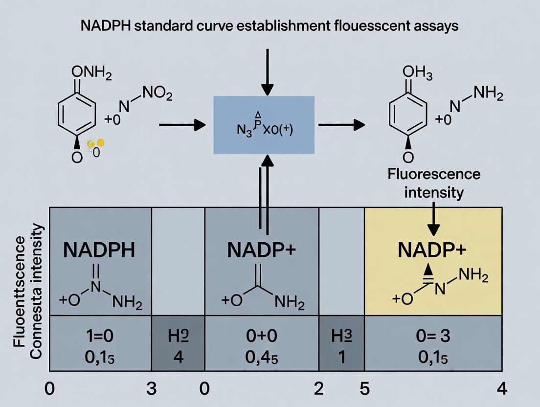
NADPH Fluorescence Standard Curve: A Step-by-Step Guide for Accurate Enzyme and Cell-Based Assays
This comprehensive guide details the establishment of a robust NADPH standard curve for fluorescent assays, a cornerstone technique in enzyme kinetics, drug discovery, and metabolic research.
NADPH Fluorescence Standard Curve: A Step-by-Step Guide for Accurate Enzyme and Cell-Based Assays
Abstract
This comprehensive guide details the establishment of a robust NADPH standard curve for fluorescent assays, a cornerstone technique in enzyme kinetics, drug discovery, and metabolic research. It provides foundational knowledge on NADPH fluorescence principles, a detailed methodological protocol for curve generation and application, essential troubleshooting strategies for common pitfalls, and validation methods to ensure data reliability and cross-platform comparability. Designed for researchers and assay development scientists, this article equips readers with the practical knowledge to generate precise, reproducible quantitative data from NADPH-dependent fluorescent readouts.
Understanding NADPH Fluorescence: The Key to Quantifying Metabolic and Enzymatic Activity
The Role of NADPH in Cellular Redox and Biosynthetic Pathways
Technical Support Center: NADPH Fluorescent Assay Troubleshooting
Frequently Asked Questions (FAQs)
Q1: My NADPH standard curve has poor linearity (R² < 0.98). What could be the cause? A: Poor linearity is often due to improper serial dilution technique or degradation of the NADPH standard stock. Ensure you are using fresh, high-purity NADPH, performing dilutions in the assay buffer (not water), and using low-protein-binding tips. Verify the pH of your buffer is stable at 7.4. Prepare the standard curve fresh for each assay run.
Q2: The fluorescence signal in my experimental wells is saturated or out of the dynamic range of the standard curve. A: This indicates an over-concentration of NADPH in your samples. Dilute your sample extract or lysate appropriately in the reaction buffer and re-run the assay. The sample NADPH concentration should fall within the mid-range of your standard curve for optimal accuracy. A typical dilution factor for cell lysates is between 5x and 20x.
Q3: I observe high background fluorescence in my negative control (no-enzyme or no-sample control). A: High background can be caused by:
- Contamination of reagents or labware with fluorescent compounds.
- Auto-oxidation of the fluorescent probe. Ensure the probe is stored correctly, protected from light, and prepared fresh.
- Impurities in the assay buffer. Use high-grade reagents and filter buffers if necessary.
Q4: My inter-assay variability (between different days) is unacceptably high. A: Key factors to standardize:
- Temperature: Perform all incubations in a pre-equilibrated, temperature-controlled plate reader or incubator.
- Incubation Time: Use a precise timer for the enzymatic reaction step.
- Reagent Thawing: Aliquot all critical reagents (enzyme, probe, co-factors) to avoid repeated freeze-thaw cycles. Thaw completely and mix gently before use.
Q5: How do I distinguish between NADPH and NADH signal in my cellular assay? A: Standard fluorescent assays often measure total reduced pyridine nucleotides (NAD(P)H). To specifically measure NADPH, you must use an enzyme-coupled assay that is selective for NADPH. For instance, use glutathione reductase (GR) and oxidized glutathione (GSSG), which is specific for NADPH. Alternatively, use a commercial NADP/NADPH extraction and quantification kit that enzymatically cycles NADP⁺ and NADPH separately.
Troubleshooting Guide: NADPH Standard Curve Establishment
| Symptom | Possible Cause | Solution |
|---|---|---|
| Low Signal-to-Noise Ratio | Degraded fluorescent probe. Old or inactive detection enzyme. | Prepare new probe aliquot. Test enzyme activity with a positive control. |
| High CV between replicates | Inconsistent pipetting, especially of viscous samples or standards. Bubbles in wells. | Use calibrated pipettes with appropriate tips. Centrifuge plate before reading. Practice consistent pipetting technique. |
| Standard curve slope differs from previous runs | Change in instrument gain settings. Lot-to-lot variation in critical reagents. | Document all instrument settings. Re-calibrate with new lot of primary standard. |
| Non-linear curve at high [NADPH] | Inner filter effect or signal saturation at high concentrations. | Use a narrower, more appropriate standard range (e.g., 0-2 µM instead of 0-10 µM). |
Key Experimental Protocol: NADPH Quantification via Enzymatic Recycling Fluorescent Assay
Objective: To accurately quantify total NADPH concentration in a cell lysate.
Principle: NADPH reduces a specific substrate (e.g., GSSG) via its cognate enzyme (GR). The resulting product (GSH) then reduces a non-fluorescent probe (e.g., Thiolite or a resazurin derivative) to a highly fluorescent product, proportional to the initial NADPH concentration.
Materials:
- NADPH Standard (e.g., 1 mM stock in Tris buffer, pH 8.0)
- Assay Buffer (100 mM Tris, 1 mM EDTA, pH 8.0)
- Glutathione Reductase (GR) from S. cerevisiae
- Oxidized Glutathione (GSSG)
- Fluorescent Thiol Probe (protected from light)
- Cell Lysis Buffer (compatible with downstream assay)
- Black 96-well microplate
Procedure:
- Standard Curve Preparation: Serially dilute NADPH stock in assay buffer to create 7 standards covering 0 µM, 0.1 µM, 0.5 µM, 1 µM, 2 µM, 4 µM, and 8 µM.
- Sample Preparation: Lyse cells in appropriate buffer. Centrifuge at 12,000g for 10 min at 4°C to remove debris. Keep samples on ice.
- Master Mix Preparation: For 1 well, combine:
- 50 µL Assay Buffer
- 2 µL GSSG (100 mM stock)
- 1 µL Fluorescent Probe (from fresh aliquot)
- 1 µL Glutathione Reductase (1 U/µL stock)
- Reaction Setup: In each well of the plate, add 46 µL of Master Mix. Add 4 µL of NADPH standard or clarified sample. Run in triplicate.
- Incubation & Detection: Mix gently, seal plate, and incubate in the dark at room temp for 30 min. Measure fluorescence (Ex/Em ~340/460 nm, verify per probe specs).
- Data Analysis: Subtract the average 0 µM standard (blank) fluorescence from all readings. Plot standard curve (fluorescence vs. [NADPH]). Use linear regression to calculate NADPH concentration in unknown samples, applying any dilution factors.
Data Presentation
Table 1: Typical NADPH Standard Curve Data for a Fluorescent Assay
| NADPH Concentration (µM) | Fluorescence (RFU, Mean ± SD, n=3) | CV (%) |
|---|---|---|
| 0.0 (Blank) | 125 ± 15 | 12.0 |
| 0.1 | 450 ± 25 | 5.6 |
| 0.5 | 1850 ± 90 | 4.9 |
| 1.0 | 3650 ± 120 | 3.3 |
| 2.0 | 7200 ± 180 | 2.5 |
| 4.0 | 14300 ± 350 | 2.4 |
| 8.0 | 28100 ± 700 | 2.5 |
Note: Linear Regression from 0.1-8.0 µM: y = 3500x + 100, R² = 0.999.
Table 2: Common Issues and Impact on Assay Parameters
| Parameter Affected | Acceptable Range | Consequence of Deviation |
|---|---|---|
| Assay Z'-factor | > 0.5 | <0.5 indicates poor assay robustness for HTS. |
| Signal-to-Background (S/B) | > 3 | Lower S/B reduces sensitivity and dynamic range. |
| Standard Curve R² | > 0.98 | Lower R² increases quantification error. |
| Intra-assay CV | < 10% | High CV indicates poor precision and pipetting error. |
Visualization
Diagram Title: NADPH Cellular Pathways: Generation and Consumption
Diagram Title: NADPH Fluorescent Assay Workflow
The Scientist's Toolkit: Research Reagent Solutions
| Item | Function & Rationale |
|---|---|
| High-Purity NADPH (Lithium Salt) | Primary standard for curve generation. Lithium salt offers superior solubility and stability in aqueous buffers compared to sodium salt. |
| Glutathione Reductase (GR), Recombinant | Enzyme for NADPH-specific detection. High specific activity ensures rapid, complete reaction kinetics and assay sensitivity. |
| Thiol-Sensitive Fluorescent Probe (e.g., Thiolite Green) | Non-fluorescent until reduced by GSH (generated by GR from GSSG using NADPH). Enables highly sensitive, low-background detection. |
| Cell Lysis Buffer with Redox Stabilizers | Must contain agents like NEM (N-ethylmaleimide) to rapidly alkylate and "freeze" the in vivo NADP/NADPH ratio upon cell disruption. |
| Low-Fluorescence, Black 96-Well Microplate | Minimizes optical crosstalk and background signal, maximizing signal-to-noise ratio for fluorescence measurements. |
| Reaction Buffer (Tris-EDTA, pH 8.0) | Optimized pH for GR activity. EDTA chelates divalent cations that might interfere with the enzymatic reaction or probe stability. |
Technical Support & Troubleshooting Center
Troubleshooting Guides
Issue: Low or No Fluorescence Signal in NADPH Assay
- Check 1: Excitation Wavelength: Verify the instrument is set to excite at ~340 nm. A common error is using 260 nm (NADPH absorbance max) for excitation.
- Check 2: Photobleaching: NADPH is moderately photostable. Avoid prolonged exposure to the excitation beam. Use shutter controls if available.
- Check 3: Quenching: Ensure buffers are free of iodide, acrylamide, or other known fluorescence quenchers. Check for heavy metal contamination.
- Check 4: Cuvette/Plate: Use quartz cuvettes or UV-transparent microplates for 340 nm excitation. Standard polystyrene plates will absorb the light.
Issue: High Background Fluorescence
- Check 1: Contamination: Test all buffer components individually. Some biological extracts (e.g., serum, lysates) contain autofluorescent compounds.
- Check 2: Light Leak: Ensure the plate reader or fluorimeter is properly sealed from ambient light during reading.
- Check 3: Buffer Impurities: Some commercial assay buffers or purified water may contain fluorescent impurities. Run a buffer-only blank.
Issue: Inconsistent Standard Curve Replicates
- Check 1: NADPH Stability: NADPH in solution degrades over time, especially at non-alkaline pH or with repeated freeze-thaw. Prepare fresh dilutions daily from a concentrated stock stored at -80°C.
- Check 2: Pipetting Accuracy: Due to the exponential nature of fluorescence, small volumetric errors in serial dilution are magnified. Use calibrated pipettes and consider intermediate dilutions.
- Check 3: Temperature: Fluorescence intensity is temperature-dependent. Allow all reagents and the plate to equilibrate to the assay temperature (e.g., 25°C) before reading.
FAQs
Q1: Why does NADPH fluoresce, but NADP+ does not? A1: Fluorescence requires the absorption of light and re-emission from an excited electron. The nicotinamide ring in NADPH is reduced, creating a conjugated π-electron system that can be excited by ~340 nm light and relax via photon emission (~460 nm). The oxidized nicotinamide ring in NADP+ lacks this specific conjugated structure, preventing efficient fluorescence.
Q2: What is the precise excitation and emission maxima for NADPH? A2: The exact maxima can shift slightly (±5-10 nm) depending on solvent, pH, and instrument. In aqueous neutral buffer, NADPH typically has an excitation maximum (λex) at 340 nm and an emission maximum (λem) at 460 nm.
Q3: Can I use the same filters/optics for NADH and NADPH measurements? A3: Yes. NADH and NADPH have nearly identical excitation/emission spectra. Therefore, you cannot spectroscopically distinguish them in a mixture without separation or using enzyme-coupled assays specific to one cofactor.
Q4: How does pH affect NADPH fluorescence? A4: Fluorescence intensity increases with pH. The quantum yield is higher for the deprotonated form of the reduced nicotinamide. For consistent quantitative assays, maintain a stable, mildly alkaline pH (e.g., pH 7.5-8.5).
Table 1: Spectral Properties of NADPH
| Property | Value | Condition / Note |
|---|---|---|
| Excitation (λ_ex) | 340 nm | Primary peak in aqueous buffer |
| Molar Extinction Coefficient (ε) at 340 nm | 6,220 M⁻¹cm⁻¹ | For quantitating stock concentration |
| Emission (λ_em) | 460 nm | Primary peak in aqueous buffer |
| Fluorescence Quantum Yield | ~0.02 | Relatively low; reference-dependent |
| Stokes Shift | ~120 nm | Large shift reduces self-quenching |
| Typical Assay Linear Range | 10 nM - 10 µM | Dependent on instrument sensitivity |
Table 2: Factors Affecting NADPH Fluorescence Intensity
| Factor | Effect on Signal | Recommended Control |
|---|---|---|
| Increased Temperature | Decreases (Quenching) | Use temperature-controlled reader |
| Decreased pH (<7.0) | Decreases | Buffer at pH 8.0 |
| Ionic Strength | Slight Decrease | Keep consistent in buffers |
| Glycerol | Increases (Viscosity) | Keep consistent if used |
Experimental Protocols
Protocol: Establishing an NADPH Fluorescence Standard Curve Objective: Generate a reliable standard curve for quantifying NADPH in enzymatic assays (e.g., dehydrogenase activity) within the context of thesis research.
Reagent Preparation:
- Stock NADPH (1 mM): Dissolve NADPH (disodium salt) in 10 mM Tris-HCl, pH 8.0. Aliquot and store at -80°C. Avoid repeated freeze-thaw.
- Assay Buffer: 50-100 mM Tris-HCl or HEPES, pH 8.0.
Serial Dilution:
- Prepare a 10 µM intermediate dilution of NADPH in assay buffer on ice.
- Perform a 1:2 or 1:1.5 serial dilution in a clear, UV-compatible microplate or quartz cuvettes to create 6-8 points covering 10 nM to 2 µM. Include a buffer-only blank.
Fluorimeter Settings:
- Excitation: 340 nm (bandwidth 5-10 nm).
- Emission: 460 nm (bandwidth 10-20 nm).
- Gain/PMT: Set using the highest standard to be ~80-90% of the instrument's maximum dynamic range.
- Temperature: 25°C (controlled).
- Integration Time: 100-500 ms.
Measurement & Analysis:
- Read the plate/cuvettes.
- Subtract the blank (buffer) value from all standards.
- Plot fluorescence intensity (RFU) vs. NADPH concentration (nM or µM).
- Fit data to a linear regression (y = mx + b). The curve is typically linear up to ~2-5 µM, after which inner filter effects may cause deviation.
Pathway & Workflow Visualizations
Title: NADPH Fluorescence Standard Curve Experimental Workflow
Title: NADPH Fluorescence Jablonski Diagram
The Scientist's Toolkit
Table 3: Essential Research Reagent Solutions for NADPH Fluorescence Assays
| Item | Function / Explanation |
|---|---|
| NADPH (Disodium Salt) | The fluorescent cofactor standard. High-purity grade ensures accurate quantitation. Store dessicated at -20°C or -80°C. |
| UV-Transparent Microplate | Allows ~340 nm excitation light to pass through. Essential for plate-based assays. Do not use standard polystyrene. |
| Quartz Cuvettes | Required for spectrophotometer/fluorimeter cuvette-based measurements at UV wavelengths. |
| Tris-HCl or HEPES Buffer (pH 8.0) | Provides a stable, mildly alkaline environment to maximize and stabilize NADPH fluorescence signal. |
| Black/Solid-Sided Microplate | Used in conjunction with UV-transparent plates to prevent optical crosstalk between wells during reading. |
| Precision Micro-pipettes & Tips | Critical for accurate serial dilution of the NADPH standard to establish a valid curve. |
| Fluorimeter with UV Lamp/LED | Instrument capable of generating ~340 nm light for excitation and detecting ~460 nm emission. Monochromators or filters are suitable. |
Troubleshooting Guides & FAQs
Q1: My NADPH standard curve shows unexpectedly low fluorescence signal. What could be wrong? A: This is often due to the instability of NADPH in solution. Ensure the assay buffer is prepared fresh, maintained at a pH of 7.0-8.0 (optimal for stability), and kept on ice. Avoid repeated freeze-thaw cycles of the NADPH stock. Also, verify that your fluorescent plate reader is set to the correct wavelengths (typically Ex ~340 nm, Em ~450-465 nm).
Q2: How can I confirm that my assay is specifically detecting NADPH and not NADH? A: Perform a specificity test. Prepare separate, identical reactions containing either NADPH or NADH at the same molar concentration. The fluorescence yield (intensity per mole) for NADPH is approximately 1.5 to 2 times higher than for NADH under standard conditions (see Table 1). If your assay is specific, the signal from the NADPH sample should be significantly higher. Using an enzyme like glutathione reductase (specific for NADPH) in a control reaction can further confirm specificity.
Q3: My background fluorescence is high, reducing the assay's sensitivity. How can I reduce it? A: High background often stems from contaminants in buffers or plate choice. Use ultrapure, low-fluorescence water and reagents. Conduct a "no-NADPH" control to identify background sources. Consider using black-walled microplates to minimize cross-talk and light scattering. Also, ensure all plasticware (tips, tubes) is certified as low-fluorescence.
Q4: The linear range of my NADPH standard curve is narrower than expected. How can I extend it? A: The linear dynamic range is typically 0.1-10 µM. If it's narrower, check for inner filter effect at high concentrations (>10 µM), where NADPH absorbs the excitation light. Dilute samples into the linear range. Alternatively, use a shorter pathlength (e.g., a low-volume plate) to mitigate this effect. Ensure your instrument's gain is optimally set to avoid signal saturation.
Q5: During kinetic assays, the fluorescence signal decreases over time. Is this normal? A: A decrease is not normal for a producing reaction and indicates instability. NADPH is susceptible to oxidation. Decrease can be caused by oxidants in the buffer or exposure to air. Include an antioxidant system like 0.1% BSA or 1-5 mM DTT in your assay buffer. Also, ensure the reaction is sealed or covered to minimize evaporation and oxidation during reading.
Q6: Can I use an NADH standard curve to quantify NADPH in my samples? A: No. Due to the difference in fluorescence quantum yield, using an NADH standard curve for NADPH quantification will introduce significant error (underestimation by ~30-50%). You must always use the correct cofactor (NADPH) to generate the standard curve for your assay. See Table 1 for quantitative differences.
Table 1: Key Fluorescence Properties of NADPH vs. NADH
| Property | NADPH | NADH | Notes |
|---|---|---|---|
| Optimal Excitation (Ex) | ~340 nm | ~340 nm | Similar for both. |
| Optimal Emission (Em) | ~450-465 nm | ~450-465 nm | Similar for both. |
| Relative Fluorescence Yield | 1.5 - 2.0 | 1.0 (Reference) | Highly dependent on pH, solvent, and instrument. NADPH is brighter. |
| Common Linear Range | 0.1 - 10 µM | 0.1 - 10 µM | Can vary with plate and reader settings. |
| pH Stability Optimum | 7.0 - 8.0 | 7.0 - 8.0 | Degrades rapidly in acidic conditions. |
| Key Specific Enzyme | Glutathione Reductase, Thioredoxin Reductase | Lactate Dehydrogenase, Alcohol Dehydrogenase | Used for validation and specificity tests. |
Experimental Protocol: Establishing a Robust NADPH Standard Curve
Objective: To generate a precise and accurate standard curve for quantifying NADPH in fluorescent enzymatic assays.
Materials: See "The Scientist's Toolkit" below.
Procedure:
- Buffer Preparation: Prepare fresh Assay Buffer (e.g., 50 mM Tris-HCl, pH 7.5, 1 mM EDTA). Filter through a 0.22 µm filter to remove particles.
- NADPH Stock Dilution: Thaw a high-concentration NADPH stock (e.g., 100 mM in neutral buffer) on ice. Dilute in Assay Buffer to create a 100 µM intermediate stock. Keep on ice and protected from light.
- Standard Series Preparation: In a low-binding, low-fluorescence microcentrifuge tube, perform a serial dilution of the 100 µM stock using Assay Buffer to create the following standard points: 0, 0.1, 0.5, 1, 2.5, 5, 7.5, and 10 µM. Prepare these immediately before use.
- Plate Loading: Pipette 100 µL of each standard concentration in triplicate into a black-walled, clear-bottom 96-well microplate.
- Fluorescence Measurement: Immediately place the plate in a pre-warmed (to assay temperature, e.g., 30°C) plate reader. Measure fluorescence with Ex = 340 nm (±10-15 nm bandwidth) and Em = 460 nm (±10-15 nm bandwidth). Use an optimal gain setting determined from a mid-range standard.
- Data Analysis: Calculate the average Relative Fluorescence Units (RFU) for each standard triplicate. Subtract the average RFU of the 0 µM (blank) standard. Plot the blank-corrected RFU (y-axis) against the NADPH concentration (x-axis). Perform linear regression analysis on the linear portion of the curve (typically 0-10 µM). The slope represents the assay sensitivity.
Diagrams
Diagram 1: NADPH vs NADH Fluorescence Spectral Overlap
Diagram 2: Specificity Validation Workflow
Diagram 3: Key Factors Affecting Assay Specificity
The Scientist's Toolkit: Essential Reagents & Materials
| Item | Function & Importance |
|---|---|
| Ultra-Pure NADPH (Tetrasodium Salt) | The gold-standard reference for creating standard curves. High purity minimizes background fluorescence and ensures accurate quantification. |
| Low-Fluorescence Assay Buffer | Typically Tris or HEPES, pH 7.5-8.0. Provides optimal enzymatic activity and cofactor stability. Must be filtered to remove particulate scatterers. |
| Black-Walled, Clear-Bottom 96-Well Plates | Black walls minimize well-to-well crosstalk of fluorescence signal. Clear bottom is compatible with readers using bottom optics. |
| Glutathione Reductase (from yeast or E. coli) | An NADPH-specific enzyme. Critical for validating assay specificity and confirming the identity of the fluorescent signal in unknown samples. |
| Fluorometer/Plate Reader | Capable of measuring kinetics with excitation ~340 nm and emission ~460 nm. Monochromators are preferred over filters for flexibility. |
| Low-Binding, Nuclease-Free Microtubes & Tips | Prevents adsorption of low-concentration NADPH standards to plastic surfaces, which can cause nonlinearity in standard curves. |
| Antioxidants (e.g., DTT, BSA) | DTT (1-5 mM) or BSA (0.1%) helps stabilize NADPH in solution by reducing oxidation, especially in long kinetic runs. |
| Precision Pipettes (P2, P20, P200, P1000) | Accurate serial dilution of NADPH standards is paramount for a reliable standard curve. Regular calibration is recommended. |
Technical Support Center: NADPH Fluorescent Assay Troubleshooting
FAQs & Troubleshooting Guides
Q1: My NADPH standard curve has poor linearity (R² < 0.98). What could be the cause? A: Poor linearity typically stems from inaccurate serial dilution, fluorophore degradation, or improper plate reader settings. Ensure: 1) Use fresh, sterile diluent (e.g., assay buffer). 2) Perform dilutions in low-binding tubes using calibrated pipettes. 3) Prepare standard curve fresh daily. 4) Verify the instrument's gain and calibration settings are optimized for the excitation/emission wavelengths (e.g., Ex/Em ~340/460 nm for common NADPH probes).
Q2: I observe high background fluorescence in my no-enzyme/no-cell control wells. A: High background can be caused by: 1) Autofluorescence of media/components: Use phenol-red-free media and pre-test all reagents. 2) Probe instability: Reconstitute fluorescent probe (e.g., resorufin-based) in anhydrous DMSO, aliquot, and store at ≤ -20°C protected from light. 3) Contamination: Ensure assay plates are free of particulates. 4) Light exposure: Perform all probe handling in minimal light.
Q3: My dehydrogenase activity assay shows low signal-to-noise ratio. A: For enzymatic reactions (e.g., using glucose-6-phosphate dehydrogenase):
- Check cofactor stability: NADP⁺ should be stored at -20°C in neutral, dry conditions.
- Optimize substrate concentration: Perform a preliminary kinetic experiment to determine Km.
- Verify enzyme integrity: Aliquot and store enzyme at recommended temperature; avoid freeze-thaw cycles.
- Include critical controls: A "no substrate" control is essential to define baseline.
Q4: Cell viability assay (MTT/XTT) results are inconsistent with the NADPH-dependent fluorescence readout. A: Discrepancies are common as assays measure different endpoints. MTT/XTT rely on mitochondrial reductase activity, while NADPH fluorescence assays often reflect total cytosolic redox capacity. For correlation:
- Cell number titration: Establish a linear range for both assays.
- Lysis consideration: Some fluorescence assays require cell lysis for accurate NADPH quantification.
- Timing: Take readings at consistent time points post-treatment.
Q5: How do I differentiate signal from NADPH vs. NADH in intracellular ROS assays? A: Use pharmacologic or genetic tools. Apply specific inhibitors: e.g., 6-aminonicotinamide (6-AN) to inhibit the pentose phosphate pathway (PPP) and lower NADPH. For a more specific readout, employ genetically encoded biosensors (e.g., iNAP sensors) that are tuned for NADPH. Always run parallel assays with an inhibitor control.
Key Experimental Protocols
Protocol 1: Establishment of a High-Quality NADPH Standard Curve for Fluorescence
- Preparation: Thaw NADPH stock (100 mM in 10 mM Tris, pH 8.0) on ice. Warm assay buffer to RT.
- Serial Dilution: In low-binding microcentrifuge tubes, perform 1:2 serial dilutions in assay buffer to create 8 points from 100 µM to 0.78 µM. Include a 0 µM blank (buffer only).
- Plate Setup: Add 50 µL of each standard in triplicate to a black, clear-bottom 96-well plate.
- Reaction Mix: Add 50 µL of probe/developer solution (e.g., 2 µM of a resazurin-based probe in buffer).
- Incubation & Reading: Incubate protected from light for 30 min at 25°C. Read fluorescence (Ex/Em as per probe specs, e.g., 540/590 nm).
- Analysis: Plot mean fluorescence intensity (FI) vs. [NADPH]. Fit a linear regression. Accept only curves with R² ≥ 0.98.
Protocol 2: Cellular NADPH Quantitation for Redox Status Assessment
- Cell Seeding: Seed cells in a 96-well plate and treat as required.
- Lysis & Extraction: Aspirate media. Add 100 µL of extraction buffer (0.1% Triton X-100, 0.1M NaOH) to lyse cells and stabilize NADPH. Incubate 10 min on ice.
- Neutralization: Add 100 µL of neutralization buffer (0.1M HCl, 0.1M Tris).
- Enzymatic Cycling Reaction:
- Prepare reaction mix: 100 µL extract + 100 µL of cycling buffer (0.1M Tris, pH 8.0, 0.5 mM MTT, 2 mM PMS, 5 mM G6P, 2 U/mL G6PDH).
- Incubate at 37°C for 10-30 min (optimize time).
- Detection: Measure absorbance at 570 nm. Calculate [NADPH] from the standard curve run in parallel with identical buffers.
Table 1: Common NADPH-Dependent Fluorescent Probes and Their Parameters
| Probe Name | Target | Excitation (nm) | Emission (nm) | Dynamic Range (NADPH) | Key Application |
|---|---|---|---|---|---|
| Resorufin-based | NADPH via reductase | 540-570 | 580-590 | 0.5 - 50 µM | Cell viability, dehydrogenase kinetics |
| CytoRed | NAD(P)H | 540 | 590 | 1 - 100 µM | General redox status |
| iNAP (genetic) | NADPH specifically | Varies (e.g., 420) | Varies (e.g., 475) | 0.1 - 100 µM in vivo | Live-cell NADPH imaging |
| Amplite Fluorimetric | NADPH | 540 | 590 | 0.1 - 10 µM | High-sensitivity detection in lysates |
Table 2: Troubleshooting Quick Reference: Symptoms & Solutions
| Symptom | Possible Cause | Immediate Action | Long-Term Fix |
|---|---|---|---|
| Low Signal | Degraded NADPH/Probe | Use fresh aliquots | Store in single-use aliquots at -80°C (NADPH) |
| High CV% | Inconsistent pipetting | Check pipette calibration | Use automated liquid handler for serial dilutions |
| Plate Edge Effect | Evaporation | Use a plate sealer | Use a humidity chamber during incubation |
| Non-linear Kinetics | Substrate depletion | Use higher [Substrate] | Perform initial velocity measurements (<10% conversion) |
The Scientist's Toolkit: Research Reagent Solutions
| Item | Function & Rationale |
|---|---|
| NADPH (Tetrasodium Salt) | The core standard; reduced form of NADP⁺. High purity (>97%) is critical for accurate standard curves. |
| Glucose-6-Phosphate Dehydrogenase (G6PDH) | Model dehydrogenase enzyme; used in enzymatic cycling assays to amplify NADPH signal. |
| Resazurin-based Fluorogenic Probe | Cell-permeable, non-fluorescent dye reduced by NADPH-dependent reductases to fluorescent resorufin. |
| Black/Clear-Bottom 96-Well Plates | Minimize cross-talk and background fluorescence while allowing for microscopic validation. |
| Phenazine Methosulfate (PMS) | Electron-coupling agent used in cycling assays to transfer electrons from NADPH to MTT/XTT. |
| 6-Aminonicotinamide (6-AN) | PPP inhibitor; negative control to decrease intracellular NADPH in ROS/viability assays. |
| NADPH Extraction Buffer (Alkaline) | Stabilizes the reduced form of NADPH during cell lysis, preventing rapid oxidation. |
| Recombinant iNAP Biosensor Plasmid | For live-cell, specific NADPH imaging; allows spatiotemporal resolution of NADPH dynamics. |
Diagrams
Title: NADPH Roles in Key Experimental Pathways
Title: NADPH Fluorescent Standard Curve Protocol
Technical Support Center & FAQs
FAQ 1: I am establishing a standard curve for NADPH using a fluorescent assay. My low-concentration standards are indistinguishable from the blank. What is the likely cause and how can I fix it?
- Answer: The most likely cause is suboptimal signal-to-noise ratio due to excessive excitation or emission slit/bandwidth settings. Wide slits increase signal but also increase background photon noise, drowning out weak signals. For NADPH fluorescence (Ex ~340 nm, Em ~460 nm), start with narrow slits (e.g., 5-10 nm). Use the highest instrument sensitivity/gain setting first, and only widen slits if the signal from your highest standard is below the detector's optimal range. Ensure your buffer and plasticware are low-fluorescence grade.
FAQ 2: My NADPH standard curve is non-linear at the higher concentration range. What should I check in my instrument settings?
- Answer: This indicates signal saturation, either from inner filter effect (IFA) or detector saturation. First, confirm your readings are within the linear dynamic range of your plate reader/fluorometer (consult the manual). For IFA, if the pathlength-corrected absorbance at the excitation wavelength is >0.1, significant signal attenuation occurs. Dilute your standards or use a shorter pathlength (e.g., low-volume plates). Optically clear, low-binding plates are essential.
FAQ 3: The fluorescence readings for my replicate NADPH standards show high variability (poor precision). What are the key troubleshooting steps?
- Answer: Follow this protocol:
- Mixing: Ensure the NADPH stock is thoroughly mixed before serial dilution.
- Pipetting: Use calibrated pipettes and reverse pipetting for viscous buffers.
- Plate Effects: Use the same plate type consistently. Wipe the bottom of the plate clean.
- Instrument: Perform a calibration check with a stable fluorophore (e.g., fluorescein). Check if the variability is position-dependent (edge effects); if so, use a temperature-controlled incubator with lid during reading.
- Settling: Read the plate immediately after mixing or after a consistent, defined delay.
FAQ 4: How do I choose between a monochromator-based and a filter-based reader for my NADPH assay development?
- Answer: The choice depends on flexibility vs. sensitivity and cost.
- Monochromators: Ideal for assay development and optimization. You can scan to find exact Ex/Em peaks (e.g., confirming NADPH peak at ~460 nm). They offer continuous wavelength selection but typically have lower light throughput.
- Filter-based: Optimal for established, high-throughput assays. Fixed filters (e.g., 340/25 nm Ex, 460/10 nm Em) provide higher light throughput and sensitivity, crucial for weak signals. Use the table below for a comparison.
Table 1: Quantitative Comparison of Key Fluorometer/Plate Reader Parameters for NADPH Assays
| Parameter | Typical Optimal Setting for NADPH | Impact on Assay | Filter-Based Reader | Monochromator-Based Reader |
|---|---|---|---|---|
| Excitation Wavelength | 340 ± 5 nm | Matches NADPH absorbance max. | Fixed (e.g., 340/25 nm) | Adjustable (e.g., 340 nm, 5-20 nm BW) |
| Emission Wavelength | 460 ± 10 nm | Captures NADPH fluorescence max. | Fixed (e.g., 460/10 nm) | Adjustable (e.g., 460 nm, 5-20 nm BW) |
| Slit/Bandwidth (BW) | 5-15 nm (Start narrow) | Wider = more signal & noise. Narrower = better resolution. | Fixed by filter (e.g., 10-25 nm) | User-adjustable (e.g., 1-20 nm) |
| Gain/PMT Voltage | Set so top standard is ~80-90% of max. | Maximizes dynamic range. | Often automated or manual | Usually manual optimization |
| Read Height/Position | Optimal for plate type (e.g., 1 mm for 96-well) | Affects signal intensity and consistency. | Manually set | Manually set |
| Cost & Throughput | -- | -- | Higher throughput, often lower cost | More flexible, often higher cost |
Experimental Protocol: Establishing a Robust NADPH Fluorescent Standard Curve
Objective: To generate a linear standard curve for the quantification of NADPH in enzymatic (e.g., dehydrogenase) assays.
Materials: See "Research Reagent Solutions" table below.
Methodology:
- Solution Preparation:
- Prepare Assay Buffer (e.g., 50-100 mM Tris or phosphate buffer, pH 7.4-8.0). Filter through a 0.22 µm membrane.
- Prepare a 1-10 mM stock of NADPH in assay buffer. Aliquot and store at -80°C. Avoid repeated freeze-thaw cycles.
- Serial Dilution:
- Thaw NADPH stock on ice. Perform a serial dilution in low-binding, clear microcentrifuge tubes using assay buffer to create 6-8 standards covering a range from below expected sample concentration to above (e.g., 0.1 µM to 50 µM).
- Prepare a blank (assay buffer only).
- Plate Setup:
- Pipette 100 µL of each standard and blank into a minimum of 3 replicate wells of a black-walled, clear-bottom 96-well plate.
- Instrument Setup & Measurement:
- Pre-warm the plate reader chamber to assay temperature (e.g., 30°C).
- Set excitation to 340 nm, emission to 460 nm.
- Set slit/bandwidth to 10 nm for both. Set gain so the highest standard reads near the instrument's maximum without saturation.
- Insert plate, run kinetic or endpoint measurement.
- Data Analysis:
- Average replicate reads for each standard.
- Subtract the average blank value from all standard averages.
- Plot corrected fluorescence (y-axis) vs. NADPH concentration (x-axis).
- Perform linear regression. A robust curve should have R² > 0.99.
Visualizations
Diagram 1: NADPH Fluorescence Assay Workflow
Diagram 2: Key Factors Affecting Signal-to-Noise Ratio (S/N)
The Scientist's Toolkit: Research Reagent Solutions
| Item | Function & Importance |
|---|---|
| High-Purity NADPH (Tetrasodium Salt) | The standard itself. Stability is critical. Store dry, desiccated at -80°C. Prepare fresh solutions frequently. |
| Low-Fluorescence, Black 96-Well Plates | Black walls minimize optical crosstalk. Clear bottom optimizes light transmission. Low-binding surface reduces analyte loss. |
| Assay-Grade Buffer Components (e.g., Tris-HCl) | High-purity reagents minimize background fluorescence. Buffer pH stabilizes NADPH (stable above pH 7). |
| 0.22 µm Syringe Filter | For filtering buffers to remove particulates that cause light scattering and increased background noise. |
| Low-Binding Microcentrifuge Tubes & Pipette Tips | Prevents adsorption of the NADPH standard to plastic surfaces during serial dilution, improving accuracy. |
| Fluorescent Reference Standard (e.g., Fluorescein) | Used for periodic instrument validation and performance checks (PMT sensitivity, wavelength accuracy). |
Protocol: Building Your NADPH Standard Curve for Microplate and Cuvette-Based Assays
Troubleshooting Guides & FAQs for NADPH Fluorescent Assay Establishment
Q1: My NADPH standard curve is non-linear, especially at higher concentrations. What could be the cause? A: This is often due to inner filter effect or fluorophore self-quenching. Ensure the absorbance of your highest standard at the excitation wavelength is below 0.1. Prepare fresh standards in assay buffer from a high-purity, lyophilized NADPH stock. Validate linearity by serial dilution.
Q2: The fluorescence signal is unstable, decaying rapidly during plate reading. A: This typically indicates reagent degradation or oxidation. Key considerations:
- Prepare NADPH stock solutions in a neutral, volatile buffer (e.g., 20 mM Tris, pH 8.0), aliquot, and store at -80°C. Avoid repeated freeze-thaw cycles.
- Ensure all buffers (especially Tris) are prepared with CO₂-free, deionized water and sparged with argon or nitrogen to minimize dissolved oxygen.
- Include a chelating agent (e.g., 1 mM EDTA) in buffers to prevent metal-catalyzed oxidation.
Q3: I observe high background fluorescence in my blank (no NADPH) wells. A: Contaminants or auto-fluorescent compounds are likely present.
- Use the highest purity water (≥18 MΩ·cm, HPLC grade).
- Filter all buffers through a 0.22 µm filter.
- Use non-fluorescent, black-walled microplates.
- Verify the purity of all buffer components (e.g., some BSA preparations can be fluorescent).
Q4: The inter-assay coefficient of variation (CV) for my standard points is >15%. A: This points to inconsistent reagent preparation or handling.
- Always prepare a master mix of all common reagents (buffer, enzyme, probe) for the entire plate.
- Use calibrated, positive-displacement pipettes for viscous reagents.
- Thaw all critical reagents (enzyme, probe) on ice and centrifuge briefly before use.
- Standardize the incubation time and temperature precisely.
Q5: How should I handle and prepare the fluorescent detection probe (e.g., resazurin, coumarin-based probes)? A: These probes are typically light-sensitive and unstable in aqueous solution.
- Reconstitute lyophilized probe in high-grade DMSO to create a concentrated (e.g., 1000X) stock. Aliquot and store at -80°C in the dark.
- Dilute the DMSO stock into assay buffer immediately before use to create the working solution. Do not store aqueous working solutions.
- Shield all solutions containing the probe from light during experiment setup.
Key Data for NADPH Standard Curve Establishment
Table 1: Recommended Storage Conditions for Critical Reagents
| Reagent | Recommended Stock Concentration | Storage Buffer/Condition | Stable For | Critical Note |
|---|---|---|---|---|
| NADPH (Primary Standard) | 10-100 mM | 20 mM Tris, pH 8.0, 1 mM EDTA; Aliquoted at -80°C | 3-6 months | Verify concentration by A340 (ε = 6220 M⁻¹cm⁻¹) after thaw. |
| Fluorescent Probe (e.g., Resazurin) | 100-500X final | Anhydrous DMSO; Aliquoted, shielded at -80°C | 1 year | Protect from light. Final DMSO in assay <0.5%. |
| Assay Buffer (e.g., Tris) | 10X concentrate | Filtered (0.22 µm), sparged with N₂, at 4°C | 1 month | Check pH after warming to assay temperature. |
| Enzyme (e.g., reductase) | Varies by activity | 50% Glycerol, -80°C | Manufacturer specified | Avoid repeated freeze-thaw; use tube racks on ice. |
Table 2: Troubleshooting Checklist for Poor Standard Curve Performance
| Symptom | Primary Check | Secondary Check | Solution |
|---|---|---|---|
| Low Signal/High EC₅₀ | Probe activity (run QC with known [NADPH]) | Photomultiplier (PMT) gain setting | Fresh probe prep; optimize PMT or probe concentration. |
| Curve Plateaus Early | Inner filter effect (Aex of top standard) | Probe/enzyme limiting | Dilute samples; increase probe/enzyme in master mix. |
| High Background | Plate type (black vs. clear) | Buffer/water contamination | Use black plates; prepare fresh, filtered buffers. |
| Poor Replicate Agreement | Pipette calibration | Evaporation during read | Service pipettes; use plate seal during incubation/read. |
Experimental Protocol: Establishing a Robust NADPH Standard Curve (Fluorometric)
Objective: To prepare a precise and accurate standard curve for quantifying NADPH in unknown samples via a coupled enzymatic fluorescent assay.
Materials:
- NADPH (high-purity, lyophilized)
- Assay Buffer: 100 mM Tris-HCl, 1 mM EDTA, pH 8.0 (filtered, degassed)
- Fluorescent Detection System (e.g., resazurin + purified diaphorase enzyme, or a commercial NADPH detection probe)
- Black 96-well microplate, non-binding surface
- Plate-reading fluorometer (with appropriate ex/em filters, e.g., 540/590 nm for resorufin)
Method:
- Buffer Preparation: Prepare 1L of 100 mM Tris buffer. Adjust to pH 8.0 at room temperature using HCl. Add 1 mL of 1 M EDTA stock. Sparge with nitrogen gas for 20 minutes. Filter through a 0.22 µm vacuum filter unit. Store at 4°C.
- NADPH Primary Stock (10 mM): Reconstitute 1 mg of lyophilized NADPH in 121 µL of pre-chilled 20 mM Tris, pH 8.0, 1 mM EDTA. Mix gently, aliquot 10 µL into PCR tubes, and flash-freeze in liquid nitrogen. Store at -80°C.
- Standard Curve Points: Thaw one aliquot of NADPH on ice. In the assay buffer, prepare a 200 µM intermediate dilution. Perform a 1:2 serial dilution in assay buffer across 8 points (e.g., 100 µM to 0.78 µM). Include a 0 µM (blank) point containing only assay buffer.
- Master Mix Preparation: Prepare a master mix containing assay buffer, the detection enzyme (e.g., 1-10 mU/mL diaphorase), and the fluorescent probe (e.g., 10-50 µM resazurin) on ice. Vortex gently and centrifuge.
- Assay Assembly: In a black microplate, add 80 µL of each NADPH standard per well, in triplicate. Initiate the reaction by adding 20 µL of the master mix to each well using a multichannel pipette. Seal the plate and incubate protected from light at the specified temperature (e.g., 37°C) for exactly 15-30 minutes.
- Measurement: Read fluorescence on a plate reader using pre-optimized wavelengths/gain settings. The signal should be stable for the duration of the scan.
- Analysis: Plot mean fluorescence intensity (Blank subtracted) vs. NADPH concentration. Fit data to a 4-parameter logistic or linear regression model. The R² value should be >0.99.
The Scientist's Toolkit: Research Reagent Solutions
Table 3: Essential Materials for NADPH Fluorescent Assays
| Item | Function & Critical Consideration |
|---|---|
| Ultra-Pure Water (≥18.2 MΩ·cm) | Solvent for all buffers; minimizes ionic and fluorescent contaminants. |
| Molecular Biology Grade Tris | Buffer for maintaining pH; low heavy metal and UV-absorbing impurity content. |
| NADPH (Tetrasodium Salt, ≥97%) | Primary standard; high purity ensures accurate curve fitting and quantification. |
| Resazurin Sodium Salt | Redox-sensitive fluorescent probe; becomes highly fluorescent resorufin upon reduction by NADPH via diaphorase. |
| Diaphorase (from C. kluyveri) | Coupling enzyme; must have high specific activity and low background reduction of probe. |
| Black, Flat-Bottom 96-Well Plate | Minimizes crosstalk and background fluorescence; ensures optimal signal detection. |
| Opaque Plate Seals | Prevents evaporation and photobleaching of light-sensitive reagents during incubation. |
| Inert Gas Canister (N₂/Ar) | For degassing buffers to prevent oxidation of NADPH and sensitive probes. |
Diagrams
Title: NADPH Fluorescent Detection Assay Workflow
Title: Key Signaling Pathway in a Coupled NADPH Detection Assay
FAQs & Troubleshooting Guide
Q1: My NADPH standard curve is not linear across the 0.1 - 10 µM range. What could be the cause? A: Non-linearity often stems from pipetting errors in serial dilution or fluorescence saturation at the high end. Ensure you are using calibrated, positive-displacement pipettes for volumes below 10 µL. Perform each dilution step in triplicate. If the upper points are flattening, the detector may be saturated; confirm the assay's linear detection limit and consider diluting your highest concentration standard.
Q2: How do I verify the accuracy of my serial dilutions? A: Validate your dilution series using a independent method. After preparing your NADPH standards, measure the absorbance at 340 nm (ε = 6220 M⁻¹cm⁻¹) in a UV-vis spectrophotometer. Calculate the actual concentration from the Beer-Lambert law (A = εcl) and compare to your target concentrations.
Q3: The fluorescence background is too high, obscuring the low-end (0.1 µM) signal. How can I reduce it? A: High background is frequently due to contaminating auto-fluorescent compounds in buffers or plate readers. Use ultrapure, HPLC-grade water and non-fluorescent microplates. Include a "no NADPH" blank containing all assay components. Ensure the plate reader chamber is clean. If the issue persists, consider using a buffer with lower background fluorescence.
Q4: My replicate data for the serial dilution shows high variability (high %CV). What steps should I take? A: High variability typically indicates inconsistent technique during liquid handling. For serial dilutions, always mix each dilution step thoroughly (e.g., 10-15 gentle pipette mixes or vortexing if compatible). Use fresh, low-retention pipette tips. Prepare the entire dilution series from the same master stock solution on the same day. Consider using an automated liquid handler for improved precision.
Q5: Should I perform a simple dilution or a serial dilution for the NADPH standard curve, and why? A: For a dynamic range spanning two orders of magnitude (0.1 to 10 µM), a serial dilution is superior. A simple dilution from a single stock for each point accumulates large volumetric errors at low concentrations. A serial dilution, where each concentration is made from the previous one, maintains higher precision and consistency across the range, which is critical for a reliable standard curve in quantitative fluorescent assays.
| Parameter | Recommended Specification | Purpose/Rationale |
|---|---|---|
| Starting Stock Conc. | 1 mM or 10 mM NADPH in buffer | Provides manageable volumes for dilution; prepare fresh or aliquot & store at -80°C. |
| Dilution Factor | 1:2 or 1:3 | Balances number of points across the range with minimal cumulative error. |
| Number of Points | 8-10 standards | Adequately defines the curve across 0.1 - 10 µM. |
| Replicates | Minimum n=3 per point | Allows statistical analysis (CV%, curve fitting error). |
| Final Volume per Well | 100-200 µL | Standard for 96-well plate assays, sufficient for reader pathlength. |
| Buffer | Assay-compatible buffer (e.g., Tris, PBS, pH 7.4) | Matches the experimental sample matrix to avoid matrix effects. |
| Verification Method | A340 absorbance measurement (A=εcl) | Independent validation of prepared standard concentrations. |
Experimental Protocol: Serial Dilution for NADPH Standard Curve
Objective: Prepare a precise serial dilution of NADPH for a standard curve ranging from 10 µM to 0.1 µM.
Materials: See "Research Reagent Solutions" below.
Procedure:
- Prepare Master Stock: Dissolve solid NADPH in assay buffer to create a 10 mM stock. Filter sterilize (0.2 µm). Verify concentration by A340 (dilute 1:100, Abs should be ~0.622).
- Create Intermediate Stock: Dilute the 10 mM stock to 100 µM using assay buffer. This is your "high standard."
- Serial Dilution Setup: a. Label 8-10 microcentrifuge tubes (e.g., S1 to S8). b. Add 300 µL of assay buffer to tubes S2 through S8. c. To tube S1, add 600 µL of the 100 µM NADPH intermediate stock. S1 is now 50 µM (for a 1:2 series) or your starting point. d. Perform a two-fold serial dilution: Transfer 300 µL from S1 to S2, mix thoroughly. Transfer 300 µL from S2 to S3, mix. Continue through tube S8. e. Discard 300 µL from the final tube (S8).
- Calculate Final Concentrations: Your series will approximate: 50, 25, 12.5, 6.25, 3.125, 1.56, 0.78, 0.39 µM. Adjust starting concentration or dilution factor to ensure coverage of 0.1 - 10 µM.
- Plate Setup: In a black, clear-bottom 96-well plate, add 100 µL of each standard concentration in triplicate. Include triplicate blanks (assay buffer only).
- Assay Execution: Add 100 µL of your detection reagent (e.g., cycling enzyme mix for enzymatic assays) to each well. Incubate as required and measure fluorescence (Ex/Em ~340/460 nm, instrument-dependent).
Diagrams
Title: Serial Dilution Workflow for NADPH Standards
Title: NADPH Fluorescence Assay Context in Research
Research Reagent Solutions
| Item | Function & Rationale |
|---|---|
| β-NADPH (Tetrasodium Salt) | The standard molecule for calibration. Reduced form is fluorescent/absorbent. High purity (>98%) is essential for accurate molar concentration. |
| Non-Fluorescent Assay Buffer (e.g., Tris-HCl, pH 8.0) | Provides a stable, chemically inert matrix for dilution and reaction. Must not absorb or emit at target wavelengths. |
| Low-Binding Microcentrifuge Tubes | Minimizes adsorption of low-concentration NADPH standards to plastic walls, preserving accuracy. |
| Positive-Displacement or Calibrated Air-Gap Pipettes | Essential for accurate volume transfer, especially in the µL range, to minimize serial dilution error. |
| Black, Clear-Bottom 96-Well Plates | Black sides minimize well-to-well crosstalk of fluorescence; clear bottoms allow absorbance verification if needed. |
| UV-vis Spectrophotometer & Cuvettes | For independent verification of stock and standard concentrations via absorbance at 340 nm. |
| Fluorescence Plate Reader | Equipped with filters/ monochromators suitable for NADPH (Ex ~340 nm, Em ~460 nm). Must have a dynamic range capable of detecting sub-micromolar signals. |
Troubleshooting Guides & FAQs
Q1: During NADPH standard curve establishment, my fluorescence signal is too weak, even with high NADPH concentrations. What should I optimize first? A: First, optimize the Gain/PMT Voltage. Incrementally increase the gain on your microplate reader until your highest standard yields a signal near 80-90% of the detector's maximum range. Avoid saturation (>95%). If the signal remains weak after maximizing gain within a non-saturating range, proceed to optimize Incubation Time. Extend the incubation time at the assay's reaction temperature (typically 25-37°C) in 2-5 minute intervals, measuring kinetics to determine when the signal plateaus.
Q2: My standard curve shows high background (low signal-to-noise ratio). How can I address this? A: High background is often tied to Temperature and reagent stability. Ensure all reagents, especially the enzyme/development mix, are kept on ice and the assay plate is maintained at a consistent, optimal temperature (e.g., 25°C) using the reader's temperature control. Temperature fluctuations can increase enzyme-independent background. Also, verify that your buffer components are fresh and free of contaminants that may auto-fluoresce.
Q3: The coefficient of variation (CV) between technical replicates for my NADPH standards is unacceptably high (>10%). What steps should I take? A: High replicate CV points to inconsistencies in Incubation Time or temperature equilibration. Ensure the microplate reader is allowed to pre-equilibrate to the set Temperature (e.g., 25°C) for at least 30 minutes. Use a plate mixer for consistent reagent mixing before reading. If using a kinetic read, confirm that the time interval between reads for each well is minimal (using simultaneous or rapid sequential reading modes).
Q4: How do I determine the optimal incubation time for my specific assay conditions? A: Perform a kinetic read. Prepare your highest NADPH standard and a blank (zero standard) in triplicate. Read fluorescence (with a moderate gain setting) every minute for 30-60 minutes at your assay Temperature. Plot signal vs. time. The optimal incubation time is where the signal for the high standard reaches a plateau while the blank signal remains stable and low. See Table 1 for example data.
Table 1: Example Kinetic Data for Incubation Time Optimization
| Time (min) | High Std Fluorescence (RFU) | Blank (RFU) | Signal-to-Blank |
|---|---|---|---|
| 5 | 15,250 | 1,050 | 14.5 |
| 10 | 28,750 | 1,100 | 26.1 |
| 15 | 35,500 | 1,150 | 30.9 |
| 20 | 36,200 | 1,200 | 30.2 |
| 25 | 36,000 | 1,250 | 28.8 |
Note: In this example, 15 minutes is optimal as the signal plateaus.
Q5: I changed the gain setting and now my standard curve linearity (R²) is poor at the lower end. What is the cause and solution? A: Excessively high Gain can introduce electronic noise that obscures low-concentration signals. Reduce the gain so that your highest standard reads between 70-85% of the detector's maximum. Re-run the standard curve. The improved signal-to-noise at the low end should restore linearity.
Detailed Protocol: Optimization of Measurement Parameters
Objective: To systematically determine the optimal Gain, Temperature, and Incubation Time for a fluorescent NADPH detection assay.
Materials: See "Research Reagent Solutions" table below.
Methodology:
- Gain/PMT Voltage Optimization:
- Prepare a single high concentration NADPH standard (e.g., top of your expected range) and assay buffer blank in a 96-well plate.
- Set the reader to a kinetic cycle at your standard assay temperature (e.g., 30°C).
- Program a series of reads with increasing gain settings (e.g., 700, 800, 900, 1000 on a typical instrument).
- Plot RFU vs. Gain. Select the highest gain that does not saturate the detector (typically RFU < 90% of max) for the high standard.
Temperature & Incubation Time Optimization (Kinetic Assay):
- Prepare a full NADPH standard curve series (e.g., 0, 0.5, 1, 2, 4, 8, 16 µM) in triplicate.
- Using the optimized gain, set the reader to perform a kinetic read every minute for 40-60 minutes.
- Repeat this experiment at two different controlled temperatures (e.g., 25°C and 37°C).
- Analyze the data to determine the timepoint at which the signal for all standards has stabilized (plateaued) and the signal-to-background ratio is maximal. Compare the performance (background, linear range) between temperatures.
Final Standard Curve Generation:
- Using the optimized parameters (Gain=X, Temperature=Y°C, Incubation Time=Z minutes), run the NADPH standard curve in quadruplicate.
- Fit the data using a linear regression model. Acceptable optimization typically yields an R² value >0.99 and a lower limit of detection (LOD) suitable for your experimental samples.
Diagrams
Diagram 1: Parameter Optimization Decision Pathway
Diagram 2: NADPH Fluorescence Assay Workflow
The Scientist's Toolkit: Research Reagent Solutions
| Item | Function in NADPH Fluorescence Assay |
|---|---|
| Recombinant Enzyme (e.g., Dihydrofolate Reductase - DHFR) | Catalyzes the reduction of a non-fluorescent substrate (e.g., Resazurin) to a fluorescent product (Resorufin), with kinetics directly proportional to NADPH concentration. |
| Fluorogenic Substrate (e.g., Resazurin) | Serves as the electron acceptor from NADPH via the enzyme, transforming from a non-fluorescent to a highly fluorescent state upon reduction. |
| Assay Buffer (Tris or PBS, pH 7.4-8.0) | Maintains optimal enzymatic activity and pH stability throughout the incubation period. May contain stabilizers like BSA. |
| NADPH Standard (High-Purity) | The critical reference molecule for generating the standard curve. Must be freshly prepared or aliquoted from stable stocks (-80°C) to avoid oxidation. |
| Black/Solid-Bottom 96/384-Well Microplates | Minimizes optical crosstalk and background fluorescence, maximizing signal-to-noise ratio for plate reader detection. |
| Temperature-Controlled Microplate Reader | Precisely maintains the optimized temperature during incubation and reading, which is crucial for reproducible enzyme kinetics and stable fluorescence signals. |
Technical Support Center
Troubleshooting Guides & FAQs
Q1: My fluorescence intensity values for the standard concentrations are erratic and do not follow a linear trend. What could be the cause? A: This is commonly due to pipetting errors or insufficient mixing of the NADPH stock solution. Ensure accurate serial dilution using calibrated pipettes. Vortex each dilution step for 10 seconds and centrifuge briefly before use. Also, check that your fluorometer cuvette is clean and free of scratches.
Q2: The fluorescence signal from my highest standard is saturated, resulting in a non-linear curve at the upper end. How should I proceed? A: Signal saturation invalidates those data points. You must repeat the standard curve, diluting your highest NADPH concentration so that all measured intensities fall within the linear dynamic range of your plate reader or fluorometer. Consult your instrument's manual to determine its optimal detection range.
Q3: The blank (0 µM NADPH) shows a high fluorescence background. How can I reduce this? A: High background often comes from contaminating fluorescence in the buffer or plate. Use ultrapure, freshly prepared assay buffer. Ensure all plasticware (tips, tubes, plates) is low-fluorescence grade. Protect NADPH standards from light exposure during preparation to prevent degradation.
Q4: After plotting, my R² value is below 0.98. Is my standard curve acceptable? A: An R² < 0.98 indicates poor fit and unreliable quantification. You should repeat the curve. Common fixes include: increasing the number of replicate readings per standard (use at least triplicates), ensuring the temperature is consistent during reading, and verifying that your NADPH stock concentration is accurate via absorbance at 340 nm (ε = 6220 M⁻¹cm⁻¹).
Q5: How should I handle outliers in my standard curve data? A: Use a statistical test like Grubb's test to identify significant outliers. Do not remove data points arbitrarily. If an outlier is confirmed and a technical error (e.g., a bubble in the well) is noted, it may be excluded. Always document any exclusion. Repeating the measurement is the most robust solution.
Table 1: Raw Fluorescence Data for NADPH Standard Curve
| NADPH Concentration (µM) | Replicate 1 (RFU) | Replicate 2 (RFU) | Replicate 3 (RFU) | Mean RFU | Std. Dev. |
|---|---|---|---|---|---|
| 0 | 105 | 98 | 102 | 101.7 | 3.5 |
| 1 | 520 | 510 | 531 | 520.3 | 10.5 |
| 2 | 1020 | 1050 | 1035 | 1035.0 | 15.0 |
| 4 | 2100 | 2150 | 2080 | 2110.0 | 35.1 |
| 8 | 4250 | 4300 | 4180 | 4243.3 | 60.3 |
| 16 | 8100 | 8250 | 8000 | 8116.7 | 125.2 |
Table 2: Linear Regression Analysis of Standard Curve
| Parameter | Value |
|---|---|
| Slope (RFU/µM) | 501.2 |
| Y-Intercept (RFU) | 95.4 |
| R² Value | 0.9987 |
| Linear Range | 0 - 16 µM |
Experimental Protocols
Protocol: NADPH Standard Curve for Fluorescent Assay
1. Reagent Preparation:
- NADPH Stock Solution (10 mM): Dissolve NADPH in assay-compatible buffer (e.g., Tris-HCl, pH 8.0). Verify concentration by measuring absorbance at 340 nm. Aliquot and store at -80°C protected from light.
- Assay Buffer: Prepare as required for your specific enzymatic assay. Filter through a 0.22 µm membrane to remove particulate matter.
2. Serial Dilution & Plate Setup:
- Perform a two-fold serial dilution of the 10 mM NADPH stock in assay buffer to create working standards: 16 µM, 8 µM, 4 µM, 2 µM, 1 µM, 0 µM (blank).
- Pipette 100 µL of each standard in triplicate into a black, clear-bottom 96-well microplate.
3. Fluorescence Measurement:
- Using a plate reader, set excitation/emission wavelengths appropriate for your assay (e.g., Ex 340 nm / Em 460 nm for NADPH autofluorescence, or the wavelengths of your coupled fluorescent probe).
- Shake the plate for 5 seconds before reading.
- Read the fluorescence of all wells at a controlled temperature (e.g., 25°C or 37°C).
4. Data Analysis & Curve Fitting:
- Calculate the mean and standard deviation for each concentration's replicates.
- Subtract the mean blank (0 µM) value from all other mean values.
- Plot the blank-corrected Mean Fluorescence Intensity (Y-axis) against the NADPH Concentration (X-axis).
- Perform linear regression analysis. The equation (y = mx + c) is used to calculate unknown NADPH concentrations in experimental samples.
Experimental Workflow Diagram
The Scientist's Toolkit: Research Reagent Solutions
Table 3: Essential Materials for NADPH Fluorescent Assays
| Item | Function & Key Consideration |
|---|---|
| High-Purity NADPH | The standard. Must be >95% pure. Verify concentration spectrophotometrically. Light and temperature sensitive. |
| Low-Fluorescence Microplate | Black plates with clear bottoms minimize cross-talk and allow bottom reading. Essential for sensitive detection. |
| Calibrated Precision Pipettes | Critical for accurate serial dilutions. Regular calibration (every 6-12 months) is mandatory. |
| Assay-Specific Buffer | Must be compatible with both the enzyme/protein of interest and fluorescence measurement (low autofluorescence). |
| Fluorescent Plate Reader | Instrument with appropriate filters/ monochromators for excitation/emission wavelengths. Temperature control is ideal. |
| Data Analysis Software | Software (e.g., GraphPad Prism, SoftMax Pro) to perform linear regression and calculate sample concentrations. |
Troubleshooting Guides & FAQs
Q1: My standard curve is not linear. The R² value is below 0.99. What should I do? A1: A low R² value typically indicates issues with pipetting accuracy, degraded standards, or instrument instability. First, ensure your NADPH stock solution is freshly prepared in the correct buffer (e.g., Tris-HCl, pH 8.0) and protected from light. Perform serial dilutions meticulously using calibrated pipettes. Verify the fluorescence plate reader is warmed up and the gain is set appropriately to avoid signal saturation at higher concentrations.
Q2: How do I definitively identify the linear range for my NADPH standard curve? A2: The linear range is where the fluorescence response is directly proportional to NADPH concentration. Prepare a wide range of standards (e.g., 0 to 2000 nM). After measurement, plot concentration (x) vs. fluorescence intensity (y). Inspect the plot for the region where the data points fall on a straight line. Statistically, it is where the residuals (difference between predicted and observed values) are randomly distributed and minimal. Use the data table below to guide your serial dilution scheme.
Q3: What is an acceptable R² value, and can I still use a curve with an R² of 0.985? A3: For quantitative assays like NADPH quantification, an R² value of ≥0.998 is considered excellent, and ≥0.990 is typically the minimum acceptable for high-confidence research. An R² of 0.985 suggests significant variance. You should investigate and repeat the standard curve. Using it for sample quantification will introduce unacceptable error into your enzymatic rate calculations for drug screening assays.
Q4: The linear regression equation gives a negative y-intercept. Is this valid? A4: A slightly negative y-intercept can be valid and often indicates minimal background fluorescence in your assay buffer. However, a large negative value may suggest an error in defining the "blank." Ensure your blank (0 nM NADPH standard) is measured correctly. The equation should be in the form y = mx + b, where 'm' is the slope (sensitivity) and 'b' is the intercept. It is critical for calculating unknown sample concentrations: [NADPH] = (Fluorescence - b) / m.
Data Presentation
Table 1: Example NADPH Standard Curve Data for Linear Range Determination
| NADPH Concentration (nM) | Mean Fluorescence Intensity (RFU) | Standard Deviation (RFU) | Within Linear Range? |
|---|---|---|---|
| 0 (Blank) | 150 | 12 | No (Anchor Point) |
| 50 | 1200 | 85 | Yes |
| 100 | 2250 | 110 | Yes |
| 250 | 5100 | 230 | Yes |
| 500 | 10100 | 450 | Yes |
| 1000 | 20500 | 900 | Yes |
| 1500 | 29800 | 1500 | Yes (Upper Limit) |
| 2000 | 35000 | 2200 | No (Signal Saturation) |
Note: Data from a hypothetical fluorescent assay (Ex/Em ~340/460 nm). The linear range in this example is 50 - 1500 nM. The resulting regression equation is y = 19.87x + 205.34, with an R² = 0.9992.
Experimental Protocol: Establishing the NADPH Standard Curve
Materials: See "The Scientist's Toolkit" below. Procedure:
- Preparation: Reconstitute high-purity NADPH in assay-compatible buffer (e.g., 0.1M Tris-HCl, pH 8.0) to create a 10 mM primary stock. Aliquot and store at -80°C. Thaw on ice for use.
- Serial Dilution: On the day of the assay, prepare a working stock (e.g., 200 µM) in buffer. Perform a serial dilution in the same buffer to create standards covering 0 nM to 2000 nM. Use low-protein-binding tubes.
- Plate Setup: Pipette 100 µL of each standard in triplicate into a black, flat-bottom 96-well microplate.
- Fluorescence Measurement: Immediately read the plate using a fluorescence microplate reader pre-warmed for at least 15 minutes. Set excitation to 340 nm and emission to 460 nm (adjust per instrument specifics). Use a gain setting that does not saturate the signal at the highest suspected concentration.
- Data Analysis: Calculate the mean RFU for each standard. Plot concentration (x-axis) vs. mean RFU (y-axis). Using statistical software, apply linear regression analysis to the data points within the visually linear region. Record the equation (y = mx + b) and the R² value.
Visualization: NADPH Standard Curve Workflow
Title: Workflow for NADPH Standard Curve Analysis
The Scientist's Toolkit: Key Research Reagent Solutions
Table 2: Essential Materials for NADPH Fluorescent Assays
| Item | Function & Rationale |
|---|---|
| High-Purity NADPH (Lyophilized) | The assay standard. Purity >98% ensures accurate concentration calibration. Stabilized formulations reduce background oxidation. |
| Black 96-Well Microplates (Flat Bottom) | Minimizes optical crosstalk and well-to-well light scattering, critical for sensitive fluorescence measurements. |
| Assay Buffer (e.g., Tris-HCl, pH 8.0) | Provides a stable, non-interfering chemical environment. Chelating agents (e.g., EDTA) may be added to inhibit metal-catalyzed NADPH degradation. |
| Precision Microplate Pipettes & Calibrated Tips | Ensumes accurate and reproducible liquid handling for serial dilutions and sample/reagent transfers. |
| Fluorescence Microplate Reader | Equipped with correct filters/ monochromators for NADPH's excitation/emission maxima (~340/460 nm). Temperature control is essential for kinetic assays. |
| Low-Protein-Binding Microcentrifuge Tubes | Prevents adsorption of low-concentration NADPH standards to tube walls, preserving concentration accuracy. |
| Statistical Analysis Software | Used to perform linear regression, calculate the R² value, and determine the linear range from the standard curve data. |
Technical Support Center
Troubleshooting Guide: Common Experimental Issues
Q1: After generating the standard curve, my unknown sample values fall outside the linear range of the curve. What should I do? A: This indicates either an improper sample dilution or an issue with the assay's dynamic range. First, ensure you have accurately prepared your NADPH serial dilutions (e.g., 0, 2.5, 5, 10, 20, 40 µM). For unknown samples, perform a dilution series (e.g., 1:2, 1:5, 1:10) in assay buffer and re-run. The calculated concentration from a dilution that falls within the linear range (R² > 0.99) should then be multiplied by the dilution factor.
Q2: I observe high background fluorescence in my negative control (no enzyme) wells, compromising sensitivity. A: High background can arise from several sources. 1) Reagent Contamination: Prepare fresh assay buffer and ensure all pipette tips and tubes are RNase/DNase free. 2) Light Exposure: NADPH and assay substrates are light-sensitive. Perform all reagent preparation in low light and use opaque-walled or foil-wrapped plates. 3) Buffer Components: Check for auto-fluorescent compounds in your reaction buffer. Include a "buffer-only" control to identify the source.
Q3: My standard curve has a poor coefficient of determination (R² < 0.98). How can I improve it? A: A poor R² suggests pipetting inaccuracies or unstable reagents. 1) Use calibrated pipettes and perform reverse pipetting for viscous solutions. 2) Ensure the NADPH stock solution is fresh, prepared in the correct buffer (e.g., 10 mM Tris-HCl, pH 8.0), and aliquoted to avoid freeze-thaw cycles. 3) Confirm the fluorescence plate reader is properly warmed up and calibrated. Take three readings per standard and average them.
Q4: How do I determine if my reaction is consuming or generating NADPH? A: The direction of change in signal relative to controls determines this. For a generation assay (e.g., enzyme activity), an increase in fluorescence over time in the sample compared to a "no substrate" control indicates NADPH generation. For a consumption assay (e.g., antioxidant capacity), a decrease in fluorescence compared to an "NADPH only" control indicates consumption. Always run appropriate controls in parallel.
Q5: The signal from my positive control is lower than expected. A: Verify the following: 1) Incubation Temperature: Ensure the plate reader or incubator is at the correct temperature (e.g., 37°C). 2) Reaction Timing: Start the reaction immediately after adding the final component and begin kinetic measurements promptly. 3) Enzyme/Reagent Viability: Check the certificates of analysis for your positive control enzyme (e.g., Glucose-6-phosphate dehydrogenase). Use fresh aliquots.
Frequently Asked Questions (FAQs)
Q: What is the optimal excitation/emission wavelength for NADPH detection? A: NADPH fluorescence is typically measured at an excitation wavelength of 340 nm and an emission wavelength of 460 nm. Confirm the optimal settings for your specific instrument and plate type, as they can vary slightly.
Q: Can I use this standard curve method for cell lysate samples? A: Yes, but cell lysates introduce complexity. You must include a sample background control (lysate without the fluorescent detection reagent) to account for auto-fluorescence. Subtract this value from your experimental readings. Clarify samples by centrifugation (e.g., 12,000 x g, 10 min, 4°C) to reduce light scattering.
Q: How should I store my assay plates during the reading process? A: For kinetic assays requiring multiple reads over 30-60 minutes, maintain temperature control using the plate reader's environmental chamber. Protect from light between reads. For endpoint assays, read immediately after the reaction is stopped.
Q: What statistical method should I use to calculate unknowns from the standard curve? A: Use linear regression (y = mx + c) from your standard curve. The sample NADPH concentration (x) is calculated as x = (y - c) / m, where y is the sample's fluorescence intensity. Most plate reader software and tools like GraphPad Prism can perform this calculation automatically.
Q: Is it necessary to run the standard curve on every plate? A: Yes. To ensure accuracy and account for day-to-day and plate-to-plate variability in reagent performance, reader sensitivity, and ambient conditions, a fresh standard curve must be included on every assay plate.
Data Presentation
Table 1: Example NADPH Standard Curve Data (Fluorescent Assay)
| NADPH Standard (µM) | Fluorescence Intensity (RFU, Mean ± SD, n=3) | Corrected RFU (Subtracted Blank) |
|---|---|---|
| 0 (Blank) | 150 ± 15 | 0 |
| 2.5 | 425 ± 25 | 275 ± 29 |
| 5.0 | 700 ± 32 | 550 ± 34 |
| 10.0 | 1250 ± 45 | 1100 ± 47 |
| 20.0 | 2350 ± 68 | 2200 ± 70 |
| 40.0 | 4450 ± 120 | 4300 ± 121 |
Linear Regression: y = 107.5x + 12.5, R² = 0.999.
Table 2: Troubleshooting Summary: Symptoms and Solutions
| Symptom | Possible Cause | Recommended Solution |
|---|---|---|
| Low signal in all wells | Expired detection reagent | Prepare fresh reagent aliquot. |
| Incorrect filter set on reader | Verify Ex/Em: 340/460 nm. | |
| High variation between replicates | Inconsistent pipetting | Use calibrated pipettes, reverse pipetting. |
| Bubbles in wells | Centrifuge plate briefly before reading. | |
| Curve is nonlinear at high [NADPH] | Signal saturation (inner filter effect) | Dilute samples to fit linear range. |
| Negative calculated concentration | High background in sample | Include and subtract sample-specific background. |
Experimental Protocols
Protocol 1: Establishment of NADPH Standard Curve
- Preparation: Prepare assay buffer (e.g., 50 mM Tris-HCl, pH 7.5). Prepare a 1 mM NADPH stock in buffer.
- Serial Dilution: Dilute the stock in buffer to create working standards: 40, 20, 10, 5, 2.5, and 0 µM.
- Plate Setup: In a black 96-well plate, add 90 µL of assay buffer per well. Add 10 µL of each standard to triplicate wells.
- Reading: Immediately read fluorescence (Ex 340 nm / Em 460 nm) on a pre-warmed plate reader.
- Analysis: Average the triplicate reads. Subtract the 0 µM standard (blank) value from all others. Plot corrected RFU vs. concentration. Perform linear regression.
Protocol 2: Calculating NADPH Generation in Unknown Enzyme Samples
- Sample Prep: Prepare enzyme sample in ice-cold assay buffer. Clarify by centrifugation (12,000 x g, 10 min, 4°C).
- Reaction Mix: In plate wells, combine 80 µL assay buffer, 10 µL substrate (specific to your enzyme), and 10 µL sample (or buffer for negative control). Run in triplicate.
- Kinetic Measurement: Immediately place plate in reader and initiate kinetic cycle (e.g., read every minute for 30 minutes at 37°C).
- Calculation: Determine the maximum linear rate of RFU increase (ΔRFU/min) for each sample. Subtract the rate of the negative control. Using the slope (m) from your standard curve, calculate NADPH generation rate: [NADPH] (µM/min) = (ΔRFU/min) / m.
Mandatory Visualization
Diagram 1: NADPH Fluorescent Assay Workflow
Diagram 2: Determining NADPH Flux in Unknown Samples
The Scientist's Toolkit: Research Reagent Solutions
| Item | Function & Explanation |
|---|---|
| NADPH (Tetrasodium Salt) | The core standard. High-purity (>95%) is essential for accurate curve generation. Reconstitute in appropriate pH buffer to prevent degradation. |
| Black/Clear Bottom 96-well Plates | Black walls minimize cross-talk; clear bottoms are compatible with some detection systems. Ensure plates are compatible with your reader. |
| Fluorescent Plate Reader | Must be capable of measuring at ~340 nm excitation and ~460 nm emission. Temperature control and kinetic software are critical. |
| Assay Buffer (e.g., Tris-HCl, PBS) | Provides consistent pH and ionic strength. Must be free of fluorescent contaminants. May contain Mg²⁺ if required for enzymatic reactions. |
| Enzyme-specific Substrate (e.g., Glucose-6-Phosphate) | For generation assays, this initiates the reaction that produces NADPH. Purity and concentration are critical for reproducible kinetics. |
| Positive Control Enzyme (e.g., G6PDH) | Validates the entire assay workflow. A known amount of enzyme should yield a predictable rate of NADPH generation. |
| Microplate Centrifuge | Used to remove bubbles from wells after reaction setup, ensuring consistent light path and readings. |
| Precision Pipettes (Multi-channel & Single-channel) | Essential for accurate, reproducible liquid handling of standards and samples, especially for high-throughput formats. |
Solving Common Problems: Enhancing Sensitivity, Linearity, and Reproducibility
Troubleshooting Guides & FAQs
Q1: My NADPH standard curve in a fluorescent assay has a very low signal-to-noise ratio. What are the most common causes?
A: Common causes include:
- Quenching: Improper buffer composition (e.g., high ionic strength) or contaminated labware (detergents, metals) can quench fluorescence.
- Probe Degradation: The fluorescent probe (e.g., resorufin, Coumarin derivatives) is light-sensitive and may have degraded if stored or handled improperly.
- Incorrect Assay pH: The enzymatic reaction generating NADPH and the fluorescence of the detection probe are often pH-sensitive. A deviation from optimal pH (typically 7.0-8.0) reduces signal.
- Substrate or Enzyme Limitation: In coupled assays, insufficient concentrations of the enzyme generating NADPH (e.g., glucose-6-phosphate dehydrogenase) or its substrate will limit signal.
- Instrument Issues: Incorrect gain/PMT voltage settings, misaligned optics, or a dirty cuvette/plate reader lens.
Q2: How can I distinguish between a problem with my NADPH generation reaction versus my fluorescence detection system?
A: Perform a direct NADPH spiking experiment.
- Prepare your standard assay mixture but omit the enzyme that generates NADPH.
- Add a known, high concentration of NADPH standard (e.g., 10 µM) directly to the well.
- Measure the fluorescence. If the signal remains low, the issue lies in the detection system (probe, buffer, instrument). If the signal is strong and as expected, the issue is upstream in the NADPH-generating reaction (enzyme activity, substrate concentration, coupling enzymes).
Q3: What are the most effective strategies to amplify a weak signal in my NADPH fluorescent assay without increasing background?
A: The primary strategy is signal amplification through enzymatic cycling.
- Principle: Use a second enzyme system to continuously regenerate NADPH from the NADP+ produced in your primary reaction, creating a fluorescent readout over time that is cumulative and more sensitive.
- Example Protocol (Phenol Red / Ampliflu Red Cycling Assay):
- Primary Reaction: Allow your standard NADPH-generating reaction (e.g., from an enzyme activity assay) to proceed to completion.
- Cycling Reaction Mix: To the same well, add:
- Amplification Enzyme: Diaphorase (0.5 - 2 U/mL).
- Cycling Substrate: Phenazine methosulfate (PMS, 10 - 50 µM).
- Probe: Ampliflu Red (Resorufin derivative, 10 - 50 µM) in assay buffer (pH 7.8-8.0).
- Measurement: Incubate at room temperature and monitor the increase in fluorescence (Ex/Em ~560/590 nm) kinetically for 10-30 minutes. The rate or total fluorescence increase is proportional to the initial NADPH amount.
Q4: My standard curve is non-linear or has a poor R² value. What should I check?
A: This indicates issues with assay robustness.
- Check 1 - Pipetting Accuracy: Use calibrated pipettes and low-retention tips for serial dilutions of the NADPH standard. For critical low-concentration points, consider preparing standards independently rather than by serial dilution.
- Check 2 - Dynamic Range: Ensure your highest standard is not saturating the detector or the detection chemistry. Perform a dilution series of your highest standard to confirm linearity.
- Check 3 - Reagent Stability: Prepare fresh NADPH stock solution for each standard curve, as it degrades in aqueous solution. Keep all components on ice.
- Check 4 - Incubation Time: For kinetic assays, ensure the reaction has reached steady-state for all concentrations before reading.
Key Experimental Protocols
Protocol 1: Establishing a Robust NADPH Standard Curve for Fluorescent Detection
Objective: To generate a reliable standard curve correlating NADPH concentration to fluorescence intensity.
Materials: See "Research Reagent Solutions" table. Procedure:
- Prepare a 1 mM NADPH stock solution in the assay buffer. Aliquot and store at -80°C. Thaw on ice for immediate use.
- Perform a 1:2 serial dilution of the NADPH stock in assay buffer across 10-12 points in a low-binding microcentrifuge tube series. The final range should typically span 0 µM to 10 µM NADPH in the assay well.
- In a black-walled, clear-bottom 96-well plate, add 80 µL of assay buffer (pH 7.4-8.0) to each well.
- Add 10 µL of each NADPH standard dilution (in triplicate) to the designated wells. For the blank, add 10 µL of buffer.
- Initiate the detection reaction by adding 10 µL of the fluorescent detection mix (e.g., containing the probe and any necessary co-factors) to all wells using a multichannel pipette. Mix gently by pipetting up and down.
- Incubate the plate at the recommended temperature (e.g., 25°C) for a precise time (e.g., 10 minutes), protected from light.
- Read fluorescence using a plate reader with appropriate filters/optics (e.g., Ex 540-560 nm / Em 580-600 nm).
- Subtract the average blank value from all readings. Plot the mean fluorescence intensity (y-axis) against the final NADPH concentration in the well (x-axis). Fit the data with a linear regression model.
Protocol 2: Signal Amplification via Enzymatic Cycling for Low NADPH Detection
Objective: To detect very low levels of NADPH (picomole to nanomole range) by amplifying the signal.
Materials: Requires all items from Protocol 1, plus Diaphorase, Phenazine Methosulfate (PMS), and Ampliflu Red. Procedure:
- Complete the primary NADPH-generating reaction (your experimental assay) in the wells of a microplate. Include your NADPH standard curve in separate wells.
- Prepare the Cycling Master Mix on ice, protected from light:
- Assay Buffer (pH 8.0): To final volume
- Diaphorase: 1 U/mL final concentration
- Phenazine Methosulfate (PMS): 30 µM final concentration
- Ampliflu Red: 20 µM final concentration
- Add 100 µL of the Cycling Master Mix to each well to stop the primary reaction and initiate the cycling reaction.
- Immediately place the plate in the pre-warmed (25°C) plate reader.
- Measure fluorescence kinetically (Ex/Em ~560/590 nm) every minute for 30-60 minutes.
- Data Analysis: Calculate the slope (rate of fluorescence increase, RFU/min) for each well during the linear phase of the reaction (typically minutes 5-25). Plot the rate (y-axis) against the known initial NADPH amount (x-axis) to generate your amplified standard curve.
Data Presentation
Table 1: Troubleshooting Low Signal in NADPH Fluorescent Assays
| Symptom | Possible Cause | Diagnostic Test | Solution |
|---|---|---|---|
| Low Signal Across All Wells | Fluorescent probe degraded | Test fresh probe lot. | Use fresh, aliquoted probe; store in dark, -20°C. |
| Instrument PMT/gain too low | Read a known fluorescent control. | Optimize instrument settings. | |
| Buffer quenching | Compare signal in water vs. buffer. | Use ultrapure water, chelators (e.g., EDTA). | |
| High Background in Blank | Contaminated reagents/labware | Run blank with each component omitted. | Use fresh buffer, clean plates with ethanol. |
| Probe auto-oxidation | Incubate probe alone in buffer. | Prepare detection mix fresh, minimize light exposure. | |
| Poor Standard Curve Linearity (R²<0.98) | Inaccurate serial dilutions | Prepare independent standard points. | Use calibrated pipettes; make independent dilutions. |
| NADPH stock degradation | Test old vs. fresh stock. | Prepare fresh NADPH stock daily from powder or -80°C aliquot. | |
| Signal Saturation at High Standards | PMT/Gain too high | Dilute top standard 1:2. | Reduce detector gain; use optimal range. |
| Increased CV% in Replicates | Inconsistent mixing | Check pipetting technique. | Mix thoroughly after all additions. |
| Signal Fades Rapidly | Photobleaching of probe | Read plate multiple times over 5 min. | Reduce light exposure; read immediately after incubation. |
Table 2: Research Reagent Solutions for NADPH Fluorescence Assays
| Item | Function & Specification | Example Product/Catalog # (for reference) |
|---|---|---|
| NADPH (Tetrasodium Salt) | The standard molecule for calibration curves. High purity (>97%) is critical. | Sigma-Aldrich, N1630 |
| Fluorescent Probe (e.g., Resorufin) | Directly binds or reacts with NADPH, emitting detectable light. Light-sensitive. | Thermo Fisher, Ampliflu Red (A25741) |
| Assay Buffer (e.g., Tris, HEPES) | Maintains optimal pH (7.0-8.5) for both enzymatic activity and fluorescence. | Prepare 50-100 mM, pH 7.4-8.0, with 1 mM EDTA. |
| Diaphorase (from C. kluyveri) | Key amplification enzyme. Transfers electrons from NADPH to an intermediate. | Sigma-Aldrich, D5540; ≥300 U/mg. |
| Phenazine Methosulfate (PMS) | Electron carrier in amplification cycle. Shuttles electrons from Diaphorase to the probe. Light-sensitive. | Sigma-Aldrich, P9625 |
| Microplate (Black, Clear Bottom) | Maximizes signal capture while allowing bottom reading. Low protein binding reduces scatter. | Corning, 3603 |
| Plate Reader with Fluorescence Capability | Must have appropriate filters/monochromators for Ex/Em of chosen probe (e.g., ~560/590 nm). | BioTek Synergy H1, Tecan Spark |
Visualizations
Troubleshooting Low Signal in NADPH Assays
NADPH Signal Amplification via Enzymatic Cycling
NADPH Fluorescent Standard Curve Workflow
Technical Support Center: Troubleshooting & FAQs
FAQ 1: Why does my NADPH standard curve become non-linear and plateau at high concentrations in a fluorescent microplate reader assay?
- Answer: This is a classic symptom of the inner filter effect (IFE). At high concentrations of the fluorophore (NADPH), the emitted fluorescence light is re-absorbed by other NADPH molecules in the solution before it can reach the detector. This quenching effect leads to a underestimation of the true fluorescence, causing the curve to deviate from linearity and plateau. It is a physical artifact, not a biochemical property.
FAQ 2: How can I experimentally distinguish between inner filter effects and other types of quenching (e.g., collisional)?
- Answer: Perform a dilution test. Prepare a high-concentration NADPH sample that shows suspected quenching. Take an aliquot and dilute it 2-5 fold in assay buffer. If the measured fluorescence of the diluted sample, when multiplied by the dilution factor, is significantly higher than the fluorescence of the original concentrated sample, IFE is the primary cause. Collisional quenching is typically concentration-independent over this range in a fixed setup.
FAQ 3: What are the best practical methods to correct for or avoid inner filter effects in my standard curve?
- Answer: Two primary approaches are recommended:
- Pathlength Correction: Use a microplate reader equipped with an absorbance-capable detector to measure the absorbance (A) at the excitation wavelength at each well. Apply the correction formula: Fcorr = Fobs * antilog[(Aex + Aem)/2], where Aex and Aem are absorbances at excitation and emission wavelengths. For NADPH (Ex ~340 nm, Em ~460 nm), A_em is often negligible.
- Geometric Dilution: Simply dilute your stock NADPH to ensure all points on your standard curve fall within the linear range (typically where absorbance at 340 nm < 0.1). Establish the curve in this range and use it to calculate the concentration of unknown samples, which should also be diluted to fall within this linear range.
Experimental Protocol: Pathlength Correction for IFE
Objective: To establish a linear NADPH standard curve accounting for inner filter effects. Materials: NADPH (disodium salt), assay buffer (e.g., Tris-EDTA, pH 8.0), black-walled clear-bottom 96-well plate, fluorescent microplate reader capable of measuring top-read fluorescence (Ex: 340nm, Em: 460nm) and bottom-read pathlength absorbance (340nm or a reference wavelength). Procedure:
- Prepare a high-concentration NADPH stock solution (e.g., 200 µM) in assay buffer. Verify concentration via A340 (ε = 6220 M⁻¹cm⁻¹).
- Perform a 2-fold serial dilution in the microplate well to create a standard series from 0 to 200 µM. Final well volume: 100 µL.
- In the plate reader, first perform an absorbance scan at 340 nm (or use a pathlength correction feature) for each well to obtain the absorbance value (A).
- Immediately after, perform a fluorescence measurement (Ex 340, Em 460) for each well to obtain the observed fluorescence (F_obs).
- Apply Correction: For each well, calculate corrected fluorescence: Fcorr = Fobs * 10^(A/2). This simplified formula assumes the average pathlength for excitation and emission is half of the total, valid for standard microplate geometries.
- Plot both Fobs vs. [NADPH] and Fcorr vs. [NADPH] to visualize the correction.
Data Presentation: Corrected vs. Uncorrected NADPH Standard Curve
Table 1: Comparison of Observed and Pathlength-Corrected Fluorescence for a NADPH Standard Series
| NADPH Concentration (µM) | Absorbance at 340 nm (A) | Observed Fluorescence (F_obs, RFU) | Corrected Fluorescence (F_corr, RFU) | % Increase from Correction |
|---|---|---|---|---|
| 3.125 | 0.002 | 1250 | 1251 | 0.1% |
| 6.25 | 0.005 | 2450 | 2458 | 0.3% |
| 12.5 | 0.010 | 4800 | 4824 | 0.5% |
| 25 | 0.020 | 9200 | 9292 | 1.0% |
| 50 | 0.042 | 16500 | 16941 | 2.7% |
| 100 | 0.085 | 25000 | 26924 | 7.7% |
| 200 | 0.170 | 32000 | 38138 | 19.2% |
Note: Data is simulated for illustrative purposes. RFU: Relative Fluorescence Units.
The Scientist's Toolkit: Key Reagent Solutions
Table 2: Essential Materials for NADPH Fluorescent Assays
| Item | Function & Rationale |
|---|---|
| High-Purity NADPH (Disodium Salt) | The fluorophore and electron donor standard. Purity is critical for accurate concentration determination via absorbance. |
| Black-Walled, Clear-Bottom Microplates | Minimizes cross-talk and background scatter between wells while allowing for pathlength absorbance measurement from below. |
| Low-Fluorescence Assay Buffer | A consistent buffer matrix (e.g., Tris, HEPES) without fluorescent contaminants, essential for maintaining stable pH and ionic strength. |
| Fluorescent Microplate Reader with Absorbance Capability | Must be capable of top fluorescence reads and bottom absorbance/pathlength reads for direct in-well IFE correction. |
| Precision Liquid Handling Tools (e.g., multichannel pipettes) | Ensures accurate and reproducible serial dilutions directly in the microplate, crucial for standard curve integrity. |
Visualization: Experimental Workflow for IFE Correction
Visualization: Mechanism of Inner Filter Effect Quenching
Troubleshooting Guides & FAQs
Q1: In our NADPH fluorescent assay for enzyme kinetics, we observe high initial background fluorescence, compromising the detection of the standard curve's lower points. What are the most common buffer components that cause this?
A: High background in NADPH assays is frequently caused by:
- Reducing agents: DTT, β-mercaptoethanol, or TCEP can autofluoresce or react with the fluorophore.
- Primary amines: Components like Tris buffer at high concentrations can interfere with certain fluorescent probes.
- Contaminants: Impurities in commercial assay buffers or water, such as phenolic compounds or trace metals.
- NADPH/NADP+ preparations: Older or impure cofactor stocks can contain fluorescent degradation products.
- Detergent micelles: Some non-ionic detergents (e.g., Triton X-100) can scatter light or form fluorescent aggregates.
Q2: How can we systematically identify which specific reagent in our complex reaction buffer is causing the interference?
A: Perform a stepwise reagent omission test.
- Prepare your complete master mix, excluding the enzyme/substrate to measure only background.
- In a 96-well plate, create a series of wells where each well omits one key buffer component (e.g., well A: no DTT, well B: no detergent, well C: no NADPH, etc.).
- Include a well with only the detection probe in assay-grade water/buffer as a baseline.
- Measure the fluorescence (Ex/Em ~340/460 nm for common NADPH detection).
- Compare the signal from each omission well to the full background signal. A significant drop pinpoints the interfering agent.
Q3: Our assay requires DTT for enzyme stability. How can we mitigate its fluorescence interference without removing it?
A: Consider these mitigation strategies:
| Strategy | Protocol Detail | Expected Outcome |
|---|---|---|
| Concentration Titration | Systematically reduce DTT concentration from 10mM to 0.5-1mM in the final assay. | Find the minimum concentration that maintains enzyme activity while lowering background. |
| Alternative Reductants | Replace DTT with Tris(2-carboxyethyl)phosphine (TCEP). TCEP is often less fluorescent and more stable. | Lower background, but requires validation of enzyme compatibility. |
| Buffer Pre-Incubation | Pre-incubate the assay buffer (with DTT) at room temp for 30 min before adding NADPH and probe. | Allows DTT autofluorescence to stabilize or decay, yielding a more stable baseline. |
| Background Subtraction | Use a calibration curve prepared in your complete assay buffer, not in pure buffer. | The standard curve's background is matched to sample background, improving accuracy. |
Q4: What experimental protocol can we use to establish a robust NADPH standard curve in the presence of inherently high but consistent buffer background?
A: Protocol for a Background-Matched NADPH Standard Curve Principle: Prepare standards in the exact matrix as your samples to correct for inner-filter and quenching effects.
Materials:
- NADPH stock solution (e.g., 100 mM in pH-buffered water, aliquoted and stored at -80°C).
- Your complete assay buffer (including all interfering agents like DTT, detergents).
- Fluorescent detection probe (if applicable, e.g., a NADPH sensor).
- Black, clear-bottom 96- or 384-well assay plates.
- Plate reader capable of fluorescence measurement (typical NADPH direct fluorescence: Ex ~340 nm, Em ~460 nm).
Method:
- Preparation: Thaw and dilute NADPH stock in assay-grade water to a 10x intermediate stock series (e.g., 0, 2, 5, 10, 20, 50, 100 µM).
- Dilution into Buffer: In a low-protein-binding microtube, dilute the 10x NADPH intermediate series 1:10 into your complete assay buffer. This creates your 1x standard curve (e.g., 0, 0.2, 0.5, 1, 2, 5, 10 µM) in the correct matrix.
- Plate Setup: Pipette 90 µL of your background control (complete assay buffer without NADPH) into the first column of wells. Pipette 90 µL of each 1x standard into subsequent wells, in triplicate.
- Measurement: Add 10 µL of probe or buffer to all wells. Mix gently by orbital shaking. Incubate for 5-10 minutes at assay temperature. Read fluorescence.
- Data Analysis: Subtract the average fluorescence of the background control (0 µM NADPH in buffer) from all standard readings. Plot corrected RFU vs. NADPH concentration. Use a linear or quadratic fit where R² > 0.99.
Q5: Are there specific fluorescent probes or detection kits for NADPH that are less susceptible to buffer interference?
A: Yes. When establishing your assay, selecting the right detection method is key. The table below compares common approaches.
Research Reagent Solutions for NADPH Detection
| Item / Reagent | Function in NADPH Assay | Key Consideration for Background |
|---|---|---|
| Direct Fluorescence (Measure NADPH intrinsic fluorescence) | Measures NADPH directly without probes. Simple, cost-effective. | Highly susceptible to buffer interference (e.g., DTT, amines). Requires ultra-pure buffers. |
| Coupled Enzymatic Cycling Probes (e.g., resazurin-based) | Uses diaphorase to cycle NADPH, reducing resazurin to fluorescent resorufin. Amplifies signal. | Probe itself can be reduced by DTT/TCEP, causing high background. Requires careful optimization of probe/enzyme concentration. |
| NADPH Sensor Dyes (Genetically encoded or chemical) | Binds NADPH, causing a spectral shift or fluorescence change. | Specificity for NADPH over NADH is variable. May be affected by ionic strength or competing molecules. |
| LC-MS/MS | Gold-standard for absolute quantification. | Not fluorescent; avoids optical interference entirely. Complex and expensive. |
Diagnostic & Mitigation Workflow Diagram
Title: Troubleshooting High Background Fluorescence Workflow
NADPH Standard Curve Establishment with Background Correction
Title: Background-Matched NADPH Standard Curve Protocol
Troubleshooting Guide & FAQs
Q1: During my NADPH standard curve assay, my replicates show high variability (CV > 15%). What are the most common pipetting-related causes? A: High inter-replicate CV is frequently tied to pipetting technique and tool calibration. Key causes include:
- Inconsistent Pre-Wetting: Not pre-wetting the pipette tip with the reagent or standard solution leads to evaporation and aspirate volume error, crucial for viscous NADPH stock solutions.
- Poor Reverse Pipetting Technique: For fluorescent assays, reverse pipetting is recommended for dispensing standards and samples, as it ensures greater accuracy with surfactant-containing buffers.
- Calibration Drift: Pipettes used for serial dilution of the NADPH standard must be calibrated quarterly. A 2% deviation at a high concentration step multiplies error down the curve.
- Tip Selection: Using low-retention tips is non-negotiable for accurate NADPH transfer.
Q2: How can I diagnose if my poor replicate data is due to a plate effect versus a pipetting error? A: Perform a plate map pattern analysis.
- Method: Prepare a single, homogenous solution of your fluorescent readout buffer or a mid-range NADPH standard. Using a single, calibrated pipette and one box of tips, dispense this identical solution into every well of the microplate.
- Analysis: After reading the plate, plot the fluorescence values by well position. A systematic gradient (e.g., left-to-right, center-to-edge) indicates a plate effect (e.g., from uneven temperature in the plate reader). Random scatter points to pipetting or tip effects. Clustering by tip column (e.g., wells A1, B1, C1) suggests a specific pipette channel issue.
Q3: What are the best practices to minimize edge effect in 96-well plates during fluorescent endpoint reads for NADPH assays? A: Edge effect (evaporation in perimeter wells) severely compromises data. Mitigation strategies include:
- Physical Sealing: Use a optically clear, pierceable seal during incubation steps, not just a lid.
- Plate Condenser: If your reader has one, use it.
- Buffer Blanket: For long incubations (>30 min), place the plate in a humidified chamber.
- Experimental Design: Exclude perimeter wells from analysis; fill them with buffer or water to create a uniform evaporation barrier for the inner experimental wells.
Q4: My standard curve has a good R² value, but my unknown sample replicates are inconsistent. What could be wrong? A: This often points to sample-specific pipetting issues or interference.
- Check Sample Viscosity: Biological samples (lysates, serum) differ from your buffer-based standards. Use reverse pipetting and consider dilution if viscosity is high.
- Allow Plate to Equilibrate: Before reading, ensure the plate has settled for 5-10 minutes post-mixing to eliminate bubbles, which cause massive fluorescence scatter.
- Check for Signal Saturation: Ensure your unknown sample fluorescence is within the dynamic range of your standard curve. Saturating the detector leads to non-linear and unreliable values.
Key Experimental Protocols
Protocol 1: Reliable NADPH Standard Curve Preparation (Reverse Pipetting)
- Materials: Certified NADPH stock (e.g., 10 mM in buffer), assay buffer, low-retention microcentrifuge tubes, calibrated pipettes (2-20 µL, 20-200 µL), low-retention filtered tips, black-walled 96-well plate.
- Pre-Wetting: Aspirate and dispense the NADPH stock solution three times with a fresh tip before making the first standard aliquot.
- Serial Dilution: In a fresh tube, perform a 1:2 serial dilution in assay buffer to create 8 points (e.g., 100 µM to 0.78 µM). Use reverse pipetting for each transfer: Depress plunger to the second stop, aspirate, dispense to the first stop. Discard tip.
- Plate Setup: Dispense 80 µL of assay buffer into all standard wells.
- Standard Addition: Using a fresh tip and reverse pipetting for each standard, add 20 µL of each NADPH standard concentration to triplicate wells. Mix by gentle plate shaking.
- Read: Immediately read fluorescence (Ex ~340 nm, Em ~460 nm). Run in triplicate.
Protocol 2: Plate Reader Qualification for Fluorescent Assays
- Homogeneity Test: As described in FAQ A2, dispense a uniform fluorescein (e.g., 100 nM) or NADPH solution across the entire plate. Read.
- Acceptance Criteria: The inter-well CV should be <5%.
- Edge Effect Test: Fill all perimeter wells with a fluorescent solution and inner wells with buffer. Read over 60 minutes with the plate reader set to your assay's incubation temperature.
- Analysis: Calculate the mean fluorescence of edge vs. interior wells over time. A divergence >10% indicates significant evaporation requiring mitigation.
Data Presentation
Table 1: Impact of Pipetting Technique on NADPH Standard Curve Precision (n=6)
| Technique | Pre-wetting | CV of Mid-range Standard (25 µM) | Curve R² Value |
|---|---|---|---|
| Forward Pipetting | No | 18.5% | 0.983 |
| Forward Pipetting | Yes | 9.2% | 0.995 |
| Reverse Pipetting | No | 12.1% | 0.991 |
| Reverse Pipetting | Yes | 4.8% | 0.999 |
Table 2: Plate Effect Analysis Using Homogeneous Fluorescein Solution
| Well Position Group | Mean Fluorescence (RFU) | Std Dev | CV | Likely Cause |
|---|---|---|---|---|
| All Wells (n=96) | 10,245 | 1,150 | 11.2% | Combined Effects |
| Inner Wells (n=60) | 10,500 | 405 | 3.9% | Pipetting Noise |
| Edge Wells (n=36) | 9,817 | 1,450 | 14.8% | Evaporation (Edge Effect) |
Visualizations
Title: Troubleshooting Workflow for Poor Replicates
Title: Plate Map Showing Combined Edge & Pipetting Effects
The Scientist's Toolkit: Research Reagent Solutions
| Item | Function in NADPH Fluorescent Assays |
|---|---|
| Certified NADPH Standard | Provides accurate reference for curve generation. Must be aliquoted and stored at -80°C to prevent degradation. |
| Low-Binding Microcentrifuge Tubes | Minimizes adsorption of NADPH and enzyme proteins to plastic surfaces during sample prep. |
| Low-Retention Filtered Pipette Tips | Essential for accuracy. The filter prevents aerosol contamination, low-retention surface ensures full dispense. |
| Black-walled, Clear-bottom 96-well Plates | Maximizes fluorescent signal (minimizes crosstalk) while allowing bottom reading if used with adherent cells. |
| Optically Clear Plate Seals | Prevents evaporation during incubation steps, which is critical for eliminating edge effects. |
| Precision-Calibrated Pipettes (2-20 µL, 20-200 µL) | Must be regularly serviced and calibrated. Critical for serial dilution accuracy. |
| Plate Reader with Temperature Control | Maintains consistent reaction kinetics. A built-in condenser is highly recommended to control evaporation. |
| Fluorescence Quencher (e.g., NiCl₂) | Used in validation experiments to confirm signal specificity is from NADPH. |
Technical Support Center: Troubleshooting NADPH Fluorescent Assays
FAQs & Troubleshooting Guides
Q1: My NADPH standard curve shows poor linearity (R² < 0.98). What could be the cause? A: Poor linearity is often related to assay buffer conditions. The primary suspects are:
- Incorrect pH: The fluorescence intensity of NADPH is highly pH-dependent. A suboptimal pH can quench fluorescence non-uniformly across concentrations.
- Inadequate Buffer Capacity: Ionic strength fluctuations between standards can alter quantum yield.
- Adhesion to Labware: NADPH can adhere to tubes, especially at low concentrations, leading to inaccurate readings.
Q2: The fluorescence signal in my kinetic assay is unstable, drifting downward over time. A: Signal instability frequently points to NADPH degradation or interference.
- Oxidation: NADPH is susceptible to oxidation in non-ideal buffers.
- Photobleaching: Repeated exposure to the excitation light source can degrade the signal.
- Enzyme Contamination: Trace dehydrogenases in impurities can consume NADPH.
Q3: Adding BSA improved my low-concentration standard points but flattened the high-end response. Why? A: BSA acts as a stabilizer and carrier protein but can also introduce inner filter effects or light scattering at high concentrations, which disproportionately quenches strong signals. Optimization of BSA concentration is critical.
Q4: My assay shows high background noise. How can I reduce it? A: High background can stem from:
- Impure Reagents: Contaminants in buffer components may autofluoresce.
- Unoptimized Ionic Strength: Very high ionic strength can increase light scattering.
- Unclean Plates or Cuvettes: Residual materials from previous experiments.
Table 1: Effect of pH on NADPH Fluorescence Intensity (Relative to pH 7.4)
| pH | Relative Fluorescence Intensity (%) | Notes |
|---|---|---|
| 6.5 | 65% | Significant quenching |
| 7.0 | 92% | Suboptimal for maximum signal |
| 7.4 | 100% | Common physiological baseline |
| 8.0 | 118% | Often optimal for max. intensity |
| 8.5 | 110% | Signal begins to drop |
| 9.0 | 85% | Increased instability |
Table 2: Impact of Common Additives on NADPH Assay Parameters
| Additive | Typical Conc. | Effect on Signal (Low [NADPH]) | Effect on Signal (High [NADPH]) | Primary Function |
|---|---|---|---|---|
| BSA | 0.1 - 1.0 mg/mL | Increases (reduces adhesion) | Can decrease (inner filter) | Stabilizer, reduces surface loss |
| DTT/TCEP | 0.5 - 1.0 mM | Slight increase | No significant effect | Antioxidant, prevents oxidation |
| Glycerol | 5% (v/v) | Slight increase | Slight increase | Protein stabilizer, reduces noise |
| Triton X-100 | 0.01% (v/v) | Variable | Variable | Prevents aggregation; use sparingly |
Detailed Experimental Protocols
Protocol 1: Establishing an Optimized NADPH Standard Curve
- Buffer Preparation: Prepare a 50 mM Tris-HCl or HEPES buffer, pH 8.0.
- Additive Stock: Add Bovine Serum Albumin (BSA) to a final concentration of 0.2 mg/mL and DTT to 0.5 mM. Filter through a 0.22 µm membrane.
- NADPH Master Stock: Prepare a 10 mM NADPH stock in the optimized assay buffer. Confirm concentration via A340 (ε = 6220 M⁻¹cm⁻¹).
- Standard Dilution Series: Perform a serial dilution in the same optimized buffer to create standards from 0.1 µM to 20 µM. Use low-binding tubes.
- Measurement: Pipette 100 µL of each standard into a black, clear-bottom 96-well plate. Measure fluorescence (Ex: 340 nm, Em: 460 nm) with a 1-minute shake before reading.
- Analysis: Plot fluorescence vs. concentration. A well-optimized curve should yield R² > 0.995.
Protocol 2: Troubleshooting Buffer Ionic Strength Effects
- Prepare three 50 mM Tris buffers, all at pH 8.0, with NaCl concentrations of 0 mM, 50 mM, and 150 mM.
- Add 0.2 mg/mL BSA to each.
- Prepare a single 5 µM NADPH solution in each buffer variant.
- Measure fluorescence intensity in triplicate for each condition.
- Compare intensities and coefficients of variation (CV%). The condition with the highest signal and lowest CV% indicates the optimal ionic strength for your system.
Visualization of Key Concepts
Title: Factors Affecting NADPH Fluorescence Signal
Title: Optimized NADPH Standard Curve Workflow
The Scientist's Toolkit: Research Reagent Solutions
| Item | Function in NADPH Assay Optimization |
|---|---|
| Ultra-Pure NADPH (Lithium Salt) | Standard molecule; lithium salt offers higher solubility and stability in aqueous buffers. |
| BSA (Fatty-Acid Free, Protease-Free) | Carrier protein that reduces adsorption of NADPH to plasticware, improving low-concentration recovery. |
| HEPES or Tris Buffer Salts | Provides stable buffering capacity in the pH 7.5-8.5 range, critical for consistent fluorescence. |
| DTT (Dithiothreitol) or TCEP | Reducing agents that prevent oxidation of NADPH to NADP⁺, maintaining signal stability. |
| Low-Binding Microcentrifuge Tubes & Plates | Surface-treated plasticware that minimizes loss of biomolecules via adsorption. |
| Black, Clear-Bottom 96-Well Plates | Minimizes crosstalk and well-to-well light scattering for optimal fluorescence readings. |
| Spectrophotometer / Plate Reader | For verifying stock concentration (A340) and measuring endpoint/kinetic fluorescence. |
| 0.22 µm Syringe Filters | For sterilizing and clarifying assay buffers to remove particulates that cause light scattering. |
Technical Support Center
Troubleshooting Guides
Issue: Inconsistent or Drifting Fluorescence Readings in NADPH Standard Curves
- Symptom: The standard curve linearity (R²) is poor, or repeated readings of the same sample decrease over time.
- Likely Cause: Degradation of the NADPH standard or reaction product due to ambient light exposure or thermal instability.
- Step-by-Step Resolution:
- Immediate Action: Shield all microplates, tubes, and stock solutions from light using aluminum foil or amber containers.
- Temperature Check: Verify that your plate reader's incubator is stable at the assay temperature (e.g., 25°C or 37°C). Avoid bench-top setup in warm rooms.
- Fresh Preparation: Discard old NADPH stock. Prepare a fresh dilution series from powder or a frozen aliquot immediately before the assay.
- Kinetic Validation: Run a control well with a known NADPH concentration. Monitor its fluorescence over the planned assay duration. A slope > -2% per minute is acceptable.
- Instrument Check: Perform a calibration check on your fluorometer to rule out instrument drift.
Issue: High Background or Poor Signal-to-Noise Ratio
- Symptom: The zero (blank) standard has unexpectedly high fluorescence, compressing the dynamic range.
- Likely Cause: Contamination of buffers or non-specific reactions. Degraded NADPH can also produce fluorescent by-products.
- Step-by-Step Resolution:
- Reagent Audit: Prepare fresh assay buffer. Use ultrapure, nuclease-free water.
- Container Check: Ensure all cuvettes or plates are clean and free of residual fluorescent compounds.
- Light Control: Confirm that the blank solution was protected from light with the same rigor as the standards.
- Chemical Stability: Add a stabilizing agent like Trehalose (e.g., 0.1% w/v) to your NADPH dilution buffer if recommended for your assay system.
FAQs
Q1: How quickly does NADPH degrade in solution under typical lab conditions? A: Degradation is highly dependent on conditions. At pH 7.4 and 25°C, unprotected from light, significant degradation (>10%) can occur within hours. When stored at 4°C in the dark, a 100 µM solution may be stable for 1-2 days. For reproducible standard curves, we recommend preparing fresh dilutions for each experiment.
Q2: What is the optimal long-term storage method for NADPH stock solutions? A: For long-term stability, prepare small aliquots (e.g., 20-50 µL) of a concentrated stock (e.g., 10-100 mM) in a neutral, sterile buffer (e.g., 10 mM Tris-HCl, pH 7.5). Flash-freeze in liquid nitrogen and store at -80°C. Avoid repeated freeze-thaw cycles. Thaw on ice and use immediately.
Q3: Does the choice of buffer affect NADPH photostability? A: Yes. Buffers that chelate metal ions (like EDTA-containing buffers) can enhance stability by reducing metal-catalyzed degradation. A common formulation is 10 mM Tris-HCl, 1 mM EDTA, pH 7.5-8.0.
Q4: In a kinetic fluorescent assay, should I pre-incubate NADPH with other reaction components? A: No. You should initiate the reaction by adding the most labile or critical component (often NADPH) last. Pre-incubating NADPH with the enzyme or other reagents at elevated temperature will lead to pre-reaction degradation and underestimate initial rates.
Q5: Are there chemical stabilizers recommended for NADPH in assay buffers? A: Low concentrations of inert stabilizing agents can be beneficial. The table below summarizes key options.
Table 1: NADPH Stability Under Various Conditions
| Condition | Temperature | Light Exposure | Time to 10% Degradation* | Recommended Use Case |
|---|---|---|---|---|
| Concentrated Aliquot | -80°C | Dark | >1 year | Long-term stock storage |
| Diluted Working Solution | 4°C | Dark | 24-48 hours | Short-term hold |
| Diluted Working Solution | 25°C | Dark | 4-8 hours | During assay execution |
| Diluted Working Solution | 25°C | Ambient Lab Light | 1-2 hours | Not Recommended |
| In Assay Buffer (with EDTA) | 37°C | Dark | 1-2 hours | Kinetic assay duration |
*Degradation measured by absorbance at 340 nm (ε = 6220 M⁻¹cm⁻¹) or fluorescence loss (Ex/Em ~340/460 nm).
Experimental Protocols
Protocol 1: Establishing a Light-Protected NADPH Standard Curve for Fluorescence Assays Objective: To generate a linear, reproducible standard curve for quantifying NADPH in enzymatic assays. Materials: See "The Scientist's Toolkit" below. Procedure:
- Preparation: Turn off overhead lights in the work area. Use task lighting directed away from samples. Wrap all tubes and plates in foil.
- Stock Thaw: Remove one aliquot of 10 mM NADPH stock from -80°C. Thaw on ice in the dark.
- Serial Dilution: Using light-protected, pre-chilled tubes and buffer, perform a serial dilution in assay buffer to create standards covering 0.1 µM to 50 µM. Keep on ice.
- Plate Setup: In a black-walled, clear-bottom 96-well plate, pipette 90 µL of assay buffer into wells A1-A12 for the blank. Add 90 µL of each standard in triplicate to subsequent rows.
- Immediate Reading: Place the plate in a pre-warmed (e.g., 37°C) plate reader. Read fluorescence (e.g., Top Read, Ex 340 nm, Em 460 nm, Cutoff 435 nm) immediately after placing in the reader (Kinetic Cycle 1).
- Analysis: Subtract the average blank value from all standards. Plot mean fluorescence vs. concentration. Perform linear regression. Acceptable curves should have R² ≥ 0.99.
Protocol 2: Testing NADPH Heat Stability for Kinetic Assays Objective: To determine the acceptable time window for a specific assay condition before NADPH degradation impacts results. Materials: Fluorometer, heating block, NADPH solution. Procedure:
- Prepare a 10 µM NADPH solution in assay buffer, keep on ice in the dark (Time 0 control).
- Aliquot 200 µL into three PCR tubes. Place one tube each on a heating block set to 25°C, 37°C, and 45°C. Shield tubes with foil.
- At time points (0, 15, 30, 60, 120 min), remove a 20 µL sample from each temperature condition and immediately dilute into 180 µL of ice-cold buffer in a black microplate.
- Measure fluorescence immediately.
- Analysis: Plot fluorescence (normalized to Time 0) vs. time for each temperature. Calculate the degradation half-life.
Diagrams
Title: Primary Degradation Pathways for NADPH
Title: Optimal Workflow for Robust NADPH Standard Curves
The Scientist's Toolkit: Key Research Reagent Solutions
Table 2: Essential Materials for NADPH Fluorescent Assays
| Item | Function & Rationale |
|---|---|
| β-Nicotinamide Adenine Dinucleotide Phosphate (NADPH), Tetrasodium Salt | The essential cofactor and standard. High-purity grade (>98%) ensures accurate quantification. |
| Black-Walled, Clear-Bottom 96-Well Microplates | Maximizes signal (fluorescence capture) while minimizing cross-talk between wells. Clear bottom allows for dual-mode readers. |
| Tris-HCl Buffer (with 1 mM EDTA), pH 7.5-8.0 | Provides a stable, metal-chelating environment to protect NADPH from metal-catalyzed degradation. |
| Trehalose Dihydrate | A stabilizer that can be added (0.1-1% w/v) to dilution buffers to protect biomolecules from thermal and dehydration stress. |
| Low-Protein-Binding Microcentrifuge Tubes (Amber) | Prevents adsorption losses and provides built-in light protection for stock solutions. |
| Plate Sealer (Foil or Optical Film) | Prevents evaporation and contamination during kinetic reads and provides an extra light barrier. |
| Fluorescent Plate Reader with Temperature Control | Enables stable, kinetic measurement of NADPH fluorescence (Ex ~340 nm, Em ~460 nm) under controlled temperature. |
Ensuring Accuracy: Validating Your NADPH Assay Against Established Methods
Technical Support Center
Troubleshooting Guides & FAQs
Q1: In our NADPH fluorescent assay, the spike-and-recovery results are consistently low (e.g., 70-85%). What could cause this, and how do we fix it? A: Low recovery in NADPH assays typically indicates interference from the sample matrix.
- Primary Cause: Matrix components (e.g., proteins, salts, biological buffers) may quench fluorescence or interact with the assay reagents, reducing the detected signal of the spiked NADPH.
- Troubleshooting Steps:
- Dilute the Sample: Perform the assay on a diluted sample. If recovery improves, it confirms matrix interference. Report the optimal dilution factor.
- Modify the Calibrator: Prepare your NADPH standard curve in a matrix that mimics your sample (e.g., assay buffer with a similar concentration of purified protein or salts). This is the matrix-matched calibration approach.
- Validate the Assay Buffer: Ensure your assay buffer is compatible. High concentrations of reducing agents or certain detergents can interfere.
- Check Instrumentation: Confirm the fluorescence plate reader or spectrophotometer is calibrated and that the correct excitation/emission filters (e.g., ~340 nm Ex / ~460 nm Em for common NADPH probes) are used.
Q2: How do we accurately determine the Limit of Detection (LOD) for our custom NADPH standard curve assay? A: The LOD is the lowest analyte concentration distinguishable from zero. It is calculated from the standard curve data.
- Standard Protocol:
- Run multiple replicates (n≥10) of your zero standard (assay buffer without NADPH).
- Calculate the mean fluorescence intensity (FI) and standard deviation (SD) of these zero samples.
- Determine the LOD using the formula: LOD = Mean(FIzero) + 3*(SDzero).
- Interpolate this FI value back onto your standard curve to express the LOD as a concentration (e.g., nM or µM NADPH).
- Critical Note: For fluorescent assays, ensure your zero standard is truly analyte-free and that background fluorescence from buffers or plates is minimal and consistent.
Q3: What is the acceptable range for spike-and-recovery and linearity in NADPH assay validation? A: Acceptance criteria are protocol-dependent but follow general bioanalytical guidelines.
- Spike-and-Recovery: Recovery of 85-115% is generally acceptable for most research applications. Recovery outside this range requires investigation and method adjustment.
- Linearity: The standard curve should have a coefficient of determination (R² ≥ 0.98). The back-calculated concentration of each standard point should be within ±15% of its nominal value (except at LLOQ, where ±20% is often used).
Summary of Key Validation Parameters
| Parameter | Target Acceptance Criteria | Calculation Method |
|---|---|---|
| Spike-and-Recovery | 85% - 115% | (Measured Concentration / Expected Concentration) x 100 |
| Standard Curve Linearity (R²) | ≥ 0.98 | Linear regression of FI vs. concentration |
| Limit of Detection (LOD) | Protocol-defined | Mean(FIzero) + 3*(SDzero) |
| Lower Limit of Quantification (LLOQ) | Signal ≥5x SD of zero | Lowest standard with precision (CV) ≤20% and accuracy ±20% |
Experimental Protocol: NADPH Spike-and-Recovery Experiment
- Prepare Samples:
- Test Matrix Sample: Your biological sample of unknown NADPH content.
- Spiked Matrix Sample: Aliquots of the test matrix spiked with a known, low, and high concentration of NADPH standard (e.g., 50 nM and 200 nM).
- Reference Standard Samples: The same low and high NADPH standards prepared in pure assay buffer (not matrix).
- Run the Assay: Process all samples (test matrix, spiked matrix, reference standards) in the same fluorescent assay run, following your established NADPH detection protocol.
- Calculate Recovery:
- Determine the measured concentration of NADPH in the unspiked matrix sample [M].
- Determine the measured concentration in the spiked matrix sample [S].
- The recovered concentration of the spike is: [Spike] = [S] – [M].
- % Recovery = ( [Spike] / Nominal Concentration of Spike ) x 100.
The Scientist's Toolkit: Key Research Reagent Solutions
| Item | Function in NADPH Fluorescent Assay |
|---|---|
| Recombinant Enzyme (e.g., G6PDH) | Generates NADPH from NADP+ in a coupled reaction for standard curve establishment. |
| Fluorogenic Probe (e.g., Resorufin-based) | Reacts specifically with NADPH to produce a highly fluorescent product. |
| NADPH Standard (High-Purity) | Used to generate the primary standard curve for absolute quantitation. |
| Assay Buffer (e.g., Tris or PBS, pH 7.4-8.0) | Provides optimal enzymatic activity and fluorescence development. |
| Matrix Mimicking Solution (e.g., BSA in buffer) | Used to prepare matrix-matched standards for validation when sample interference is high. |
| Low-Fluorescence Microplate | Minimizes background signal for sensitive fluorescence measurement. |
Diagrams
Title: Spike-and-Recovery Experimental Workflow
Title: NADPH Generation and Fluorescent Detection Pathway
Troubleshooting Guides & FAQs
Q1: Our fluorometric NADPH assay shows a high signal, but the corresponding UV-Vis absorbance at 340 nm is unexpectedly low. What could be the cause? A: This discrepancy often indicates an interfering fluorescent compound in your sample or buffer. First, run a blank of your sample matrix in the fluorometer (ex/em ~340/460 nm for NADPH) without the enzymatic reaction. If the blank signal is high, purify your sample or change buffers (e.g., avoid amines like Tris which can fluoresce). Second, check the photomultiplier tube (PMT) voltage on your fluorometer; a very high setting can amplify noise. Validate your fluorometer with a standard quinine sulfate solution.
Q2: When establishing a standard curve, the UV-Vis absorbance data shows excellent linearity (R² > 0.99), but the fluorescence data is non-linear at higher concentrations. How should we troubleshoot? A: This is typically due to the inner filter effect (IFE) in fluorescence. High concentrations of NADPH absorb the excitation light (340 nm), attenuating the signal. Protocol to correct/test:
- Dilute your highest NADPH standard to fall within the linear range of the UV-Vis assay (A340 < 1).
- Re-measure fluorescence. If linearity is restored, IFE is confirmed.
- For the full curve, apply an inner filter effect correction formula: Fcorr = Fobs * antilog[(Aex + Aem)/2], where Aex and Aem are the absorbances at the excitation and emission wavelengths.
- Alternatively, always use a low, non-saturating concentration range for the fluorescent standard curve.
Q3: The signal-to-noise ratio is poor in our fluorescent assay compared to the UV-Vis assay. What steps can improve this? A: Key steps include:
- Plate/ Cuvette: Use black-walled, clear-bottom microplates to minimize cross-talk. For cuvettes, use the same orientation.
- Background Subtraction: Include a "no enzyme" control for each sample and subtract its value.
- Reader Settings: Optimize integration time and number of flashes. Increase the bandwidth for fluorescence (often to 20 nm) to capture more light, but ensure it doesn't introduce interference.
- Reagent Quality: Ensure all reagents (especially enzymes) are centrifuged before use to avoid light-scattering particulates.
Q4: We need to correlate absolute NADPH concentrations from a fluorescent plate reader assay with a traditional UV-Vis spectrophotometer. What is the best validation protocol? A: Use the following cross-validation protocol:
- Prepare a master dilution series of NADPH in your assay buffer.
- Part A (UV-Vis): Measure absorbance at 340 nm in a spectrophotometer using a quartz cuvette. Use the extinction coefficient (ε = 6220 M⁻¹cm⁻¹) to calculate precise concentration for each point. This is your reference.
- Part B (Fluorescence): From the same dilution series, measure fluorescence (ex 340±20 nm, em 460±20 nm) in your plate reader.
- Analysis: Plot UV-Vis-derived concentration (x) vs. Fluorescence RFU (y). Fit the curve. A strong linear fit validates the fluorescent assay for quantifying unknown samples when used with a fluorescent standard curve.
Data Presentation
Table 1: Comparison of UV-Vis and Fluorescence Assay Characteristics for NADPH Quantification
| Parameter | UV-Vis Absorbance at 340 nm | Fluorometric Detection (ex/em ~340/460 nm) |
|---|---|---|
| Primary Measurement | Photon Absorption | Photon Emission |
| Sensitivity | ~0.1 µM (in cuvette) | ~1-10 nM |
| Dynamic Range | ~2 log units (A=0.1-1.0) | ~3-4 log units (but may require IFE correction) |
| Key Interference | Any absorbing substance at 340 nm (e.g., proteins, other chromophores) | Inner Filter Effect, quenchers, ambient light, fluorescent contaminants |
| Sample Throughput | Low (cuvette) to Medium (plate) | High (microplate reader) |
| Key Advantage | Direct, absolute quantification via ε. Robust and simple. | High sensitivity, ideal for kinetic studies and low-volume samples. |
Table 2: Example Cross-Validation Data for an NADPH Standard Curve
| NADPH Nominal (µM) | UV-Vis A₃₄₀ | [NADPH] from UV-Vis* (µM) | Fluorescence (RFU) | Fluorescence (RFU, IFE-Corrected) |
|---|---|---|---|---|
| 50 | 0.311 | 50.0 | 15500 | 15800 |
| 25 | 0.155 | 24.9 | 8200 | 8200 |
| 12.5 | 0.078 | 12.5 | 4300 | 4300 |
| 6.25 | 0.039 | 6.3 | 2250 | 2250 |
| 3.13 | 0.019 | 3.1 | 1150 | 1150 |
| Linearity (R²) | 0.9998 | — | 0.9985 | 0.9995 |
*Calculated using Beers Law: [NADPH] = (A₃₄₀ / 6220) * 10⁶; Pathlength = 1 cm.
Experimental Protocols
Protocol 1: Establishing a Primary NADPH Standard Curve via UV-Vis Absorbance
- Materials: NADPH (disodium salt), molecular biology-grade buffer (e.g., 50 mM Tris-HCl, pH 8.0), quartz cuvette (1 cm pathlength), UV-Vis spectrophotometer.
- Prepare a 1 mM NADPH stock in buffer. Verify concentration: Dilute 50 µL to 1 mL, measure A₃₄₀. [NADPH] = (A₃₄₀ * Dilution Factor) / 6.22.
- Serially dilute the stock in buffer to create standards covering 0-100 µM.
- Blank the spectrophotometer with buffer.
- Measure A₃₄₀ for each standard. Plot A₃₄₀ vs. nominal concentration. Slope should be ~0.00622 µM⁻¹.
Protocol 2: Cross-Validation of Fluorescent NADPH Assay with UV-Vis Gold Standard
- Materials: NADPH standard from Protocol 1, black 96-well plate, fluorescence microplate reader.
- Using the same dilution series from Protocol 1 Step 3, pipette 100 µL of each standard and blank into the microplate (in triplicate).
- Fluorescence Measurement: Set reader to top-mode reading. Set excitation to 340±20 nm, emission to 460±20 nm. Gain/PMT may need optimization using the mid-range standard.
- Data Processing: Average replicate RFU values. Subtract blank RFU.
- Validation Plot: Create a scatter plot with UV-Vis-derived concentration (from Table 2) on the x-axis and blank-subtracted RFU on the y-axis. Perform linear regression. An R² > 0.99 indicates successful cross-validation.
Diagrams
Cross-Validation Workflow for NADPH Assays
NADPH Detection Methods Contrast
The Scientist's Toolkit: Research Reagent Solutions
Table 3: Essential Materials for NADPH Assay Cross-Validation
| Item | Function & Rationale |
|---|---|
| NADPH (Disodium Salt), High Purity | The standard itself. Must be >95% pure, stored dry at -20°C. Verify concentration spectrophotometrically before use. |
| Quartz Cuvette (1 cm pathlength) | Essential for accurate UV-Vis measurement below 350 nm; plastic or glass cuvettes absorb significantly at 340 nm. |
| Black, Clear-Bottom 96-Well Plates | Minimizes well-to-well crosstalk and background fluorescence in plate reader assays while allowing bottom reading. |
| Tris or Phosphate Assay Buffer | Provides stable pH. Note: Tris can fluoresce; phosphate is preferred for ultra-sensitive fluorescent assays. |
| Fluorescence Microplate Reader | Must have a ~340 nm excitation filter/ monochromator and a ~460 nm emission filter. Capable of kinetic reads. |
| Quinine Sulfate in H₂SO₄ | A standard fluorophore used for calibrating and verifying the sensitivity of the fluorescence detector. |
Assessing Interference from Sample Matrices (Cell Lysates, Tissue Homogenates)
Technical Support Center
Troubleshooting Guides & FAQs
FAQ 1: Why is my NADPH standard curve linear in buffer but becomes non-linear or shifts when generated in my sample matrix (e.g., liver homogenate)? Answer: This is classic matrix interference. Components in complex biological samples (like proteins, lipids, nucleic acids, or endogenous enzymes) can quench fluorescence, cause background fluorescence, or chemically interact with assay reagents. This alters the relationship between NADPH concentration and the fluorescent signal. You must always prepare standard curves in a matrix that matches your samples.
FAQ 2: My sample fluorescence signal is outside the range of the standard curve. What should I do? Answer: First, ensure you are using a matrix-matched standard curve. If the signal is still out of range, you must adjust your sample preparation.
- Signal too high (above curve): Dilute your sample with the appropriate homogenization buffer. Re-assay and multiply the result by the dilution factor. See Table 1 for dilution guide.
- Signal too low (below curve): Concentrate your sample using a speed vacuum concentrator or protein precipitation followed by resuspension in a smaller volume. Alternatively, increase the sample volume added to the assay, if the protocol allows.
FAQ 3: How do I determine if my sample matrix is causing fluorescence quenching or enhancement? Answer: Perform a standard addition (spike-recovery) experiment.
- Prepare a sample aliquot with a known, low concentration of NADPH (or NADH, if converting).
- Spike another aliquot of the same sample with a known high concentration of NADPH.
- Compare the measured concentrations to the expected values. Significant deviations (>±15%) indicate interference. See Protocol 1.
FAQ 4: What are the best practices for preparing a matrix-matched standard curve for tissue homogenates? Answer:
- Prepare a "blank" matrix pool by combining equal volumes of homogenate from multiple samples.
- Centrifuge the pool at high speed (e.g., 15,000 x g, 30 min, 4°C) to remove particulates.
- Treat this supernatant (e.g., with charcoal, acid, or specific enzymes) to deplete endogenous NAD(P)H. Validate depletion by assay.
- Use this processed, analyte-depleted matrix to serially dilute your NADPH standard stock. Do not use buffer for standards when samples are in matrix.
Experimental Protocols
Protocol 1: Standard Addition (Spike-Recovery) Test for Matrix Interference Objective: To quantitatively assess the effect of a sample matrix on NADPH detection accuracy. Materials: Test sample (cell lysate/homogenate), NADPH standard stock, assay buffer, fluorescence plate reader. Procedure:
- Divide your test sample into four equal aliquots.
- Spike them as follows:
- Aliquot 1: No spike (original sample).
- Aliquot 2: Spike with a low concentration of NADPH (e.g., final = 1 µM).
- Aliquot 3: Spike with a mid-range concentration of NADPH (e.g., final = 5 µM).
- Aliquot 4: Spike with a high concentration of NADPH (e.g., final = 10 µM).
- Perform your fluorescent NADPH assay on all four aliquots in triplicate.
- Calculate the recovered NADPH concentration for spiked samples:
[Spiked Sample] – [Unspiked Sample]. - Calculate % Recovery:
(Measured Spike Concentration / Expected Spike Concentration) * 100.
Table 1: Troubleshooting Data Summary
| Issue | Likely Cause | Diagnostic Test | Solution |
|---|---|---|---|
| Curve shift (Altered slope/intercept) | Fluorescence quenching/enhancement | Standard addition (Spike-recovery) | Use matrix-matched standards |
| High background signal | Endogenous fluorophores, auto-oxidation | Measure "no-NADPH" matrix blank | Matrix depletion (charcoal, filtration), antioxidant addition |
| Poor linearity (R² < 0.98) | Signal saturation, protease/phosphatase activity | Check instrument gain, review sample prep | Dilute sample, add protease inhibitors, use fresh samples |
| Low signal intensity | Enzyme degradation, incorrect buffer pH | Check sample pH, run positive control | Optimize lysis buffer, include stabilizing agents (BSA) |
Table 2: Example Spike-Recovery Results in Liver Homogenate
| Spike Added (µM) | Expected [NADPH] (µM) | Measured [NADPH] (µM) | % Recovery | Conclusion |
|---|---|---|---|---|
| 0.0 | Endogenous level | 0.5 | N/A | Baseline |
| 1.0 | 1.5 | 1.3 | 86.7% | Mild interference |
| 5.0 | 5.5 | 4.6 | 83.6% | Consistent interference |
| 10.0 | 10.5 | 8.7 | 82.9% | Significant under-recovery |
Diagrams
Title: Troubleshooting Matrix Interference Workflow
Title: Sources and Effects of Matrix Interference
The Scientist's Toolkit: Key Research Reagent Solutions
| Item | Function in NADPH Fluorescent Assays |
|---|---|
| NADPH Standard (High Purity) | Primary standard for curve generation. Must be prepared fresh or aliquoted and stored at -80°C to prevent degradation. |
| Depleted Matrix Pool | Processed sample matrix (charcoal-treated, filtered) used to prepare standard curves, matching sample environment. |
| Protease/Phosphatase Inhibitor Cocktail | Added to homogenization buffers to prevent degradation of enzymes involved in NADPH generation/consumption. |
| BSA (Bovine Serum Albumin) | Often added to standard diluents and assay buffers to stabilize proteins and reduce non-specific adsorption to tubes/plates. |
| Fluorometric Assay Buffer (Specific) | Optimized buffer (correct pH, ionic strength) for the specific enzymatic or chemical reaction generating the fluorescent signal. |
| Charcoal (Activated) | Used to adsorb and remove small molecules like endogenous NAD(P)H from matrix pools for standard preparation. |
| Antioxidants (e.g., DTT, Trolox) | Added to prevent oxidation of NADPH or the fluorescent probe, especially in long assays or with complex matrices. |
| Acid/Base Solution (e.g., PCA, KOH) | For rapid denaturation of enzymes in samples to "freeze" the NADPH/NADP+ ratio at time of collection. |
Benchmarking Different Fluorescent Dyes and Probes (Resazurin, Coumarin Boronic Acid).
Technical Support Center
Frequently Asked Questions (FAQs)
Q: During NADPH standard curve establishment, my resazurin assay shows high background fluorescence in the negative control (no NADPH). What could be the cause? A: High background is often due to chemical or photochemical reduction of resazurin. Ensure your assay buffer is degassed to remove dissolved oxygen, which can cause auto-reduction. Protect assay plates from direct light during preparation and incubation. Check for contaminating reductants in your enzyme or sample preparations.
Q: The signal from my coumarin boronic acid (CBA) probe for H₂O₂ is lower than expected. How can I optimize it? A: CBA reactivity is pH-dependent, with optimal performance around pH 7.4. Verify your buffer pH. Secondly, the reaction kinetics are slower than enzymatic probes; extend your incubation time (e.g., 60-120 minutes) and ensure the reaction is conducted at 37°C. Also, confirm the absence of strong antioxidants in your sample that may scavenge H₂O₂.
Q: My NADPH standard curve is non-linear, especially at higher concentrations. How do I fix this? A: This indicates signal saturation or inner filter effect. For fluorescent probes like resazurin, the linear range is typically below 10 µM NADPH. Ensure you are using appropriate concentrations. For higher ranges, dilute your standards or samples. Always use a black-walled, clear-bottom microplate to minimize optical crosstalk.
Q: I observe inconsistent results between replicates when using the coupled enzyme system (e.g., Glucose-6-Phosphate Dehydrogenase). What are the likely sources of this variability? A: Inconsistent pipetting of the multi-component enzyme mix is a common culprit. Prepare a master mix of all coupling enzymes and co-factors, vortex gently but thoroughly, and then dispense. Ensure all reagents, especially enzymes and NADP⁺, are thawed completely and kept on ice after thawing. Check enzyme activity if they have been through multiple freeze-thaw cycles.
Q: Can I use resazurin and CBA probes in the same experimental well to measure two different metabolites? A: It is not recommended. The excitation/emission spectra of resorufin (resazurin's product; ~560/590 nm) and coumarin (~385/460 nm) have sufficient separation, but the chemical reactions will interfere. Resazurin reduction is non-specific and can be triggered by various reductants generated in the sample, potentially giving false readings for the H₂O₂-specific CBA signal.
Troubleshooting Guide
| Symptom | Possible Cause | Recommended Solution |
|---|---|---|
| Poor Signal-to-Noise Ratio (Resazurin) | 1. Old or degraded resazurin stock.2. Suboptimal incubation time/temperature.3. Inactive enzyme or sample. | 1. Prepare fresh stock solution in PBS or dH₂O, aliquot, and store at -20°C protected from light.2. Optimize time/temperature (e.g., 37°C for 30-60 min).3. Include a positive control (e.g., known concentration of NADPH) to validate system. |
| Unstable Fluorescence Signal (CBA) | 1. Photobleaching of the fluorescent product.2. Fluctuations in plate reader temperature. | 1. Read plates immediately after incubation. If storing, keep in dark at 4°C.2. Allow plate reader to equilibrate to set temperature (usually 37°C) before reading. |
| High Inter-well Variation | 1. Evaporation during incubation.2. Bubbles in wells during pipetting. | 1. Use a microplate with a lid or a sealing film during incubation.2. Centrifuge the plate briefly (e.g., 500 x g for 1 min) before reading. |
| Standard Curve Fails Linear Fit (R² < 0.98) | 1. Errors in serial dilution of NADPH standard.2. Pipetting inaccuracies at very low volumes.3. Probe concentration is limiting. | 1. Perform fresh serial dilutions using calibrated pipettes. Use low-binding tubes.2. For volumes < 5 µL, use pipettes with appropriate range and technique.3. Increase the concentration of the fluorescent probe in the master mix (ensure it's not causing inner filter effect). |
Experimental Protocol: Establishing an NADPH Standard Curve Using Resazurin
Objective: To generate a standard curve for quantifying NADPH in unknown samples via its reduction of resazurin to fluorescent resorufin.
Materials: See "The Scientist's Toolkit" below.
Method:
- Prepare NADPH Standards: Perform a two-fold serial dilution of NADPH (e.g., from 10 µM to 0.156 µM) in your assay buffer (e.g., 50 mM PBS, pH 7.4). Include a 0 µM (blank) standard.
- Prepare Resazurin Working Solution: Dilute the 10 mM resazurin stock in assay buffer to a final concentration of 50 µM.
- Assay Setup: In a black-walled, clear-bottom 96-well plate, add 80 µL of each NADPH standard or unknown sample per well, in triplicate.
- Initiate Reaction: Add 20 µL of the 50 µM resazurin working solution to each well. Gently shake the plate to mix.
- Incubation: Cover the plate with foil or a lid and incubate at 37°C for 30 minutes in the dark.
- Fluorescence Measurement: Using a plate reader, measure fluorescence with excitation at 560 nm and emission at 590 nm.
- Data Analysis: Subtract the average fluorescence of the blank (0 µM NADPH) from all other readings. Plot the corrected fluorescence against NADPH concentration. Fit the data with a linear regression model.
Visualization
Diagram 1: NADPH Detection Pathways
Diagram 2: Standard Curve Workflow
The Scientist's Toolkit: Research Reagent Solutions
| Item | Function/Explanation |
|---|---|
| Resazurin Sodium Salt | A cell-permeable blue dye that is non-fluorescent. Upon reduction (e.g., by NADPH), it converts to pink, highly fluorescent resorufin. Acts as the terminal electron acceptor in the assay. |
| Coumarin Boronic Acid (CBA) | A chemoselective fluorescent probe for hydrogen peroxide (H₂O₂). Oxidation by H₂O₂ converts the boronic ester to a phenol, releasing highly fluorescent coumarin. |
| β-Nicotinamide Adenine Dinucleotide Phosphate (NADPH) | The reduced cofactor standard. Used to generate the primary standard curve for quantifying enzymatic activity that produces NADPH. |
| Glucose-6-Phosphate (G6P) & Glucose-6-Phosphate Dehydrogenase (G6PDH) | Components of a coupled enzyme system. G6PDH uses G6P to reduce NADP⁺ to NADPH, allowing indirect measurement of substrates or inhibitors affecting this pathway. |
| Black-walled, Clear-bottom 96-well Plates | Minimizes optical crosstalk and background fluorescence between wells during plate reading, critical for sensitive fluorescent assays. |
| Phosphate Buffered Saline (PBS), pH 7.4 | A physiologically compatible assay buffer that maintains enzyme stability and probe reactivity. |
| Dimethyl Sulfoxide (DMSO), anhydrous | High-quality solvent for preparing concentrated stock solutions of hydrophobic probes like CBA and resazurin. |
Troubleshooting Guides and FAQs
Q1: My NADPH standard curve has poor linearity (R² < 0.98) on my microplate reader. What could be the cause? A: Poor linearity is often due to pipetting errors, well-to-well crosstalk, or incorrect instrument settings. Ensure you are using calibrated, high-precision pipettes for serial dilution. Check that the microplate reader's gain/PMT voltage is not set too high, causing signal saturation at the top standard. For fluorescent NADPH assays, confirm the selected filters (e.g., Excitation ~340 nm, Emission ~460 nm) are correct and that you are using a black-walled plate to minimize optical crosstalk between wells.
Q2: I get different fluorescence intensity values for the same sample when comparing my cuvette fluorometer and microplate reader. Which one should I trust? A: Direct numerical comparison is invalid due to fundamental differences in optical paths, measurement volumes, and detector sensitivity. Each instrument requires its own standard curve. The microplate reader data is generally more reliable for high-throughput sample comparison under identical conditions. The cuvette fluorometer may offer higher sensitivity for low-volume or low-concentration samples. Consistency within an experiment is key; choose one platform and use it for the entire assay.
Q3: There is high background noise in my microplate reader data for low NADPH concentrations. How can I improve the signal-to-noise ratio? A: High background can stem from plate autofluorescence or contaminating fluorescent compounds. Use high-quality, low-fluorescence microplates. Include a reagent blank (all components except NADPH) and subtract its value from all readings. Increase the number of flashes or integration time per well on the reader to improve signal averaging. Ensure all buffers and reagents are prepared with ultrapure, filtered water.
Q4: The fluorescence signal in my cuvette is unstable, drifting over time during measurement. What should I do? A: Signal drift is often thermal or photobleaching-related. NADPH is light-sensitive. Perform measurements quickly with the shutter closed between readings. Use a temperature-controlled cuvette holder to maintain constant sample temperature. Mix the sample gently but thoroughly in the cuvette before measurement. Ensure the cuvette is meticulously clean and free of scratches on the optical faces.
Q5: When establishing a standard curve, should I use the same type of cuvette/microplate for both standards and samples? A: Absolutely. This is critical. The material (e.g., quartz, glass, plastic), optical clarity, and path length significantly affect fluorescence readings. Always use identical vessel types for standards and unknowns. For microplate readers, ensure the entire curve and samples are on the same plate to control for inter-plate variation.
Data Presentation: Quantitative Comparison Table
Table 1: Key Parameter Comparison for Fluorescent NADPH Assays
| Parameter | Microplate Reader (96-well) | Cuvette-Based Fluorometer | Implication for NADPH Assay |
|---|---|---|---|
| Sample Volume | 50 - 200 µL | 200 - 2000 µL (standard) | Microplate readers conserve precious NADPH standard and assay reagents. |
| Throughput | High (96+ samples parallel) | Low (1 sample serial) | Microplate readers enable full standard curve & samples in one run, improving consistency. |
| Path Length | Variable (~0.5-0.7 cm vertical) | Fixed (usually 1.0 cm) | Cuvette provides a defined, longer path, potentially yielding higher signal intensity. |
| Mixing Capability | Limited (requires pre-mixing) | Excellent (via stir bar or pipetting) | Kinetic assays measuring NADPH generation may benefit from cuvette mixing. |
| Sensitivity (Typical) | Good | Very High (larger vol., optimized optics) | Cuvette may be better for detecting very low [NADPH] in endpoint assays. |
| Reagent Cost per Read | Low | High | Microplate format is more economical for large-scale or screening studies. |
| Inter-assay Consistency | High (all samples same conditions) | Moderate (subject to cuvette variance) | Microplate reader minimizes vessel-to-vessel variability. |
Experimental Protocols
Protocol 1: Establishing an NADPH Fluorescence Standard Curve on a Microplate Reader
- Reagent Preparation: Prepare a 1 mM NADPH stock solution in assay buffer (e.g., Tris-HCl, pH 8.0). Verify concentration spectrophotometrically (A340, ε = 6220 M⁻¹cm⁻¹).
- Serial Dilution: Perform a 1:2 serial dilution of the stock in assay buffer across 10-12 tubes to create standards ranging from 100 µM down to ~0.1 µM.
- Plate Loading: Aliquot 100 µL of each standard concentration into a minimum of 3 replicate wells of a black-walled, clear-bottom 96-well microplate. Include buffer-only blanks.
- Reader Setup: Set the microplate reader temperature to 25°C or 37°C as required. Configure fluorescence parameters: Ex = 340-360 nm, Em = 450-460 nm, bandwidths 10-20 nm. Set gain to auto or manually adjust using the highest standard to avoid saturation.
- Measurement: Read the plate using a top-down optic. Perform 5-10 flashes per well and an integration time of 50-100 µs.
- Data Analysis: Average replicate wells. Subtract the average blank value from all readings. Plot mean fluorescence intensity (RFU) vs. NADPH concentration and fit a linear regression.
Protocol 2: Kinetic NADPH Consumption/Generation Assay on a Cuvette Fluorometer
- Instrument Setup: Turn on fluorometer and allow lamp to warm up (15-30 min). Set excitation to 340 nm, emission to 460 nm, slit widths to 5-10 nm. Install a magnetic stirrer in the cuvette chamber if kinetic mixing is needed.
- Baseline Acquisition: Place 1.0 mL of assay buffer (containing all reaction components except the enzyme/initiating agent) in a quartz cuvette. Record the baseline fluorescence for 30-60 seconds.
- Reaction Initiation: Carefully remove the cuvette, add a small volume (10-50 µL) of the enzyme or substrate solution to initiate the reaction that generates/consumes NADPH. Mix thoroughly by inversion or with a stir bar, and quickly return to the instrument.
- Kinetic Recording: Immediately start recording fluorescence (in RFU) over time (e.g., 5-10 minutes). The instrument software should allow for continuous or interval-based data collection.
- Data Analysis: Export time (x-axis) and RFU (y-axis) data. Convert RFU to [NADPH] using a separately run standard curve performed on the same instrument with identical settings. Calculate the reaction rate (slope of the linear phase).
Diagrams
Diagram 1: Workflow for NADPH Assay Platform Selection
Diagram 2: Key Factors Affecting Fluorescence Signal Fidelity
The Scientist's Toolkit: Research Reagent Solutions
Table 2: Essential Materials for Fluorescent NADPH Assays
| Item | Function/Benefit | Key Consideration |
|---|---|---|
| β-Nicotinamide adenine\ndinucleotide phosphate (NADPH) | The fluorescent standard. Reduced form fluoresces (Ex~340 nm, Em~460 nm); oxidized form (NADP⁺) does not. | Light and heat-sensitive. Aliquot and store at -20°C or -80°C. Verify concentration via A340 before use. |
| Black-walled, clear-bottom\n96-well microplates | Maximizes signal capture from sample while minimizing well-to-well optical crosstalk in plate readers. | Ensure plates are compatible with your reader (e.g., bottom geometry). Low-autofluorescence plates are best. |
| Quartz (or UV-transparent)\ncuvettes | Provides optimal UV light transmission for 340 nm excitation in cuvette fluorometers. Essential for accurate readings. | Use high-quality, matched cuvettes. Handle by top edges; clean meticulously with appropriate solvents. |
| Low-fluorescence Assay Buffer | Provides a stable, non-interfering chemical environment for the reaction (e.g., Tris, HEPES, PBS). | Filter through 0.22 µm filter to remove particulates that cause light scattering. Check buffer autofluorescence. |
| Precision Micropipettes\n(2 µL - 1000 µL) | Accurate serial dilution and reagent transfer is critical for a reliable standard curve. | Regularly calibrated. Use positive displacement pipettes for viscous or volatile liquids if needed. |
| Enzyme or Detection System | Component that generates or consumes NADPH in the experimental assay (e.g., dehydrogenases, NADPH-dependent enzymes). | Specific activity and purity must be known. Prepare fresh or aliquot to avoid freeze-thaw cycles. |
Importance of Standard Curve Freshness and Inter-Day Reproducibility Checks
Technical Support Center
Troubleshooting Guides & FAQs
Q1: Why did my NADPH fluorescent assay signal decrease dramatically compared to yesterday's run, even though I used the same protocol and reagents?
A: This is a classic symptom of an expired or degraded standard curve. The NADPH standard is highly susceptible to oxidation and degradation upon repeated freeze-thaw cycles or prolonged storage in solution. A fresh standard must be prepared daily from a high-quality, lyophilized stock. First, repeat the assay with a freshly prepared standard curve. If the signal is restored, the issue was standard degradation. If the signal remains low, check the fluorescence plate reader's lamp hours and calibrate with the appropriate fluorescent validation plate.
Q2: How often should I perform an inter-day reproducibility check for my NADPH assay?
A: We recommend a formal inter-day reproducibility check every time you begin a new experimental series or at minimum once per month during ongoing research. This involves running a full standard curve and a set of 2-3 control samples (low, mid, high concentration) across three separate days. Calculate the coefficient of variation (CV%) for the control samples between days. A CV >15% indicates significant inter-day drift requiring investigation.
Q3: My standard curve R² value is >0.99, but my sample quantifications are inconsistent. What could be wrong?
A: A high R² value only confirms a good fit of your standard points to a curve; it does not confirm the accuracy of the standard concentrations themselves. Your primary standard stock concentration may be incorrectly calculated or the NADPH may have degraded prior to dilution. Furthermore, matrix effects from your sample buffer (e.g., different pH, salt content) can quench fluorescence differently than your standard buffer. Always spike a known amount of NADPH into your sample matrix to check for recovery (ideally 90-110%).
Q4: What specific factors cause NADPH standard curves to degrade or become unreliable?
A: The key factors are:
- Oxidation: NADPH is oxidized to NADP+ by atmospheric oxygen, especially in aqueous solutions.
- Light Exposure: The fluorescent properties can degrade with prolonged light exposure.
- Temperature Fluctuations: Working solutions left at room temperature for >2 hours degrade significantly.
- Repeated Freeze-Thaw: Aliquots of stock solution should be single-use. Avoid more than one freeze-thaw cycle.
- Contamination: Bacterial or enzymatic contamination can consume NADPH.
Data Presentation: Impact of Standard Curve Freshness
Table 1: Effect of Standard Curve Age on NADPH Quantification Accuracy
| Standard Solution Age | Nominal NADPH (nM) | Mean Measured (nM) | % Deviation | Intra-assay CV% |
|---|---|---|---|---|
| Freshly Prepared (Day 0) | 100 | 98.5 | -1.5% | 2.1% |
| 500 | 505.3 | +1.1% | 1.8% | |
| Stored 4°C for 24h | 100 | 85.2 | -14.8% | 3.5% |
| 500 | 421.7 | -15.7% | 4.7% | |
| After 3 Freeze-Thaws | 100 | 72.1 | -27.9% | 6.9% |
| 500 | 352.4 | -29.5% | 7.3% |
Table 2: Inter-Day Reproducibility Check Results for a NADPH Fluorescent Assay
| Control Sample | Day 1 (nM) | Day 2 (nM) | Day 3 (nM) | Mean (nM) | Std Dev | Inter-Day CV% |
|---|---|---|---|---|---|---|
| Low QC (50 nM) | 48.9 | 52.1 | 47.3 | 49.4 | 2.44 | 4.9% |
| Mid QC (250 nM) | 247.5 | 255.8 | 240.2 | 247.8 | 7.82 | 3.2% |
| High QC (800 nM) | 810.2 | 788.5 | 775.9 | 791.5 | 17.4 | 2.2% |
Experimental Protocols
Protocol 1: Daily Preparation of Fresh NADPH Standard Curve
- Reconstitution: Briefly centrifuge a vial of lyophilized NADPH. Reconstitute with assay buffer (e.g., Tris-HCl, pH 8.0) to create a 10 mM primary stock. Vortex gently.
- Aliquoting: Immediately aliquot the primary stock into single-use, low-protein-binding microcentrifuge tubes. Store at -80°C.
- Working Stock: Thaw one aliquot on ice. Dilute in assay buffer to create a 100 µM intermediate working stock.
- Standard Curve Points: Perform a serial dilution in assay buffer from the 100 µM stock to generate these points: 0, 0.1, 0.5, 1, 5, 10, 20, 50 µM. Use immediately. Do not store for future use.
Protocol 2: Performing an Inter-Day Reproducibility Check
- Day 1: Prepare a fresh standard curve (as per Protocol 1). Prepare three quality control (QC) samples in your experimental sample matrix at low, mid, and high concentrations within the curve's range. Run the assay in triplicate for both standards and QCs. Record the calculated concentrations for the QCs.
- Day 2 & 3: Repeat the entire process from Day 1 using freshly prepared standard curves and QC samples from independent stock dilutions.
- Analysis: For each QC level, calculate the mean, standard deviation, and CV% across the three days. An inter-day CV% <15% is generally acceptable for biological assays.
Mandatory Visualization
Title: Fresh vs. Old Standard Curve Workflow Comparison
Title: NADPH Fluorescence and Degradation Pathway
The Scientist's Toolkit: Research Reagent Solutions
Table 3: Essential Materials for Reliable NADPH Standard Curves
| Item | Function & Importance | Key Consideration |
|---|---|---|
| Lyophilized NADPH (High-Purity) | Primary standard. Accuracy begins with a certified, high-purity compound. | Purchase from reputable suppliers. Verify purity (>98%) and storage conditions. |
| Assay Buffer (e.g., Tris, PBS) | Diluent for standards and samples. Provides consistent pH and ionic strength. | De-gas buffer to reduce oxygen. Include 0.1% BSA or similar to reduce adsorption. |
| Low-Protein-Binding Tubes | For storing aliquots of NADPH stock and making dilutions. | Minimizes adsorption of the standard to tube walls, which lowers effective concentration. |
| Single-Channel & Multi-Channel Pipettes | For accurate serial dilutions and reagent dispensing. | Calibrate regularly. Use reverse pipetting for viscous or volatile solutions. |
| Black/Solid-Bottom 96/384-Well Plates | Microplate for running the fluorescent assay. | Use black plates to minimize cross-talk and background fluorescence between wells. |
| Fluorescent Microplate Reader | Instrument to measure excitation/emission. | Ensure lamp has <1000 hours of use. Regularly calibrate with fluorescent reference standards. |
Conclusion
Establishing a rigorous NADPH fluorescence standard curve is not a mere preliminary step but a critical determinant of data integrity in a wide array of biochemical and cellular assays. By mastering the foundational principles, adhering to a meticulous protocol, proactively troubleshooting, and rigorously validating the assay, researchers can transform a simple fluorescent signal into robust, quantitative insights into enzyme function, metabolic flux, and drug mechanism of action. As high-content screening and live-cell imaging advance, the principles outlined here will remain fundamental. Future directions include adapting these methods for real-time, in vivo NADPH monitoring and integrating them with multi-omics approaches to provide a systems-level understanding of cellular redox biology, paving the way for novel therapeutic interventions in cancer, metabolic disorders, and aging.