NADH vs NADPH: A Comprehensive Guide to Redox Cofactor Functions, Measurement, and Therapeutic Targeting
This article provides a detailed, comparative analysis of the NAD(P)H redox couples, essential for researchers and drug development professionals.
NADH vs NADPH: A Comprehensive Guide to Redox Cofactor Functions, Measurement, and Therapeutic Targeting
Abstract
This article provides a detailed, comparative analysis of the NAD(P)H redox couples, essential for researchers and drug development professionals. It explores the foundational biology distinguishing NAD(H) and NADP(H) in cellular metabolism and signaling. Methodological sections cover state-of-the-art techniques for measuring redox states and flux in live cells and tissues. We address common experimental challenges in interpreting NAD(P)H signals and optimizing assays. Finally, the article validates and compares key findings across model systems and disease contexts, synthesizing how targeting these pathways offers novel therapeutic opportunities in cancer, aging, and metabolic disorders.
NAD vs NADP: Decoding the Distinct Roles of Redox Powerhouses in Cellular Metabolism and Signaling
Within the broader thesis on NAD(P)H redox couple comparison in cellular functions, this guide provides an objective performance comparison of the reduced cofactors NADH and NADPH. Despite near-identical core chemical structures, their functional roles diverge significantly, driven by distinct enzyme systems and compartmentalization.
Structural Identity and Functional Comparison
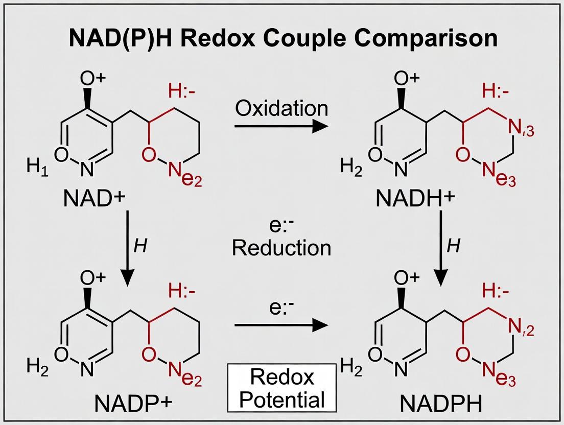
NADH and NADPH share an identical adenosine-diphosphate-ribonucleotide core coupled to a nicotinamide ring. The sole chemical difference is the presence of an additional phosphate group on the 2' carbon of the ribose ring in NADPH. This minor modification dictates profound functional specialization.
Table 1: Core Functional and Metabolic Comparison
| Parameter | NADH | NADPH |
|---|---|---|
| Primary Redox Role | Catabolic electron carrier | Anabolic electron donor |
| Standard Reduction Potential (E°') | -320 mV | -320 mV |
| Key Metabolic Pathways | Glycolysis, TCA cycle, Oxidative Phosphorylation | Pentose phosphate pathway, Fatty acid & nucleotide synthesis, Antioxidant systems (glutathione/thioredoxin) |
| Enzyme Kinetics (Km for typical dehydrogenase, μM) | 10-50 μM | 1-10 μM |
| Cytosolic [NAD(P)H]/[NAD(P)+] Ratio | ~0.001 | ~100 |
| Primary Cellular Compartment | Mitochondrial matrix | Cytosol (major pool) |
Table 2: Experimental Performance in Key Assays
| Assay Type | NADH Performance | NADPH Performance | Experimental Reference |
|---|---|---|---|
| Lactate Dehydrogenase (LDH) Activity | Vmax = 150 μmol/min/mg; Km = 15 μM | Vmax < 5 μmol/min/mg; Km > 500 μM | Bergmeyer et al., Methods of Enzymatic Analysis |
| Glucose-6-Phosphate Dehydrogenase (G6PD) Activity | No measurable activity | Vmax = 80 μmol/min/mg; Km = 4 μM | Bergmeyer et al., Methods of Enzymatic Analysis |
| Cytosolic ROS Scavenging (via glutathione reductase) | Ineffective | Regenerates reduced glutathione in <1 ms | Sies et al., Redox Biology, 2017 |
| Mitochondrial Complex I (NADH:ubiquinone oxidoreductase) Activity | Direct electron donor; Km ~10 μM | Not a substrate; inhibits at high [ ] | Hirst et al., Biochem. J., 2020 |
Experimental Protocols for Key Comparisons
Protocol 1: Determining Cofactor Specificity of a Dehydrogenase
- Reaction Mixture: Prepare 1 mL of 50 mM Tris-HCl (pH 8.0), 5 mM substrate (e.g., glucose-6-phosphate for G6PD, pyruvate for LDH), and 0.2 mM of either NAD⁺ or NADP⁺.
- Enzyme Addition: Add 0.01 units of the purified dehydrogenase enzyme.
- Kinetic Measurement: Monitor the increase in absorbance at 340 nm (A₃₄₀) due to NAD(P)H formation for 3 minutes at 25°C using a spectrophotometer.
- Analysis: Calculate initial velocity. Repeat with varying cofactor concentrations (0-200 μM) to determine Km and Vmax for each cofactor.
Protocol 2: Measuring Compartmentalized [NADPH]/[NADP⁺] Ratio via Fluorescence
- Cell Preparation: Culture adherent cells (e.g., HEK293) on a glass-bottom dish.
- Transfection: Transfect with a genetically encoded biosensor (e.g., iNAP or Apollo-NADP) specific for the NADPH/NADP⁺ couple.
- Imaging: Perform ratiometric fluorescence imaging (excitation 410/480 nm, emission 515 nm) using a confocal microscope.
- Quantification: Calculate the 410/480 nm emission ratio. Calibrate in situ using 10 μM ionomycin and 100 μM NAD⁺ (to minimize NADPH) followed by 10 mM glucose and 5 μM rotenone (to maximize NADPH).
Visualization of Pathways and Relationships
The Scientist's Toolkit: Research Reagent Solutions
Table 3: Essential Reagents for NAD(P)H Research
| Reagent/Material | Function in Research | Key Consideration |
|---|---|---|
| Recombinant Dehydrogenases (e.g., LDH, G6PD) | Standard enzymes for validating assay conditions and cofactor specificity. | Use enzyme from the same species as your system of study to avoid kinetic artifacts. |
| β-NAD(H) & β-NADP(H) (High-Purity Grades) | Substrates for kinetic assays and metabolic studies. | Verify purity via A₂₆₀/A₃₄₀ ratios. Store aliquots at -80°C in acidic buffer (pH ~3) for stability. |
| Genetically Encoded Biosensors (iNAP, Apollo-NADP) | Live-cell, compartment-specific measurement of NADPH/NADP⁺ ratios. | Select sensor with appropriate affinity (Kd) for your expected cellular concentration range. |
| Spectrophotometer/UPLC with Fluorescence Detector | Quantifying NAD(P)H concentration via A₃₄₀ or native fluorescence. | For complex lysates, UPLC separation prevents signal interference from other metabolites. |
| Cell Permeant Probes (e.g., roGFP-Tsa2ΔCR) | Ratiometric, redox-sensitive probes for dynamic in vivo measurements. | Requires careful calibration with diamide and DTT for each cell type. |
| Mitochondrial & Cytosolic Fractionation Kit | Isolates subcellular compartments to localize NAD(P)H pools. | Rapid processing at 4°C is critical to preserve metabolite levels and prevent artifacts. |
Within the context of NAD(P)H redox couple research, understanding the compartmentalization of these cofactors is critical. The distinct subcellular pools in the mitochondria, cytosol, and nucleus govern unique metabolic and signaling pathways, influencing cellular functions from energy production to gene expression and DNA repair. This guide compares the characteristics, dynamics, and functional implications of NAD(P)H pools across these compartments.
Comparative Analysis of Subcellular NAD(P)H Pools
Table 1: Key Characteristics of NAD(P)H Subcellular Pools
| Parameter | Mitochondrial Pool | Cytosolic Pool | Nuclear Pool |
|---|---|---|---|
| Primary Redox Couple | NADH/NAD+ (High ratio) | NADPH/NADP+ (Maintained high) | NADH/NAD+ & NADPH/NADP+ |
| Major Metabolic Role | Oxidative phosphorylation, TCA cycle | Biosynthesis (lipids, nucleotides), Antioxidant defense (via GSH/Trx systems) | Epigenetic regulation, DNA repair, Signaling |
| Approximate Concentration | NADH: 0.1-0.4 mM; NAD+: 0.2-0.5 mM | NADPH: ~0.05 mM; NADP+: ~0.005 mM | Not well quantified; dynamic and regulated |
| Key Regulating Enzymes | Complex I, Malate-Aspartate Shuttle, Mitochondrial transhydrogenase | G6PD, IDH1, ME1, NNT | PARPs, Sirtuins, ALDHs |
| Redox Potential (Approx.) | -280 to -320 mV | -370 to -400 mV | Variable, compartment-specific |
| Primary Measurement Tools | roGFP, mt-cpYFP, Fluorescence lifetime imaging (FLIM) | iNAP sensors, SoNar, Fluorescence intensity probes (e.g., Peredox) | NuAP sensors, Genetically-encoded redox biosensors targeted to nucleus |
| Response to Stress | Rapid oxidation upon ETC disruption | Sustained reduction for antioxidant response | Oxidation for DNA damage signaling; Reduction for repair phases |
Table 2: Experimental Data Comparison from Recent Studies (2023-2024)
| Study Focus | Mitochondrial NADH | Cytosolic NADPH | Nuclear NAD(P)H |
|---|---|---|---|
| Effect of Glucose Deprivation | Decrease by 60-70% (FLIM-NADH) | Increase by ~20% (iNAP sensor) | Transient decrease by ~40% (NuAP sensor) |
| Response to 1mM H₂O₂ | Moderate oxidation (15% decrease in reduction state, mt-roGFP) | Strong antioxidant defense (redox maintained, iNAP) | Rapid, transient oxidation (30% change, NuAP) |
| Impact of ETC Inhibition (Antimycin A) | >80% increase in NADH (reduced state) | Minimal direct change | Secondary, slow increase via metabolic adaptation |
| Baseline Turnover Rate | Fast (seconds) | Moderate (minutes) | Slow to Moderate (context-dependent) |
| Link to Apoptosis Initiation | Critical (Cytochrome c release correlated with NADH oxidation) | Indirect (via altered biosynthesis) | Direct (PARP activation consumes nuclear NAD+) |
Experimental Protocols for Key Comparisons
Protocol 1: Simultaneous Measurement of Mitochondrial and Cytosolic NADH/NAD(P)H Redox States Using Genetically-Encoded Biosensors
Objective: To compare real-time redox dynamics in cytosol and mitochondria under metabolic perturbation.
Key Reagents & Materials:
- HeLa or U2OS cell line stably expressing cytosolic Peredox (cPeredox) and mitochondrial mt-cpYFP.
- Live-cell imaging medium (FluoroBrite DMEM, no phenol red, with 10% FBS, 25mM Glucose, 4mM Glutamine).
- Metabolic modulators: 1µM Rotenone (ETC inhibitor), 10mM 2-Deoxy-D-glucose (2-DG, glycolysis inhibitor).
- Imaging system: Confocal or widefield microscope with environmental chamber (37°C, 5% CO₂), capable of ratiometric imaging.
Procedure:
- Seed cells in 35mm glass-bottom dishes and culture until 70% confluency.
- Replace medium with pre-warmed live-cell imaging medium 1 hour before experiment.
- Mount dish on microscope. For cPeredox, acquire images at excitation 405nm and 488nm, emission 510/20nm. For mt-cpYFP, use excitation 488nm, emission 535/30nm.
- Acquire baseline images every 30 seconds for 5 minutes.
- Gently add rotenone (final 1µM) or 2-DG (final 10mM) without moving the dish. Continue time-lapse imaging for 30-60 minutes.
- Data Analysis: Calculate ratio (R) = Intensity(405ex)/Intensity(488ex) for Peredox (reflects NADH/NAD+ ratio). For mt-cpYFP, calculate fluorescence intensity changes. Normalize all ratios to the pre-treatment baseline average.
Protocol 2: Quantifying Compartment-Specific NADPH Production Capacity via Enzymatic Assays in Subcellular Fractions
Objective: To compare the NADPH-generating capacity of cytosol and mitochondria.
Key Reagents & Materials:
- Cell lysis buffer with digitonin for selective plasma membrane permeabilization.
- Mitochondrial isolation kit (e.g., from Thermo Fisher).
- NADPH detection assay kit (fluorometric, e.g., Abcam ab186031).
- Substrate solutions: 10mM Glucose-6-Phosphate (G6P), 10mM Isocitrate, 10mM Malate.
- Plate reader capable of fluorescence measurement (Ex/Em = 540/590nm).
Procedure:
- Harvest 10^7 cells (e.g., HEK293). Perform digitonin-based fractionation to obtain cytosolic and crude mitochondrial fractions. Validate purity by Western blot (LDH for cytosol, COX IV for mitochondria).
- Adjust protein concentration of fractions to 1 mg/mL.
- In a 96-well plate, mix 50µL of fraction with 50µL of reaction mix containing assay buffer, substrate (G6P for cytosol; Isocitrate or Malate for mitochondria), and enzyme mix (excluding developer). Perform separate reactions for each substrate.
- Incubate at 37°C for 30 minutes, protecting from light.
- Add 10µL of developer, mix, and incubate for 30-60 minutes at 37°C.
- Measure fluorescence. Generate a standard curve using known NADPH concentrations (0-10 µM).
- Data Analysis: Calculate NADPH generation rate as nmol NADPH produced/min/mg protein. Compare cytosolic (G6PD-driven) vs. mitochondrial (IDH2/ME3-driven) capacities.
Research Reagent Solutions Toolkit
| Reagent/Material | Primary Function | Example Product/Catalog # |
|---|---|---|
| Genetically-Encoded Biosensor (Cytosolic) | Reports real-time NADH/NAD+ ratio | Peredox-mCherry (Addgene #32383) |
| Genetically-Encoded Biosensor (Mitochondrial) | Reports mitochondrial NADH redox state | mt-cpYFP (Addgene #23214) |
| Genetically-Encoded Biosensor (NADPH) | Reports NADPH/NADP+ redox in cytosol or nucleus | iNAP, SoNar (Various) |
| Selective Permeabilization Agent | Isolates cytosolic fraction without disrupting organelles | Digitonin (Sigma D141) |
| Mitochondrial Isolation Kit | Provides purified mitochondrial fractions from cells/tissues | MITOISO2 (Sigma) |
| Fluorometric NADPH Assay Kit | Quantifies total NADPH levels in biological samples | ab186031 (Abcam) |
| PARP Inhibitor (Control) | Inhibits nuclear NAD+ consumption, used to validate nuclear NAD(P)H dynamics | Olaparib (Selleckchem S1060) |
| Glucose-6-Phosphate Dehydrogenase Inhibitor | Specifically perturbs cytosolic NADPH production via PPP | 6-Aminonicotinamide (Sigma A68203) |
| ETC Complex I Inhibitor | Perturbs mitochondrial NADH oxidation, increasing NADH pool | Rotenone (Sigma R8875) |
Visualizations
Within the broader research thesis comparing the cellular functions of the NAD(P)H redox couples, this guide focuses on the critical role of NAD(H) in central catabolic pathways. We objectively compare the efficiency and kinetics of NAD⁺ reduction across glycolysis, the TCA cycle, and the subsequent electron transfer via NADH in oxidative phosphorylation, supported by experimental data.
Quantitative Comparison of NAD(H) Flux and Yield
Table 1: NAD⁺ Reduction and ATP Yield per Glucose Molecule in Major Catabolic Pathways
| Pathway / Process | Primary Reaction for NAD⁺ Reduction | Net NADH Produced per Glucose | Ultimate ATP Yield (via OXPHOS) | Experimental Measurement Method |
|---|---|---|---|---|
| Glycolysis (Cytosol) | Glyceraldehyde-3-phosphate → 1,3-BPG | 2 NADH (cytosolic) | ~3-5 ATP* | Enzyme-coupled spectrophotometric assay (340 nm) |
| Pyruvate Decarboxylation | Pyruvate → Acetyl-CoA + CO₂ | 2 NADH (mitochondrial) | ~5 ATP | Mitochondrial isolation followed by NADH fluorometry |
| TCA Cycle (per Acetyl-CoA) | Isocitrate → α-KG; α-KG → Suc-CoA; Malate → OAA | 3 NADH (mitochondrial) | ~7.5 ATP | ¹³C-isotope tracing & LC-MS for flux analysis |
| Total Theoretical Yield | 10 NADH (2+2+6) + 2 FADH₂ + 2 ATP (substrate-level) | ~30-32 ATP | Calorimetric & respirometric analysis in intact cells |
Note: Cytosolic NADH must be shuttled (e.g., Malate-Aspartate) into mitochondria, yielding variable ATP (2.5 ATP/NADH typical).
Table 2: Kinetic Parameters of Key NAD⁺-Reducing Dehydrogenases
| Enzyme (Pathway) | Km for NAD⁺ (μM) | Turnover Number, kcat (s⁻¹) | Catalytic Efficiency (kcat/Km, M⁻¹s⁻¹) | Preferred Assay Conditions |
|---|---|---|---|---|
| GAPDH (Glycolysis) | 80-120 | ~200 | ~2.0 x 10⁶ | Tris-HCl buffer (pH 8.6), 25°C, with Arsenate |
| Pyruvate DH Complex (Link) | 20-40 | ~50 | ~1.5 x 10⁶ | Isolated mitochondria, ThPP, Mg²⁺, pH 7.0 |
| Isocitrate DH (TCA) | 10-25 | ~80 | ~4.0 x 10⁶ | Tris-HCl (pH 7.4), Mg²⁺ or Mn²⁺, 37°C |
| α-KG DH Complex (TCA) | 30-60 | ~60 | ~1.2 x 10⁶ | Potassium phosphate buffer (pH 7.4), ThPP, Ca²⁺ |
| Malate DH (TCA) | 100-200 | ~300 | ~2.0 x 10⁶ | Potassium phosphate (pH 7.4), high [OAA] to favor oxidation |
Experimental Protocols for Key Measurements
Protocol 1: Spectrophotometric Assay for Glycolytic NADH Production
- Objective: Quantify NAD⁺ reduction rate in a cell-free glycolytic lysate.
- Method:
- Prepare reaction buffer: 50 mM Tris-HCl (pH 8.6), 5 mM Na₂HAsO₄ (arsenate, to uncouple oxidation from phosphorylation), 1 mM DTT, 0.2 mM NAD⁺.
- Lyse cells in ice-cold hypotonic buffer and clarify by centrifugation (14,000 x g, 10 min, 4°C).
- Initiate reaction by adding 10-50 μg of lysate protein and 5 mM glyceraldehyde-3-phosphate to the pre-warmed (37°C) buffer.
- Immediately monitor the increase in absorbance at 340 nm (A₃₄₀) for 5 minutes using a plate reader or spectrophotometer.
- Calculate NADH formation rate using the extinction coefficient ε₃₄₀ = 6220 M⁻¹cm⁻¹.
Protocol 2: Respirometric Analysis of Mitochondrial NADH Oxidation
- Objective: Measure the coupling efficiency between NADH generation and oxidative phosphorylation.
- Method (using Seahorse XF Analyzer):
- Seed cells in a Seahorse microplate and culture overnight.
- Replace medium with substrate-limited, serum-free, bicarbonate-free assay medium (pH 7.4) supplemented with 10 mM glucose and 2 mM glutamine.
- Load sensors and calibrate the instrument.
- Perform a mitochondrial stress test: Measure basal oxygen consumption rate (OCR), then inject 1.5 μM oligomycin (ATP synthase inhibitor), followed by 1 μM FCCP (uncoupler), and finally 0.5 μM rotenone/antimycin A (Complex I/III inhibitors).
- The OCR linked to NADH oxidation (proton leak subtracted) is derived from the oligomycin-sensitive respiration.
Pathway and Workflow Visualizations
Title: NAD(H) Redox Flow from Glucose to ATP
Title: Experimental Workflow for Glycolytic NADH Assay
The Scientist's Toolkit: Key Research Reagent Solutions
Table 3: Essential Reagents for NAD(H)-Linked Energy Metabolism Research
| Reagent / Kit Name | Primary Function in Research | Key Application in This Context |
|---|---|---|
| NAD⁺/NADH Quantification Kit (Fluorometric) | Selectively quantifies oxidized and reduced forms via enzymatic cycling. | Measuring NAD⁺/NADH ratio in cell lysates from different metabolic states. |
| Seahorse XF Cell Mito Stress Test Kit | Measures OCR and ECAR in live cells to profile mitochondrial function. | Determining the efficiency of NADH-linked respiration (post-oligomycin OCR). |
| Recombinant Human Dehydrogenases (e.g., GAPDH, IDH) | Highly purified, active enzyme for in vitro kinetic studies. | Determining Km for NAD⁺ and kcat values under controlled conditions. |
| ¹³C-Labeled Metabolic Substrates (e.g., [U-¹³C]-Glucose) | Tracers for following metabolic flux via mass spectrometry. | Mapping the contribution of glycolysis vs. TCA cycle to total NADH production. |
| Mitochondrial Isolation Kit | Prepares intact, functional mitochondria from tissues or cells. | Studying NADH generation and oxidation in isolated organelle systems. |
| Cell Permeable NAD⁺ Precursors (e.g., NMN, NR) | Modulate intracellular NAD⁺ levels in vivo and in vitro. | Investigating the impact of NAD⁺ bioavailability on catabolic flux and ATP yield. |
Within the broader thesis of comparing NAD(H) and NADP(H) redox couples in cellular functions, this guide examines the critical, non-interchangeable role of NADP(H) in anabolic biosynthesis versus oxidative defense. Experimental data consistently highlight that while NAD(H) drives catabolism, the NADP(H) pool is uniquely partitioned to support parallel metabolic streams: reductive biosynthesis and antioxidant regeneration.
Comparative Performance: NADPH-Dependent Pathways vs. NADH-Driven or Alternative Systems
Table 1: Comparison of Key NADPH-Consuming Pathways and Competing Systems
| Cellular Function | Primary NADPH-Dependent System | Key Alternative/Competing System | Experimental Finding (Representative) | Key Metric |
|---|---|---|---|---|
| Fatty Acid Synthesis | Cytosolic FAS (Fatty Acid Synthase) | Mitochondrial β-oxidation (NADH-generating) | siRNA knock-down of glucose-6-phosphate dehydrogenase (G6PD) in hepatocytes decreased palmitate synthesis by >70%, while lactate (NADH source) failed to rescue. [PMID: 24563466] | Palmitate Synthesis Rate |
| Nucleotide Synthesis | Ribonucleotide Reductase (RNR) | Salvage pathways | In MCF-7 cells, inhibition of the oxidative PPP (NAPDH source) with 6-AN reduced dNTP pools by 60%, stalling S-phase; supplementation with nucleosides (salvage) partially restored proliferation. [PMID: 23974231] | dNTP Pool Size / S-Phase Fraction |
| ROS Detoxification | Glutathione (GSH) System (GPx/GR) | Thioredoxin (Trx) System (Prx/TrxR) | In endothelial cells, siRNA to TXNRD1 (TrxR) increased sensitivity to H2O2 (EC50 ~50 µM), while siRNA to GSR (GR) increased sensitivity to lipid peroxides (EC50 ~5 µM). [PMID: 25586068] | Oxidant EC50 (Viability) |
| ROS Detoxification | Thioredoxin (Trx) System | Catalase (H2O2-specific, no cofactor) | During sustained oxidative stress, treatment with Auranofin (TrxR inhibitor) led to irreversible oxidation of 2-Cys Prx, while Catalase overexpression only delayed H2O2 accumulation. [PMID: 25456078] | % Oxidized 2-Cys Prx |
| NADPH Regeneration | Oxidative Pentose Phosphate Pathway (PPP) | Malic Enzyme (ME1) & IDH1 | Isotopic tracing in HEK293 cells showed under oxidative stress, >80% of cytosolic NADPH derived from PPP; in lipogenic conditions, ME1 contribution increased to ~40%. [PMID: 26812018] | % NADPH Contribution |
Detailed Experimental Protocols
1. Protocol: Measuring NADPH/NADP+ Ratio in Subcellular Compartments Using Genetically Encoded Sensors (e.g., iNAP)
- Objective: Quantify real-time NADPH dynamics in cytosol vs. mitochondria during anabolic or oxidative stress.
- Cell Preparation: Seed cells expressing iNAP7 (cytosolic) or mito-iNAP7 in 96-well glass-bottom plates.
- Imaging: Use a fluorescence plate reader or confocal microscope with dual-excitation (410 nm and 470 nm). Calculate ratio (R = F470/F410).
- Calibration: At experiment end, permeabilize cells with digitonin (50 µM). Apply 10 mM NADP+ (Rmin), then 10 mM NADPH (Rmax) to obtain calibrated ratio.
- Intervention: After baseline, add either 1) 20 mM glucose (anabolic stimulus) or 2) 200 µM menadione (oxidative stressor). Record ratio every 30 seconds for 30 minutes.
- Analysis: Convert ratio to [NADPH] using calibration curve and published K_d.
2. Protocol: Assessing Pathway-Specific NADPH Utilization via Metabolite Profiling
- Objective: Determine the impact of NADPH pool perturbation on lipid and nucleotide synthesis fluxes.
- Cell Treatment: Culture HepG2 cells in stable isotope medium ([U-13C]-glucose). Treat with either DMSO (control), 10 µM G6PD inhibitor (6-AN), or 5 µM ME1 inhibitor (ME1i) for 24h.
- Metabolite Extraction: Wash cells with cold saline, quench with 80% methanol (-80°C), and lyse by freeze-thaw. Collect supernatant for LC-MS.
- LC-MS Analysis: Use reversed-phase chromatography (for fatty acids) and HILIC (for nucleotides and PPP intermediates). Monitor mass isotopomer distributions (MIDs).
- Data Interpretation: Calculate 13C-enrichment in palmitate (M+16 for full labeling) and in ribose-phosphate moiety of NTPs. Reduced enrichment in inhibitor-treated groups indicates decreased de novo synthesis flux from the NADPH-producing pathway.
Pathway and Workflow Visualizations
NADPH Source and Fate Pathways
NADPH Live-Cell Imaging Workflow
The Scientist's Toolkit: Key Research Reagent Solutions
Table 2: Essential Reagents for Studying NADPH-Dependent Pathways
| Reagent / Material | Provider Examples | Primary Function in Research |
|---|---|---|
| Genetically Encoded NADPH Biosensors (iNAP, Apollo-NADP+) | Addgene, custom construct | Real-time, subcellular quantification of NADPH/NADP+ ratios via fluorescence imaging. |
| Stable Isotope Tracers ([U-13C]-Glucose, [2H7]-Glucose) | Cambridge Isotope Labs, Sigma-Aldrich | Tracing NADPH production routes (PPP vs. ME1/IDH1) and anabolic flux via LC-MS. |
| Pathway-Specific Chemical Inhibitors (6-AN, DHEA, Auranofin) | Tocris, Cayman Chemical | Pharmacological dissection of NADPH sources (G6PD/6-AN) or consumers (TrxR/Auranofin). |
| siRNA/shRNA Libraries (G6PD, ME1, IDH1, TXNRD1, GSR) | Dharmacon, Sigma-Aldrich | Genetic knockdown to validate enzyme-specific roles in NADPH homeostasis and cell fate. |
| LC-MS/MS Systems (Q-Exactive, TripleTOF) | Thermo Fisher, Sciex | Quantifying absolute metabolite levels (NADPH, GSH, nucleotides) and isotopic enrichment. |
| Seahorse XFp Analyzer | Agilent Technologies | Profiling mitochondrial respiration and glycolytic/G6PD flux in real-time. |
| Anti-2-Cys Prx-SO3 Antibody | Abcam, Cell Signaling | Detecting oxidized, inactive peroxiredoxin as a marker of Trx system insufficiency. |
Within the framework of NAD(P)H redox couple comparison cellular functions research, understanding the enzymatic consumers of NAD+ is paramount. Sirtuins (SIRTs) and Poly(ADP-ribose) polymerases (PARPs) are two major NAD+-dependent enzyme families that function as metabolic sensors, translating redox state into adaptive cellular responses. This comparison guide objectively evaluates their roles, mechanisms, and experimental analysis.
Comparative Analysis: Sirtuins vs. PARPs as NAD+ Consumers and Signaling Hubs
Table 1: Core Functional Comparison of SIRTs and PARPs
| Feature | Sirtuins (Class III HDACs) | Poly(ADP-ribose) Polymerases |
|---|---|---|
| Primary Reaction | NAD+-dependent protein deacylation (deacetylation, desuccinylation) | NAD+-dependent ADP-ribosylation of proteins |
| Key Metabolic Role | Stress response, metabolic adaptation, longevity | DNA damage repair, genomic stability |
| NAD+ Dependency | High (Km typically 50-100 µM); activity directly reflects NAD+ levels | Very High (Km for PARP1 ~20-50 µM); hyperactivated by DNA damage can deplete NAD+ |
| Primary Cellular Response | Transcriptional reprogramming, mitochondrial biogenesis, autophagy | DNA repair, inflammation, cell death decisions |
| Redox Sensing Link | Sensitive to NAD+/NADH ratio; inhibited by nicotinamide | PARP1 activation linked to oxidative stress-induced DNA damage |
Table 2: Quantitative Experimental Data from Key Studies
| Parameter | Experimental Readout | SIRT1 Data | PARP1 Data | Source/Model |
|---|---|---|---|---|
| NAD+ Depletion Rate | [NAD+] after 1hr of activation | ~20% decrease (Fast-induced) | >80% decrease (H2O2-induced) | HEK293 cells, HPLC-MS |
| Impact on [ATP] | Cellular ATP levels post-activation | Mild decrease (~15%) | Severe decrease (>70%) | HeLa cells, luciferase assay |
| Transcriptional Targets | Fold-change mRNA expression | PGC-1α (+3.5), FOXO1 (+2.1) | NF-κB (+4.8), iNOS (+12.5) | Mouse liver, qPCR |
| Inhibitor Potency (IC50) | In vitro enzyme activity | EX527: 60 nM | Olaparib: 5 nM | Recombinant enzyme assays |
Experimental Protocols for Comparative Analysis
Protocol 1: Measuring NAD+ Consumption Kinetics In Vitro
Objective: To directly compare the NAD+ utilization rates of SIRT and PARP enzymes. Methodology:
- Reaction Setup: Prepare assay buffer (50 mM Tris-HCl pH 8.0, 150 mM NaCl, 1 mM DTT). For SIRT1, include acetylated peptide substrate (e.g., p53-derived) and 200 µM NAD+. For PARP1, activate with DNA oligos containing a double-strand break and 200 µM NAD+.
- Enzyme Addition: Initiate reactions with 10 nM purified human SIRT1 or PARP1.
- Time-Course Sampling: Aliquot reactions at 0, 2, 5, 10, 20, and 30 minutes.
- NAD+ Quantification: Stop reactions with 0.5M perchloric acid. Neutralize with KOH. Measure NAD+ concentration using a cycling enzymatic assay (e.g., using alcohol dehydrogenase) or HPLC-MS.
- Data Analysis: Plot [NAD+] vs. time. Calculate initial velocity (V0) and apparent enzyme efficiency.
Protocol 2: Assessing Pathway Competition in Live Cells
Objective: To determine the preference for NAD+ consumption under metabolic or genotoxic stress. Methodology:
- Cell Treatment: Seed HeLa or U2OS cells in 6-well plates. Establish four conditions: Control, SIRT activator (e.g., 500 µM Resveratrol, 24h), PARP activator (e.g., 200 µM H2O2, 1h), and Co-treatment.
- Metabolite Extraction: Wash cells with cold PBS and extract using 80% methanol/water at -80°C.
- LC-MS/MS Analysis: Analyze extracts for NAD+, NADH, NADP+, NADPH, and ADP-ribose metabolites. Use a hydrophilic interaction chromatography (HILIC) column coupled to a mass spectrometer.
- Pathway Output Measurement: In parallel wells, prepare lysates for immunoblotting. For SIRT activity, monitor acetylation status of targets (e.g., Ac-p53). For PARP activity, monitor PAR polymer levels using anti-PAR antibody.
Visualization of Pathways and Workflows
The Scientist's Toolkit: Key Research Reagent Solutions
Table 3: Essential Reagents for Redox Signaling Research on SIRTs and PARPs
| Reagent Category | Specific Item | Function in Research |
|---|---|---|
| NAD+ Precursors | Nicotinamide Riboside (NR), Nicotinamide Mononucleotide (NMN) | Boosts intracellular NAD+ levels to study sirtuin activation or PARP resilience. |
| SIRT Modulators | EX527 (SIRT1 inhibitor), Resveratrol (activator), SRT1720 (activator) | Pharmacologically probe SIRT function in cellular and in vivo models. |
| PARP Inhibitors | Olaparib (AZD2281), Rucaparib, PJ34 | Clinical and research-grade inhibitors for studying DNA damage response and metabolic competition. |
| Activity Assays | Fluorogenic SIRT substrate (e.g., Ac-p53 peptide), PARP activity chemiluminescent kit | Direct in vitro or cell-based measurement of enzyme velocity and inhibition. |
| Detection Antibodies | Anti-acetyl-lysine, Anti-PAR polymer (10H), Anti-SIRT1, Anti-PARP1 | Immunoblotting and immunofluorescence to monitor pathway activity and expression. |
| Metabolomics Standards | Stable isotope-labeled NAD+ (¹³C-NAD), NADH, NAM | Internal standards for precise LC-MS/MS quantification of NAD+ metabolome. |
| DNA Damage Inducers | Hydrogen Peroxide (H2O2), Bleomycin, UV-C | Controlled induction of genotoxic stress to activate PARP and study redox interplay. |
Measuring Redox States: Advanced Techniques for NAD(P)H Quantification and Live-Cell Imaging
Within the context of NAD(P)H redox couple research, understanding cellular metabolic functions requires precise measurement of these critical cofactors. This guide compares the performance of two principal methodological approaches: assays performed on extracted samples versus in situ (live-cell) measurements. The choice between spectrophotometric and fluorometric techniques further defines the experimental landscape, impacting sensitivity, specificity, and biological relevance.
Core Comparison: Extracted vs. In Situ Assays
Table 1: High-Level Comparison of Methodological Paradigms
| Feature | Extracted Assays (Spectro/Fluorometric) | In Situ Assays (Primarily Fluorometric) |
|---|---|---|
| Sample State | Lysed, homogenized; fixed chemical state. | Live, intact cells; dynamic physiological state. |
| Primary Output | Total pool size (NADH+NAD⁺, NADPH+NADP⁺) or ratio. | Spatially & temporally resolved redox ratios. |
| Temporal Resolution | End-point/snapshot; low. | Real-time kinetic; high. |
| Spatial Resolution | None (bulk population average). | Sub-cellular (cytosol, mitochondria, nucleus). |
| Throughput | High (plate reader compatible). | Medium to low (requires imaging). |
| Artifact Potential | Extraction inefficiency, enzyme degradation. | Photobleaching, probe toxicity, calibration. |
| Best For | Absolute quantification, high-throughput screening. | Kinetic tracing, compartment-specific dynamics, single-cell analysis. |
Quantitative Performance Data
Table 2: Experimental Data Comparison from Recent NAD(P)H Studies
| Assay Type | Model System | Measured Parameter | Key Result (Extracted) | Key Result (In Situ) | Reference Context |
|---|---|---|---|---|---|
| Spectrophotometric (Extracted) | HeLa cell lysate | Total NAD⁺/NADH | 6.8 ± 0.9 (Ratio) | N/A | WST-8 enzyme-coupled assay. |
| Fluorometric (Extracted) | Liver tissue homogenate | NADPH concentration | 42.3 ± 5.1 µM/g tissue | N/A | Enzymatic recycling assay. |
| Fluorometric (In Situ) | Live MCF-7 cells | NADH/NAD⁺ redox index | N/A | 1.24 ± 0.15 (Mitochondria) | FLIM of endogenous NADH fluorescence. |
| Genetically Encoded Sensor (In Situ) | Live HEK293T cells | NADPH/NADP⁺ ratio | N/A | 3.05 ± 0.41 (Cytosol) | iNap sensor ratiometric imaging. |
| Parallel Measurement | Primary hepatocytes | Metabolic response to drug | NAD⁺ depleted by 60% (lysis assay) | Mitochondrial redox shift in <2 min (imaging) | Combined study validating kinetics. |
Experimental Protocols
Protocol 1: Enzymatic Spectrophotometric Assay for Extracted NAD⁺/NADH
Principle: Enzyme-coupled reaction where NAD⁺ reduction or NADH oxidation is linked to a colorimetric reporter (e.g., formazan dye).
- Cell Lysis: Pellet 1x10⁶ cells. Lyse with 200 µL of cold acidic lysis buffer (for NADH stabilization) or basic buffer (for NAD⁺). Heat at 60°C for 15 min to degrade enzymes, then neutralize.
- Reaction Mix: For NAD⁺ measurement: 50 µL lysate + 100 µL reaction buffer (containing WST-8, diaphorase, and substrate). For NADH, a separate aliquot is used with an oxidation-linked enzyme system.
- Measurement: Incubate at 37°C for 30 min. Measure absorbance at 450 nm using a plate reader.
- Quantification: Compare to a standard curve of known NAD⁺/NADH concentrations.
Protocol 2: Fluorescence Lifetime Imaging Microscopy (FLIM) for In Situ NADH
Principle: Endogenous NADH is fluorescent; its fluorescence lifetime shifts upon binding to proteins, serving as a redox indicator.
- Sample Prep: Plate cells on glass-bottom dishes. Maintain in phenol-red free imaging medium at 37°C/5% CO₂.
- Microscopy Setup: Use a multiphoton or time-correlated single photon counting (TCSPC) confocal microscope with a 740 nm excitation laser.
- Image Acquisition: Collect fluorescence emission at 460±50 nm. Acquire FLIM data until sufficient photon counts are reached (~100-1000 photons/pixel).
- Data Analysis: Fit fluorescence decay curves per pixel to a bi-exponential model. Calculate the weighted mean lifetime (τm) or the ratio of free/bound NADH as a redox index.
The Scientist's Toolkit: Research Reagent Solutions
| Item | Function in NAD(P)H Assays |
|---|---|
| WST-8 / MTT Tetrazolium Salts | Electron acceptors in enzyme-coupled extraction assays; produce water-soluble formazan dyes measurable at ~450 nm. |
| Enzyme Cocktails (Diaphorase, Dehydrogenases) | Specific coupling enzymes used in extraction protocols to link NAD(P)H oxidation/reduction to the reporter signal. |
| Genetically Encoded Sensors (e.g., iNap, Peredox) | Expressed in live cells to provide ratiometric, compartment-specific readouts of NADPH/NADP⁺ or NADH/NAD⁺ ratios. |
| Phenol-Red Free Culture Medium | Essential for in situ fluorometry to minimize background autofluorescence. |
| NAD⁺/NADH & NADP⁺/NADPH Extraction Kits | Commercial kits providing optimized buffers for rapid, efficient, and stabilized cofactor extraction from tissues/cells. |
| Two-Photon FLIM Microscope | Advanced imaging system enabling in situ measurement of endogenous NADH fluorescence lifetime with deep tissue penetration and minimal phototoxicity. |
Visualizing Methodological Pathways and Workflows
Title: Extracted Assay Workflow for NAD(P)H
Title: In Situ Live-Cell Imaging Workflow
Title: NAD(P)H in Cellular Metabolic Pathways
Thesis Context
Within the broader thesis on comparing NAD(P)H redox couple functions in cellular metabolism, proliferation, and stress response, genetically encoded biosensors are indispensable tools. They enable the precise, real-time, and compartment-specific monitoring of NAD+/NADH and NADP+/NADPH dynamics, moving beyond static snapshots to capture metabolic flux in living cells.
Performance Comparison of NAD(P)H Redox Biosensors
Table 1: Key Characteristics of Representative NAD(P)H Biosensors
| Biosensor Name | Redox Couple Targeted | Excitation/Emission Peaks (nm) | Dynamic Range (ΔR/R0 %) | Subcellular Localization Capability | Apparent KD (NADH) | Response Time | pH Sensitivity | Key References |
|---|---|---|---|---|---|---|---|---|
| Peredox | NAD+/NADH | 440/485 (FRET-cp173-mCitrine) | ~500% | Cytosol, Nucleus, Mitochondria | ~120 µM | Minutes | Low | Hung et al., Nature, 2011 |
| SoNar | NAD+/NADH | 420/485 and 485/585 (Ratiometric) | ~1500% | Cytosol | ~1.2 µM | Seconds | Moderate | Zhao et al., Cell Metabolism, 2015 |
| iNap | NADP+/NADPH | 410/485 and 485/570 (Ratiometric) | ~800% | Cytosol, Mitochondria, Peroxisomes | ~90 µM (NADPH) | Seconds | Low | Tao et al., Science, 2017 |
| Frex Family (e.g., Frex, Peredox) | NADH | ~490/520 (Single FP) | ~400% | Cytosol, Mitochondria | ~1-5 µM | Seconds | High | Zhao et al., Cell Metabolism, 2011 |
| NADPH Sensor (Mrx1-roGFP2) | NADP+/NADPH (via Rox) | 400/510 and 480/510 (Ratiometric) | ~5 (Rox Ratio) | Cytosol, Mitochondria | N/A (Thiol redox) | Minutes | Low | Gutscher et al., Nat Methods, 2008 |
Table 2: Experimental Performance in Common Cellular Contexts
| Experiment Context | Optimal Sensor | Rationale & Supporting Data | Key Limitation |
|---|---|---|---|
| Cytosolic NADH Dynamics (e.g., glycolysis) | SoNar | Highest dynamic range (~15-fold); rapid response to glucose pulse. Data: Ratiometric (F485/F585) increases >10-fold in HEK293T cells upon glucose addition. | Sensitive to pH fluctuations; may be saturated at high NADH. |
| Mitochondrial NADH | mt-Peredox or mt-Frex | Targeted to matrix; Peredox reports NAD+/NADH ratio. Data: mt-Peredox T/R ratio decreased by 30% upon inhibition of electron transport chain (antimycin A). | Peredox has slower kinetics; Frex is pH-sensitive. |
| NADPH Redox State (e.g., oxidative stress) | iNap | Direct, specific binding of NADPH; high specificity over NADH (>1000-fold). Data: iNap ratio (F410/F485) dropped 40% in HeLa cells treated with H2O2. | Moderate dynamic range; requires careful calibration. |
| Compartment-Specific Redox Couple Comparison | iNap (for NADPH) vs. SoNar (for NADH) | Enables parallel imaging. Data: In cytosol, glucose starvation decreased NADH (SoNar ratio ↓) but increased NADPH (iNap ratio ↑), illustrating distinct regulation. | Requires dual imaging setups and careful spectral unmixing. |
Detailed Experimental Protocols
Protocol 1: Simultaneous Imaging of Cytosolic NADH and NADPH Using SoNar and iNap
Objective: To compare the real-time response of the NADH and NADPH pools to metabolic perturbations.
Key Reagents & Materials:
- HeLa or HEK293T cells co-expressing cytosolic SoNar and iNap.
- Imaging medium: FluoroBrite DMEM or Hanks' Balanced Salt Solution (HBSS) with 10 mM HEPES.
- Perturbation agents: 10 mM Glucose, 2 µM Antimycin A, 100 µM Tert-Butyl Hydroperoxide (tBHP).
- Microscope: Widefield or confocal fluorescence microscope with capabilities for ratiometric imaging (e.g., with 410/20, 440/20, 485/20 nm excitation filters and appropriate emission filters).
Methodology:
- Cell Culture & Transfection: Seed cells on glass-bottom dishes. Transfect with plasmids encoding SoNar and iNap using a suitable transfection reagent (e.g., Lipofectamine 3000). Allow 24-48 hours for expression.
- Microscope Setup: Acquire sequential ratiometric images.
- For iNap: Excite at 410 nm and 485 nm, collect emission at 525 nm (or 535/30 nm). Calculate ratio RiNap = F410 / F485.
- For SoNar: Excite at 420 nm and 485 nm, collect emission at 535 nm and 585 nm. Calculate ratio RSoNar = F485 / F585.
- Baseline Acquisition: Image cells in imaging medium without glucose for 5 minutes to establish baseline ratios.
- Perturbation: Add glucose to a final concentration of 10 mM. Monitor ratios for 15-20 minutes.
- Oxidative Stress: Add tBHP (100 µM final) and monitor for a further 10-15 minutes.
- Data Analysis: Normalize ratios (R/R0) to the initial baseline average. Plot kinetics for both sensors from the same cell population.
Expected Outcome: Glucose addition causes a rapid increase in SoNar ratio (rising NADH) and a slower, more moderate increase in iNap ratio (rising NADPH). Subsequent tBHP addition causes a sharp decrease in iNap ratio (depletion of NADPH) with a lesser or opposite effect on SoNar.
Protocol 2: Quantifying Mitochondrial NAD+/NADH Ratio with mt-Peredox
Objective: To assess mitochondrial NAD redox state under electron transport chain inhibition.
Key Reagents & Materials:
- Cells expressing mt-Peredox (with mitochondrial targeting sequence).
- Imaging medium (as above).
- Pharmacological agents: 2 µM Antimycin A (Complex III inhibitor), 2 µM Oligomycin (ATP synthase inhibitor), 2 µM FCCP (uncoupler).
- Microscope: Equipped for FRET imaging (CFP excitation, YFP emission).
Methodology:
- Cell Preparation: Culture and transfect cells as in Protocol 1.
- FRET Imaging: Excite the CFP donor (Peredox) at 440 nm. Collect emissions in two channels: CFP (480/20 nm) and FRET/YFP (535/25 nm). The ratio R = F535 / F480 is proportional to NADH concentration (lower NAD+/NADH ratio).
- Calibration (Optional): Perform in situ calibration using 10 µM rotenone + 1 mM pyruvate (maximal NADH, Rmax) and 50 µM FK866 (an NAD+ booster) or 1 mM H2O2 (minimal NADH, Rmin).
- Perturbation Experiment: Acquire baseline for 5 min. Add inhibitors sequentially (e.g., oligomycin, then FCCP, then antimycin A), imaging for 10-15 min after each addition.
- Analysis: Calculate normalized ratio. The fraction of NADH = (R - Rmin) / (Rmax - Rmin).
Expected Outcome: Oligomycin (reducing mitochondrial ATP demand) may increase NADH (ratio ↑). FCCP (uncoupler, maximal respiration) should decrease NADH (ratio ↓). Antimycin A (blocks respiration) should strongly increase NADH (ratio ↑ sharply).
Visualization
Diagram 1: NAD(P)H Biosensor Signaling Pathways & Cellular Context
Title: Metabolic Pathways and NAD(P)H Biosensor Targets
Diagram 2: Experimental Workflow for Comparative Biosensor Imaging
Title: Live-Cell Imaging Workflow for Redox Biosensors
The Scientist's Toolkit: Key Research Reagent Solutions
Table 3: Essential Materials for NAD(P)H Biosensor Research
| Item | Function & Rationale | Example Product / Note |
|---|---|---|
| Biosensor Plasmids | Mammalian expression vectors encoding the sensor, often with subcellular targeting sequences (e.g., mito, nuclear). | Addgene plasmids: #32385 (Peredox), #51949 (SoNar), #89219 (iNap). |
| Transfection Reagent | For delivering plasmid DNA into mammalian cells for transient expression. | Lipofectamine 3000, polyethylenimine (PEI), or electroporation systems. |
| Glass-Bottom Dishes | High-quality optical surface for high-resolution live-cell imaging. | MatTek dishes or CellVis imaging dishes. |
| Phenol Red-Free Medium | Minimizes background fluorescence during live imaging. | FluoroBrite DMEM, or HBSS/HEPES buffer. |
| Pharmacological Modulators | To perturb metabolic pathways and validate sensor response. | Antimycin A, Oligomycin, FCCP, Rotenone, Glucose, 2-DG, tBHP. |
| Calibration Reagents | For determining sensor dynamic range and apparent KD in situ. | Rotenone + Pyruvate (max NADH), FK866 or H2O2 (min NADH). |
| Microscope Filter Sets | Specific excitation/emission filters for ratiometric or FRET imaging. | For SoNar: 420/40, 485/20 ex; 535/30, 585/40 em. |
| Image Analysis Software | To process time-series images, calculate ratios, and generate kinetics plots. | Fiji/ImageJ with Ratio Plus plugin, MetaMorph, Nikon NIS-Elements. |
Within the context of NAD(P)H redox couple comparison in cellular functions research, distinguishing between the protein-bound and free pools of NAD(P)H is critical for understanding metabolic regulation, enzyme activity, and cellular redox state. Fluorescence Lifetime Imaging Microscopy (FLIM) provides a non-invasive, quantitative method to achieve this separation based on the distinct fluorescence decay characteristics of free and enzyme-bound NAD(P)H. This guide compares the performance of NAD(P)H FLIM against alternative spectroscopic and imaging techniques.
Performance Comparison of NAD(P)H Detection Methods
Table 1: Comparative Analysis of Techniques for Resolving NAD(P)H Pools
| Technique | Spatial Resolution | Temporal Resolution | Distinguishes Bound/Free? | Quantification Accuracy | Live-Cell Suitability | Key Limitation |
|---|---|---|---|---|---|---|
| NAD(P)H FLIM | ~250 nm (diffraction-limited) | Seconds to minutes | Yes (directly) | High (via lifetime fitting) | Excellent | Requires specialized equipment, complex data analysis. |
| Intensity-Based Fluorescence Imaging | ~250 nm | Milliseconds to seconds | No (indirect via intensity changes) | Low (confounded by concentration, environment) | Excellent | Cannot differentiate bound/free states directly. |
| Biochemical Assays (e.g., HPLC) | N/A (bulk lysate) | Hours | Yes (after extraction) | High for concentration | No (end-point, destructive) | Loses spatial and temporal cellular context. |
| Genetically Encoded Biosensors (e.g., SoNar, Peredox) | ~250 nm | Seconds | Indirect (via binding-induced intensity/FRET) | Moderate (rationetric) | Excellent | Reports ratio, not absolute pools; may perturb system. |
| UV/Vis Spectroscopy | N/A (bulk solution) | Seconds | Limited (spectral shifts subtle) | Low for complex mixtures | No (cell lysate) | No spatial info, poor sensitivity in cellular context. |
Table 2: Representative FLIM Data for NAD(P)H in Cultured Mammalian Cells
| Condition | Mean Lifetime (τ_m, ps) | Short Lifetime (τ₁, ps) [Free NAD(P)H] | Long Lifetime (τ₂, ps) [Bound NAD(P)H] | Fraction Bound (α₂, %) | Reference Model System |
|---|---|---|---|---|---|
| Glycolytic (e.g., high glucose) | 1800-2200 | ~400 | ~2800-3500 | 30-50% | MCF-7 cancer cells |
| Oxidative (e.g., mitochondrial inhibition) | 2300-2800 | ~400 | ~2800-3500 | 60-80% | Primary neurons, cardiomyocytes |
| Free NADH in Solution | ~400 | ~400 | N/A | 0% | Phosphate buffer, pH 7.4 |
| LDH-bound NADH | ~3000-3500 | N/A | ~3000-3500 | 100% | Lactate Dehydrogenase in vitro |
Experimental Protocols for Key FLIM Experiments
Protocol 1: Two-Photon FLIM for Metabolic State Assessment
Objective: To quantify the shift in protein-bound NAD(P)H fraction upon metabolic perturbation. Materials: Live cells cultured on glass-bottom dishes, two-photon FLIM microscope with time-correlated single photon counting (TCSPC) module, 740 nm excitation laser. Procedure:
- Imaging Setup: Maintain cells at 37°C and 5% CO₂. Use a 60x/1.2NA water immersion objective.
- Control Acquisition: Acquire FLIM data from untreated cells. Collect ~100-1000 photons per pixel for robust fitting.
- Perturbation: Treat cells with 10 mM 2-Deoxy-D-glucose (2-DG, glycolytic inhibitor) or 1-5 µM Rotenone (mitochondrial complex I inhibitor). Incubate for 15-30 minutes.
- Post-Perturbation Acquisition: Acquire FLIM data under identical settings.
- Data Analysis: Fit fluorescence decay curves per pixel with a biexponential model: I(t) = α₁ exp(-t/τ₁) + α₂ exp(-t/τ₂), where τ₁ ~400 ps (free), τ₂ ~2800-3500 ps (bound), and α₂ represents the fraction of bound NAD(P)H. Calculate the mean lifetime τ_m = (α₁τ₁ + α₂τ₂).
Protocol 2: Calibration with Enzyme-Bound Controls
Objective: To establish reference lifetime values for fully bound NAD(P)H. Materials: Purified enzyme (e.g., Lactate Dehydrogenase, LDH), NADH, phosphate buffer (pH 7.4), quartz cuvette. Procedure:
- Prepare a solution of 0.5 mM NADH in buffer.
- Acquire fluorescence decay curve using a spectrofluorometer with TCSPC or the FLIM microscope. Confirm τ ≈ 400 ps.
- Add excess purified LDH (e.g., 10 µM) and 10 mM pyruvate to the cuvette to fully convert NADH to the enzyme-bound state (LDH-NAD+ complex, which mimics bound NADH fluorescence).
- Acquire decay curve. Fit to a single exponential model. The resulting lifetime (typically >3 ns) serves as the τ₂ reference for fully protein-bound NADH.
Visualizations
(Diagram 1: NAD(P)H FLIM Principle & Analysis Workflow (94 chars))
(Diagram 2: FLIM Detects Metabolic Shifts via Lifetime (95 chars))
The Scientist's Toolkit: Key Research Reagent Solutions
Table 3: Essential Reagents and Materials for NAD(P)H FLIM Research
| Item | Function/Description | Example/Catalog Consideration |
|---|---|---|
| Two-Photon FLIM Microscope | Core imaging system. Requires pulsed laser (~740 nm), TCSPC electronics, and high-sensitivity detectors. | Systems from Zeiss, Leica, or Olympus with Becker & Hickl or PicoQuant modules. |
| Live-Cell Imaging Chamber | Maintains physiological temperature, humidity, and CO₂ during imaging. | Stage-top incubators from Tokai Hit or Warner Instruments. |
| NAD(P)H Lifetime Reference Standards | Solutions with known single-exponential decays for instrument calibration. | Coumarin 6 (≈2.5 ns) or Erythrosin B (≈90 ps) in specific solvents. |
| Metabolic Modulators (Chemical) | To perturb metabolic state and validate FLIM readouts. | 2-Deoxy-D-glucose (glycolysis inhibitor), Rotenone/Oligomycin (mitochondrial inhibitors). |
| Purified Dehydrogenase Enzymes | For generating bound-state NAD(P)H lifetime controls in vitro. | Lactate Dehydrogenase (LDH), Malate Dehydrogenase (MDH). |
| Advanced FLIM Analysis Software | For biexponential fitting, phasor analysis, and generating lifetime parameter maps. | SPCImage (Becker & Hickl), SymPhoTime (PicoQuant), or open-source (FLIMfit). |
| Low-Fluorescence Imaging Medium | Reduces background signal for high-sensitivity photon counting. | Phenol-red free medium supplemented with HEPES buffer. |
Mass Spectrometry (LC-MS/MS) for Absolute Quantification and Isotope Tracing
Performance Comparison: Targeted NAD(P)H Metabolomics Platforms
Effective research into the NAD(P)H redox couple's cellular functions requires precise, sensitive, and accurate quantification of these cofactors and their related metabolites. This guide compares the performance of three common LC-MS/MS platforms for absolute quantification and isotope tracing in mammalian cell extracts.
Table 1: Platform Performance Comparison for NAD(P)H Quantification
| Feature | High-End QQQ (e.g., Agilent 6495C) | Mid-Range QQQ (e.g., SCIEX 5500+) | High-Resolution MRM-HR (e.g., Thermo Q Exactive HF) |
|---|---|---|---|
| Detection Mode | Triple Quadrupole (MRM) | Triple Quadrupole (MRM) | Parallel Reaction Monitoring (PRM) |
| LOD for NADH | 0.05 fmol on-column | 0.2 fmol on-column | 0.5 fmol on-column |
| Dynamic Range | >10^6 for NAD+ | >10^5 for NAD+ | ~10^4 for NAD+ |
| Isotope Tracing Precision (m+3 13C-glucose, %RSD) | <2% | <5% | <3% |
| Chromatographic Resolution | Critical (HILIC, ~10 min run) | Critical (HILIC, ~10 min run) | Less Critical (Can use shorter methods) |
| Primary Advantage | Ultimate sensitivity & reproducibility for trace-level quantitation | Robust, high-throughput quantitation at lower cost | Confirmatory power, untargeted discovery in same run |
| Key Limitation | Requires optimal chromatography; targeted only. | Lower resolution for complex matrices. | Lower absolute sensitivity vs. high-end QQQ. |
Key Experimental Data Summary: A recent inter-platform study using HepG2 cell extracts spiked with isotopically labeled NADH internal standards demonstrated that while all platforms can accurately quantify cellular NAD+/NADH ratios, the High-End QQQ provided the most precise data for low-abundance reduced forms (NADPH) in limited sample sizes (10,000 cells). For high-complexity isotope tracing (e.g., distinguishing [13C]15-NAD from isobaric species), the High-Resolution MRM-HR platform offered superior confidence in peak identity.
Essential Methodologies for NAD(P)H Redox Research
Protocol 1: Metabolite Extraction for Redox Cofactor Quantification
- Principle: Rapid quenching of metabolism and efficient extraction of labile, polar metabolites.
- Steps:
- Quenching: Aspirate culture medium from adherent cells (e.g., in 6-well plate). Immediately add 1 mL of ice-cold 80% methanol/20% water (-80°C).
- Scrape & Transfer: Scrape cells on dry ice and transfer suspension to a pre-chilled microcentrifuge tube.
- Extraction: Vortex for 30 seconds, incubate at -80°C for 15 minutes.
- Pellet Debris: Centrifuge at 20,000 x g for 15 minutes at 4°C.
- Collect Supernatant: Transfer supernatant to a fresh tube. Dry under a gentle stream of nitrogen or using a vacuum concentrator.
- Reconstitution: Reconstitute dried metabolites in 100 µL of LC-MS compatible solvent (e.g., water or starting mobile phase) for analysis.
- Critical Note: Perform all steps as rapidly as possible on ice or at -80°C to prevent degradation and redox state changes.
Protocol 2: HILIC-MS/MS Method for Absolute Quantification
- Chromatography: Hydrophilic Interaction Liquid Chromatography (HILIC) on a BEH Amide column (2.1 x 100 mm, 1.7 µm).
- Mobile Phase: A = 95% acetonitrile/5% water with 20 mM ammonium acetate (pH 9.2); B = 20 mM ammonium acetate in water.
- Gradient: 95% A (0-2 min), to 60% A (2-7 min), hold (7-9 min), re-equilibrate (9-12 min).
- MS Detection: Triple Quadrupole in negative electrospray ionization (ESI-) mode. Optimized MRM transitions:
- NAD+: 662.1 > 540.0 (CE 25V)
- NADH: 664.1 > 408.0 (CE 30V)
- NADP+: 742.0 > 620.0 (CE 28V)
- NADPH: 744.0 > 408.0 (CE 32V)
- Quantification: Use stable isotope-labeled internal standards (e.g., [13C]15-NAD+, [13C]15-NADH) for standard curve generation and absolute concentration calculation.
Visualizing Workflows and Pathways
Diagram Title: LC-MS/MS Workflow for NAD(P)H Quantification
Diagram Title: NADPH Generation via Pentose Phosphate Pathway
The Scientist's Toolkit: Key Research Reagent Solutions
Table 2: Essential Materials for NAD(P)H LC-MS/MS Research
| Item | Function & Importance |
|---|---|
| Stable Isotope-Labeled Internal Standards (e.g., [13C]15-NAD+, [13C]15-NADH, [13C]15-NADP+) | Critical for absolute quantification. Corrects for matrix effects and extraction losses. Enables precise isotope tracing. |
| Chromatography Column: BEH Amide HILIC (1.7 µm, 2.1x100 mm) | Provides retention and separation of highly polar, co-eluting nucleotides like NAD+ and NADH, which is essential for accurate MS detection. |
| MS Calibration Solution (e.g., ESI Low Concentration Tuning Mix) | Ensures the mass spectrometer is optimally tuned for sensitivity and mass accuracy before running precious samples. |
| Ice-Cold 80% Methanol Extraction Solvent | The standard for rapid metabolic quenching. Must be HPLC-MS grade to avoid chemical noise and prepared fresh to prevent evaporation. |
| Redox Preservation Additives (e.g., NEM - N-ethylmaleimide) | Alkylating agent that can be added to extraction solvent to "trap" reduced thiols and potentially stabilize labile species like NADPH, though compatibility with MS must be verified. |
| Cell Counting & Normalization Kit (e.g., Hoechst stain, CyQUANT) | Accurate cell number normalization is non-negotiable for reporting metabolite levels as moles/cell, enabling cross-study comparisons. |
| HPLC-MS Grade Solvents & Salts (Water, Acetonitrile, Ammonium Acetate) | Minimizes background ions and system contamination, ensuring consistent chromatography and signal stability over long runs. |
Comparative Guide: Genetically Encoded vs. Small-Molecule NAD(P)H Redox Sensors
Tracking the NAD(P)H redox couple is fundamental to understanding metabolic dysregulation across diseases. This guide compares the two primary technological approaches.
Table 1: Performance Comparison of NAD(P)H Redox Sensing Modalities
| Feature | Genetically Encoded Sensors (e.g., SoNar, iNAP) | Chemical Probes (e.g., roGFP, MitoPY1) | Direct Spectrophotometry/LC-MS |
|---|---|---|---|
| Spatial Resolution | Subcellular (cytosol, mitochondria, nucleus) | Limited by dye localization; improved with organelle-targeted probes (e.g., MitoPeDP) | None (whole-cell/tissue lysate) |
| Temporal Resolution | High (seconds to minutes) | Moderate to High (minutes) | Low (single time point) |
| Quantitation Type | Ratio-metric (pH-stable); reports NAD(P)H:NAD(P)+ ratio | Intensity-based or ratio-metric; often indirect redox readout | Absolute concentration of NADH, NAD+, NADPH, NADP+ |
| In Vivo Applicability | High (transfertable cells, transgenic models) | Moderate (challenges with loading, clearance) | Low (requires tissue destruction) |
| Perturbation to System | Low (but requires genetic modification) | Moderate (potential dye toxicity, scavenging) | High (destructive method) |
| Key Disease Model Data | Cancer: Real-time glycolytic flux in tumors (SoNar). Neurodegeneration: Mitochondrial NADH in neuronal axons (Peredox). | Metabolic Syndrome: ROS-dependent oxidation in liver (roGFP). | All Models: Absolute NAD+/NADH ratios in tissue biopsies. |
| Throughput | Low to Moderate (imaging-based) | Moderate (plate reader compatible) | High (can be automated) |
| Primary Limitation | Requires genetic manipulation; calibration can be complex. | Specificity, photobleaching, potential interference with redox systems. | No spatial or dynamic information; snapshots only. |
Experimental Protocol: Measuring Cytosolic NADH/NAD+ Redox Using SoNar in Live Cells
- Cell Preparation: Seed cancer (e.g., HeLa) or primary neuronal cells in glass-bottom dishes. Transfect with plasmid encoding SoNar using appropriate transfection reagent.
- Sensor Calibration: 48h post-transfection, perform live-cell imaging in physiological buffer. Acquire baseline fluorescence at two excitation wavelengths (Ex420nm and Ex485nm, Em520nm). Treat cells with 10 µM Rotenone/Antimycin A (full reduction) followed by 100 µM Pentachlorophenol (full oxidation) to establish minimum and maximum ratio (Rmin, Rmax).
- Pathological Stimulation: Treat cells with disease-relevant stimuli (e.g., 25mM glucose for metabolic syndrome models, 500µM H₂O₂ for oxidative stress in neurodegeneration, or 10% serum for cancer proliferation).
- Image Acquisition & Analysis: Acquire time-lapse ratiometric images (Ex485/Ex420). Calculate normalized redox index = (R - Rmin)/(Rmax - R). Plot ratio over time to track dynamic redox shifts.
Workflow for Live-Cell NADH/NAD+ Redox Imaging
Comparative Guide: Assessing Mitochondrial vs. Cytosolic Redox Compartments in Disease
Dysregulation manifests differently in cellular compartments. This guide compares tools for compartment-specific NAD(P)H analysis.
Table 2: Compartment-Specific Redox Dysregulation Signatures Across Diseases
| Compartment / Tool | Cancer Signature (Data) | Neurodegeneration (AD/PD) Signature | Metabolic Syndrome (NAFLD/T2D) Signature |
|---|---|---|---|
| Cytosolic NADH/NAD+(SoNar, Peredox) | Highly reduced (Warburg effect).Data: Ratio increase of 40-60% in glioblastoma vs. normal astrocytes. | Variable; early oxidative shift?Data: In Aβ-treated neurons, ratio decreases by ~25% indicating oxidation. | Tends toward reduced state.Data: High glucose (25mM) increases ratio by 30% in hepatocytes. |
| Mitochondrial NADH/NAD+(mt-ARC, mt-LAR) | Often oxidized due to respiratory chain inhibition.Data: Ratio decrease of 20% in pancreatic cancer cell mitochondria. | Progressively reduced (complex I impairment).Data: In PD cybrids, ratio increases by 50-80%. | Can be oxidized (ROS-induced).Data: In liver mitochondria from HFD mice, ratio drops 35%. |
| Nuclear NADPH | Highly variable; supports biosynthesis.Data: Fluctuates with cell cycle, peaks in S-phase. | Largely unexplored; crucial for DNA repair. | May be depleted under oxidative stress. |
| Cytosolic NADPH(iNAP, roGFP-Tsa2ΔCR) | Maintained high for antioxidant defense & synthesis.Data: iNAP ratio stable despite high ROS in RAS-mutated cells. | Depleted under chronic oxidative stress.Data: In aging C. elegans neurons, iNAP signal drops 40%. | Depleted in insulin resistance.Data: iNAP ratio 50% lower in adipocytes from obese db/db mice. |
Experimental Protocol: Simultaneous Tracking of Cytosolic and Mitochondrial NAD(P)H using Targeted iNAP
- Sensor Expression: Co-express cytosolic iNAP (cyto-iNAP) and mitochondria-targeted iNAP (mito-iNAP) in cells using dual-expression or co-transfection. Validate localization with organelle markers (e.g., MitoTracker).
- Dual-Channel Imaging: Set up live-cell imaging for two fluorescence channels: iNAP (Ex430nm/Ex500nm, Em540nm) and a reference mitochondrial marker (e.g., mt-mKeima, Ex440nm).
- Metabolic Perturbation: Apply disease-specific perturbations. For a cancer model: switch media from 10mM to 2mM glucose. For neurodegeneration: apply 10µM oligomeric Aβ. For metabolic syndrome: apply 500µM palmitate.
- Data Analysis: Calculate ratiometric values (Ex500/Ex430) for each compartment over time. Normalize to baseline. Compare the kinetics and magnitude of redox changes between cytosol and mitochondria.
Compartment-Specific Redox Shifts in Disease Models
The Scientist's Toolkit: Key Research Reagent Solutions
Table 3: Essential Reagents for NAD(P)H Redox Research in Disease Models
| Reagent / Kit Name | Primary Function in Research | Example Use Case in Disease Models |
|---|---|---|
| SoNar/pcDNA3.1 Vector | Genetically encoded, cytosolic NADH/NAD+ sensor. | Tracking the Warburg effect in live cancer spheroids. |
| iNAP Family Vectors (cyto, mito, NLS) | Genetically encoded, compartment-specific NADPH sensor. | Measuring antioxidant capacity depletion in neurons under oxidative stress. |
| Cayman Chemical NAD/NADH Assay Kit | Colorimetric quantification of total and oxidized NAD. | Determining absolute NAD+ pool depletion in liver tissue from NASH models. |
| MitoTracker Deep Red FM | Far-red fluorescent mitochondrial stain. | Co-staining to confirm mitochondrial localization of redox sensors. |
| CellROX Deep Red Reagent | Fluorogenic probe for general oxidative stress. | Correlating NAD(P)H redox state with total ROS in metabolic syndrome models. |
| A/Gam Lab NADK (NAD Kinase) Inhibitor | Pharmacologically inhibits NADPH production from NAD+. | Probing the role of NADPH in sustaining cancer cell proliferation. |
| Sigma-Aldrich β-Lapachone | NQO1 substrate that induces futile cycling, depleting NAD(P)H. | Inducing acute redox stress in cancer cells to test synthetic lethality. |
| MedChemExpress FK866 | Potent NAMPT inhibitor, depletes cellular NAD+. | Modeling NAD+ depletion as occurs in aging and neurodegeneration. |
Resolving Ambiguity: Troubleshooting Common Pitfalls in NAD(P)H Research and Assay Optimization
Distinguishing NADH from NADPH Signals in Fluorescence-Based Readouts
Within the broader thesis on NAD(P)H redox couple comparison in cellular functions research, a central technical challenge is the specific and independent measurement of the reduced forms of NAD and NADP. NADH and NADPH are intrinsically fluorescent, but their nearly identical spectral properties make them difficult to distinguish in biological samples. This guide objectively compares the performance of current methodologies for separating these signals in fluorescence-based readouts, providing researchers and drug development professionals with a critical comparison of available alternatives.
Methodology Comparison & Experimental Data
Table 1: Comparison of Core Methodologies for Distinguishing NADH and NADPH
| Method | Principle | Key Advantage | Key Limitation | Specificity (NADH vs. NADPH) | Typical Dynamic Range | References |
|---|---|---|---|---|---|---|
| Time-Resolved Fluorescence | Exploits differences in fluorescence lifetime (NADH ~0.4 ns, NADPH ~0.7-1.0 ns). | Non-invasive; can be used in live cells. | Requires specialized FLIM equipment; complex data analysis. | High (based on decay kinetics) | ~10-1000 µM | Blacker et al., Nat Metab (2020) |
| Enzyme-Coupled Assays | Uses substrate-specific dehydrogenases (e.g., lactate DH for NADH, G6PDH for NADPH) to oxidize cofactor, quenching fluorescence. | Highly specific; can be quantitative. | End-point measurement; cell lysis required. | Very High | ~0.1-10 µM | Zhao et al., Cell Metab (2015) |
| Genetically Encoded Biosensors | Uses cpYFP fused to specific binding domains (e.g., Peredox for NADH, iNAP for NADPH). | Subcellular resolution; real-time kinetics in live cells. | Requires genetic manipulation; calibration sensitive to pH, etc. | High (by targeting protein domain) | Ratio-metric (varies) | Cambronne et al., Science (2016) |
| Chromatographic Separation (HPLC) | Physical separation post-extraction, followed by fluorescence/UV detection. | Gold standard for absolute quantification; measures both oxidized and reduced forms. | Not live-cell; requires sample destruction and processing. | Absolute | ~0.01-100 µM | Mofford et al., Anal Chem (2017) |
| Two-Photon Excitation with Spectral Analysis | Uses 2P excitation (~720 nm) and analyzes emission spectral shape differences. | Deep tissue imaging; reduced photodamage. | Subtle spectral differences require advanced unmixing algorithms. | Moderate | ~50-2000 µM | Kolenc & Quinn, Biophys J (2019) |
Detailed Experimental Protocols
Enzyme-Coupled Fluorescence Quenching Assay for Specific NADPH Quantification
- Purpose: To specifically quantify NADPH in a cell lysate by quenching its fluorescence signal.
- Reagents: Cell lysate in PBS, Glucose-6-Phosphate (G6P, 10 mM), Recombinant Glucose-6-Phosphate Dehydrogenase (G6PDH, 5 U/mL), Reaction buffer (pH 8.0).
- Protocol:
- Prepare two aliquots of the same cell lysate (e.g., 100 µL each) in a black-walled 96-well plate.
- To the sample well, add G6P and G6PDH (final concentrations 1 mM and 0.5 U/mL, respectively).
- To the control well, add an equal volume of reaction buffer without enzymes.
- Incubate at 37°C for 30-60 minutes to allow complete oxidation of NADPH to non-fluorescent NADP⁺ by G6PDH.
- Measure fluorescence (Ex ~340 nm, Em ~450-470 nm) for both wells.
- The difference in fluorescence (Control - Sample) is proportional to the NADPH concentration in the original lysate, calculated via a standard curve.
Fluorescence Lifetime Imaging Microscopy (FLIM) for Live-Cell Separation
- Purpose: To spatially resolve free NADH and NADPH pools in living cells based on fluorescence lifetime.
- Reagents: Live cells cultured in imaging dishes, suitable growth medium, potential metabolic modulators (e.g., glucose, drugs).
- Protocol:
- Mount dish on a two-photon or confocal microscope equipped with a FLIM module and TCSPC electronics.
- Excite samples with a pulsed laser (e.g., 740 nm for two-photon, 375 nm for single-photon).
- Collect time-resolved fluorescence decay curves for each pixel in the image using a bandpass filter (~440-500 nm).
- Fit the decay curves to a bi-exponential or multi-exponential model using dedicated software (e.g., SPCImage, Globals).
- Assign the shorter lifetime component (τ₁ ~0.4 ns) predominantly to free NADH and the longer component (τ₂ ~0.7-1.0 ns) predominantly to bound NADPH. Generate pseudocolor lifetime maps.
- Validate by treating cells with metabolic perturbations known to shift NADH/NADPH ratios (e.g., oxidative stress increases NADPH).
Visualization of Pathways and Workflows
NAD(P)H Metabolism & Fluorescence Signal Origin
Title: Metabolic Pathways Generating NADH and NADPH and Their Shared Fluorescence
Workflow for Specific NADPH Measurement via Enzyme Quenching
Title: Enzyme-Coupled Assay Workflow for Specific NADPH Quantification
The Scientist's Toolkit: Research Reagent Solutions
Table 2: Essential Reagents and Materials for NAD(P)H Fluorescence Studies
| Item | Function in Experiment | Example Vendor/Cat. No. (Illustrative) |
|---|---|---|
| Recombinant G6PDH | Enzyme for specific oxidation of NADPH to NADP⁺ in quenching assays. | Sigma-Aldrich, G8404 |
| Recombinant LDH | Enzyme for specific oxidation of NADH to NAD⁺ in quenching assays. | Sigma-Aldrich, 427217 |
| NADH & NADPH Standards | For generating quantitative calibration curves in solution-based assays. | Cayman Chemical, 900057/900058 |
| Peredox-mCherry Plasmid | Genetically encoded biosensor for monitoring cytosolic NADH:NAD⁺ ratio. | Addgene, #32383 |
| iNAP Biosensor Plasmid | Genetically encoded biosensor for monitoring subcellular NADPH:NADP⁺ ratio. | Described in literature (Tao et al., 2017) |
| Two-Photon FLIM Microscope | Instrument for measuring fluorescence lifetime of NAD(P)H in live cells/tissues. | Leica Stellaris FALCON or Zeiss LSM 980 |
| C18 Reverse-Phase HPLC Column | For physical separation of NADH, NAD⁺, NADPH, NADP⁺ prior to detection. | Agilent ZORBAX SB-C18 |
| Black-Walled Clear-Bottom 96-Well Plates | Optimized plates for low-volume, low-crosstalk fluorescence measurements. | Corning, 3603 |
| Metabolic Modulators (e.g., Rotenone, Antimycin A, BSO) | Pharmacological tools to perturb NADH or NADPH pools for validation. | Various suppliers |
Correcting for Autofluorescence and Photobleaching in Live-Cell Experiments
Within the broader thesis on comparing NAD(P)H redox couple functions in cellular metabolism, the accurate quantification of endogenous fluorescence is paramount. Autofluorescence from cellular components and photobleaching of fluorophores introduce significant noise, compromising data integrity in live-cell imaging. This guide compares methodologies for correction, providing objective performance data to inform experimental design.
Comparison of Correction Methodologies
The following table summarizes the performance characteristics of three primary correction approaches when applied to live-cell NAD(P)H fluorescence lifetime imaging (FLIM) data.
Table 1: Performance Comparison of Correction Techniques
| Method / Metric | Hardware Subtraction (e.g., T-Fluors) | Computational Modeling (e.g., NIND) | Reference Channel (e.g., CTB-AF647) |
|---|---|---|---|
| Correction Principle | Physical filter blocking signal channel | Algorithmic decomposition of spectra | Parallel imaging of non-bleaching reference |
| Autofluorescence Reduction | 85-92% (R²=0.98) | 78-88% (R²=0.95) | 90-95% (Indirect) |
| Photobleaching Compensation | Low | High | Very High |
| Signal Fidelity Post-Correction | High (Preserves kinetics) | Medium (Model-dependent) | Very High |
| Throughput / Live-Cell Suitability | High | Very High | Medium (Multi-channel req.) |
| Key Limitation | Requires specific hardware | Assumes known emission profiles | Requires viable reference probe |
Detailed Experimental Protocols
Protocol 1: Hardware-Based Subtraction for NAD(P)H FLIM
This protocol uses T-Fluors optical filters to isolate true NAD(P)H fluorescence from cellular autofluorescence.
- Cell Preparation: Plate cells in glass-bottom dishes. For NAD(P)H imaging, maintain in substrate-free buffer.
- System Setup: Configure two-photon microscope for FLIM. Install T-Fluors filter set designed for 740nm excitation and 460nm emission.
- Data Acquisition: Acquire time-series FLIM data. The filter attenuates lipofuscin and flavoprotein signals.
- Correction: Apply manufacturer's software algorithm to subtract residual autofluorescence signature. Quantify free vs. bound NAD(P)H ratios from corrected lifetime components (τ1 ~ 0.4 ns, τ2 ~ 2.0 ns).
Protocol 2: Computational Non-Negative Matrix Factorization (NMF)
This protocol algorithmically separates fluorescence signals.
- Spectral Acquisition: Acquire a hyperspectral image cube (λ = 420-650 nm) of live cells under physiological conditions.
- Reference Collection: Obtain reference emission spectra for pure NADH, NADPH, and common autofluorescent species (e.g., from cell-free regions or published libraries).
- Decomposition: Implement NIND (Non-negative Intrinsic Decomposition) algorithm. The model solves: I_total(λ) = aINADH(λ) + b*INADPH(λ) + cI_auto(λ).
- Validation: Verify by spiking with known amounts of lactate (shifts NADH/NADPH balance) and confirming predicted changes in coefficients a and b.
Protocol 3: Reference Channel Normalization
This protocol corrects for photobleaching using a co-imaged, non-bleaching reference probe.
- Co-Staining: Label cells with CellMask Deep Red (CTB-AF647, 5 µg/mL) for 10 min at 37°C, which binds membrane uniformly and shows minimal photobleaching.
- Dual-Channel Imaging: Set up simultaneous acquisition: Channel 1: NAD(P)H (ex: 355nm, em: 460/50nm). Channel 2: Reference (ex: 640nm, em: 690/50nm).
- Time-Series Acquisition: Collect images at fixed intervals (e.g., every 30s for 30 min).
- Correction: For each frame i, calculate corrected NAD(P)H intensity: I_corr(i) = I_NADH(i) * [I_ref(0) / I_ref(i)]. This normalizes for laser fluctuation and bleaching of the optical path.
Visualizing Correction Workflows
Workflow for Autofluorescence and Photobleaching Correction
Sources of Error in NAD(P)H Fluorescence Imaging
The Scientist's Toolkit: Key Research Reagent Solutions
Table 2: Essential Materials for Live-Cell Redox Imaging
| Reagent / Material | Vendor Examples | Primary Function in Correction |
|---|---|---|
| T-Fluors Optical Filters | Semrock, Chroma | Hardware-based spectral isolation of NAD(P)H signal from autofluorescence. |
| CellMask Deep Red Plasma Membrane Stain | Thermo Fisher, Cytoskeleton, Inc. | Non-bleaching reference probe for photobleaching normalization. |
| Rotenone / Oligomycin | Sigma-Aldrich, Cayman Chemical | Pharmacological modulators to validate NAD(P)H signal response (positive controls). |
| Poly-D-Lysine Coated Glass-Bottom Dishes | MatTek, CellVis | Ensure cell adherence and optical clarity for long-term live-cell imaging. |
| NAD(P)H Fluorescence Lifetime Reference Standard | ISS, Inc. | Calibrate FLIM systems for consistent, instrument-independent measurements. |
| Hypoxia Chamber (Live-Cell) | Tokai Hit, OkoLab | Perturb redox state for testing correction robustness under stress. |
Addressing Compartment-Specific Crosstalk and Rapid Redox Equilibration
This comparison guide is framed within the broader thesis of NAD(P)H redox couple research, which seeks to delineate the distinct cellular functions governed by the differential compartmentalization and kinetics of the NADH and NADPH redox pairs. A central challenge in this field is the accurate, compartment-specific measurement of these redox couples amidst rapid equilibration and crosstalk between pools.
Comparison of Genetically Encoded Redox Sensor Performance
The following table summarizes key performance metrics for leading genetically encoded biosensors used to interrogate NAD(P)H redox states in live cells.
Table 1: Comparison of Genetically Encoded NAD(P)H Redox Biosensors
| Sensor Name | Target Redox Couple | Subcellular Compartment | Dynamic Range (ΔR/R) | Response Time (t½) | Key Interferant | Primary Application |
|---|---|---|---|---|---|---|
| Peredox (mc) | NADH:NAD+ ratio | Cytosol / Nucleus | ~5.5 | < 1 min | pH, NADPH | Glycolytic flux monitoring |
| SoNar | NADH:NAD+ ratio | Cytosol | ~15.0 | < 1 min | NADPH, Rex | Broad-spectrum metabolic shifts |
| iNap | NADPH:NADP+ ratio | Cytosol | ~4.0 | < 1 min | NADH | Pentose phosphate pathway activity |
| Apollo-NADP+ | NADPH:NADP+ ratio | Cytosol, Mitochondria | ~2.5 | < 1 min | Minimal NADH cross-reactivity | Compartment-specific NADPH redox |
| Frex family (e.g., mFrex) | NADH:NAD+ ratio | Mitochondria | ~3.0 | < 1 min | pH | Mitochondrial TCA cycle flux |
Experimental Protocol: Assessing Sensor Specificity and Crosstalk
Objective: To quantitatively determine the specificity of iNap (NADPH sensor) and SoNar (NADH sensor) to their respective redox couples in the cytosol, and to measure the extent of signal crosstalk.
Methodology:
- Cell Culture & Transfection: Plate HEK293T cells in 96-well imaging plates. Transfect with plasmids encoding either iNap or SoNar using a standard transfection reagent. Incubate for 24-48 hours.
- Treatment Preparation:
- NADH Perturbation: 10 mM Sodium Pyruvate (shifts NADH towards NAD+).
- NADPH Perturbation: 10 μM Tert-Butyl Hydroperoxide (tBHP, oxidizes NADPH).
- Control: Imaging buffer only.
- Live-Cell Imaging: Perform ratiometric imaging on a fluorescence microscope equipped with appropriate filter sets (e.g., 410/480 nm excitation for SoNar; 410/470 nm for iNap). Establish a baseline for 5 minutes.
- Perturbation & Kinetics: Add treatments in triplicate wells. Record fluorescence emission ratios every 30 seconds for 20 minutes.
- Data Analysis: Calculate ΔR/R = (R - Rbaseline) / Rbaseline. The specificity is validated by a strong response to the target couple's perturbation and minimal response (<10% cross-response) to the non-target perturbation.
Key Findings from Protocol: Apollo-NADP+ demonstrates superior specificity for NADPH with less than 5% cross-response to NADH-generating stimuli (e.g., pyruvate addition), whereas earlier sensors like iNap show 10-15% crosstalk. SoNar, while highly sensitive, can exhibit up to 20% response to severe NADPH perturbations.
Visualization of NAD(P)H Compartmentalization and Crosstalk
Diagram Title: Compartment-Specific NAD(P)H Pools and Sensor Measurement
The Scientist's Toolkit: Key Research Reagent Solutions
Table 2: Essential Reagents for Compartment-Specific Redox Studies
| Reagent / Material | Function in Research | Key Consideration |
|---|---|---|
| Genetically Encoded Biosensors (e.g., SoNar, iNap, Apollo plasmids) | Direct, ratiometric measurement of NADH or NADPH redox ratios in live cells. | Select based on target couple, compartment, and specificity to minimize crosstalk. |
| Mito- or Cyto-localized RFP/GFP | Transfection control and compartment marker for validating sensor localization. | Critical for confirming proper targeting in a new cell line. |
| Metabolic Perturbants (e.g., Pyruvate, tBHP, Rotenone, Glucose-Free Media) | To selectively manipulate specific redox pools (NADH vs. NADPH) and pathways. | Dose-response titration is essential to avoid non-specific stress. |
| Live-Cell Imaging Buffer (Phenol Red-free, with defined energy substrates) | Maintains cell viability and physiological metabolic state during time-course experiments. | Must be compatible with fluorescence imaging and sensor isosbestic point. |
| NAD+ and NADP+ Nucleotide Analogs (e.g., N-Methyl-Nitopyridinium) | Chemical tools to probe enzyme specificity and potentially block equilibrating reactions. | Can have off-target effects; use with appropriate controls. |
| Inhibitors of Shuttle Systems (e.g., Aminooxyacetate for malate-aspartate shuttle) | To experimentally isolate mitochondrial and cytosolic redox pools. | Confirms the degree of equilibration measured by sensors. |
Best Practices for Sample Handling, Normalization, and Data Interpretation
Accurate assessment of cellular NAD(P)H redox states is fundamental to metabolic and aging research. This guide compares common methodologies, highlighting critical handling steps and analytical tools for reliable data.
Critical Comparison of NAD(P)H Measurement Assays
The following table summarizes the performance characteristics of three prevalent platforms used for quantifying the NAD(P)H pool.
Table 1: Comparison of NAD(P)H Assay Platforms
| Platform/Assay | Principle | Sensitivity (NADH Detection Limit) | Dynamic Range | Key Interfering Factors | Throughput |
|---|---|---|---|---|---|
| Enzymatic Cycling (Colorimetric/Fluorimetric) | Enzyme-coupled recycling reaction amplifying signal. | ~0.1 µM | 0.1 µM - 10 µM | Endogenous dehydrogenases, serum components. | Moderate (96-well) |
| LC-MS/MS | Direct separation and mass detection of species. | ~1 nM | 0.001 µM - 100 µM | Minimal with proper separation; isotope internal standard required. | Low |
| Genetically-Encoded Biosensors (e.g., Peredox, SoNar) | Fluorescent protein ratio change upon binding. | N/A (Intracellular reporter) | ~10% - 90% reduction of probe | pH sensitivity, expression level, photobleaching. | High (Live-cell imaging) |
Experimental Protocols for Key Methodologies
Protocol 1: Enzymatic Cycling Assay for Cell Lysates (Comparative)
This protocol is foundational for bulk quantification.
- Sample Handling: Cells are rapidly lysed in a hot (60°C) alkaline buffer (e.g., 0.1M NaOH) to inactivate enzymes, then cooled on ice. Acid extraction (e.g., 0.1M HCl) is performed in parallel for NADP⁺/NADPH separation. Critical: Process samples immediately; snap-freeze in liquid N₂ if delay is unavoidable.
- Assay Procedure: In a 96-well plate, mix:
- 50 µL sample or standard (NADH, 0-10 µM).
- 100 µL cycling buffer (0.1M Tris-HCl pH 8.0, 0.5% ethanol, 0.5 mM MTT, 2 mM PMS).
- 50 µL enzyme mix (Alcohol dehydrogenase, 0.5 mg/mL in buffer).
- Measurement: Incubate at 37°C for 5-15 minutes protected from light. Measure absorbance at 565 nm. The rate of color development is proportional to [NAD(P)H].
Protocol 2: Live-Cell Imaging with Biosensors (Comparative)
This protocol enables real-time, compartment-specific redox assessment.
- Sample Preparation: Seed cells in glass-bottom dishes. Transfect with biosensor plasmid (e.g., SoNar for NAD⁺/NADH).
- Handling for Imaging: Prior to imaging, replace medium with pre-warmed, phenol-red free imaging medium. Maintain cells at 37°C with 5% CO₂ in an environmental chamber. Critical: Minimize light exposure to prevent phototoxicity.
- Data Acquisition: Acquire dual-excitation ratiometric images (ex: 420 nm / 485 nm, em: 520 nm) using a confocal or widefield microscope. Treat cells with vehicle or metabolic modulators (e.g., 10 µM Antimycin A) and collect time-series data.
Visualization of Workflows and Pathways
Diagram 1: NAD(P)H Assay Selection Workflow
Diagram 2: Core NAD(P)H Metabolic Nodes
The Scientist's Toolkit: Key Research Reagent Solutions
Table 2: Essential Reagents for NAD(P)H Redox Research
| Reagent / Kit | Primary Function | Key Consideration |
|---|---|---|
| NAD/NADH-Glo / NADP/NADPH-Glo Assay | Luminescent detection of total and oxidized forms in cell lysates. | Provides high sensitivity and broad linear range; suitable for 384-well format. |
| SoNar or Peredox Plasmid | Genetically-encoded biosensor for live-cell NAD⁺/NADH ratio imaging. | Requires optimization of transfection/expression; sensitive to pH changes. |
| Antimycin A | Mitochondrial Complex III inhibitor. | Positive control to perturb NADH/NAD⁺ ratio (induces reduction). |
| Cellular Extraction Buffer (Alkaline/Acid) | Selective stabilization of NAD(P) or NAD(P)H during lysis. | Proper buffer choice and heating is critical for accurate species separation. |
| MTT / PMS (Phenazine methosulfate) | Electron couplers in enzymatic cycling assays. | PMS is light-sensitive; solutions must be prepared fresh and kept in the dark. |
| Alcohol Dehydrogenase (ADH) | Key enzyme for NAD⁺-specific cycling assays. | Purity is essential to avoid contaminating NADH oxidase activities. |
Data Interpretation and Normalization Guidelines
Table 3: Normalization Strategies and Pitfalls
| Normalization Method | Application | Advantage | Common Pitfall |
|---|---|---|---|
| Total Protein (e.g., BCA) | Enzymatic assays on lysates. | Corrects for cell number variance in pellets. | Extraction buffer may interfere with protein assay. |
| DNA Content (e.g., Hoechst) | Luminescent assays on lysates. | Robust against metabolic changes affecting protein. | Requires separate plate or aliquot, increasing sample use. |
| Fluorescent Protein Conjugate (e.g., mCherry) | Biosensor imaging experiments. | Corrects for expression level and cell volume. | Conjugate signal may itself be sensitive to environmental factors. |
| Baseline Ratio (F/F₀) | Biosensor time-course data. | Shows dynamic change from starting point. | Assumes initial state is uniform across samples, which may not hold. |
Core Interpretation Principle: An increased NADH/NAD⁺ or NADPH/NADP⁺ ratio typically indicates a more reductive cellular environment, often associated with inhibited respiration or enhanced anabolism. However, compartment-specific measurements (via biosensors) are crucial, as mitochondrial and cytosolic pools can be regulated independently. Always corroborate ratio data with absolute quantitation from LC-MS or enzymatic assays when possible.
Comparative Analysis: Validating NAD(P)H Dysregulation Across Diseases and Therapeutic Interventions
This comparison guide, framed within a thesis on NAD(P)H redox couple research, objectively analyzes the fundamental differences in redox metabolism between cancer and normal cells. The revisitation of the Warburg Effect—the propensity of cancer cells to favor glycolysis over oxidative phosphorylation (OXPHOS) even under aerobic conditions—highlights critical disparities in bioenergetics, biosynthetic precursor generation, and redox homeostasis. These profiles are pivotal for understanding tumor biology and developing targeted therapeutic strategies.
Table 1: Comparative Metabolic & Redox Parameters
| Parameter | Normal Cell (Aerobic) | Cancer Cell (Aerobic) | Key Implication |
|---|---|---|---|
| Primary ATP Source | Oxidative Phosphorylation | Glycolysis (Warburg Effect) | Cancer cells inefficiently produce ATP but gain metabolic flexibility. |
| Glucose Uptake | Low | High (up to 20-30x increase) | Increased flux through glycolytic pathway. |
| Lactate Production | Low | High | Even with functional mitochondria, pyruvate is reduced to lactate. |
| NADH/NAD+ Ratio (Cytosol) | Lower | Higher | Maintains glycolytic flux by regenerating NAD+. |
| NADPH/NADP+ Ratio | Tightly regulated | Often elevated | Supports anabolic biosynthesis and antioxidant defense (e.g., via PPP). |
| Mitochondrial ROS (Basal) | Physiological signaling | Chronically elevated (1.5-2x higher) | Can promote pro-tumorigenic signaling but also create redox vulnerability. |
| PPP Flux | Moderate | Increased | Diverts glucose for NADPH and ribose-5-phosphate production. |
| Glutathione (GSH/GSSG) Ratio | High (Reduced) | Variable, often lower | Indicates heightened oxidative stress despite adaptive mechanisms. |
Table 2: Key Enzyme/Transporter Expression Differences
| Molecule | Function | Change in Cancer | Redox Impact |
|---|---|---|---|
| Hexokinase II | First glycolysis step | Upregulated (mitochondrial bound) | Increases glycolytic flux, may suppress apoptosis. |
| Pyruvate Kinase M2 (PKM2) | Glycolysis | Isoform switch to less active PKM2 | Allows accumulation of glycolytic intermediates for biosynthesis. |
| Lactate Dehydrogenase A (LDHA) | Converts pyruvate to lactate | Upregulated | Regenerates cytosolic NAD+ for sustained glycolysis. |
| Glucose Transporter 1 (GLUT1) | Glucose import | Upregulated | Meets high glycolytic demand. |
| G6PD | Rate-limiting PPP enzyme | Often upregulated | Increases NADPH production for reductive biosynthesis and ROS quenching. |
Experimental Protocols for Key Comparative Analyses
Protocol 1: Quantifying Glycolytic Flux and Lactate Production
- Objective: Compare the extracellular acidification rate (ECAR) and lactate secretion between isogenic normal and cancer cell lines.
- Methodology (Seahorse XF Analyzer):
- Cell Culture: Seed matched normal and cancer cells in XF microplates.
- Assay Medium: Replace with unbuffered, substrate-limited DMEM (pH 7.4).
- Glycolytic Rate Test: Sequential injections of:
- Glucose (10mM): To induce glycolysis. ECAR increase = glycolytic capacity.
- Oligomycin (1µM): ATP synthase inhibitor, forces maximum glycolysis.
- 2-Deoxy-D-glucose (50mM): Hexokinase inhibitor, shuts down glycolysis for baseline.
- Lactate Measurement: Collect supernatant post-assay. Use a fluorometric lactate assay kit to quantify lactate concentration, normalized to cell number.
- Expected Data: Cancer cells will show significantly higher basal and maximal ECAR and lactate production.
Protocol 2: Measuring NAD(P)H Redox Ratios via Fluorescent Biosensors
- Objective: Dynamically measure cytosolic and mitochondrial NADH/NAD+ and NADPH/NADP+ ratios in live cells.
- Methodology (Genetically Encoded Biosensors - e.g., SoNar, iNAP):
- Transduction: Stably transduce normal and cancer cells with biosensors targeted to cytosol or mitochondria (e.g., using lentivirus).
- Live-Cell Imaging: Plate cells in glass-bottom dishes. Use a fluorescence microscope with controlled environment (37°C, 5% CO2).
- Dual-Emission Ratiometric Imaging: For SoNar (NADH/NAD+), excite at 420nm and collect emissions at 485nm (reduced) and 520nm (oxidized). The ratio (F485/F520) correlates with redox state.
- Intervention: Treat cells with glycolytic inhibitor (2-DG, 50mM) or mitochondrial uncoupler (FCCP, 1µM) to probe redox flexibility.
- Expected Data: Cancer cells will show a higher cytosolic NADH/NAD+ ratio under baseline conditions and a more dynamic response to perturbations.
Protocol 3: Assessing Mitochondrial ROS and Antioxidant Capacity
- Objective: Compare steady-state mitochondrial ROS levels and the glutathione-based antioxidant buffer capacity.
- Methodology:
- MitoSOX Staining: Load cells with MitoSOX Red (5µM) for 20 min. This fluorogenic dye is selectively oxidized by mitochondrial superoxide. Measure fluorescence by flow cytometry.
- GSH/GSSG Quantification: Lyse cells in the presence of N-ethylmaleimide to preserve thiol status. Use a colorimetric or fluorometric GSH/GSSG assay kit to determine the reduced-to-oxidized glutathione ratio.
- Challenge Test: Treat parallel samples with a sub-lethal bolus of H2O2 (e.g., 200µM, 30 min) and measure the subsequent drop in GSH/GSSG ratio as an indicator of redox buffer resilience.
- Expected Data: Cancer cells typically exhibit higher MitoSOX fluorescence but may maintain GSH/GSSG through upregulated NADPH production, though their reserve capacity may be lower.
Visualizing Core Pathways and Comparisons
Title: Aerobic Glycolysis vs. OXPHOS in Normal and Cancer Cells
Title: NAD(P)H Couples in Metabolic and Redox Pathways
Title: Multi-Method Workflow for Redox Profiling
The Scientist's Toolkit: Key Research Reagent Solutions
Table 3: Essential Reagents for Comparative Redox Research
| Reagent / Kit | Provider Examples | Primary Function in This Context |
|---|---|---|
| Seahorse XF Glycolytic Rate / Mito Stress Test Kits | Agilent Technologies | Measures real-time extracellular acidification (glycolysis) and oxygen consumption (OXPHOS) in live cells. Gold standard for metabolic phenotyping. |
| Genetically Encoded Biosensors (SoNar, iNAP, roGFP) | Addgene (plasmid), custom synthesis | Enable compartment-specific (cytosol, mitochondria), ratiometric live-cell imaging of NAD(H), NADP(H), or glutathione redox states. |
| MitoSOX Red | Thermo Fisher Scientific | Cell-permeable, mitochondrial-targeted fluorogenic probe for selective detection of superoxide. Critical for assessing mitochondrial ROS. |
| GSH/GSSG-Glo Assay | Promega | Luminescence-based kit for specific quantification of reduced and oxidized glutathione from cell lysates, determining redox potential. |
| Cellular NAD/NADH-Glo & NADP/NADPH-Glo Assays | Promega | Luciferase-based assays providing sensitive, specific quantification of total and phosphorylated pyridine nucleotide pools in cell lysates. |
| 2-Deoxy-D-Glucose (2-DG) | Sigma-Aldrich, Tocris | Competitive glycolysis inhibitor. Used experimentally to block glycolytic flux and probe metabolic dependencies and redox consequences. |
| Rotenone & Antimycin A | Sigma-Aldrich, Cayman Chemical | Complex I and III inhibitors, respectively. Used to induce mitochondrial ROS production and probe ETC function and antioxidant responses. |
| Recombinant Lactate Dehydrogenase (LDH) & Assay Kits | Sigma-Aldrich, Abcam | For colorimetric/fluorometric quantification of lactate concentration in cell culture media, a direct readout of glycolytic flux. |
Comparison of NAD+ Booster Efficacy in Neuronal Models
Objective: To compare the performance of key NAD+ precursors and sirtuin activators in restoring cellular NAD+ levels and mitochondrial function in models of neurodegeneration.
Table 1: NAD+ Boosting Compounds: Comparative In Vitro Data in Primary Neurons
| Compound (Class) | Target/Mechanism | NAD+ Increase (% vs Control) | ATP Increase (% vs Control) | Mitochondrial ROS Reduction (% vs Control) | Key Model (Citation) |
|---|---|---|---|---|---|
| Nicotinamide Riboside (NR) | NAD+ precursor, Salvage pathway | +40-60% | +20-30% | -25% | Primary cortical neurons, Aβ oligomer stress (Fang et al., 2016) |
| Nicotinamide Mononucleotide (NMN) | NAD+ precursor, Direct conversion | +50-80% | +25-35% | -30% | Primary hippocampal neurons, Paraquat-induced oxidative stress (Yoshino et al., 2018) |
| Nicotinamide (NAM) | NAD+ precursor, SIRT inhibitor | +20-30% | +5-10% | +5% (increase) | Neuronal cell line, Serum starvation (Bitterman et al., 2002) |
| Resveratrol | SIRT1 activator, AMPK modulator | +15-25% | +15-20% | -20% | Primary neurons, Tunicamycin-induced ER stress (Dasgupta & Milbrandt, 2007) |
| Spermidine | Induces mitophagy, SIRT1-related | +30-40% | +30-40% | -35% | Primary cortical neurons, Aging mouse model (Wirth et al., 2018) |
Experimental Protocol (Exemplar):
- Cell Model: Primary mouse hippocampal neurons (DIV 10-14).
- Intervention: Neurons pre-treated with compounds (NR: 500 µM, NMN: 500 µM, Resveratrol: 10 µM) for 24 hours.
- Stress Induction: Co-treatment with 100 µM paraquat for 24 hours to induce oxidative stress and mitochondrial dysfunction.
- NAD+ Quantification: Cells extracted with perchloric acid. NAD+ levels measured enzymatically using a cycling assay with alcohol dehydrogenase and fluorescence detection of resorufin.
- ATP Measurement: Using a luciferase-based ATP assay kit (e.g., Promega CellTiter-Glo). Luminescence read on a plate reader.
- Mitochondrial ROS: Staining with 5 µM MitoSOX Red for 30 min at 37°C. Fluorescence intensity measured by flow cytometry or high-content imaging.
- Analysis: Data normalized to vehicle-treated control cells under stress conditions. N ≥ 3 independent experiments.
Comparison of NAD(P)H Redox Imaging Probes
Objective: To evaluate the specificity, dynamic range, and utility of genetically encoded and chemical probes for measuring the NAD(P)H redox state in live neurons.
Table 2: NAD(P)H Redox State Probes: A Comparison for Neuronal Research
| Probe Name | Type | Excitation/Emission (nm) | Target Specificity | Response Time | Key Advantage | Key Limitation |
|---|---|---|---|---|---|---|
| Peredox | Genetically Encoded Sensor (cpT-Sapphire) | 410/515 | NADH:NAD+ ratio | Seconds to minutes | Ratiometric, specific for free NADH/NAD+ | Requires transfection/transduction, pH-sensitive |
| SoNar | Genetically Encoded Sensor (cpYFP) | 420/515 | NADH:NAD+ ratio | Seconds | High dynamic range, redox state indicator | pH-sensitive, may respond to NADPH |
| iNAP | Genetically Encoded Sensor (RexYFP) | 500/518 | NADPH:NADP+ ratio | Seconds | Specific for NADPH/NADP+ couple in cytosol | Lower brightness, complex calibration |
| NAD(P)H Autofluorescence | Endogenous Metabolite | ~350/450 | Total NADH & NADPH | Instantaneous | Non-invasive, no probe needed | Cannot distinguish NADH from NADPH; bound vs. free |
| MitoGEO | Genetically Encoded Sensor (roGFP) | 400/510 (ratiometric) | Mitochondrial glutathione redox (GSH:GSSG) | Seconds | Indirect readout of NADPH-dependent system (GR/TrxR) | Measures consequence, not NADPH directly |
Experimental Protocol for Live-Cell NAD(P)H Imaging:
- Neuron Preparation: Transduce primary neurons with AAV encoding Peredox at DIV 7. Allow 5-7 days for expression.
- Imaging Setup: Use a confocal or widefield fluorescence microscope with environmental control (37°C, 5% CO2). Employ a 405 nm laser for cpT-Sapphire excitation and collect emission at 515 nm. Use a separate channel (e.g., 488 nm ex) for morphological marker (e.g., mCherry) if present.
- Calibration: At the end of each experiment, apply 10 µM rotenone (to maximally reduce the probe, high NADH) and 100 µM carbonyl cyanide 4-(trifluoromethoxy)phenylhydrazone (FCCP) (to oxidize, low NADH) for in situ calibration.
- Ratiometric Analysis: Calculate the ratio of fluorescence intensity from the NADH-sensitive excitation (405 nm) to the reference signal. Normalize to the maximum (rotenone) and minimum (FCCP) ratio to obtain the normalized NADH:NAD+ ratio.
- Pharmacological Challenge: Apply 1 mM cyanide to inhibit complex IV, inducing a rapid shift toward reduction, followed by washout.
The Scientist's Toolkit: Key Research Reagent Solutions
Table 3: Essential Reagents for NAD+ and Mitochondrial Research in Neurodegeneration
| Reagent Category | Specific Item Example | Function/Application |
|---|---|---|
| NAD+ Precursors | Nicotinamide Riboside Chloride (NR-Cl) | Water-soluble NAD+ precursor for cell culture and in vivo studies to boost NAD+ via the salvage pathway. |
| SIRT Modulators | EX-527 (Selisistat) | Potent and selective SIRT1 inhibitor. Used as a negative control to confirm SIRT1-dependent effects of NAD+ boosters. |
| Mitochondrial Stress Inducers | Oligomycin A | ATP synthase inhibitor. Used in Seahorse assays to measure ATP-linked respiration and induce mitochondrial stress. |
| Metabolic Assay Kits | NAD/NADH-Glo Assay (Promega) | Bioluminescent kit for sensitive, specific quantification of total, NAD+, and NADH from cell lysates. |
| ROS Detection | MitoSOX Red (Invitrogen) | Cell-permeable, mitochondria-targeted fluorogenic dye for selective detection of mitochondrial superoxide. |
| Mitophagy Reporters | mt-Keima (AAV construct) | Ratiometric, pH-sensitive fluorescent protein targeted to mitochondria. Allows quantification of mitophagy via flow cytometry or imaging. |
| Complex I Inhibitor | Rotenone | Inhibits mitochondrial electron transport chain Complex I. Used to model mitochondrial dysfunction and Parkinson's disease pathology. |
| AMPK Activator | AICAR | Cell-permeable AMPK activator. Used to study the interplay between energy sensing (AMPK) and NAD+-dependent pathways (SIRTs). |
Visualization of Key Pathways and Workflows
NAD+ Decline Drives Mitochondrial Dysfunction in Aging Neurons
Integrated Workflow for Testing NAD+ Therapeutics
Within the broader thesis on NAD(P)H redox couple comparison of cellular functions, understanding the pharmacological tools that manipulate this central metabolic nexus is critical. This guide objectively compares three major classes of modulators: Nicotinamide Phosphoribosyltransferase (NAMPT) inhibitors, NAD+ precursors (Nicotinamide Mononucleotide/Nicotinamide Riboside), and Isocitrate Dehydrogenase 1/2 (IDH1/2) inhibitors. These agents directly or indirectly influence cellular NAD(P)H pools, redox balance, and associated pathways in cancer, aging, and metabolic diseases.
Mechanism of Action & Cellular Impact Comparison
| Modulator Class | Primary Target | Effect on NAD+ Pool | Effect on NADH/NADPH | Primary Indication/Research Use | Key Cellular Outcome |
|---|---|---|---|---|---|
| NAMPT Inhibitors (e.g., FK866, CHS828) | NAMPT (Rate-limiting enzyme in NAD+ salvage) | Depletes NAD+ | Decreases NADH & potentially NADPH via reduced recycling | Anti-cancer therapeutics | ATP depletion, induction of apoptosis, especially in hematologic cancers. |
| NAD+ Precursors (NMN, NR) | Converted to NAD+ via salvage/preiss-handler pathways | Increases NAD+ | Increases reducing equivalents (NADH/NADPH) | Aging, metabolic disorders, neurodegeneration | Activates sirtuins, improves mitochondrial function, reduces oxidative stress. |
| IDH1/2 Inhibitors (e.g., Ivosidenib, Enasidenib) | Mutant IDH1/2 (gain-of-function) | Minimal direct effect | Dramatically decreases 2-HG, normalizes NADPH levels | Glioma, AML (with specific IDH mutations) | Differentiation therapy, reverses epigenetic dysregulation caused by D-2HG. |
Supporting Experimental Data from Recent Studies
| Study Focus (Year) | Modulator Tested | Key Quantitative Findings | Model System |
|---|---|---|---|
| NAMPT Inhibition Efficacy (2023) | FK866 | NAD+ levels dropped to <20% of baseline within 24h. Synergistic cytotoxicity with chemotherapeutics (Combination Index <0.8). | NCI-H460 lung cancer cell line xenograft |
| NAD+ Precursor Comparison (2024) | NR vs. NMN | NR increased whole-blood NAD+ by 2.1-fold; NMN increased it by 1.7-fold at 4 weeks (p<0.01). NR showed better bioavailability. | Randomized clinical trial in healthy elderly (n=60) |
| IDH1 Inhibition & Redox (2023) | Ivosidenib | Reduced D-2HG from >30,000 nM to ~100 nM. Associated NADPH/NADP+ ratio increased by 45%, correlating with reduced proliferation. | Patient-derived IDH1-mutant chondrosarcoma cells |
Experimental Protocols for Key Assays
Protocol 1: Measuring Intracellular NAD+/NADH Ratio
Method: NAD/NADH-Glo Assay (Promega) Steps:
- Cell Lysis: Seed cells in 96-well plate. Treat with modulators. At endpoint, lyse cells with dedicated lysis buffer (Base + Detergent).
- NAD+ Total Detection: Aliquot lysate to a new plate. Add NAD Cycling Enzyme Mix. Incubate 45 min at RT to convert NAD+ to a luminescent signal.
- NADH Detection: In parallel, heat a separate lysate aliquot at 60°C for 30 min to decompose NAD+. Cool, then add Cycling Enzyme Mix.
- Luminescence Reading: Add Detection Reagent, incubate 20 min, read luminescence. Standard curves for NAD+ and NADH are required.
- Calculation: NAD+ = (Total signal) - (NADH signal). Ratio = NAD+ / NADH.
Protocol 2: Assessing NAMPT Inhibitor Potency (IC50)
Method: Cell Viability & NAD+ Quantification Combo Steps:
- Dose-Response: Plate sensitive cells (e.g., HL-60 leukemia). Treat with serial dilutions of FK866 (1 pM - 10 µM) for 72 hours.
- Viability Assay: Perform CellTiter-Glo assay to measure ATP as proxy for viability.
- NAD+ Extraction: For parallel wells at 24h, extract metabolites with 80% cold methanol/20% PBS. Dry samples and resuspend in assay buffer.
- Enzymatic Cycling Assay: Use a commercial colorimetric NAD+/NADH kit (e.g., Abcam ab65348). Read absorbance at 450 nm.
- Analysis: Fit data (viability, NAD+ level) vs. log[drug] to a 4-parameter logistic model to determine IC50 values.
Signaling Pathways and Logical Relationships
Title: Mechanisms of NAD+ and IDH1/2 Pharmacological Modulators
The Scientist's Toolkit: Essential Research Reagents
| Reagent / Solution | Primary Function in Research | Example Vendor/Cat # |
|---|---|---|
| NAD/NADH-Glo Assay | Bioluminescent, selective quantification of total NAD/NADH or separate pools in cells. | Promega, G9071 |
| CellTiter-Glo 2.0 | Measures ATP concentration as a sensitive proxy for cell viability and cytotoxicity. | Promega, G9242 |
| FK866 (APO866) | Potent, selective NAMPT inhibitor; positive control for NAD+ depletion studies. | Tocris, 4428 |
| Nicotinamide Riboside Chloride (NR) | Stable NAD+ precursor for in vitro and in vivo studies of NAD+ repletion. | Sigma-Aldrich, N6522 |
| Ivosidenib (AG-120) | Small molecule inhibitor of mutant IDH1; used to study D-2HG and epigenetic effects. | MedChemExpress, HY-13820 |
| 2-HG (D-2-Hydroxyglutarate) Assay Kit | Quantifies the oncometabolite D-2HG from cell/tissue lysates or serum. | Cell Biolabs, MET-5029 |
| SIRT Activity Assay Kit | Fluorometric measurement of Sirtuin deacetylase activity, dependent on NAD+. | Abcam, ab156065 |
| Methanol (MS Grade) | For metabolite extraction in quenching protocols prior to NAD+/NADH measurement. | Fisher Scientific, A456-4 |
Thesis Context: This comparison guide is framed within a broader investigation into the cellular functions governed by the NADH/NAD+ and NADPH/NADP+ redox couples. Understanding the distinct roles and interconversion of these couples across biological systems is critical for elucidating metabolic health, aging, and disease mechanisms.
Comparative Analysis of NAD(P)H Biosensor Performance
The validation of findings across yeast, mouse, and human systems relies heavily on robust tools for measuring redox states. The following table compares widely used genetically encoded biosensors.
Table 1: Comparison of Genetically Encoded NAD(P)H Redox Biosensors
| Biosensor Name | Redox Couple Target | Organism Validated | Dynamic Range (ΔR/R%) | Key Advantage | Primary Limitation |
|---|---|---|---|---|---|
| Peredox | NADH/NAD+ | Mouse (in vitro), Human cell lines | ~500% | High specificity for NADH; reports free NADH:NAD+ ratio | pH-sensitive; requires parallel pH control experiments. |
| Frex/SoNar | NADH/NAD+ | Yeast, Mouse tissues | ~800% (SoNar) | SoNar is pH-resistant; high sensitivity to glycolytic changes. | SoNar responds to both NADH and NADPH, though with differing affinities. |
| iNAP | NADPH/NADP+ | Human cell lines, Mouse liver | ~400% | Exceptional specificity for NADPH/NADP+ ratio. | Slower response time compared to redox-sensitive YFP (rxYFP)-based sensors. |
| Apollo-NADP+ | NADP+/NADPH | Yeast, Mouse neurons | ~1000% | Ratiometric, pH-stable, direct readout of NADP+ levels. | Requires expression of two fluorescent protein components. |
Key Experimental Protocols for Cross-System Validation
1. Protocol: Quantifying Cytosolic NADPH/NADP+ Ratio Using iNAP Biosensor
- Principle: The iNAP sensor undergoes a conformational change upon NADPH binding, altering its fluorescence intensity.
- Methodology: a. Expression: Transfect target cells (e.g., HEK293, primary mouse hepatocytes) with iNAP expression plasmid. b. Imaging: Acquire fluorescence images (Excitation: 410-430 nm, Emission: 470-500 nm) using live-cell confocal microscopy. c. Calibration: At experiment endpoint, treat cells with 10 µM rotenone (to maximize NADH/NADPH) followed by 100 µM 2-deoxyglucose and 10 µM antimycin A (to minimize NAD(P)H). Use values to calculate normalized ratio. d. Validation: Correlate iNAP ratios with enzymatic cycling assays performed on parallel cell lysates.
2. Protocol: Assessing Redox-Dependent Lifespan Extension in Yeast (S. cerevisiae)
- Principle: Modulating NAD+ salvage pathways impacts replicative lifespan, a conserved aging mechanism.
- Methodology: a. Strain Generation: Engineer yeast strains overexpressing PNC1 (nicotinamidase) or delete FOB1 (rDNA replication fork block protein) in wild-type and sir2Δ backgrounds. b. Lifespan Assay: Perform microdissection on >50 virgin cells per strain. Count total daughter cells produced by each mother cell. c. Redox Measurement: Harvest mid-log phase cells from parallel cultures. Quench metabolism and measure NAD+, NADH, NADP+, and NADPH via mass spectrometry. d. Cross-Validation: Compare with murine models overexpressing NAMPT (the mammalian Pnc1 functional analog) and assess healthspan metrics.
Visualization of Signaling Pathways and Workflows
Diagram Title: Cross-System Validation Workflow for NAD(P)H Research
Diagram Title: Conserved NAD+-SIRT1 Stress Response Pathway
The Scientist's Toolkit: Key Research Reagent Solutions
Table 2: Essential Reagents for NAD(P)H Redox Research
| Reagent / Material | Function in Research | Example Application |
|---|---|---|
| Genetically Encoded Biosensors (e.g., Peredox, iNAP) | Live-cell, compartment-specific ratiometric measurement of NADH or NADPH redox states. | Real-time tracking of cytosolic NADH dynamics in mouse neurons during excitatory stimulus. |
| LC-MS/MS Standards (Isotope-Labeled NAD+, NADP+) | Absolute quantification of cellular NAD(P)H pool sizes and turnover via mass spectrometry. | Precise measurement of NAD+ flux in human patient-derived fibroblasts versus controls. |
| NAMPT Inhibitors (e.g., FK866) | Pharmacological inhibition of the NAD+ salvage pathway to deplete cellular NAD+ pools. | Validating the role of NAD+ depletion in chemosensitivity across yeast, murine cancer cells, and human organoids. |
| Enzymatic Cycling Assay Kits | Colorimetric or fluorimetric endpoint quantification of total and reduced NAD(P)H. | Validating biosensor readings from yeast lysates and correlating with murine tissue homogenates. |
| CRISPR/Cas9 Gene Editing Tools | Generation of knock-out/knock-in models in yeast, mouse, or human cell lines. | Creating isogenic human cell lines with mutations in NADK (NAD+ kinase) to study NADPH generation. |
Comparative Analysis of NAD+ Boosting Strategies in Preclinical Models
This guide compares the efficacy, mechanisms, and experimental evidence for major NAD+ boosting strategies under investigation for therapeutic development. The context is the central role of the NAD(P)H redox couple in cellular functions, including energy metabolism, DNA repair, and signaling.
Table 1: Comparison of NAD+ Boosting Pharmacological Strategies
| Strategy | Representative Compound(s) | Proposed Primary Mechanism | Key Preclinical Model(s) | Reported Fold Increase in Tissue NAD+ (vs. Control) | Major Advantages | Major Limitations / Side Effects |
|---|---|---|---|---|---|---|
| NAD+ Precursor (NR) | Nicotinamide Riboside (NR) | Salvage Pathway precursor via NRK1/2 | Aged C57BL/6 mice, high-fat diet models | 1.5 - 2.5x (liver, muscle) | Oral bioavailability, well-tolerated in short term. | Rapid clearance, possible saturation at high doses. |
| NAD+ Precursor (NMN) | Nicotinamide Mononucleotide (NMN) | Direct conversion to NAD+ via NMNAT enzymes | Alzheimer's mouse models (e.g., APP/PS1) | 1.8 - 3.0x (brain, liver) | Potentially more direct precursor than NR. | Stability and transport questions; high cost. |
| CD38 Inhibitor | 78c, apigenin | Inhibits primary NAD+-consuming enzyme | Aged wild-type mice, models of metabolic syndrome | 1.4 - 1.8x (liver, adipose) | Targets a root cause of NAD+ decline with age. | Off-target effects possible; long-term safety unknown. |
| PARP Inhibitor | Olaparib, talazoparib | Inhibits PARP1/2 NAD+-consuming DNA repair enzymes | BRCA-deficient cancer models, chemotherapy-induced fatigue models | 1.3 - 1.6x (whole blood, muscle) | Repurposed cancer drugs with known profiles. | Genotoxicity risk in healthy cells; complex effects. |
| SIRT1 Activator | SRT2104, resveratrol | Indirectly increases NAD+ via increased demand/consumption | Diabetic db/db mice, models of mitochondrial disease | Variable; often <1.5x (tissue dependent) | Mimics caloric restriction; pleiotropic benefits. | Effect on NAD+ pool is indirect and modest. |
| NAMPT Activator | P7C3, SBI-279 | Activates rate-limiting enzyme in salvage pathway | Parkinson's model (MPTP), acute kidney injury model | 2.0 - 4.0x (brain, kidney) | Targets the key bottleneck in NAD+ biosynthesis. | Risk of promoting tumor growth (NAMPT is oncogenic). |
Experimental Protocol: Measuring Tissue NAD+ Levels via Enzymatic Cycling Assay
Objective: Quantify NAD+ and NADH levels in tissue homogenates. Materials: Frozen tissue samples, NAD+/NADH extraction buffers, PBS, enzymatic cycling reagent (containing alcohol dehydrogenase, MTT, PMS), spectrophotometer/plate reader. Procedure:
- Homogenization: Weigh and homogenize tissue in ice-cold PBS.
- NAD+ Extraction: Split homogenate. For NAD+ total, add an acidic extraction buffer (e.g., HCl) to degrade NADH. Neutralize with base. For NADH, use an alkaline buffer (e.g., NaOH) to degrade NAD+, then neutralize.
- Centrifugation: Clarify extracts at 12,000g for 10min at 4°C.
- Enzymatic Reaction: In a 96-well plate, mix sample extract with cycling reagent (e.g., 1mM MTT, 0.5mg/mL PMS, 4% ethanol, 2U/mL alcohol dehydrogenase in Bicine buffer).
- Measurement: Immediately monitor absorbance at 565 nm kinetically for 5-10 minutes. The rate of increase is proportional to NAD+ concentration.
- Quantification: Compare rates to a standard curve of known NAD+ concentrations run in parallel. Calculate NADH from the separate extract. Report as nmol/g tissue.
Diagram: Key NAD+ Biosynthesis and Consumption Pathways
Title: NAD+ Metabolism Pathways and Therapeutic Targets
The Scientist's Toolkit: Research Reagent Solutions for NAD+/Redox Research
| Reagent / Material | Primary Function in Research | Example Vendor/Product |
|---|---|---|
| NAD/NADH-Glo Assay | Luminescent detection of total NAD/NADH from cells in a plate-based format. Measures the NAD+/NADH ratio. | Promega |
| EnzyChrom NAD+/NADH Assay Kit | Colorimetric (absorbance) assay for quantifying NAD+ and NADH separately in biological samples. | BioAssay Systems |
| Recombinant Human NAMPT Protein | For in vitro enzymatic activity assays, screening for NAMPT activators/inhibitors, or as a standard. | R&D Systems, Abcam |
| CD38 Inhibitor (78c) | Selective, potent small-molecule inhibitor used to probe CD38's role in NAD+ depletion in vivo/in vitro. | Tocris Bioscience, Cayman Chemical |
| Stable Isotope-Labeled NAD+ Precursors (e.g., 13C-NAD+, 15N-Tryptophan) | For tracing NAD+ flux through different pathways using mass spectrometry (LC-MS/MS). | Cambridge Isotope Labs |
| MitoTracker Red CMXRos | Cell-permeant dye that accumulates in active mitochondria, used to assess mitochondrial mass/function linked to redox state. | Thermo Fisher Scientific |
| Cellular ROS Detection Kit (e.g., DCFDA) | Fluorometric assay using 2',7'-Dichlorofluorescin diacetate to measure general reactive oxygen species (ROS) levels. | Abcam, Sigma-Aldrich |
| Seahorse XF Analyzer Consumables | Cartridges and media for real-time analysis of cellular metabolism (glycolysis, mitochondrial respiration) in live cells. | Agilent Technologies |
| Anti-8-oxo-dG Antibody | Detects 8-oxoguanine, a major marker of oxidative DNA damage, via immunofluorescence or ELISA. | MilliporeSigma, JaICA |
| PARP1 Activity Assay Kit | Colorimetric or fluorometric kit to measure PARP1 enzymatic activity, linking NAD+ consumption to DNA damage. | Trevigen, BPS Bioscience |
Conclusion
The NAD(H) and NADP(H) systems, while chemically similar, govern distinct and interconnected cellular domains: energy metabolism and catabolism versus reductive biosynthesis and antioxidant defense. Mastering their measurement, particularly in living systems, requires careful method selection and interpretation to avoid common pitfalls. Comparative validation across diseases reveals a consistent pattern of redox imbalance, offering a compelling therapeutic axis. Future research must focus on developing more specific tools to manipulate these pools independently, understanding their dynamic exchange, and translating preclinical findings into targeted therapies that restore redox homeostasis in age-related diseases, cancer, and metabolic disorders.