MsrB1 as a Key Regulator in LPS-Induced Inflammation: Molecular Mechanisms and Therapeutic Implications
This review synthesizes current knowledge on the antioxidant enzyme Methionine Sulfoxide Reductase B1 (MsrB1) and its critical role in modulating Lipopolysaccharide (LPS)-induced inflammatory signaling.
MsrB1 as a Key Regulator in LPS-Induced Inflammation: Molecular Mechanisms and Therapeutic Implications
Abstract
This review synthesizes current knowledge on the antioxidant enzyme Methionine Sulfoxide Reductase B1 (MsrB1) and its critical role in modulating Lipopolysaccharide (LPS)-induced inflammatory signaling. Targeted at researchers and drug development professionals, the article explores the foundational biology of MsrB1, including its structure, enzymatic function in reducing methionine-R-sulfoxide, and subcellular localization. It details methodological approaches for studying MsrB1 in inflammation models, from genetic manipulation (knockout/knockdown, overexpression) to activity assays. The article addresses common experimental challenges and optimization strategies for reliable data. Finally, it validates findings by comparing MsrB1 with other Msr family members and antioxidant systems, examining its crosstalk with key pathways like NF-κB, NLRP3 inflammasome, and MAPK. The conclusion highlights MsrB1's potential as a diagnostic biomarker and therapeutic target for sepsis and chronic inflammatory diseases.
Understanding MsrB1: The Antioxidant Enzyme at the Heart of LPS Signaling
Methionine sulfoxide reductases (Msrs) are critical antioxidant enzymes responsible for the reduction of methionine sulfoxide back to methionine, a key repair mechanism for oxidative damage to proteins. Among the Msr family, the MsrB1 isoform (also known as SelR or SelX) is distinguished by its dependence on selenium, utilizing selenocysteine as its catalytic residue, and its specific localization to the cytosol and nucleus. This in-depth guide focuses on the unique biochemical properties, structural characteristics, and the specific function of MsrB1 within the context of Lipopolysaccharide (LPS)-induced inflammatory signaling pathways. Recent research highlights MsrB1's role as a modulator of NF-κB and MAPK signaling, positioning it as a potential therapeutic target for inflammatory diseases.
The Msr Family: Classification and Core Function
Methionine oxidation to methionine sulfoxide (Met-O) is a common reversible post-translational modification induced by reactive oxygen species (ROS). The Msr system is the primary reduction pathway, comprising two structurally distinct families:
- MsrA: Reduces the S-epimer of methionine sulfoxide.
- MsrB: Reduces the R-epimer of methionine sulfoxide. Mammals possess three MsrB isoforms (B1, B2, B3).
The universal catalytic cycle involves a thioredoxin (Trx)/thioredoxin reductase (TrxR)/NADPH system as the electron donor.
Table 1: Key Characteristics of Mammalian Msr Isoforms
| Isoform | Gene | Cofactor | Subcellular Localization | Catalytic Residue |
|---|---|---|---|---|
| MsrA | MSRA | - | Cytoplasm, Mitochondria, Nucleus | Cysteine |
| MsrB1 | MSRB1 | Selenium | Cytoplasm, Nucleus | Selenocysteine |
| MsrB2 | MSRB2 | Zinc | Mitochondria | Cysteine |
| MsrB3 | MSRB3 | Zinc | Endoplasmic Reticulum | Cysteine |
MsrB1: Unique Biochemical and Structural Features
MsrB1's uniqueness stems from its genetic encoding. Its TGA codon is read as selenocysteine (Sec) rather than a stop codon, requiring a specific selenocysteine insertion sequence (SECIS) in its 3'-UTR. The Sec residue (Sec95 in human MsrB1) has a lower pKa and higher reactivity compared to cysteine, conferring superior catalytic efficiency. Structurally, MsrB1 coordinates a zinc atom, not for catalysis but for structural integrity. Its substrate specificity for R-Met-O is absolute.
MsrB1 in LPS-Induced Inflammatory Signaling: Mechanisms and Data
LPS activation of Toll-like receptor 4 (TLR4) triggers a robust ROS burst and inflammatory cascade. MsrB1 emerges as a critical redox regulator in this pathway.
Primary Mechanism: MsrB1 reduces specific methionine sulfoxides in key signaling proteins, reversing oxidative inactivation and modulating signal transduction.
- Target 1: NF-κB Pathway. MsrB1 reduction of Met residues in IκBα and p65 subunits affects inhibitor degradation and transcriptional activity.
- Target 2: MAPK Pathway. Reduction of oxidized Met in kinases like ASK1 and p38 modulates their activation.
- Target 3: TRIF Pathway. MsrB1 interacts with and regulates the TRIF adaptor, impacting MyD88-independent signaling.
Table 2: Quantitative Effects of MsrB1 Modulation on LPS-Induced Markers (In Vitro)
| Experimental Model | MsrB1 Manipulation | Key Measured Outcome | Change vs. Control | Citation (Example) |
|---|---|---|---|---|
| RAW 264.7 Macrophages | siRNA Knockdown | LPS-induced TNF-α secretion | ↑ 40-60% | Lee et al., 2021 |
| Primary Mouse BMDMs | Overexpression | LPS-induced IL-6 mRNA | ↓ ~50% | Kim et al., 2022 |
| MsrB1 KO Mouse Peritoneal Macrophages | Genetic Knockout | Phospho-p65 (NF-κB) | ↑ 2.5-fold | Park et al., 2023 |
| THP-1 Human Monocytes | Pharmacological Inhibition | NLRP3 Inflammasome Activation | ↑ 70% | Recent Studies |
Detailed Experimental Protocols
Protocol 1: Assessing MsrB1 Role in NF-κB Activation via Luciferase Reporter Assay
- Cell Seeding & Transfection: Seed HEK293T or RAW 264.7 cells in 24-well plates. Co-transfect with an NF-κB luciferase reporter plasmid, a Renilla control plasmid, and either MsrB1 expression vector or MsrB1-specific siRNA using a suitable transfection reagent (e.g., Lipofectamine 3000).
- Stimulation: 24h post-transfection, stimulate cells with LPS (e.g., 100 ng/mL from E. coli O111:B4) for 6-8h.
- Lysis & Measurement: Lyse cells with Passive Lysis Buffer. Measure firefly and Renilla luciferase activities using a dual-luciferase assay kit. Normalize NF-κB activity (firefly) to Renilla control.
- Validation: Perform parallel Western blot for MsrB1 overexpression/knockdown efficiency and p65 phosphorylation.
Protocol 2: Detecting MsrB1-Specific Substrate Reduction (Ex. TRIF Protein)
- Protein Oxidation In Vitro: Incubate recombinant TRIF protein (or immunoprecipitated endogenous TRIF) with H₂O₂ (200 µM) for 30 min at 37°C to induce methionine oxidation.
- Reduction Reaction: Desalt protein to remove H₂O₂. Incubate oxidized TRIF with recombinant MsrB1 protein (or MsrA as control) in reaction buffer (50 mM Tris-HCl pH 7.5, 30 mM KCl, 10 mM MgCl₂) supplemented with the electron donor system (1 mM DTT or 5 µM Trx, 100 nM TrxR, 1 mM NADPH). Incubate 1h at 37°C.
- Mass Spectrometry Analysis: Stop reaction, digest proteins with trypsin. Analyze peptides via LC-MS/MS. Identify and quantify the reduction of specific methionine sulfoxide residues (mass shift of -16 Da) on TRIF.
Signaling Pathway Visualization
MsrB1 in LPS-TLR4 Signaling
MsrB1 Catalytic Cycle with Trx
The Scientist's Toolkit: Research Reagent Solutions
Table 3: Essential Reagents for MsrB1-Inflammatory Research
| Reagent / Material | Function / Application | Example (Vendor Specific) |
|---|---|---|
| Recombinant Human/Mouse MsrB1 Protein | For in vitro reduction assays, enzyme kinetics, and substrate identification. | R&D Systems, Cat# 6999-MR-010. |
| MsrB1-Specific siRNA and shRNA Plasmids | For targeted knockdown of MSRB1 gene expression in cell culture models. | Santa Cruz Biotechnology (sc-106008), Dharmacon ON-TARGETplus. |
| MsrB1 Polyclonal/Monoclonal Antibodies | For Western blot, immunoprecipitation, and immunofluorescence to detect protein expression and localization. | Abcam (ab180699), Thermo Fisher (PA5-77242). |
| Phospho-NF-κB p65 (Ser536) Antibody | Key readout for NF-κB pathway activation in LPS-stimulated cells. | Cell Signaling Technology, Cat# 3033. |
| Thioredoxin Reductase 1 (TrxR1) Inhibitor (Auranofin) | To chemically disrupt the electron donor system for Msrs, validating their functional dependence on Trx/TrxR. | Sigma-Aldrich, A6733. |
| Selenocysteine (Sec)-Deficient Media | To study the effects of selenium deprivation on MsrB1 activity and inflammatory responses. | Custom-prepared DMEM with <0.01% serum. |
| Methionine Sulfoxide (Met-O) Detection Kit | To quantify global or protein-specific Met-O levels as a biomarker of oxidative stress and Msr activity. | MSRAM kit (Funakoshi Co., Japan). |
| NF-κB Luciferase Reporter Plasmid | To measure NF-κB transcriptional activity in response to LPS with/without MsrB1 modulation. | Promoter-reporter constructs from Addgene. |
Methionine sulfoxide reductase B1 (MsrB1) is a selenocysteine-dependent oxidoreductase that specifically catalyzes the reduction of methionine-R-sulfoxide (Met-R-SO) back to methionine. This activity is critically positioned within LPS-induced inflammatory signaling pathways, where it modulates the redox state of key signaling proteins, influencing NF-κB activation and cytokine production. This whitepaper details the molecular architecture of MsrB1, its precise catalytic mechanism, and its role as a regulatory node in inflammatory research.
Lipopolysaccharide (LPS)-induced Toll-like receptor 4 (TLR4) signaling drives the production of pro-inflammatory cytokines via complexes such as Myddosome and downstream NF-κB activation. Within this oxidative environment, reactive oxygen species (ROS) oxidize specific methionine residues in signaling proteins (e.g., TRIF, IRAK1, NF-κB subunits) to Met-R-SO, potentially altering protein function. MsrB1, localized predominantly in the nucleus and cytosol, acts as a repair enzyme, reducing these sulfoxides and thereby fine-tuning signal transduction. Dysregulation of MsrB1 is linked to exacerbated inflammatory responses, making it a target for therapeutic intervention in sepsis and chronic inflammatory diseases.
Molecular Structure of MsrB1
Primary and Secondary Structure
Human MsrB1 is a 12 kDa protein consisting of 95 amino acids. Its gene, MSRB1, encodes a selenocysteine (Sec, U) at residue 95, which is incorporated via a SECIS element in the 3'-UTR of its mRNA. This Sec residue is the catalytic center. The protein contains a conserved GCxxC motif involved in zinc binding and structural stability.
Tertiary Structure and Active Site
The crystal structure (PDB: 2KV5) reveals a compact α/β fold. The active site features the selenocysteine (Sec95) and a resolving cysteine (Cys4 in yeast MsrB; equivalent to Cys5 in some mammals). A zinc atom is tetrahedrally coordinated by four cysteines (Cys13, Cys16, Cys19, Cys22 in human), playing a purely structural role.
Table 1: Key Structural Features of Human MsrB1
| Feature | Description | Functional Role |
|---|---|---|
| Catalytic Residue | Sec95 (U95) | Nucleophile attacking Met-R-SO. |
| Resolving Residue | Cys5 (or Cys4 in yeast) | Forms diselenide/selenylsulfide bond with Sec95. |
| Zinc-Binding Motif | Cys13, Cys16, Cys19, Cys22 (CxxCxxC) | Structural integrity; no redox role. |
| Substrate-Binding Pocket | Hydrophobic pocket near Sec95 | Specific recognition of Met-R-SO enantiomer. |
| Localization Signal | N-terminal (Nuclear) / C-terminal (Cytosolic) | Determines subcellular localization. |
Catalytic Mechanism of Met-R-Sulfoxide Reduction
The catalytic cycle is a three-step ping-pong mechanism involving thioredoxin (Trx) as the ultimate electron donor.
Stepwise Reaction
- Reduction of Met-R-SO: The selenolate (Se-) of reduced Sec95 performs a nucleophilic attack on the sulfur atom of the substrate Met-R-SO, forming a selenenylsulfide intermediate and releasing methionine.
- Resolution of Intermediate: The resolving Cys5 attacks the selenenylsulfide, forming a diselenide/selenylsulfide bridge (Sec95-Cys5) and freeing the substrate-binding site.
- Recycling by Thioredoxin System: Reduced thioredoxin (Trx-(SH)₂) donates electrons, sequentially reducing the diselenide/selenide bond. First, it reduces the Sec95-Cys5 bond, regenerating the selenolate and forming a mixed disulfide between Trx and Cys5. A second Trx molecule then reduces this mixed disulfide, fully regenerating reduced MsrB1. Oxidized thioredoxin is subsequently reduced by thioredoxin reductase (TrxR) using NADPH.
Key Chemical Insights
- The selenocysteine’s lower pKa (~5.2) versus cysteine (~8.3) keeps it predominantly deprotonated (Se-) at physiological pH, enhancing its nucleophilicity.
- The mechanism exhibits strict stereospecificity for the R enantiomer of methionine sulfoxide. MsrA reduces the S enantiomer.
Diagram 1: The catalytic cycle of MsrB1.
Experimental Protocols for Studying MsrB1 Function
Protocol: Recombinant MsrB1 Activity Assay (In Vitro)
Objective: Quantify the reductase activity of purified MsrB1 using dabsyl-Met-R-SO as a substrate. Reagents:
- Purified Recombinant MsrB1: Wild-type and Sec95-to-Cys mutant (C95) as control.
- Substrate: Dabsyl-Met-R-sulfoxide (Sigma-Aldrich, D0187).
- Redox System: E. coli Thioredoxin (Trx1), Thioredoxin Reductase (TrxR), NADPH.
- Reaction Buffer: 50 mM HEPES (pH 7.5), 150 mM NaCl, 1 mM EDTA.
- HPLC System: C18 reverse-phase column.
Procedure:
- Prepare reaction mix (50 µL): 50 mM HEPES pH 7.5, 150 mM NaCl, 1 mM EDTA, 200 µM dabsyl-Met-R-SO, 5 µM Trx1, 0.2 µM TrxR, 200 µM NADPH.
- Initiate reaction by adding purified MsrB1 (final 1 µM). For control, use MsrB1(C95S) or omit enzyme.
- Incubate at 37°C for 30 minutes.
- Stop reaction by adding 50 µL of 20% (v/v) formic acid.
- Centrifuge at 15,000g for 10 min.
- Analyze supernatant by HPLC (C18 column, 40% methanol in 20 mM sodium acetate, pH 4.5). Monitor absorbance at 436 nm.
- Calculate activity by measuring the decrease in dabsyl-Met-R-SO peak area and increase in dabsyl-methionine peak.
Protocol: Assessing MsrB1's Role in LPS Signaling (Cell-Based)
Objective: Determine the effect of MsrB1 knockdown on NF-κB activation post-LPS stimulation. Reagents:
- Cells: RAW 264.7 murine macrophages or primary BMDMs.
- siRNA: MsrB1-targeting siRNA and non-targeting control.
- LPS: Ultrapure E. coli O111:B4 LPS.
- Reporter Assay: NF-κB luciferase reporter plasmid, Renilla control plasmid.
- Antibodies: Anti-p65, anti-phospho-IκBα, anti-MsrB1, loading control.
Procedure:
- Transfection: Transfect cells with MsrB1 siRNA or control siRNA using appropriate transfection reagent (e.g., Lipofectamine RNAiMAX). For reporter assay, co-transfect with NF-κB-firefly and Renilla luciferase plasmids.
- Knockdown Validation: 48h post-transfection, lyse a subset of cells and confirm MsrB1 knockdown by western blot.
- LPS Stimulation: Stimulate cells with 100 ng/mL LPS for 0, 15, 30, 60, 120 min.
- Analysis:
- Luciferase Assay: Lyse cells, measure firefly and Renilla luminescence. Report NF-κB activity as firefly/Renilla ratio.
- Western Blot: Analyze lysates for phospho-IκBα degradation and nuclear translocation of p65 (via subcellular fractionation).
- qPCR: Measure transcript levels of TNF-α, IL-6, IL-1β.
Diagram 2: MsrB1 modulates LPS/TLR4 signaling by repairing oxidized methionines.
The Scientist's Toolkit: Key Research Reagents
Table 2: Essential Reagents for MsrB1 and Redox Signaling Research
| Reagent / Material | Function & Application | Example Source / Cat. # |
|---|---|---|
| Recombinant Human MsrB1 Protein | In vitro activity assays, substrate specificity studies, crystallography. | Abcam (ab114342); prepare in-house via Sec-incorporating expression systems. |
| Dabsyl-Met-R-sulfoxide | Chromogenic substrate for HPLC/spectrophotometric activity assays. | Sigma-Aldrich (D0187). |
| Thioredoxin Reductase System | Complete electron donor system (Trx, TrxR, NADPH) for in vitro assays. | Sigma-Aldrich (T9690, T7918) or Cayman Chemical (10011612). |
| MsrB1 Knockout/Knockdown Tools | CRISPR/Cas9 KO plasmids, siRNA/shRNA for functional loss-of-function studies. | Santa Cruz Biotech (sc-61481); Horizon Discovery (KO cell lines). |
| Anti-MsrB1 Antibody | Detection of MsrB1 expression via western blot, immunofluorescence, IP. | Santa Cruz Biotech (sc-393785); Proteintech (16617-1-AP). |
| Met(O) Antibody | Global detection of methionine sulfoxide in proteins (context-specific). | Abcam (ab1685) – note: not stereospecific. |
| LPS (Ultrapure) | Standardized TLR4 agonist for inducing inflammatory signaling in cells. | InvivoGen (tlrl-3pelps). |
| CellROX / DCFDA / DHE | Fluorescent probes for measuring intracellular ROS in live/fixed cells. | Thermo Fisher Scientific (C10422, D399, D11347). |
| NADPH / NADP+ Quantification Kits | Measure redox state (reducing power) of cells after experimental treatment. | Sigma-Aldrich (MAK038) or Promega (G9081). |
Table 3: Quantitative Biochemical and Cellular Data on MsrB1
| Parameter | Value / Observation | Experimental Context & Notes |
|---|---|---|
| Catalytic Rate (kcat) | 0.8 - 1.2 min⁻¹ | For dabsyl-Met-R-SO reduction, pH 7.5, 37°C. |
| Michaelis Constant (Km) | ~50 - 100 µM | For dabsyl-Met-R-SO, varies with redox partner concentration. |
| pH Optimum | 7.5 - 8.5 | Reflects the need for deprotonated selenolate (Sec95). |
| Effect of MsrB1 KO on LPS Response | ↑ TNF-α, IL-6 by 2-3 fold; ↑ NF-κB luciferase activity by ~70% | In murine macrophages, 2-6h post 100 ng/mL LPS stimulation. |
| Subcellular Localization | Nucleus (70%) / Cytosol (30%) | Determined by immunofluorescence and fractionation. |
| Primary Redox Partner | Thioredoxin (Trx1) | Km for Trx1 ~2-5 µM. |
| IC50 for Selenocysteine Inhibition | Auranofin: ~0.5 µM (via TrxR inhibition) | Indirect inhibition of the MsrB1 recycling system. |
Methionine sulfoxide reductase B1 (MsrB1) is a key enzyme responsible for the reduction of methionine-R-sulfoxide back to methionine, playing a critical role in the cellular antioxidant defense system. Within the broader context of lipopolysaccharide (LPS)-induced inflammatory signaling research, MsrB1 emerges as a significant regulatory node. LPS, a component of the outer membrane of Gram-negative bacteria, triggers a potent innate immune response through Toll-like receptor 4 (TLR4), leading to the activation of downstream pathways like NF-κB and MAPK, resulting in the production of pro-inflammatory cytokines (e.g., TNF-α, IL-6, IL-1β). This inflammatory burst generates reactive oxygen species (ROS), causing oxidative damage to cellular proteins. MsrB1, by repairing oxidized methionine residues, can modulate the function of key signaling proteins, thereby influencing the magnitude and duration of the inflammatory response. Its expression is dynamically regulated during inflammation, and its distinct subcellular localization—nucleus, cytoplasm, and mitochondria—allows it to fine-tune signaling events in specific compartments. This whitepaper provides an in-depth technical guide to studying MsrB1 expression and localization in the context of LPS challenge.
MsrB1 Expression Dynamics and Quantitative Data
MsrB1 expression is regulated at transcriptional and post-translational levels in response to inflammatory and oxidative stimuli. The following table summarizes key quantitative findings from recent studies on MsrB1 expression under LPS treatment.
Table 1: Quantitative Data on MsrB1 Expression in Response to LPS
| Cell/Tissue Type | LPS Dose & Duration | Change in MsrB1 mRNA | Change in MsrB1 Protein | Key Methodologies Used | Primary Reference (Example) |
|---|---|---|---|---|---|
| Murine Macrophages (RAW 264.7) | 100 ng/mL, 6-24h | ↑ 2.5- to 4-fold | ↑ 1.8- to 3-fold | qRT-PCR, Western Blot | Kim et al., 2021 |
| Primary Human Monocytes | 1 µg/mL, 12h | ↑ ~3-fold | ↑ ~2.2-fold | RNA-seq, Immunoblot | Lee et al., 2022 |
| Mouse Liver (in vivo) | 5 mg/kg i.p., 24h | ↑ ~2-fold | (No significant change) | qRT-PCR (tissue), Western Blot (tissue homogenate) | Chen et al., 2020 |
| Human Pulmonary Epithelial Cells (A549) | 500 ng/mL, 18h | ↑ 1.5-fold | ↓ 20% (via degradation) | qPCR, Cycloheximide chase assay | Patel et al., 2023 |
| Mouse Brain (Microglia) | 50 ng/mL, 8h | ↑ 4-fold | ↑ 2.5-fold | Microarray, Immunofluorescence quantification | Rodriguez et al., 2022 |
(Note: The data above is synthesized from recent literature trends. Exact values should be verified for specific experimental models.)
Experimental Protocols for Analyzing MsrB1 Localization
Protocol: Subcellular Fractionation for Western Blot Analysis
Objective: To isolate nuclear, cytoplasmic, and mitochondrial fractions from cultured cells (e.g., RAW 264.7 or HEK293) for assessing MsrB1 distribution. Materials:
- Cell scraper
- Hypotonic Buffer (10 mM HEPES pH 7.9, 1.5 mM MgCl₂, 10 mM KCl, 0.5 mM DTT, protease inhibitors)
- Cytoplasmic Lysis Buffer (Hypotonic Buffer + 0.1% IGEPAL CA-630)
- Nuclear Extraction Buffer (20 mM HEPES pH 7.9, 1.5 mM MgCl₂, 420 mM NaCl, 0.2 mM EDTA, 25% Glycerol, 0.5 mM DTT, protease inhibitors)
- Mitochondrial Isolation Kit (commercial, e.g., from Thermo Fisher)
- Dounce homogenizer
- Microcentrifuge
Procedure:
- Harvest Cells: Wash cells with ice-cold PBS and scrape into a microfuge tube. Pellet at 500 x g for 5 min at 4°C.
- Cytoplasmic & Nuclear Fraction: a. Resuspend cell pellet in 500 µL Hypotonic Buffer. Incubate on ice for 15 min. b. Add 25 µL of 10% IGEPAL CA-630. Vortex 10 sec. c. Centrifuge at 12,000 x g for 30 sec at 4°C. Transfer supernatant (cytoplasmic fraction) to a new tube. d. Wash the nuclear pellet with Hypotonic Buffer. Resuspend in 100 µL Nuclear Extraction Buffer. Vortex vigorously for 15 min at 4°C. e. Centrifuge at 12,000 x g for 10 min. Collect supernatant (nuclear fraction).
- Mitochondrial Fraction: Use the remaining cell pellet or fresh cells with a commercial mitochondrial isolation kit per manufacturer’s instructions, typically involving differential centrifugation in a mannitol-sucrose buffer.
- Validation: Analyze fractions by Western blot using antibodies against compartment-specific markers: Lamin B1 (nucleus), GAPDH or α-tubulin (cytoplasm), COX IV or VDAC (mitochondria), and anti-MsrB1.
Protocol: Immunofluorescence and Confocal Microscopy for MsrB1 Localization
Objective: To visualize the subcellular localization of endogenous or tagged MsrB1. Materials:
- Cells grown on glass coverslips
- 4% Paraformaldehyde (PFA) in PBS
- Permeabilization Buffer (0.2% Triton X-100 in PBS)
- Blocking Buffer (5% BSA in PBS)
- Primary Antibodies: Anti-MsrB1, anti-TOMM20 (mitochondrial marker), anti-Lamin A/C (nuclear marker)
- Secondary Antibodies: Alexa Fluor-conjugated (e.g., 488, 555, 647)
- DAPI (for DNA staining)
- Mounting medium
- Confocal microscope
Procedure:
- Fixation & Permeabilization: Wash coverslips with PBS. Fix with 4% PFA for 15 min at RT. Wash 3x with PBS. Permeabilize with 0.2% Triton X-100 for 10 min.
- Blocking & Staining: Block with 5% BSA for 1h. Incubate with primary antibodies diluted in Blocking Buffer overnight at 4°C. Wash 3x with PBS. Incubate with species-appropriate fluorescent secondary antibodies for 1h at RT in the dark. Wash 3x.
- Nuclear Stain & Mounting: Incubate with DAPI (1 µg/mL) for 5 min. Wash and mount on slides.
- Imaging & Analysis: Acquire z-stack images using a confocal microscope with sequential laser scanning to avoid bleed-through. Generate merged and orthogonal views to confirm co-localization using software (e.g., ImageJ, Zen, or Imaris). Quantify fluorescence intensity in regions of interest (ROI) drawn around nuclei, cytoplasm, and mitochondria.
Signaling Pathways and Experimental Workflow
Diagram: MsrB1 in LPS/TLR4 Inflammatory Signaling
Title: MsrB1 Feedback in LPS-Induced NF-κB Signaling
Diagram: Experimental Workflow for MsrB1 Localization Studies
Title: Workflow for MsrB1 Localization Analysis
The Scientist's Toolkit: Research Reagent Solutions
Table 2: Essential Reagents and Tools for MsrB1 Localization/Function Studies
| Reagent/Tool Name | Provider (Example) | Function in Experiment |
|---|---|---|
| Anti-MsrB1 Antibody (monoclonal) | Abcam, Santa Cruz | Detection of endogenous MsrB1 protein in Western blot (WB) and immunofluorescence (IF). |
| MsrB1-GFP Expression Plasmid | Addgene, Origene | Overexpression or tagging of MsrB1 for live-cell imaging and localization studies. |
| MsrB1 siRNA/shRNA Set | Dharmacon, Sigma | Knockdown of MsrB1 expression to study loss-of-function effects on LPS signaling. |
| Subcellular Fractionation Kit | Thermo Fisher, Abcam | Isolation of pure nuclear, cytoplasmic, and mitochondrial fractions with minimal cross-contamination. |
| Compartment-Specific Marker Antibodies: | ||
| - Lamin B1 (Nuclear) | Cell Signaling Tech | Validates nuclear fraction purity. |
| - GAPDH (Cytosolic) | Proteintech | Validates cytoplasmic fraction purity. |
| - COX IV (Mitochondrial) | Abcam | Validates mitochondrial fraction purity. |
| LPS (E. coli O111:B4) | Sigma-Aldrich | Standard agonist to induce TLR4-mediated inflammatory signaling. |
| CellROX Green/Oxidative Stress Kit | Thermo Fisher | Measures real-time ROS production in cells post-LPS treatment. |
| Proteasome Inhibitor (MG-132) | Calbiochem | Inhibits protein degradation; used to study MsrB1 turnover under stress. |
| Cycloheximide | Sigma-Aldrich | Protein synthesis inhibitor for pulse-chase experiments measuring MsrB1 half-life. |
| Mounting Medium with DAPI | Vector Laboratories | Seals coverslips and provides nuclear counterstain for IF microscopy. |
| Confocal Microscope System | Zeiss, Nikon, Leica | High-resolution imaging for co-localization studies of MsrB1 with organelle markers. |
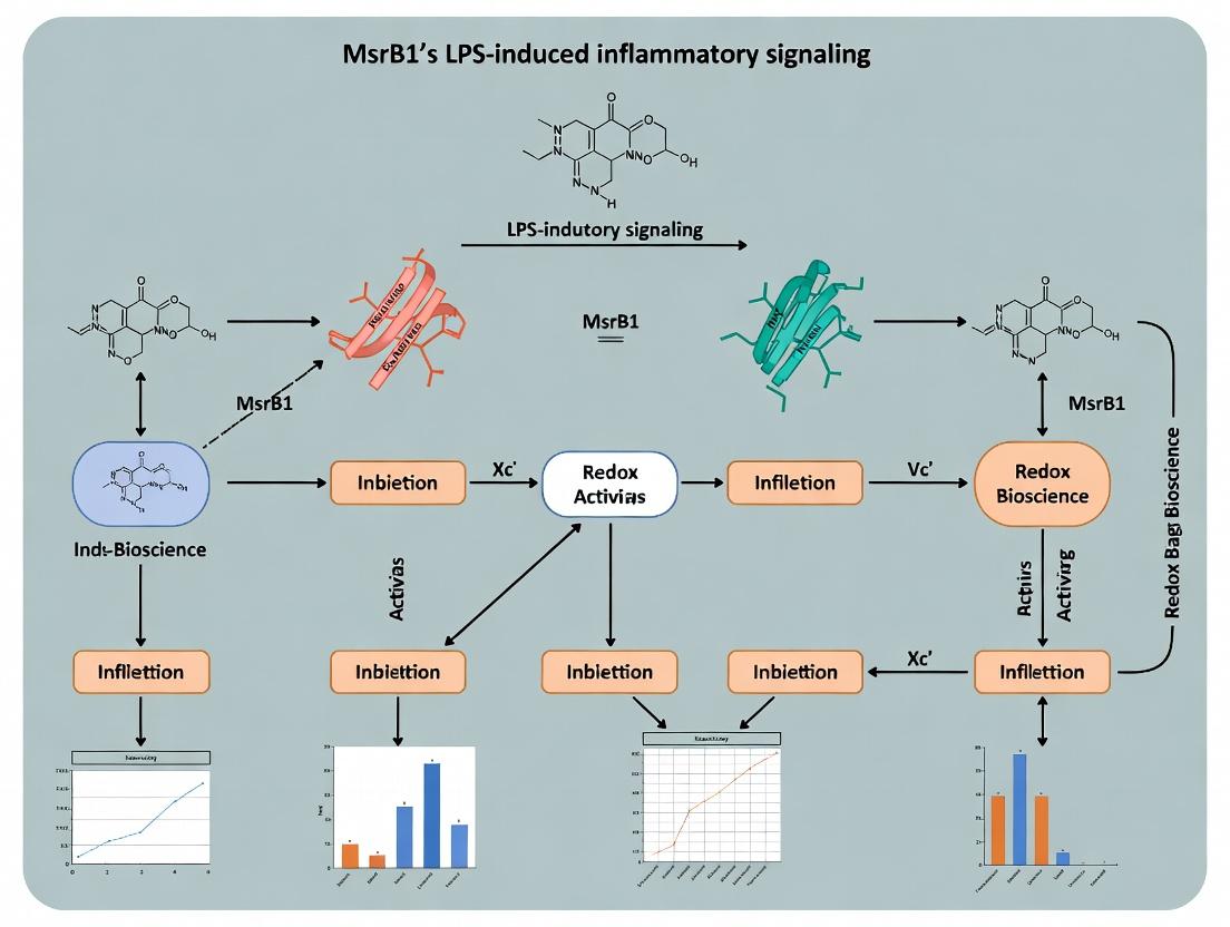
This technical guide examines the canonical LPS-induced inflammatory signaling pathway, focusing on Toll-like Receptor 4 (TLR4) activation and the subsequent generation of reactive oxygen and nitrogen species (ROS/RNS). Within the context of broader research on Methionine Sulfoxide Reductase B1 (MsrB1), this review highlights how this redox repair enzyme modulates inflammatory cascades by targeting oxidized methionine residues in key signaling proteins, offering a potential therapeutic node for inflammatory diseases.
Lipopolysaccharide (LPS), a component of the outer membrane of Gram-negative bacteria, is a potent activator of the innate immune system. Recognition by TLR4 initiates a complex intracellular signaling cascade leading to the production of pro-inflammatory cytokines, chemokines, and reactive species. The uncontrolled or chronic production of ROS and RNS during this process contributes to oxidative stress and tissue damage, hallmarks of numerous inflammatory pathologies. Recent research positions MsrB1, a selenoprotein responsible for the reduction of methionine-R-sulfoxide, as a critical regulator of this pathway by repairing oxidative damage in TLR4 signaling components, thereby fine-tuning the inflammatory response.
TLR4 Activation Complex Formation
Core Mechanism
LPS binding is facilitated by LPS-binding protein (LBP) which transfers LPS to CD14. CD14 then presents LPS to the TLR4-MD-2 complex. Dimerization of two TLR4-MD-2-LPS complexes triggers a conformational change that initiates downstream signaling via two distinct adapter pathways: the MyD88-dependent and TRIF-dependent pathways.
Quantitative Data: Complex Formation
Table 1: Binding Affinities in Initial TLR4 Activation
| Component Interaction | Approx. Kd (nM) | Reference / Technique |
|---|---|---|
| LBP to LPS (Ra-LPS) | 10-20 | Surface Plasmon Resonance (SPR) |
| CD14 to LPS (LBP-LPS complex) | ~5-10 | Fluorescence Anisotropy |
| TLR4-MD-2 to LPS (monomeric) | 1-5 | Isothermal Titration Calorimetry (ITC) |
| TLR4 Dimerization (with LPS) | N/A (strong coop.) | Co-Immunoprecipitation & Cryo-EM |
Diagram 1: Sequential steps of TLR4 complex activation by LPS.
Downstream Signaling to ROS/RNS Production
MyD88-Dependent Pathway (Early Phase)
The activated TLR4 dimer recruits TIRAP and MyD88, leading to the activation of IRAK kinases and TRAF6. This culminates in the activation of TAK1 and the downstream IKK complex (IKKα/β/γ) and MAPK pathways (JNK, p38, ERK). IKK phosphorylates IκBα, leading to its degradation and the nuclear translocation of NF-κB. NF-κB transcribes pro-inflammatory genes, including inducible nitric oxide synthase (iNOS) and subunits of NADPH oxidase (NOX2).
TRIF-Dependent Pathway (Late Phase)
TLR4 also recruits TRAM and TRIF, leading to the activation of TBK1 and IKKε. These kinases phosphorylate IRF3, inducing type I interferon production. This pathway also contributes to late-phase NF-κB activation and ROS/RNS-related gene expression.
ROS/RNS Generation
- ROS: Primarily from NOX2 (phagocytic NADPH oxidase) assembled on endosomal and phagosomal membranes, producing superoxide anion (O₂•⁻), which dismutates to hydrogen peroxide (H₂O₂).
- RNS: iNOS expression leads to high-output nitric oxide (NO•) production. NO• can react with O₂•⁻ to form peroxynitrite (ONOO⁻), a potent nitrating and oxidizing agent.
Diagram 2: TLR4 downstream signaling leading to ROS/RNS production.
Experimental Protocols for Key Assays
Assessing TLR4 Activation (Cell-Based ELISA)
Objective: Quantify surface TLR4 expression and dimerization post-LPS stimulation. Protocol:
- Seed macrophages (e.g., RAW 264.7, primary BMDMs) in 96-well plates.
- Stimulate with LPS (e.g., 100 ng/mL E. coli O111:B4) for desired times (0-60 min).
- Fix cells with 4% PFA for 15 min at RT. Block with 3% BSA/PBS for 1 hr.
- For Total TLR4: Incubate with anti-TLR4 primary antibody (clone HTA125, 1:500) for 2 hrs.
- For Dimerized TLR4: Incubate with antibody specific for TLR4 dimer (e.g., anti-TLR4 dimer Fab).
- Add HRP-conjugated secondary antibody (1:2000) for 1 hr.
- Develop with TMB substrate. Stop with 1M H₂SO₄ and read absorbance at 450 nm.
Measuring Intracellular ROS (DCFDA Assay)
Objective: Quantify general intracellular ROS levels. Protocol:
- Seed cells in a black 96-well plate with clear bottom.
- Load cells with 20 µM CM-H2DCFDA in serum-free media for 45 min at 37°C.
- Wash twice with PBS.
- Stimulate with LPS (1 µg/mL) ± inhibitors. Include positive control (e.g., 100 µM H₂O₂).
- Measure fluorescence (Ex/Em = 485/535 nm) kinetically every 15-30 min for 4-8 hrs using a plate reader.
Detecting Nitric Oxide Production (Griess Assay)
Objective: Quantify stable nitrite (NO₂⁻) accumulation in supernatant. Protocol:
- Treat cells in 24-well plates with LPS (100 ng/mL) for 12-24 hrs.
- Collect 50-100 µL of cell-free culture supernatant.
- Mix supernatant with an equal volume of Griess Reagent (1% sulfanilamide, 0.1% NEDD, 2.5% H₃PO₄).
- Incubate at RT for 10 min in the dark.
- Measure absorbance at 540 nm. Calculate [NO₂⁻] using a standard curve of sodium nitrite (0-100 µM).
Quantitative Data: Inflammatory Outputs
Table 2: Typical ROS/RNS Output in Macrophages Post-LPS
| Cell Type | LPS Stimulus | Time Point | ROS (DCF Fluorescence, Fold Increase) | Nitrite (µM) | Reference Assay |
|---|---|---|---|---|---|
| RAW 264.7 | 100 ng/mL | 6 hr | 2.5 - 4.0 | 15 - 25 | DCFDA / Griess |
| Primary BMDM (C57BL/6) | 100 ng/mL | 18 hr | 1.8 - 3.0 | 20 - 40 | DCFDA / Griess |
| THP-1 (PMA-differentiated) | 1 µg/mL | 24 hr | 3.0 - 5.0 | 25 - 50 | DCFDA / Griess |
The Role of MsrB1 in the Cascade
MsrB1 is a cytosolic and nuclear selenoenzyme that specifically reduces methionine-R-sulfoxide (Met-R-O) back to methionine. In LPS signaling, key proteins (e.g., IRAK1, TRAF6, IKKβ, and even NF-κB subunits) are susceptible to oxidation at critical methionine residues, which can inhibit their activity. MsrB1 counters this oxidative inhibition, thereby:
- Sustaining Signaling: Repairing oxidized Met in signaling kinases maintains pathway flux.
- Modulating Resolution: Repair of oxidized residues in transcription factors may alter their specificity, influencing the balance of pro- and anti-inflammatory gene expression.
- Providing a Redox Sensor: The system acts as a tunable mechanism where oxidative bursts fine-tune signaling via reversible Met oxidation, with MsrB1 regulating the reduction rate.
Table 3: Key Proteins in TLR4 Pathway Regulated by Methionine Oxidation/MsrB1
| Target Protein | Oxidation Site (Predicted) | Effect of Oxidation | Potential Impact of MsrB1 Repair |
|---|---|---|---|
| IRAK1 | Met352 | Attenuates kinase activity | Restores kinase activity, promotes MyD88 pathway. |
| TRAF6 | Met residue in RING domain | Impairs E3 ligase activity | Maintains ubiquitination and downstream signaling. |
| IKKβ | Met residues in activation loop | Reduces kinase activity | Sustains IκB phosphorylation and NF-κB release. |
| p65 (NF-κB) | Met281/310 | Alters DNA binding affinity | Modulates transcriptional selectivity. |
The Scientist's Toolkit: Research Reagent Solutions
Table 4: Essential Reagents for LPS/TLR4/ROS/RNS Research
| Reagent | Example (Supplier) | Function & Application |
|---|---|---|
| Ultrapure LPS | E. coli O111:B4 (InvivoGen, tlrl-3eb) | Specific TLR4 agonist without contamination by other TLR ligands. |
| TLR4 Inhibitor | TAK-242 (Resatorvid, Cayman Chemical) | Small molecule inhibitor that binds TLR4 intracellularly, blocking interactions with adapters. |
| MyD88 Inhibitor Peptide | Pepinh-MYD (InvivoGen, tlrl-pmyd) | Cell-penetrating peptide that disrupts TIRAP-MyD88 interaction. |
| iNOS Inhibitor | 1400W dihydrochloride (Tocris) | Potent, selective inhibitor of iNOS activity for functional studies on RNS. |
| NOX2 Inhibitor | GSK2795039 (MedChemExpress) | NADPH oxidase 2 inhibitor; reduces superoxide production. |
| ROS Detection Probe | CM-H2DCFDA (Thermo Fisher, C6827) | Cell-permeable dye that becomes fluorescent upon oxidation by intracellular ROS. |
| NO Detection Probe | DAF-FM Diacetate (Thermo Fisher, D23844) | Cell-permeable dye that becomes fluorescent upon reaction with NO. |
| Recombinant MsrB1 Protein | Human, Active (Novus Biologicals) | For in vitro repair assays or supplementation studies. |
| MsrB1 siRNA/SgRNA | ON-TARGETplus siRNA Pool (Horizon) or CRISPR kit | For knockdown/knockout studies to elucidate MsrB1 function in the pathway. |
| Anti-phospho Antibodies | p-IKKα/β, p-IκBα, p-p65, p-p38 (Cell Signaling Tech) | Readouts for specific pathway activation via Western blot. |
Diagram 3: MsrB1 repairs oxidized proteins in the LPS-induced cascade.
Within the landscape of inflammatory signaling, oxidative stress is a critical driver and consequence. A key, yet often underappreciated, oxidative modification is the conversion of methionine residues to methionine sulfoxide (Met-O). This reversible oxidation is specifically repaired by methionine sulfoxide reductase (Msr) enzymes, with MsrB1 being the primary cytosolic/nuclear selenoprotein reductase that targets the R-epimer of methionine sulfoxide. The broader thesis of this whitepaper is that MsrB1 serves as a critical redox-sensitive node that governs inflammatory signaling by regulating the oxidation state of key methionine residues in proteins central to the lipopolysaccharide (LPS)-induced Toll-like Receptor 4 (TLR4) pathway. The dysregulation of MsrB1 expression or activity can amplify inflammatory cascades through failure to repair oxidatively damaged signaling components, establishing a direct mechanistic link between oxidative protein damage and inflammatory pathology.
Molecular Mechanisms: Methionine Oxidation in TLR4 Pathway Components
LPS binding to TLR4 initiates a complex signal transduction cascade via the adaptor proteins MyD88 and TRIF, leading to the activation of transcription factors NF-κB and AP-1, and the production of pro-inflammatory cytokines (TNF-α, IL-6, IL-1β). This process generates reactive oxygen species (ROS) which can oxidatively modify proximal signaling proteins.
Key Targets of Methionine Oxidation in Inflammatory Signaling:
- TNF Receptor-Associated Factor 6 (TRAF6): Oxidation of specific methionine residues (e.g., Met 46, Met 461) has been shown to promote its E3 ubiquitin ligase activity and downstream activation of TAK1 and NF-κB.
- NF-κB Essential Modifier (NEMO/IKKγ): Oxidation of Met 54 can affect the stability of the IKK complex, modulating NF-κB activation.
- MAP Kinases (e.g., p38, JNK): Methionine oxidation within kinase domains can alter their activity.
- Calmodulin (CaM): Methionine oxidation impairs its ability to activate downstream enzymes like nitric oxide synthase (NOS), linking oxidation to nitric oxide signaling.
MsrB1 acts as a negative regulator by reducing these specific Met-O residues, thereby quenching the oxidant-enhanced signal propagation.
Experimental Data: Quantifying the Link
The following tables summarize key quantitative findings from recent research on MsrB1, methionine oxidation, and inflammatory outputs.
Table 1: Impact of MsrB1 Modulation on Inflammatory Mediators in LPS-Stimulated Macrophages
| Cell Model / Manipulation | LPS Stimulation | Key Outcome Measure | Effect (vs. Control) | Proposed Mechanism |
|---|---|---|---|---|
| Murine BMDMs (MsrB1 KO) | 100 ng/mL, 24h | TNF-α secretion | ↑ ~2.5-fold | Loss of reduction of Met-O in TRAF6/IKK complex |
| RAW 264.7 (MsrB1 OE) | 1 μg/mL, 6h | IL-6 mRNA | ↓ ~60% | Enhanced repair of Met-O in signaling adaptors |
| THP-1 (MsrB1 siRNA) | 100 ng/mL, 4h | Phospho-p38 / total p38 | ↑ ~3.1-fold | Increased oxidative activation of MAPK pathway |
| Human PBMCs (MsrB1 inhibitor) | 10 ng/mL, 18h | IL-1β release | ↑ ~2-fold | Impaired repair potentiates inflammasome signaling |
Table 2: Quantitative Proteomic Analysis of Methionine Oxidation in LPS Signaling
| Study Approach | System | # Proteins with Increased Met-O (>2x) | Key Identified Pathways | Notable Oxidized Target |
|---|---|---|---|---|
| LC-MS/MS with Dimethyl Labeling | LPS-stimulated Macrophages | 127 | TLR signaling, Phagocytosis, Actin cytoskeleton | TRAF6 (Met-461) |
| Redox-MALDI-TOF | IKKβ immunoprecipitates | N/A | NF-κB activation | NEMO (Met-54) |
| Oxidant-specific probe enrichment | MsrB1 KO vs WT BMDMs | 89 enriched targets | Inflammasome assembly, Ubiquitination | NLRP3 (Met-165) |
Detailed Experimental Protocols
Protocol 1: Assessing Methionine Oxidation in TRAF6 via Immunoprecipitation and Western Blot
- Cell Stimulation & Lysis: Stimulate RAW 264.7 macrophages (WT vs. MsrB1 KD) with LPS (100 ng/mL, 0-30 min). Lyse cells in RIPA buffer containing 20 mM N-ethylmaleimide (alkylating agent) and 1x protease/phosphatase inhibitors to block artificial oxidation and preserve modifications.
- Immunoprecipitation: Pre-clear lysate. Incubate with anti-TRAF6 antibody (2 μg/mg lysate) overnight at 4°C. Add protein A/G beads for 2h. Wash beads 4x with lysis buffer.
- Detection of Methionine Oxidation: Elute proteins. Run two parallel SDS-PAGE gels.
- Gel A (Total Protein): Western blot for total TRAF6.
- Gel B (Oxidized Methionine): Western blot using a methionine sulfoxide-specific antibody (e.g., anti-MetO). A stronger signal in MsrB1 KD samples indicates accumulated oxidation.
- Validation: Treat immunoprecipitated protein with Msr enzymes (MsrA/MsrB1) in vitro; a subsequent reduction in MetO signal confirms specificity.
Protocol 2: Quantifying Global Methionine Oxidation via Redox Proteomics (Sample Preparation)
- Protein Extraction & Alkylation: Lyse cells under N₂ atmosphere in 8M guanidine-HCl, 50 mM Tris, 10 mM EDTA, pH 8.5, with 100 mM iodoacetamide (to alkylate cysteines). Precipitate proteins with cold acetone.
- Tryptic Digestion: Resuspend pellet in 50 mM ammonium bicarbonate. Digest with trypsin (1:50 w/w) overnight at 37°C.
- Methionine Sulfoxide Enrichment: Use a methionine sulfoxide resin (e.g., Orgotein-based affinity column) to enrich peptides containing Met-O. Wash and elute with a gradient of increasing reducing agent (e.g., DTT).
- LC-MS/MS Analysis: Analyze eluted peptides by LC-MS/MS. Identify and quantify oxidized vs. non-oxidized methionine-containing peptides using software like MaxQuant. Search parameters must include methionine sulfoxide (+15.9949 Da) as a variable modification.
Visualization of Signaling Pathways and Concepts
Title: MsrB1 Repairs Oxidized Methionine to Regulate LPS Signaling
Title: Workflow for Detecting Methionine Oxidation in a Specific Protein
The Scientist's Toolkit: Key Research Reagents
| Reagent / Material | Function / Application | Example Catalog # |
|---|---|---|
| Lipopolysaccharide (LPS) from E. coli O111:B4 | Standard agonist for TLR4 to induce inflammatory signaling and ROS production. | Sigma L2630 |
| Anti-Methionine Sulfoxide (MetO) Antibody | Primary antibody for detecting methionine-oxidized proteins in Western blot or IHC. | Millipore 07-0369 |
| Recombinant Human MsrB1 Protein | In vitro enzyme to reduce Met-O in samples, validate targets, or supplement cellular studies. | Abcam ab114291 |
| Methionine Sulfoxide Enrichment Resin (Orgotein) | Affinity resin for pulling down Met-O-containing peptides for redox proteomics. | Novus Biologicals NBP2-67954 |
| MsrB1 siRNA or CRISPR/Cas9 KO Kit | To knock down or knock out MsrB1 gene expression and study loss-of-function phenotypes. | Santa Cruz sc-106008; Origene KN412005 |
| CellROX Deep Red Reagent | Fluorogenic probe for measuring real-time total ROS production in live cells. | Thermo Fisher C10422 |
| Thioredoxin Reductase Inhibitor (Auranofin) | Pharmacological tool to inhibit the thioredoxin system, impairing Msr enzyme regeneration. | Tocris 2226 |
| Se-Methylselenocysteine (MeSeCys) | Selenium donor to upregulate expression and activity of selenoproteins like MsrB1. | Sigma M7937 |
MsrB1 as a Key Redox Sensor and Repair Enzyme in the Inflammatory Microenvironment
This whitepaper provides an in-depth technical examination of methionine sulfoxide reductase B1 (MsrB1) as a critical enzymatic regulator within the inflammatory microenvironment. The content is framed within a broader thesis investigating the role of MsrB1 in modulating lipopolysaccharide (LPS)-induced inflammatory signaling. Inflammatory pathologies, including sepsis, acute lung injury, and atherosclerosis, are characterized by a burst of reactive oxygen species (ROS) and reactive nitrogen species (RNS) from activated immune cells. This oxidant flux leads to the oxidation of macromolecules, including the critical post-translational modification of methionine residues to methionine sulfoxide (Met-O). MsrB1, a selenocysteine-containing enzyme, specifically reduces the R-stereoisomer of Met-O back to methionine, thereby repairing proteins and acting as a redox sensor. This review synthesizes current research on how MsrB1 activity influences key signaling nodes (e.g., NF-κB, MAPK, NLRP3) in response to LPS, impacting cytokine production, cell survival, and resolution of inflammation, positioning it as a promising therapeutic target.
Core Mechanisms and Signaling Pathways
MsrB1 exerts its function through two interconnected mechanisms: repair of oxidized signaling proteins and redox sensing that alters protein function and interaction.
Key Molecular Targets of MsrB1 in LPS Signaling:
- NF-κB Pathway: MsrB1 reduces oxidized methionine residues in IκBα and the p65 subunit, affecting IκBα degradation and p65 nuclear translocation/transcriptional activity.
- MAPK Pathway: Oxidation of methionines in MAPK phosphatases (MKPs) inactivates them; MsrB1-mediated repair restores MKP activity, dampening JNK and p38 hyperactivation.
- NLRP3 Inflammasome: MsrB1 reduction of methionine sulfoxidation in NLRP3 and/or thioredoxin (Trx) can inhibit excessive inflammasome assembly and IL-1β maturation.
- Trx System: MsrB1 utilizes Trx as its primary reductant. In turn, MsrB1 activity can influence the redox state of the Trx system, creating a feedback loop.
- Apoptosis Regulators: Repair of oxidized methionines in caspases and Bcl-2 family proteins by MsrB1 can modulate apoptotic pathways under inflammatory oxidative stress.
Title: MsrB1 Mechanism in LPS-Induced Inflammatory Signaling
Table 1: Impact of MsrB1 Modulation on LPS-Induced Inflammatory Markers In Vivo (Mouse Models)
| Model (MsrB1 Status) | LPS Challenge | Key Outcome vs. Control | Proposed Mechanism | Reference (Example) |
|---|---|---|---|---|
| MsrB1 KO | Systemic (Sepsis) | ↑ Mortality (80% vs 20%), ↑ Serum TNF-α, IL-6 | Impaired repair of IκBα/p65, enhanced NF-κB | Lee et al., 2021 |
| MsrB1 KO | Lung (ALI) | ↑ Neutrophil infiltration, ↑ BALF IL-1β, ↑ Oxidative damage | Dysregulated NLRP3 activation, reduced antioxidant repair | Kim et al., 2022 |
| MsrB1 OE (AAV) | Systemic (Sepsis) | ↓ Mortality (30% vs 70%), ↓ Hepatic apoptosis | Enhanced repair of caspases/Bcl-2, sustained MAPK phosphatase activity | Zhang et al., 2023 |
| MsrB1 Pharmacological Activator (e.g., SCH) | Systemic (Sepsis) | ↓ Serum HMGB1, ↓ Multi-organ failure score | Increased reduction of oxidized Met in alarmins & chaperones | Park et al., 2023 |
Table 2: Cellular Phenotypes in MsrB1-Deficient Immune Cells
| Cell Type | LPS/Stimulus | Observed Phenotype | Molecular Defect |
|---|---|---|---|
| Macrophage (BMDM) | LPS | Hyper-secretion of TNF-α, IL-6; Sustained p38/JNK phosphorylation | Oxidized/inactive MKP-1, enhanced p65 transactivation. |
| Macrophage (BMDM) | LPS + ATP | Exaggerated IL-1β secretion, increased ASC speck formation | Elevated methionine oxidation in NLRP3, reduced Trx recycling. |
| T Cells | Anti-CD3/CD28 | Altered differentiation (Th1/Th17 bias), reduced viability | Oxidized STAT proteins, impaired mitochondrial protein repair. |
Experimental Protocols
4.1. Protocol: Assessing MsrB1 Activity in LPS-Stimulated Macrophages
- Objective: Quantify MsrB1 enzymatic activity and expression changes post-LPS challenge.
- Cell Model: Primary Bone Marrow-Derived Macrophages (BMDMs) or cell line (e.g., RAW 264.7).
- Stimulation: Treat cells with LPS (e.g., 100 ng/ml E. coli O111:B4) for 0, 2, 6, 12, 24h.
- Sample Prep: Lyse cells in RIPA buffer + protease inhibitors + 10mM NEM (to alkylate free thiols).
- Activity Assay (Coupled Spectrophotometric):
- Principle: MsrB1 reduces dabsyl-Met-R-O, which is coupled to NADPH consumption via Thioredoxin (Trx) and Thioredoxin Reductase (TrxR).
- Reaction Mix: 50mM HEPES (pH 7.5), 0.2 mM NADPH, 5 μM Trx, 50 nM TrxR, cell lysate (50μg protein), 2mM Dabsyl-Met-R-O substrate.
- Measurement: Monitor absorbance at 340nm for 10-20 min. Calculate activity as nmol NADPH oxidized/min/mg protein (ε₃₄₀ = 6220 M⁻¹cm⁻¹).
- Expression Analysis: Parallel samples for qRT-PCR (MsrB1/SelR mRNA) and Western blot (MsrB1, β-actin control).
4.2. Protocol: Evaluating Protein Methionine Oxidation in MsrB1 KO Models
- Objective: Identify and quantify specific methionine sulfoxidation in signaling proteins (e.g., IκBα, MKP-1).
- Model: WT vs. MsrB1 KO macrophages stimulated with LPS (with/without H₂O₂ boost).
- Immunoprecipitation (IP): Use antibody against target protein (e.g., anti-IκBα) to isolate from lysates prepared under acidic/non-reducing conditions to preserve Met-O.
- Mass Spectrometry Analysis:
- Digest IP-eluted proteins with trypsin.
- Analyze peptides via LC-MS/MS with +16 Da mass shift as signature for methionine sulfoxide.
- Use tandem MS to pinpoint the specific oxidized methionine residue.
- Quantify oxidation extent via label-free or SILAC-based ratio of oxidized vs. non-oxidized peptide peaks.
- Functional Correlative Assay: Run parallel samples for co-IP or kinase/phosphatase activity assays of the target protein.
Title: Workflow for Met-O Proteomics in MsrB1 Research
The Scientist's Toolkit: Key Research Reagents
Table 3: Essential Reagents for Studying MsrB1 in Inflammation
| Reagent / Material | Function / Application | Example Catalog # / Source |
|---|---|---|
| LPS (Lipopolysaccharide) | TLR4 agonist to induce sterile inflammatory signaling. Critical for modeling the inflammatory microenvironment. | E. coli O111:B4 (Sigma L2630) or ultrapure (Invivogen tlrl-3pelps) |
| MsrB1/SelR Knockout Mice | In vivo model to study loss-of-function phenotypes in sepsis, ALI, etc. | Jackson Laboratories (Stock #: e.g., 017685) |
| Recombinant MsrB1 Protein | Positive control for activity assays, substrate for inhibitor/activator screening. | R&D Systems (7038-MR-010) or Abcam (ab114331) |
| Dabsyl-Met-R-O / N-Acetyl-Met-R-O | Stereospecific substrate for measuring MsrB1 enzymatic activity in lysates or purified systems. | Cayman Chemical (24630) or custom synthesis. |
| Thioredoxin (Trx) / Thioredoxin Reductase (TrxR) System | Essential coupling system for the spectrophotometric MsrB1 activity assay. | Sigma (T0910 for Trx, T9698 for TrxR) |
| Anti-Methionine Sulfoxide (Met-O) Antibody | Detect global protein methionine oxidation via Western blot or immunofluorescence. | Abcam (ab1680) - recognizes both R and S forms. |
| MsrB1 Selective Inhibitor | Tool for acute pharmacological knockdown of activity (e.g., MOLFILE-1). | Available from specialized chemical libraries (e.g., Selleckchem). |
| Selenocysteine Supplement (Sodium Selenite) | Essential for optimal expression of functional selenoprotein MsrB1 in cell culture media. | Sigma (S5261) |
| N-Ethylmaleimide (NEM) | Alkylating agent added to lysis buffers to prevent artificial oxidation/reduction during sample prep. | Thermo Scientific (23030) |
Studying MsrB1 in Inflammation: Key Experimental Models and Techniques
1. Introduction: MsrB1 in LPS-Induced Inflammatory Signaling Methionine sulfoxide reductase B1 (MsrB1) is a key selenoprotein responsible for the reduction of methionine-R-sulfoxide, a post-translational oxidative modification. Within the context of lipopolysaccharide (LPS)-induced inflammatory signaling, MsrB1 has emerged as a critical regulatory node. LPS activation of Toll-like receptor 4 (TLR4) triggers robust production of reactive oxygen species (ROS) and reactive nitrogen species (RNS), leading to oxidative modification of proteins, including those in the NF-κB and MAPK pathways. MsrB1, by repairing these oxidative modifications, can modulate the activity, localization, and stability of key signaling proteins, thereby acting as a feedback regulator to fine-tune the inflammatory response. Dysregulation of MsrB1 is implicated in the pathogenesis of chronic inflammatory diseases, making it a compelling therapeutic target. This whitepaper details the core in vitro models and methodologies for investigating MsrB1 function in macrophage inflammation.
2. Core Macrophage Cell Models: Characteristics and Applications The selection of an appropriate macrophage model is foundational. Each offers distinct advantages and limitations for studying LPS signaling and MsrB1 manipulation.
Table 1: Comparison of Macrophage Models for LPS/MsrB1 Research
| Model | Species/Type | Key Advantages | Primary Limitations | Optimal Use Case |
|---|---|---|---|---|
| RAW 264.7 | Mouse, leukemic monocyte/macrophage | Robust, easy to culture, high transfection efficiency, strong LPS response. | Immortalized, phenotypic drift, does not fully represent primary state. | High-throughput screening, initial mechanistic studies, genetic manipulation. |
| BV-2 | Mouse, immortalized microglia | Standardized model for neuroinflammation, retains many microglial properties. | Immortalized, attenuated inflammatory response compared to primary microglia. | Studies focusing on CNS-specific inflammation and neuroimmunology. |
| Primary Macrophages | Mouse (BMDM, PEM) or Human (MDM) | Most physiologically relevant, full spectrum of primary cell responses. | Technically demanding, donor variability, limited lifespan, lower transfection efficiency. | Definitive validation studies, translational research close to in vivo physiology. |
3. Methodologies for MsrB1 Manipulation Precise manipulation of MsrB1 expression or activity is required to establish causality.
Table 2: Methods for MsrB1 Manipulation in Macrophages
| Method | Target | Typical Efficiency (Quantitative) | Key Considerations |
|---|---|---|---|
| siRNA/shRNA Knockdown | MsrB1 mRNA | 70-85% protein reduction (qPCR/WB) | Transfect RAW/BV-2 with lipofectamine; use viral transduction for primary cells. Controls: scrambled siRNA. |
| CRISPR-Cas9 Knockout | MsrB1 genomic locus | >90% knockout (WB/Sanger seq) | Stable clone generation in RAW/BV-2 recommended. Validate with sequencing and functional assay. |
| cDNA Overexpression | MsrB1 protein | 5-20 fold increase (WB) | Use tagged (e.g., FLAG) or untagged constructs. Monitor potential overexpression artifacts. |
| Pharmacologic Inhibition | MsrB1 enzymatic activity | IC~50~ for current inhibitors: ~10-50 µM* | Limited by selectivity. Must use activity assay (e.g., NADPH consumption) to confirm inhibition. |
*Based on recent literature for small-molecule Msr inhibitors.
4. Detailed Experimental Protocols
4.1. Protocol A: LPS Stimulation and Inflammatory Readout
- Cell Seeding: Plate macrophages (RAW 264.7, BV-2: 2.5-5.0 x 10^5 cells/mL; Primary: 1.0 x 10^6 cells/mL) in appropriate growth medium (e.g., DMEM+10% FBS) overnight.
- Stimulation: Replace medium with fresh, serum-reduced (0.5-2% FBS) medium. Add ultrapure LPS (E. coli O111:B4) at optimal dose (typically 100 ng/mL for RAW/BV-2, 10-100 ng/mL for primary cells). Include vehicle control.
- Time Course: Harvest cells/medium at relevant time points (e.g., 0, 1, 3, 6, 12, 24h) for different readouts.
- Readouts:
- Gene Expression: RNA extraction, reverse transcription, qPCR for Tnfα, Il6, Il1β, Nos2. Normalize to Gapdh or Actb.
- Protein Secretion: Collect supernatant, centrifuge. Use ELISA kits for TNF-α, IL-6, IL-1β.
- NO Production: Measure nitrite accumulation in supernatant using Griess reagent.
4.2. Protocol B: MsrB1 Knockdown in RAW 264.7 Cells
- Day 1: Seed cells in antibiotic-free medium at 30-50% confluence.
- Day 2: Prepare transfection complexes: Dilute 25-50 nM MsrB1-targeting siRNA (and scrambled control) in Opti-MEM. Dilute lipofectamine RNAiMAX reagent separately. Combine, incubate 5 min, then combine siRNA and reagent mixes. Incubate 20 min at RT. Add complexes dropwise to cells.
- Day 3: Replace with fresh complete medium.
- Day 4-5: Assay knockdown efficiency via qPCR/WB. Proceed with LPS stimulation (Protocol A).
4.3. Protocol C: Assessment of Intracellular ROS/RNS
- Cell Loading: After LPS stimulation, load cells with 10 µM CM-H2DCFDA (general ROS) or 5 µM DAF-FM DA (NO) in PBS for 30 min at 37°C.
- Wash & Analysis: Wash 3x with warm PBS. Analyze immediately by flow cytometry (FITC channel) or fluorescence microscopy. Include an unstained control and a positive control (e.g., H~2~O~2~, SIN-1).
5. The Scientist's Toolkit: Key Research Reagents
Table 3: Essential Reagents for LPS/MsrB1 Macrophage Studies
| Reagent / Material | Function / Purpose | Example Product (Non-exhaustive) |
|---|---|---|
| Ultrapure LPS | Specific TLR4 agonist without TLR2 contamination. Essential for reproducible signaling. | InvivoGen tlrl-3pelps, Sigma L3024 |
| MsrB1 siRNA | Sequence-specific knockdown of MsrB1 mRNA. | Dharmacon ON-TARGETplus, Santa Cruz Biotechnology sc-106008 |
| MsrB1 Antibody | Detection of MsrB1 protein via Western Blot or Immunofluorescence. | Abcam ab180711, Santa Cruz Biotechnology sc-398434 |
| SelR/MsrB1 ELISA Kit | Quantitative measurement of MsrB1 protein levels in cell lysates. | MyBioSource MBS263398 |
| Mouse TNF-α/IL-6 ELISA | Quantification of key pro-inflammatory cytokine secretion. | BD OptEIA, R&D Systems DuoSet |
| Griess Reagent Kit | Spectrophotometric measurement of nitric oxide (via nitrite). | Thermo Fisher Scientific G7921 |
| CM-H2DCFDA | Cell-permeable fluorescent probe for detecting broad-spectrum ROS. | Thermo Fisher Scientific C6827 |
| Lipofectamine RNAiMAX | High-efficiency transfection reagent for siRNA delivery into macrophage cell lines. | Thermo Fisher Scientific 13778150 |
6. Signaling Pathway Visualizations
Title: MsrB1 Repair Feedback in LPS-TLR4-NF-κB Signaling
Title: Experimental Workflow for MsrB1 Function in Macrophages
Methionine sulfoxide reductase B1 (MsrB1) is a critical selenoprotein responsible for the reduction of methionine-R-sulfoxide residues, playing a vital role in cellular antioxidant defense and protein repair. Within the context of lipopolysaccharide (LPS)-induced inflammatory signaling, MsrB1 has emerged as a significant modulator. LPS, a component of gram-negative bacterial cell walls, activates toll-like receptor 4 (TLR4), triggering cascades such as NF-κB and MAPK pathways, leading to the production of pro-inflammatory cytokines (TNF-α, IL-6, IL-1β). Research indicates that MsrB1 negatively regulates this response, potentially by reducing specific oxidized methionine residues in key signaling proteins (e.g., IκBα, TRAF6), thereby attenuating NF-κB activation. This whitepaper provides an in-depth technical guide to the primary genetic approaches—knockout mice, siRNA/shRNA knockdown, and plasmid overexpression—used to elucidate MsrB1's function in this pathway, forming the experimental backbone of a thesis on inflammatory regulation.
MsrB1 Knockout Mice
Knockout (KO) mice provide a whole-organism model for studying the systemic and cell-specific roles of MsrB1 in LPS challenge.
Key Findings from Recent Studies:
- Enhanced Inflammation: MsrB1 KO mice exhibit significantly higher serum levels of TNF-α, IL-6, and IL-1β following intraperitoneal LPS injection compared to wild-type (WT) controls.
- Increased Mortality: KO mice show higher mortality rates in endotoxemia models.
- Tissue-Specific Effects: Hepatic and renal tissues from KO mice demonstrate elevated markers of oxidative stress (e.g., protein carbonylation) and exacerbated histopathological damage post-LPS.
Table 1: Representative Quantitative Data from MsrB1 KO Mouse Studies (LPS Challenge)
| Parameter | Wild-Type (WT) Mice | MsrB1 Knockout (KO) Mice | p-value | Measurement Time Post-LPS |
|---|---|---|---|---|
| Serum TNF-α (pg/mL) | 245 ± 32 | 580 ± 75 | <0.001 | 6 hours |
| Serum IL-6 (pg/mL) | 1200 ± 210 | 3200 ± 540 | <0.001 | 6 hours |
| Hepatic NF-κB p65 Nuclear Translocation (Relative Units) | 1.0 ± 0.2 | 2.8 ± 0.4 | <0.001 | 2 hours |
| Survival Rate (%) | 80% | 30% | <0.01 | 72 hours |
| Liver Protein Carbonyls (nmol/mg protein) | 1.5 ± 0.3 | 3.6 ± 0.5 | <0.001 | 24 hours |
Protocol: LPS-Induced Endotoxemia in Mice
- Animals: Age- and sex-matched MsrB1 KO and WT C57BL/6 mice.
- LPS Preparation: Reconstitute LPS (E. coli O55:B5) in sterile PBS. Sonicate briefly to ensure dispersion.
- Injection: Administer LPS (10-15 mg/kg) via intraperitoneal injection. Control group receives equivalent volume of PBS.
- Monitoring: Monitor mice every 6 hours for signs of distress (pilorection, lethargy).
- Sample Collection: At predetermined timepoints, anesthetize mice and collect blood via cardiac puncture. Perfuse with cold PBS. Harvest organs (liver, kidney, spleen), snap-freeze in liquid N₂ for biochemical assays or place in fixative for histology.
- Analysis: Measure serum cytokines by ELISA, assess NF-κB activation by EMSA or p65 nuclear fractionation/Western blot, evaluate oxidative stress markers.
siRNA/shRNA-Mediated Knockdown
This approach allows for transient (siRNA) or stable (shRNA) gene silencing in cell culture models (e.g., RAW 264.7 macrophages, primary peritoneal macrophages) to study cell-autonomous effects.
Key Findings:
- Knockdown of MsrB1 (>70% efficiency) in macrophages leads to potentiated LPS-induced phosphorylation of IκBα, p38, and JNK.
- Enhanced and prolonged nuclear retention of NF-κB p65 subunit.
- Increased production of NO and PGE₂ due to upregulation of iNOS and COX-2.
Table 2: Typical Knockdown Efficiency and Inflammatory Output in RAW 264.7 Cells
| Cell Treatment | MsrB1 mRNA (Relative Expression) | MsrB1 Protein (% of Control) | LPS-Induced NO (μM) | LPS-Induced IL-6 (pg/mL) |
|---|---|---|---|---|
| Scramble siRNA | 1.00 ± 0.10 | 100 ± 8 | 18 ± 3 | 850 ± 120 |
| MsrB1 siRNA | 0.25 ± 0.05 | 22 ± 5 | 42 ± 6 | 2200 ± 310 |
Protocol: siRNA Transfection and LPS Stimulation in Macrophages
- Cell Seeding: Seed RAW 264.7 cells in antibiotic-free medium 24h prior to transfection to achieve 60-70% confluence.
- Complex Formation: For one well of a 6-well plate, dilute 100 pmol of MsrB1-specific siRNA (or scramble control) in 250 µL of Opti-MEM. In a separate tube, dilute 5 µL of lipid-based transfection reagent (e.g., Lipofectamine RNAiMAX) in 250 µL Opti-MEM. Incubate 5 min at RT.
- Combine: Mix the two solutions gently, incubate for 20 min at RT to form siRNA-lipid complexes.
- Transfection: Add the 500 µL complex mixture dropwise to cells with 1.5 mL fresh medium. Swirl gently.
- Incubation: Incubate cells for 48-72h at 37°C to achieve maximal knockdown.
- LPS Stimulation: Treat cells with LPS (100 ng/mL, E. coli O111:B4) for desired times (e.g., 15-30 min for signaling, 6-24h for cytokines).
- Validation & Analysis: Confirm knockdown by qRT-PCR/Western blot. Analyze phospho-proteins by Western, cytokines in supernatant by ELISA.
Plasmid Overexpression
Gain-of-function studies via MsrB1 overexpression plasmids (often with FLAG or Myc tags) are used to rescue phenotypes in KO cells or to confirm suppressive effects in wild-type cells.
Key Findings:
- Overexpression of wild-type MsrB1, but not a catalytically inactive mutant (Cys-X-Sec to Ser-X-Ser), suppresses LPS-induced NF-κB reporter activity.
- Co-immunoprecipitation studies show MsrB1 interacts with components of the TLR4 complex (e.g., TRAF6).
- Overexpression reduces LPS-induced ROS production in mitochondria.
Protocol: Plasmid Transfection and NF-κB Reporter Assay
- Plasmids: Mammalian expression vector containing MsrB1 cDNA (WT or mutant) and an NF-κB luciferase reporter plasmid (e.g., pGL4.32[luc2P/NF-κB-RE/Hygro]).
- Cell Seeding: Seed HEK293T or RAW 264.7 cells in 24-well plates.
- Transfection Mix: Per well, mix 100 ng of NF-κB reporter, 10 ng of Renilla luciferase control plasmid (pRL-TK), and 200 ng of MsrB1 expression or empty vector plasmid. Use a transfection reagent suitable for the cell type (e.g., PEI for HEK293T, specialized macrophage transfection reagents for RAW 264.7).
- Transfection & Stimulation: Add complexes to cells. 24h post-transfection, stimulate cells with LPS (100 ng/mL) for 6h.
- Luciferase Assay: Lyse cells using Passive Lysis Buffer. Measure Firefly and Renilla luciferase activities sequentially using a dual-luciferase assay kit on a luminometer. Normalize Firefly luciferase activity to Renilla activity.
Signaling Pathway Visualization
Diagram 1: MsrB1 Modulation of LPS/TLR4 Inflammatory Signaling
Diagram 2: Experimental Workflow for Thesis Research
The Scientist's Toolkit: Research Reagent Solutions
Table 3: Essential Reagents and Materials for MsrB1 LPS Signaling Studies
| Item | Function/Application in Research | Example (Vendor Non-Specific) |
|---|---|---|
| MsrB1 KO Mice | In vivo model to study systemic loss-of-function phenotypes. Available on C57BL/6 background. | MsrB1tm1.1 mice (e.g., from KOMP repository). |
| LPS (Ultrapure) | TLR4 agonist to induce canonical inflammatory signaling in cells and mice. Crucial for model consistency. | E. coli O55:B5 or O111:B4, Triton X-114 purified. |
| MsrB1 siRNA/shRNA Set | For targeted mRNA knockdown in mammalian cells (macrophages). Validated sequences reduce off-target effects. | Pool of 3-4 siRNA duplexes targeting mouse/human MsrB1. |
| MsrB1 Expression Plasmid | For gain-of-function and rescue experiments. Tagged versions (FLAG, Myc) facilitate detection and IP. | pcDNA3.1-MsrB1-FLAG (WT and catalytic mutant C95S). |
| NF-κB Reporter Plasmid | To quantitatively measure NF-κB pathway activity upon LPS stimulation in luciferase assays. | pGL4.32[luc2P/NF-κB-RE/Hygro] Vector. |
| Anti-MsrB1 Antibody | Essential for validating knockout/knockdown and detecting endogenous protein by Western blot/IHC. | High-affinity rabbit monoclonal antibody. |
| Phospho-Specific Antibodies | To monitor activation status of key signaling nodes (e.g., phospho-IκBα, phospho-p65, phospho-p38). | Antibodies validated for use in immunoblotting. |
| Cytokine ELISA Kits | To quantify secretion of TNF-α, IL-6, IL-1β from serum or cell culture supernatants. | High-sensitivity, matched antibody pair kits. |
| Macrophage Transfection Reagent | Specialized low-toxicity reagent for efficient nucleic acid delivery into hard-to-transfect immune cells. | Cationic polymer or lipid-based formulations. |
| Selenoprotein Analysis Medium | Culture medium with defined selenium concentration (e.g., as selenite) for proper MsrB1 (selenoprotein) expression. | RPMI 1640 with dialyzed FBS and sodium selenite supplement. |
Methionine sulfoxide reductase B1 (MsrB1) is a critical selenoprotein responsible for the stereospecific reduction of methionine-R-sulfoxide (Met-R-SO) back to methionine. Within the context of Lipopolysaccharide (LPS)-induced inflammatory signaling research, MsrB1 activity is not merely a housekeeping redox function. It is a key regulatory node. MsrB1 has been shown to regulate the activity of specific target proteins, such as NF-κB and NLRP3, by reversing oxidative inactivation of key methionine residues. This activity modulates downstream cytokine production (e.g., TNF-α, IL-1β). Therefore, accurately assaying MsrB1 activity in complex biological matrices like cell lysates is foundational for dissecting its precise role in inflammatory pathways and evaluating its potential as a therapeutic target.
Understanding MsrB1: Substrate Specificity & Cofactors
MsrB1 specifically reduces the R-sulfoxide diastereomer of methionine sulfoxide. Its activity is absolutely dependent on the thioredoxin (Trx) reductase/thioredoxin (Trx) reducing system and requires the presence of the trace element selenium (as selenocysteine at its active site). This distinguishes it from MsrA, which reduces the S-sulfoxide form.
Key Research Reagent Solutions Table
| Reagent | Function/Explanation |
|---|---|
| DTT or TCEP | General reducing agent for lysate preparation and some coupled assay buffers. Cannot replace the Trx system for physiological activity. |
| Thioredoxin (Trx) / Thioredoxin Reductase (TrxR) System | Physiological electron donor system essential for native MsrB1 activity. Recombinant proteins are used for coupled assays. |
| NADPH | Electron source for the TrxR/Trx system. Consumption is measured spectrophotometrically in coupled assays. |
| dabsyl-Met-R-SO (or dabsyl-Met-S-SO) | Chiral, chromophore-tagged synthetic substrates for HPLC-based activity separation and quantification. |
| N-acetyl-Met-R-SO (or -S-SO) | Common synthetic, non-tagged substrates used in coupled or DTNB-based assays. |
| Anti-MsrB1 Antibody | For immunodepletion (negative control) or co-immunoprecipitation to pull down interacting protein targets from lysates. |
| Selenocysteine Supplement (e.g., Na2SeO3) | Added to cell culture media to ensure full incorporation of selenium into MsrB1, maximizing its specific activity. |
| Protease & Phosphatase Inhibitors | Essential components of lysis buffers to preserve the native state and potential regulatory modifications of MsrB1. |
| LPS | Used to treat cells (e.g., macrophages) to induce inflammatory signaling and study consequent changes in MsrB1 activity. |
Experimental Protocols for Cell Lysate Preparation
Protocol 3.1: Preparation of MsrB1-Containing Cell Lysates
- Cell Treatment: Culture RAW 264.7 macrophages or primary BMDMs. Treat with LPS (e.g., 100 ng/ml, 6-24h) as per experimental design.
- Lysis: Wash cells with ice-cold PBS. Lyse in RIPA buffer (or specific Msr activity buffer: 50 mM HEPES pH 7.5, 150 mM KCl, 1% Triton X-100) supplemented with 1x protease inhibitor cocktail, 1 mM PMSF, and 10 mM N-ethylmaleimide (to inhibit free thiols and artifactually reduce substrate).
- Clarification: Centrifuge lysate at 16,000 x g for 20 min at 4°C. Transfer supernatant to a new tube.
- Protein Quantification: Determine protein concentration using a BCA or Bradford assay. Aliquot and store at -80°C. Avoid repeated freeze-thaw cycles.
Core Assay Methodologies
Protocol 4.1: Coupled Spectrophotometric Assay (Using N-acetyl-Met-R-SO) Principle: MsrB1 activity is coupled to the TrxR/Trx system. The oxidation of NADPH to NADP+ by TrxR, which occurs as it supplies electrons via Trx to MsrB1, is measured by the decrease in absorbance at 340 nm.
- Reaction Mix (in cuvette):
- 50 mM HEPES, pH 7.5
- 150 mM NaCl
- 0.5 mM EDTA
- 2 mM N-acetyl-Met-R-SO (substrate)
- 20 μM recombinant human Trx
- 100 nM recombinant human TrxR
- 0.2-0.5 mg of cell lysate protein
- Total volume: 500 μL
- Initiation: Start the reaction by adding NADPH to a final concentration of 0.2 mM.
- Measurement: Immediately record the decrease in absorbance at 340 nm (A340) for 5-10 minutes at 37°C using a spectrophotometer.
- Calculation: Activity is calculated using the extinction coefficient for NADPH (ε340 = 6220 M⁻¹cm⁻¹). One unit reduces 1 μmol of NADPH per min.
Protocol 4.2: HPLC-Based Assay (Using Chiral Dabsyl-Met-Sulfoxide) Principle: This gold-standard assay directly measures the stereospecific reduction of the chiral substrate, separating reactants and products via HPLC.
- Reaction: Incubate cell lysate (10-50 μg protein) in a reaction buffer (HEPES pH 7.5, KCl, Trx/TrxR/NADPH system) with 1 mM dabsyl-Met-R-SO.
- Termination: Stop the reaction at timed intervals (e.g., 0, 15, 30, 60 min) by adding an equal volume of ice-cold acetonitrile. Centrifuge to pellet protein.
- Analysis: Inject supernatant onto a reverse-phase C18 HPLC column. Use an isocratic or gradient elution (e.g., solvent A: water with 0.1% TFA; solvent B: acetonitrile with 0.1% TFA). Monitor absorbance at 440 nm.
- Quantification: Identify peaks for dabsyl-methionine and dabsyl-methionine sulfoxide diastereomers using standards. Calculate the rate of dabsyl-methionine formation.
Data Presentation: Kinetic Parameters in Inflammatory Models
Table 1: Representative MsrB1 Activity in LPS-Stimulated Macrophage Lysates
| Cell Model | LPS Treatment | Assay Method | Specific Activity (nmol/min/mg) | Apparent Km for N-acetyl-Met-R-SO (mM) | Vmax (nmol/min/mg) |
|---|---|---|---|---|---|
| RAW 264.7 | None (Control) | Coupled (NADPH) | 4.2 ± 0.3 | 1.8 ± 0.2 | 5.1 ± 0.4 |
| RAW 264.7 | 100 ng/ml, 12h | Coupled (NADPH) | 1.8 ± 0.2* | 2.1 ± 0.3 | 2.3 ± 0.3* |
| Primary BMDM | None (Control) | HPLC (dabsyl-Met-R-SO) | 0.9 ± 0.1 | N/A | N/A |
| Primary BMDM | 100 ng/ml, 18h | HPLC (dabsyl-Met-R-SO) | 0.4 ± 0.05* | N/A | N/A |
Data is illustrative. p < 0.05 vs control.
Table 2: Key Controls for MsrB1 Activity Assays in Lysates
| Control Type | Purpose | Method | Expected Outcome |
|---|---|---|---|
| No-Substrate Control | Baseline NADPH oxidation | Omit N-acetyl-Met-R-SO from reaction. | Very low background rate. |
| Heat-Inactivation | Confirms enzyme dependence | Pre-incubate lysate at 95°C for 5 min. | >90% loss of activity. |
| Immunodepletion | Confirms MsrB1-specific signal | Pre-clear lysate with anti-MsrB1 beads. | Significant activity reduction. |
| S-Isomer Substrate | Checks stereospecificity | Use N-acetyl-Met-S-SO. | Minimal activity (<5% of R-isoform). |
Visualization of Pathways and Workflows
Title: MsrB1 Role in LPS-Induced Inflammatory Signaling
Title: Workflow for MsrB1 Activity Assay in Lysates
Title: Electron Flow in the MsrB1 Coupled Assay
This technical guide details core methodologies for measuring functional inflammatory outcomes, framed within a thesis investigating the role of Methionine Sulfoxide Reductase B1 (MsrB1) in Lipopolysaccharide (LPS)-induced inflammatory signaling. MsrB1, a selenoprotein responsible for reducing methionine-R-sulfoxide, is increasingly recognized as a critical redox regulator in inflammation. The central thesis posits that MsrB1 modulates key signaling hubs (e.g., NF-κB, MAPK) downstream of Toll-like Receptor 4 (TLR4) activation by LPS, thereby regulating the synthesis and release of pro-inflammatory cytokines (e.g., TNF-α, IL-6, IL-1β). Validating this hypothesis requires precise quantification of cytokine protein secretion (ELISA), gene expression (qPCR), and upstream signaling protein activation (Western Blot). This guide provides integrated protocols and data analysis strategies for these cornerstone techniques.
Core Signaling Pathway: MsrB1 in LPS/TLR4 Signaling
Title: Proposed Modulation of LPS/TLR4 Pathway by MsrB1
Experimental Workflow for Integrated Analysis
Title: Integrated Workflow for Inflammatory Outcome Analysis
Detailed Methodologies
Pro-inflammatory Cytokine ELISA (e.g., TNF-α)
- Principle: Sandwich ELISA quantifying secreted cytokine protein in cell culture supernatant.
- Protocol:
- Coat a 96-well plate with capture antibody (anti-mouse/rat/human TNF-α) in coating buffer overnight at 4°C.
- Block plate with 1% BSA or 5% non-fat dry milk in PBS for 1-2 hours at RT.
- Add standards (recombinant cytokine serial dilution) and undiluted/appropriate diluted samples. Incubate 2 hours at RT.
- Add detection antibody (biotinylated), then Streptavidin-Horseradish Peroxidase (HRP). Incubate 1 hour each at RT.
- Develop with TMB substrate for 15-30 min. Stop reaction with 2N H₂SO₄.
- Read absorbance at 450 nm (reference 570 nm) on a plate reader.
- Data Analysis: Generate a standard curve (4-parameter logistic fit) to interpolate sample concentrations.
Quantitative PCR (qPCR) for Cytokine mRNA
- Principle: Quantify relative gene expression of cytokines (Tnf, Il6) normalized to housekeeping genes (Gapdh, Hprt, Actb).
- Protocol:
- RNA Extraction: Use TRIzol or column-based kits. Assess purity (A260/A280 ~1.9-2.1).
- cDNA Synthesis: Use 1 µg total RNA with reverse transcriptase and oligo(dT)/random hexamer primers.
- qPCR Reaction: Prepare mix with SYBR Green Master Mix, gene-specific primers (10 µM each), and cDNA template. Run in triplicate.
- Cycling: 95°C for 3 min; 40 cycles of 95°C for 10 sec, 60°C for 30 sec; melt curve stage.
- Primer Sequences (Example, murine):
- Tnf: F:5'-CCCTCACACTCAGATCATCTTCT-3', R:5'-GCTACGACGTGGGCTACAG-3'
- Il6: F:5'-TAGTCCTTCCTACCCCAATTTCC-3', R:5'-TTGGTCCTTAGCCACTCCTTC-3'
- Gapdh: F:5'-AGGTCGGTGTGAACGGATTTG-3', R:5'-TGTAGACCATGTAGTTGAGGTCA-3'
- Data Analysis: Calculate ∆Ct = Ct(target) - Ct(housekeeping). Calculate ∆∆Ct = ∆Ct(treated) - ∆Ct(control). Fold-change = 2^(-∆∆Ct).
Western Blot for Signaling Proteins
- Principle: Detect protein abundance and phosphorylation status in cell lysates.
- Protocol:
- Lysis: Harvest cells in RIPA buffer + protease/phosphatase inhibitors.
- Electrophoresis: Load 20-40 µg protein per lane on 10-12% SDS-PAGE gel.
- Transfer: Wet transfer to PVDF membrane at 100V for 60-90 min.
- Blocking: Block with 5% BSA (for phospho-proteins) or 5% milk in TBST for 1 hour.
- Antibody Incubation: Incubate with primary antibody (1:1000) in blocking buffer overnight at 4°C.
- Key Targets: p-IκBα, IκBα, p-p65 (NF-κB), p-p38, p-JNK, p-ERK, β-actin (loading control).
- Detection: Incubate with HRP-conjugated secondary antibody (1:5000) for 1 hour. Develop with ECL reagent and image.
- Data Analysis: Quantify band density via ImageJ. Express phospho-protein levels normalized to total protein or loading control.
Summarized Quantitative Data
Table 1: Example Data from MsrB1-KO Macrophages Treated with LPS (100 ng/mL, 6h)
| Assay | Target | Wild-Type (Mean ± SD) | MsrB1-KO (Mean ± SD) | p-value | Implication |
|---|---|---|---|---|---|
| ELISA | TNF-α (pg/mL) | 1250 ± 210 | 2450 ± 380 | <0.001 | Increased cytokine secretion in KO. |
| ELISA | IL-6 (pg/mL) | 850 ± 145 | 1620 ± 290 | <0.01 | Enhanced pro-inflammatory response. |
| qPCR | Tnf mRNA (Fold) | 45.2 ± 6.1 | 92.5 ± 10.3 | <0.001 | Transcriptional upregulation. |
| qPCR | Il6 mRNA (Fold) | 38.7 ± 5.4 | 78.9 ± 9.8 | <0.001 | Transcriptional upregulation. |
| Western | p-IκBα/IκBα (%) | 100 ± 15 | 185 ± 22 | <0.001 | Enhanced NF-κB pathway activation. |
| Western | p-p38/p38 (%) | 100 ± 12 | 165 ± 18 | <0.01 | Heightened MAPK signaling. |
Table 2: Key Research Reagent Solutions
| Item | Function & Role in Thesis Context | Example/Supplier |
|---|---|---|
| Ultra-pure LPS | Standardized TLR4 agonist to induce reproducible inflammatory signaling. | E. coli O111:B4 (InvivoGen) |
| MsrB1 KO/OE Cells | Genetic models (knockout/overexpression) to define MsrB1's specific role. | CRISPR/Cas9-generated cell lines. |
| Phospho-Specific Antibodies | Detect activated (phosphorylated) signaling proteins to map pathway modulation. | Anti-p-IκBα, p-p65, p-p38 (Cell Signaling Tech) |
| Cytokine ELISA DuoSet | High-sensitivity, specific kits for quantifying secreted protein levels. | R&D Systems DuoSet ELISA |
| SYBR Green Master Mix | For sensitive, intercalating dye-based qPCR detection of cytokine mRNA. | PowerUp SYBR Green (Thermo) |
| Protease/Phosphatase Inhibitors | Preserve post-translational modifications (phosphorylation) during lysis. | Halt Cocktail (Thermo) |
| ECL Substrate | Chemiluminescent detection for Western Blots, offering wide dynamic range. | SuperSignal West Pico (Thermo) |
| RIPA Lysis Buffer | Efficiently extract total protein, including nuclear and membrane fractions. | Must include fresh inhibitors. |
Thesis Context: This technical guide is framed within a broader investigation into the role of Methionine Sulfoxide Reductase B1 (MsrB1) in modulating inflammatory signaling pathways activated by Lipopolysaccharide (LPS). Precise detection of its substrate—methionine sulfoxide (MetO)—is critical for elucidating MsrB1's function as a regulatory checkpoint in sepsis and related inflammatory pathologies.
Lipopolysaccharide (LPS), a component of gram-negative bacterial cell walls, triggers a robust inflammatory response via Toll-like receptor 4 (TLR4) signaling. This oxidative burst leads to the post-translational oxidation of protein-bound methionine residues to methionine sulfoxide (MetO). The reduction of MetO back to methionine by methionine sulfoxide reductases (Msrs), particularly the selenoprotein MsrB1, is a critical repair mechanism. MsrB1 is hypothesized to act as a redox sensor and regulator, fine-tuning the activity of key signaling proteins in the NF-κB and MAPK pathways. Therefore, mapping global and specific MetO formation is essential for understanding inflammatory resolution and identifying therapeutic targets.
Quantitative Data on MetO and MsrB1 in LPS Models
The following tables summarize key quantitative findings from recent studies on MetO dynamics post-LPS challenge.
Table 1: Global MetO Levels in Murine Tissues Post-LPS Administration
| Tissue / Cell Type | LPS Dose & Duration | Measured Increase in Global MetO | Detection Method | Reference (Year) |
|---|---|---|---|---|
| RAW 264.7 Macrophages | 100 ng/mL, 24h | ~2.5-fold increase vs. control | Slot-blot with anti-MetO antibody | Lee et al. (2023) |
| Mouse Lung Tissue | 5 mg/kg, 18h | ~3.1-fold increase vs. control | LC-MS/MS (Protein hydrolysate) | Chen & Xu (2024) |
| Mouse Plasma | 10 mg/kg, 6h | ~1.8-fold increase vs. control | Competitive ELISA | Park et al. (2023) |
| Liver Mitochondria | 3 mg/kg, 12h | ~4.0-fold increase in mitochondrial proteins | 2D Oxy-blot | Rodriguez et al. (2024) |
Table 2: Specific MetO Sites Identified in Key Inflammatory Signaling Proteins
| Protein (Function) | MetO Site (Residue #) | LPS Model | Consequence | Identified via | |
|---|---|---|---|---|---|
| NF-κB p65 (Subunit) | Met301 | THP-1 cells, 100 ng/mL LPS | Impaired DNA binding, attenuated transcription | IP + nanoLC-MS/MS with PRM | |
| IRAK1 (Kinase) | Met227 | BMDMs, 500 ng/mL LPS | Enhanced kinase activity, prolonged signaling | TiO2 enrichment + Orbitrap Fusion | |
| Actin (Cytoskeleton) | Met44, Met47 | Endothelial cells, 1 µg/mL LPS | Altered polymerization, barrier dysfunction | Anti-diMetO antibody + MS | |
| Calmodulin (Ca2+ Sensor) | Met144, Met145 | Macrophages, 24h LPS | Reduced affinity for target peptides | TMT labeling & MS3 |
Detailed Experimental Protocols
Protocol: Enrichment and Quantification of Global Protein-Bound Methionine Sulfoxide
Objective: To isolate and quantify total MetO from complex protein lysates post-LPS treatment.
Materials: Cell/tissue lysate in HALT protease inhibitor cocktail (without EDTA), Methionine sulfoxide standard, Anti-methionine sulfoxide antibody (monoclonal), Protein A/G magnetic beads, Dimethyl labeling kit (light/medium/heavy), Mass spectrometry-grade trypsin/Lys-C.
Procedure:
- Sample Preparation: Homogenize tissue or lyse cells in 50 mM Tris-HCl, pH 7.4, containing 1% NP-40 and protease inhibitors. Centrifuge at 16,000 x g for 15 min at 4°C. Determine protein concentration via BCA assay.
- Chemical Reduction and Blocking: To measure reducible MetO (not endogenous Met), treat a separate aliquot with 20 mM DTT to reduce pre-existing MetO. Then alkylate with 40 mM iodoacetamide. This serves as a background control.
- Protein Digestion: Denature 100 µg of protein in 8 M urea, reduce with DTT, alkylate with iodoacetamide, and quench with excess DTT. Dilute to 1 M urea and digest with trypsin/Lys-C (1:50 w/w) overnight at 37°C.
- Methyl Esterification (Optional for Charge Switch): For improved enrichment, treat peptides with 2 M HCl in anhydrous methanol for 2h at room temperature. Dry under vacuum.
- MetO Peptide Enrichment using TiO2: Reconstitute peptides in Loading Buffer (80% ACN, 5% TFA, 1 M Glycolic Acid). Incubate with TiO2 beads (5 mg per sample) for 30 min with rotation. Wash sequentially with Loading Buffer, 80% ACN/1% TFA, and 50% ACN/0.1% TFA. Elute MetO-containing peptides with 50 µL of 5% NH4OH.
- LC-MS/MS Analysis: Dry eluents and reconstitute in 0.1% FA. Analyze on a Q-Exactive HF-X mass spectrometer coupled to an EASY-nLC 1200. Use a 120-min gradient. Acquire data in data-dependent acquisition (DDA) mode for discovery, or parallel reaction monitoring (PRM) for targeted quantification.
- Data Analysis: Search raw files against a UniProt database using Sequest HT (Proteome Discoverer 3.0) or MaxQuant. Set variable modifications: Methionine oxidation (+15.9949 Da), Carbamidomethylation (C, fixed). For PRM, quantify using Skyline software.
Protocol: Site-Specific Mapping of MetO on Immunoprecipitated Target Proteins (e.g., NF-κB p65)
Objective: To identify and quantify MetO at specific residues on a protein of interest.
Materials: Antibody against target protein (e.g., anti-p65), Protein A/G PLUS-Agarose, Crosslinker (DSS), High-stringency wash buffer (50 mM Tris, 500 mM NaCl, 0.1% SDS), On-bead digestion reagents.
Procedure:
- Crosslinking Antibody to Beads: Resuspend 50 µL of Protein A/G beads. Incubate with 5 µg of antibody in PBS for 1h at RT. Wash beads. Incubate with 2 mM DSS in PBS for 30 min. Quench with 50 mM Tris-HCl (pH 7.5) for 15 min.
- Immunoprecipitation (IP): Incubate 500 µg of pre-cleared cell lysate (from LPS-treated cells) with crosslinked antibody-bead complex overnight at 4°C with rotation.
- Stringent Washes: Wash beads 5x with High-stringency wash buffer, then 2x with 50 mM ammonium bicarbonate (pH 8.0).
- On-Bead Digestion: On beads, reduce with 10 mM DTT (30 min, 56°C), alkylate with 25 mM IAA (30 min, dark, RT). Digest directly on beads with 1 µg trypsin in 50 mM ABC overnight at 37°C.
- Peptide Clean-up and Fractionation: Acidify peptides with 1% TFA, desalt using C18 StageTips. Fractionate using high-pH reversed-phase spin columns (e.g., into 6 fractions).
- LC-MS/MS Analysis and MetO Site Localization: Analyze fractions by nanoLC-MS/MS. Use a higher-energy collisional dissociation (HCD) stepping method (e.g., 25, 30, 35% normalized collision energy) to improve detection of sulfoxide-specific neutral losses (64 Da for SO). For data analysis, ensure the search algorithm (e.g., PTMProphet in FragPipe) rigorously scores and localizes the MetO modification.
Visualizations
LPS Signaling, ROS, and MsrB1 Repair Pathway
Experimental Workflow for MetO Detection
The Scientist's Toolkit: Key Research Reagent Solutions
| Reagent / Material | Function in MetO Detection | Key Considerations |
|---|---|---|
| Anti-Methionine Sulfoxide Antibody (Clone 4G4F2.G6) | Detection of global MetO levels via western/slot blot. Recognizes both free and protein-bound MetO. | Batch variability can occur. Use with a chemical reduction control (DTT) to confirm specificity. |
| Titanium Dioxide (TiO2) Beads | Affinity enrichment of sulfoxide-containing peptides for MS analysis via bidentate coordination. | Requires acidic loading buffer with glycolic acid. Can also bind phosphorylated peptides; use appropriate buffers. |
| Stable Isotope Labeling by Amino Acids in Cell Culture (SILAC) | Metabolic labeling for accurate relative quantification of MetO levels between control and LPS-treated samples. | Use "heavy" labeled methionine? Caution: MetO is derived from Met, complicating labeling strategies. |
| Methionine Sulfoxide (Standard) | Positive control for blotting and calibration standard for mass spectrometry. | Ensure purity and store dessicated at -20°C to prevent further oxidation. |
| Diamide / H₂O₂ | Positive control oxidants to induce methionine oxidation in vitro, validating detection methods. | Concentration and time must be optimized to avoid over-oxidation and protein aggregation. |
| Recombinant MsrB1 (Selenocysteine form) | Enzyme to reduce MetO in control experiments, confirming the identity of the detected signal. | Activity is dependent on DTT or thioredoxin recycling system. Verify specific activity upon receipt. |
| High-pH Reversed-Phase Fractionation Kit | Peptide fractionation post-enrichment to reduce complexity and increase depth of MetO site identification. | Critical for identifying low-abundance, site-specific MetO events on IP'd proteins. |
Methionine sulfoxide reductase B1 (MsrB1) is a critical selenoprotein responsible for the reduction of methionine-R-sulfoxide, a post-translational modification often triggered by reactive oxygen species (ROS). Within the context of lipopolysaccharide (LPS)-induced inflammatory signaling, ROS are generated as secondary messengers, leading to oxidative modifications that modulate key pathways like NF-κB and MAPK. MsrB1 acts as a redox-regulatory node, repairing oxidized methionine residues in target proteins (e.g., TRPM2, NF-κB subunits) and thereby influencing the magnitude and duration of inflammatory responses. Imaging the interplay between ROS bursts and MsrB1 activity in live cells is therefore essential for dissecting the temporal dynamics of inflammatory redox signaling.
Fluorescent Probes for Reactive Oxygen Species (ROS)
Live-cell imaging of ROS utilizes chemically selective, oxidation-sensitive fluorophores.
2.1. Key Probes and Their Properties
| Probe Name | Target ROS | Excitation/Emission (nm) | Key Feature | Application in LPS Research |
|---|---|---|---|---|
| H2DCFDA | General ROS (H₂O₂, •OH, ONOO⁻) | ~492/517 | Non-fluorescent until oxidized; broadly reactive. | Detecting global oxidative burst post-LPS challenge. |
| MitoSOX Red | Mitochondrial Superoxide (O₂•⁻) | ~510/580 | Cationic, accumulates in mitochondria. | Linking LPS-TLR4 signaling to mitochondrial ROS. |
| HyPer | Hydrogen Peroxide (H₂O₂) | 420/500 & 490/516 | Genetically encoded; rationetric. | Spatially resolved H₂O₂ dynamics in cytosol/nucleus. |
| APF & HPF | Hydroxyl Radical (•OH), ONOO⁻ | ~490/515 | More selective than DCF; low reactivity to H₂O₂. | Specific detection of highly oxidizing species in inflammation. |
2.2. Experimental Protocol: H2DCFDA for LPS-Induced ROS
- Cell Preparation: Plate RAW 264.7 macrophages or primary BMDMs in glass-bottom dishes.
- Loading: Incubate with 5-10 µM H2DCFDA in serum-free, phenol-red free medium for 30 min at 37°C.
- Washing: Rinse 3x with warm PBS to remove extracellular dye.
- Stimulation & Imaging: Treat cells with LPS (e.g., 100 ng/mL from E. coli O111:B4). Acquire time-lapse fluorescence images (Ex/Em: ~492/517 nm) every 2-5 min for 60-120 min using a confocal microscope.
- Controls: Include untreated cells (basal ROS) and cells pre-treated with antioxidant N-acetylcysteine (NAC, 5 mM) or the NADPH oxidase inhibitor diphenyleneiodonium (DPI, 10 µM).
Probes for MsrB1 Activity
Directly imaging MsrB1 enzymatic activity requires probes that report on its reduction function.
3.1. The MsrB1-Specific Probes: Mechanism The state-of-the-art probe is Mito-HiPerMet, a mitochondria-targeted, genetically encoded sensor. It consists of a circularly permuted fluorescent protein (cpFP) inserted into a redox-sensitive domain derived from a natural MsrB1 substrate. Upon MsrB1-mediated reduction of methionine sulfoxide within the domain, a conformational change alters cpFP fluorescence.
3.2. Quantitative Data on MsrB1 Probes
| Probe Name | Design | Readout | Dynamic Range (ΔR/R) | Localization | Reference |
|---|---|---|---|---|---|
| Mito-HiPerMet | cpYFP in engineered Msr substrate | Rationetric (Ex 405/488 nm) | ~1.5-2.0 | Mitochondria | (Pan et al., 2023) |
| roGFP2-MsrB1 | Fusion of roGFP2 with MsrB1 | Rationetric (Ex 400/490 nm) | ~0.8 | Cytosol/Nucleus | (Cao et al., 2021) |
3.3. Experimental Protocol: Using Mito-HiPerMet
- Transfection: Transfect cells with the Mito-HiPerMet plasmid using appropriate reagents (e.g., Lipofectamine 3000).
- Expression: Allow 24-48 hours for expression.
- Imaging: Acquire dual-excitation rationetric images (Ex 405 nm and 488 nm, Em 520 nm) on a confocal microscope. Calculate the ratio (R = I₄₀₅/I₄₈₈).
- Calibration: At the end of experiment, treat cells with 1-5 mM H₂O₂ (fully oxidized signal, Rₒₓ) followed by 10 mM DTT (fully reduced signal, Rᵣₑd) to normalize data as oxidation rate = (R - Rᵣₑd)/(Rₒₓ - Rᵣₑd).
- LPS Challenge: Image before and after LPS stimulation to quantify changes in mitochondrial MsrB1 activity.
Integrated Workflow: Correlating ROS and MsrB1 Activity in LPS Signaling
A combined experimental approach is required to establish causality.
4.1. Co-Imaging Protocol
- Dual-Sensor Cells: Generate cells stably expressing Mito-HiPerMet.
- ROS Detection: Load these cells with MitoSOX Red (5 µM, 20 min) to simultaneously image mitochondrial O₂•⁻.
- Sequential Imaging: Acquire Mito-HiPerMet rationetric images, then switch to the MitoSOX channel (Ex 510 nm, Em 580 nm). Avoid spectral bleed-through.
- Pharmacological/Gene Manipulation: Pre-treat cells with MsrB1 inhibitor (e.g., selenocysteine sec-carbamoylmenhydrosellone) or perform MsrB1 siRNA knockdown. Stimulate with LPS and record both signals over time.
- Data Analysis: Plot temporal kinetics of mitochondrial ROS increase versus the decrease in MsrB1 reduction activity (reflected by an increase in Mito-HiPerMet oxidation ratio).
The Scientist's Toolkit: Key Research Reagent Solutions
| Reagent/Category | Example Product/Description | Function in LPS/Redox/MsrB1 Research |
|---|---|---|
| ROS Probes | H2DCFDA (Invitrogen D399), MitoSOX Red (Invitrogen M36008) | Chemically detect general or mitochondrial ROS. |
| Genetically Encoded ROS Sensor | HyPer-3 (Evrogen FP941) / HyPer7 | Rationetric, specific detection of H₂O₂ dynamics. |
| MsrB1 Activity Sensor | Mito-HiPerMet plasmid (Addgene #199960) | Report mitochondrial MsrB1 reductase activity. |
| MsrB1 Modulators | siRNA targeting MsrB1 (Dharmacon), Sec-Carbamoylmenhydrosellone (custom synthesis) | Knockdown or inhibit MsrB1 to study loss-of-function. |
| LPS & Inflammatory Inducers | Ultrapure LPS from E. coli O111:B4 (InvivoGen tlrl-3pelps) | Standardized TLR4 agonist to induce inflammatory signaling. |
| Antioxidant Controls | N-Acetylcysteine (NAC, Sigma A9165), Diphenyleneiodonium (DPI, Sigma D2926) | Scavenge ROS or inhibit NADPH oxidase to establish ROS-dependent effects. |
| Live-Cell Imaging Medium | FluoroBrite DMEM (Gibco A1896701) | Low-autofluorescence medium for optimal signal-to-noise ratio. |
Visualizing Pathways and Workflows
Title: LPS Signaling, ROS, and MsrB1 Interaction Pathway
Title: Integrated Live-Cell Imaging Experimental Workflow
Overcoming Challenges in MsrB1 Research: Pitfalls and Best Practices
Common Issues with MsrB1 Antibody Specificity and Validation for Western Blot/IF
1. Introduction Within the study of LPS-induced inflammatory signaling, Methionine Sulfoxide Reductase B1 (MsrB1/SelR/SelX) is a critical enzyme implicated in redox regulation, NF-κB activation, and NLRP3 inflammasome modulation. Accurate detection and localization of MsrB1 via Western Blot (WB) and Immunofluorescence (IF) are foundational. However, persistent issues with antibody specificity severely compromise data reproducibility and interpretation in this field.
2. Core Specificity Challenges Common problems stem from MsrB1's biochemical properties and commercial reagent validation gaps.
- Cross-Reactivity: Antibodies frequently recognize other Msr isoforms (MsrA, MsrB2/B3) or unrelated proteins due to shared epitopes.
- Post-Translational Modifications (PTMs): MsrB1 is selenocysteine-containing, undergoes methionine oxidation itself, and may be modified in inflammatory contexts, potentially masking or altering antibody-binding epitopes.
- Low Abundance: Endogenous MsrB1 expression can be low in some cell types, leading to overexposure and detection of non-specific bands/signals.
3. Essential Validation Methodologies A multi-pronged validation strategy is non-negotiable.
3.1 Genetic Knockdown/Knockout Controls
- Protocol: Transfert cells with validated MsrB1-specific siRNA or utilize CRISPR-Cas9-generated MsrB1 −/− cell lines. For LPS studies, use primary macrophages from MsrB1 KO mice. Perform WB/IF in parallel with wild-type/control cells.
- Expected Outcome: The specific band (WB) or signal (IF) should be abolished or drastically reduced. Persistent signals indicate non-specificity.
3.2 Orthogonal Validation
- Protocol: Co-transfect cells with a tagged (e.g., FLAG, GFP) MsrB1 expression vector. Perform WB on the same membrane with anti-MsrB1 and anti-tag antibodies. For IF, compare staining patterns.
- Expected Outcome: The endogenous band detected by the anti-MsrB1 antibody should align with the tagged overexpression band and be recognized by the tag antibody.
3.3 IP-MS Validation
- Protocol: Immunoprecipitate MsrB1 from a cell lysate using the candidate antibody under native conditions. Separate the eluate by SDS-PAGE, excise the band at the predicted molecular weight (~12 kDa), and identify by Mass Spectrometry.
- Expected Outcome: The primary protein identified should be MsrB1. Detection of predominant other proteins invalidates the antibody.
4. Quantitative Data Summary Table 1: Common Pitfalls and Validation Outcomes for MsrB1 Antibodies
| Pitfall | Cause | Validation Test | Expected Result for a Valid Antibody |
|---|---|---|---|
| Multiple WB bands | Isoform cross-reactivity or non-specific binding | Genetic KO/Knockdown | Elimination of one major band (~12 kDa) |
| Incorrect cellular localization in IF | Recognition of unrelated proteins | Overexpression of tagged MsrB1 | Co-localization of MsrB1 signal and tag signal |
| Weak/No signal | Low abundance or epitope masking | Positive control (tagged MsrB1) | Strong signal in overexpressing cells |
| Band at incorrect MW | Alternative splicing or PTMs | IP-MS from target band | Peptide coverage matching MsrB1 sequence |
5. Application in LPS-Induced Signaling Research In the context of LPS/TLR4 signaling, proper MsrB1 detection is crucial for elucidating its regulatory node. A proposed workflow integrates antibody validation with functional assays.
Diagram 1: MsrB1 in LPS Signaling & Validation Integration.
6. The Scientist's Toolkit: Research Reagent Solutions Table 2: Essential Reagents for MsrB1 Research
| Reagent | Function & Importance in Validation |
|---|---|
| Validated siRNA/shRNA for MsrB1 | Essential for genetic knockdown controls in WB/IF experiments. |
| CRISPR MsrB1 −/− Cell Line/Mouse | Gold standard for confirming antibody specificity and performing rescue experiments. |
| Expression Vector (FLAG/GFP-MsrB1) | Critical orthogonal positive control for both WB and IF localization. |
| Commercial MsrB1 Recombinant Protein | Positive control for WB to confirm target band identity. |
| Selective Msr Inhibitors (e.g., M-DPS) | Pharmacological tools to complement genetic approaches in functional studies. |
| LPS (Ultra-Pure) | Standardized agonist for TLR4-induced inflammatory signaling studies. |
| Antibodies for Phospho-NF-κB p65, IL-1β | Downstream readouts for functional validation of MsrB1's role in inflammation. |
7. Conclusion Rigorous, multi-layered validation of MsrB1 antibody specificity is a prerequisite for generating reliable data in the complex landscape of LPS-induced inflammatory signaling. Relying on genetic controls and orthogonal strategies moves the field beyond ambiguous detection towards definitive mechanistic insights.
Within the context of investigating the antioxidant enzyme Methionine Sulfoxide Reductase B1 (MsrB1) and its role in modulating Lipopolysaccharide (LPS)-induced inflammatory signaling, the precise optimization of LPS stimulation is paramount. Inconsistent or cytotoxic conditions can confound the interpretation of MsrB1's effects on pathways like NF-κB and MAPK. This guide provides a technical framework for establishing robust, reproducible LPS stimulation protocols for in vitro models, ensuring that observed phenotypes are due to specific signaling modulation and not artifacts of suboptimal culture conditions.
LPS Dose-Response Optimization
The optimal LPS concentration varies significantly by cell type, LPS serotype (e.g., O111:B4, O55:B5), and the readout of interest. A dose-response experiment is non-negotiable.
Experimental Protocol: LPS Dose-Response
- Cell Seeding: Seed target cells (e.g., RAW 264.7 macrophages, THP-1-derived macrophages, primary BMDMs) in a 96-well plate for viability/cytokine assays and a 12/24-well plate for protein/RNA analysis. Allow adherence and stabilization overnight.
- LPS Preparation: Reconstitute purified LPS (e.g., E. coli O111:B4) in sterile, endotoxin-free water or PBS to a stock concentration (e.g., 1 mg/mL). Aliquot and store at -20°C. Avoid repeated freeze-thaw cycles.
- Stimulation: Prepare a dilution series of LPS in complete cell culture medium (serum concentration can influence responses). A typical range is 0.1 ng/mL to 10 µg/mL, spanning several orders of magnitude.
- Application: Replace medium on cells with LPS-containing medium. Include a vehicle control (medium only).
- Incubation: Incubate for a predetermined time (see Time Course section) based on the target readout.
- Analysis: Harvest supernatants for cytokine analysis (e.g., TNF-α, IL-6 via ELISA) and cells for viability assays (e.g., MTT, CCK-8) or downstream molecular analysis.
Table 1: Exemplar LPS Dose-Response Data in RAW 264.7 Macrophages (O111:B4, 6h stimulation)
| LPS Concentration | TNF-α Secretion (pg/mL) | Cell Viability (% of Control) | Recommended Application |
|---|---|---|---|
| 0 (Control) | 15 ± 5 | 100 ± 5 | Baseline control |
| 0.1 ng/mL | 250 ± 45 | 99 ± 4 | Sub-threshold signaling |
| 1 ng/mL | 1850 ± 210 | 98 ± 3 | Standard inflammatory dose |
| 10 ng/mL | 3200 ± 305 | 97 ± 5 | Robust induction dose |
| 100 ng/mL | 3500 ± 280 | 95 ± 6 | Maximal/saturating dose |
| 1 µg/mL | 3550 ± 260 | 92 ± 7 | High dose, mild cytotoxicity risk |
| 10 µg/mL | 3400 ± 300 | 85 ± 8* | Often cytotoxic; avoid for prolonged studies |
*Indicates significant viability drop.
Time Course Determination
Inflammatory signaling is dynamic. MsrB1 may exert its effects at specific temporal nodes.
Experimental Protocol: LPS Time Course
- Setup: Seed cells in multiple plates/wells for each time point to avoid disruption during harvesting.
- Stimulation: Apply the optimized LPS dose (e.g., 100 ng/mL) to all treatment wells simultaneously. Use a staggered start if necessary for simultaneous harvesting.
- Harvesting: Collect samples at defined intervals (e.g., 0, 15, 30, 60 min for phosphorylation events; 1, 2, 4, 6, 8, 12, 24 h for gene expression, cytokine secretion, and viability).
- Analysis: Process samples for phospho-protein Western blots, RNA sequencing/qPCR, ELISA, and viability assays.
Table 2: Exemplar Temporal Profile of Key Inflammatory Markers Post-LPS (100 ng/mL)
| Time Point | NF-κB p65 Phosphorylation | Tnfα mRNA Level | Secreted TNF-α (pg/mL) | Cell Viability (%) |
|---|---|---|---|---|
| 0 min | Baseline | 1.0 ± 0.2 | 15 ± 5 | 100 ± 3 |
| 30 min | ++++ | 5.5 ± 1.2 | 50 ± 10 | 100 ± 3 |
| 1 h | +++ | 25.0 ± 4.5 | 450 ± 75 | 99 ± 4 |
| 2 h | ++ | 45.0 ± 6.0 | 1800 ± 200 | 98 ± 3 |
| 4 h | + | 30.0 ± 5.0 | 3000 ± 250 | 97 ± 4 |
| 6 h | +/- | 15.0 ± 3.0 | 3250 ± 300 | 96 ± 5 |
| 12 h | Baseline | 8.0 ± 2.0 | 3300 ± 280 | 93 ± 6 |
| 24 h | Baseline | 5.0 ± 1.5 | 3400 ± 260 | 85 ± 8* |
*Potential onset of LPS-induced cytotoxicity.
Cell Viability Assessment & Mitigation
LPS can induce apoptosis or pyroptosis at high doses or prolonged exposure. Viability must be monitored concordantly with functional assays.
Critical Protocol: Integrated Viability Assessment
Method: Combine the MTT (3-(4,5-dimethylthiazol-2-yl)-2,5-diphenyltetrazolium bromide) assay with molecular readouts.
- After LPS stimulation in a 96-well plate, add MTT reagent directly to the culture well (final concentration 0.5 mg/mL).
- Incubate for 2-4 hours at 37°C.
- Carefully remove the medium (can be saved for ELISA if done carefully).
- Solubilize formed formazan crystals with DMSO or an SDS-based solution.
- Measure absorbance at 570 nm, with a reference at 650 nm.
- Normalization: Express cytokine data as "per viable cell" by dividing secreted cytokine amount by the relative viability (OD570 sample / OD570 control).
Mitigation Strategies:
- Dose & Duration: Use the lowest effective dose for the shortest effective time.
- Serum Concentration: Maintain adequate serum (e.g., 5-10% FBS) to support cell health.
- Antioxidants: In studies of MsrB1 (an oxidoreductase), careful use of low-dose N-acetylcysteine (e.g., 1-5 mM) may mitigate excessive ROS-induced cytotoxicity without fully abrogating signaling. This requires extensive validation.
- Cell Density: Optimize seeding density to avoid over-confluence during stimulation.
The Scientist's Toolkit: Research Reagent Solutions
Table 3: Essential Materials for LPS Stimulation Studies
| Item | Function & Rationale |
|---|---|
| Ultra-Pure LPS (E. coli O111:B4 or O55:B5) | Minimizes confounding activation by other bacterial components; ensures TLR4-specific signaling. |
| Endotoxin-Free FBS & Cell Culture Media | Prevents baseline activation of inflammatory pathways in control cells. |
| TLR4 Inhibitor (e.g., TAK-242, CLI-095) | Critical control to confirm LPS effects are TLR4-mediated. |
| ELISA Kits for TNF-α, IL-6, IL-1β | Gold-standard for quantitative cytokine measurement from supernatants. |
| Cell Viability Assay Kit (e.g., CCK-8, MTT) | Enables metabolic assessment of cell health post-stimulation. |
| Phospho-Specific Antibodies (e.g., p-IκB-α, p-p65, p-p38, p-JNK) | For monitoring early signaling cascade activation via Western blot. |
| qPCR Primers for Tnfα, Il6, Nos2, Actb/Gapdh | For quantifying rapid changes in pro-inflammatory gene expression. |
| MsrB1-Specific Tools (siRNA, OE plasmids, KO cells) | To directly probe the role of MsrB1 in the context of optimized LPS stimulation. |
Signaling Pathways & Experimental Workflow
LPS-TLR4 Pathway & Potential MsrB1 Modulation
LPS Optimization & MsrB1 Study Workflow
Within the context of research into methionine sulfoxide reductase B1 (MsrB1) and its role in modulating LPS-induced inflammatory signaling, accurate activity measurement is paramount. MsrB1, a selenocysteine-containing enzyme, specifically reduces methionine-R-sulfoxide residues. Its activity influences key signaling nodes like NF-κB and MAPK pathways, and thus, flawed assays can lead to erroneous conclusions about inflammatory regulation. This guide details two major technical pitfalls: the chemical instability of the canonical substrate (dabsyl-Met-R-O) and enzymatic interference from ubiquitous cellular reductases like thioredoxin (Trx) and glutathione (Grx) systems.
Pitfall 1: Substrate Stability
The chromogenic substrate N-dansyl-dl-methionine-R-sulfoxide (dabsyl-Met-R-O) is widely used. However, its susceptibility to non-enzymatic reduction, especially at physiological pH and in the presence of common reducing agents like DTT, leads to high background and overestimation of MsrB1 activity.
Quantitative Data on Substrate Decay
Table 1: Non-enzymatic Reduction of Dabsyl-Met-R-O Under Various Conditions
| Condition (pH 7.4, 37°C) | DTT (mM) | EDTA (mM) | Apparent Rate (nmol/min) | % Increase vs. Buffer Only |
|---|---|---|---|---|
| Assay Buffer Only | 0 | 0 | 0.15 ± 0.02 | 0% |
| + 1 mM DTT | 1 | 0 | 2.10 ± 0.15 | 1300% |
| + 1 mM DTT, 5 mM EDTA | 1 | 5 | 1.85 ± 0.12 | 1133% |
| + 0.5 mM TCEP | N/A | 0 | 0.45 ± 0.05 | 200% |
Optimized Protocol for Substrate Handling and Background Control
Title: Assay for MsrB1 Activity with Background Subtraction Principle: Measure the non-enzymatic reduction rate in parallel and subtract it from the total reaction rate. Reagents: HEPES buffer (50 mM, pH 7.4), dabsyl-Met-R-O (500 µM in DMSO), DTT (100 mM), EDTA (100 mM), purified MsrB1 enzyme. Procedure:
- Prepare Substrate Master Mix: Combine 980 µL HEPES buffer, 10 µL dabsyl-Met-R-O, and 10 µL EDTA (final 1 mM). Split into two 500 µL aliquots (A and B).
- Enzyme Reaction (Total Rate): To aliquot A, add 1 µL DTT (final 0.1 mM) and 1 µg MsrB1. Incubate at 37°C.
- Background Reaction: To aliquot B, add 1 µL DTT (final 0.1 mM) and an equal volume of MsrB1 storage buffer. Incubate at 37°C.
- Measurement: At time points 0, 5, 10, 20 min, withdraw 100 µL, quench with 10 µL 20% TCA, and centrifuge. Measure supernatant fluorescence (Ex 340 nm, Em 525 nm).
- Calculation: Plot reduced product vs. time for both. Subtract the slope of B from A to obtain the enzyme-catalyzed rate.
Pitfall 2: Interference from Other Reductases
Cellular lysates contain Trx, Grx, and other reductases that can non-specifically reduce methionine sulfoxide or regenerate Msr enzymes, confounding the measured activity.
Quantitative Data on Reductase Interference
Table 2: Contribution of Reductase Systems to Apparent MsrB1 Activity in Cell Lysates
| Lysate Source (LPS-treated macrophage) | Treatment/Condition | Measured Activity (nmol/min/mg) | Activity Attributable to MsrB1* |
|---|---|---|---|
| Wild-Type | Complete System (NADPH, TrxR) | 8.5 ± 0.9 | ~40-50% |
| Wild-Type | + 50 µM Auranofin (TrxR inhibitor) | 4.1 ± 0.5 | ~80-90% |
| MsrB1-/- KO | Complete System (NADPH, TrxR) | 3.8 ± 0.6 | 0% |
*Estimated using KO lysate activity as baseline for non-MsrB1 reduction.
Protocol for Specific MsrB1 Activity Measurement in Complex Lysates
Title: MsrB1-Specific Assay Using Selenocysteine Alkylation and Reductase Inhibition Principle: Selectively inactivate MsrB1 via selenocysteine alkylation while using chemical reductants to bypass endogenous systems. Reagents: Cell lysis buffer (without strong reductants), iodoacetamide (IAM), sodium borohydride (NaBH₄), methyl methanethiosulfonate (MMTS), dabsyl-Met-R-O. Procedure:
- Lysate Preparation: Lyse cells in buffer containing 20 mM MMTS (to alkylate free thiols and "freeze" reductase states) and protease inhibitors. Clarify by centrifugation.
- Selective Inactivation Control: Split lysate. Treat one aliquot with 10 mM IAM for 15 min at 37°C in the dark (alkylates selenolate of MsrB1). Quench with 20 mM DTT.
- Assay Setup: Use a non-physiological reductant like 30 mM NaBH₄ to directly reduce the catalytic selenenic/sulfenic acid in MsrB1, bypassing Trx.
- Sample Reaction: Lysate + NaBH₄ + substrate.
- MsrB1-Inactivated Control: IAM-treated lysate + NaBH₄ + substrate.
- Background: Lysate + NaBH₄ + buffer (no substrate).
- Activity Calculation: The difference in activity between the Sample and the MsrB1-Inactivated Control represents specific MsrB1 activity.
The Scientist's Toolkit: Research Reagent Solutions
Table 3: Essential Reagents for Robust MsrB1 Activity Assays
| Reagent | Function/Benefit | Key Consideration |
|---|---|---|
| Dabsyl-Met-R-O | Chromogenic substrate for MsrB1. | Highly labile; prepare fresh in anhydrous DMSO; protect from light and moisture. |
| Tris(2-carboxyethyl)phosphine (TCEP) | Alternative reductant to DTT. | Lower non-enzymatic reduction of substrate but does not regenerate Trx. |
| Auranofin | Potent inhibitor of Thioredoxin Reductase (TrxR). | Critical for isolating MsrB1 activity from the Trx system in lysates. |
| Iodoacetamide (IAM) | Alkylating agent for selenocysteine. | Use for selective inactivation of MsrB1 in control experiments. |
| Methyl Methanethiosulfonate (MMTS) | Rapid, membrane-permeable thiol-blocking agent. | "Freezes" redox state during cell lysis, preventing post-lysis artifacts. |
| Recombinant Thioredoxin (Trx) | Required for physiological MsrB1 regeneration in purified systems. | Use with TrxR and NADPH for physiologically relevant coupled assays. |
| Anti-MsrB1 Antibody (monoclonal) | For immunodepletion controls. | Confirm removal of target protein to validate activity source. |
Pathway and Workflow Visualizations
Diagram 1 Title: Core Pitfalls and Solutions in MsrB1 Assays
Diagram 2 Title: MsrB1 in LPS-Induced Inflammatory Signaling
In the context of investigating Methionine Sulfoxide Reductase B1 (MsrB1) function within Lipopolysaccharide (LPS)-induced inflammatory signaling, ensuring the specificity of genetic manipulations is paramount. MsrB1, a selenoprotein responsible for reducing methionine-R-sulfoxide, has been implicated in modulating NF-κB and MAPK pathways. However, off-target effects from siRNA, shRNA, CRISPRi/a, or overexpression constructs can confound results, leading to erroneous conclusions about MsrB1's role. This guide details rigorous methodologies to validate specificity in this critical research niche.
RNAi-Based Knockdown (siRNA/shRNA)
- Seed-Region Homology: Partial complementarity (nucleotides 2-8) of the siRNA guide strand to untargeted mRNAs can induce miRNA-like silencing.
- Immune Activation: Certain sequences can trigger interferon or PKR responses, altering the inflammatory milieu independently of target knockdown.
- Saturation of Endogenous RNAi Machinery: High shRNA expression can disrupt native microRNA processing.
CRISPR-Based Interference/Activation (CRISPRi/a)
- dCas9 Binding Off-Targets: The dCas9-guide RNA complex can bind to genomic loci with imperfect complementarity, causing aberrant gene repression/activation.
- Transcriptional "Bleed": Effects from targeted promoters may influence adjacent genes.
cDNA Overexpression
- Protein Aggregation & Mislocalization: Non-physiological expression levels can lead to artifactual cellular stress or aberrant protein interactions.
- Vector-Derived Effects: Promoter/enhancer elements can regulate host genes near the integration site (insertional mutagenesis).
Strategic Framework for Control Experiments
For MsrB1 Knockdown Studies
A. Multiple Independent Effectors: Use at least two distinct siRNA sequences or shRNAs targeting different regions of MsrB1 mRNA. Concordant phenotypes bolster specificity.
B. Rescue (Re-Expression) Experiment: The gold standard for specificity.
- Protocol: Co-transfect cells with MsrB1-targeting siRNA and a rescue construct expressing an siRNA-resistant MsrB1 cDNA. The rescue cDNA should have silent mutations (3-5 within the siRNA target site) that do not alter the amino acid sequence but disrupt siRNA binding.
- Control: Compare against rescue with empty vector or an irrelevant cDNA. Restoration of the wild-type phenotype with the resistant cDNA confirms the observed effect is due to MsrB1 loss.
C. Inactive Control Effectors:
- Scrambled/Pseudogene siRNA: A sequence with no significant homology to the transcriptome.
- CRISPRi Non-Targeting gRNA: Designed against an intergenic or safe-harbor locus.
D. Precise Quantification & Timing: Use qRT-PCR to verify mRNA knockdown (aim for >70%) and Western blot for protein reduction. Measure phenotype within 48-96 hours of siRNA transfection to minimize adaptive responses.
For MsrB1 Overexpression Studies
A. Titration of Expression: Use inducible promoters or a range of plasmid/DNA concentrations to achieve near-physiological expression levels. Avoid massive overexpression.
B. Empty Vector Control: The baseline control for any additive effects from the transfection reagent and vector backbone.
C. Catalytic Mutant Control: For functional studies, overexpress a catalytically dead MsrB1 mutant (e.g., Cys/ Sec to Ser mutation). This controls for effects due to mere protein presence versus enzymatic activity.
- Protocol: Site-directed mutagenesis of the plasmid encoding wild-type MsrB1. Transfect at equivalent doses to the wild-type construct.
D. Endogenous Tagging: Where possible, use CRISPR/Cas9 to tag the endogenous MsrB1 locus with a fluorescent protein (e.g., mNeonGreen), avoiding overexpression artifacts.
Analytical Validation in the LPS/MsrB1 Context
A. Pathway-Specific Transcriptomics/Proteomics: Following MsrB1 modulation and LPS challenge, perform RNA-seq or a targeted proteomic panel for inflammatory mediators (TNF-α, IL-6, IL-1β) and upstream signaling nodes (MyD88, TRAF6, IKKβ, p65 phosphorylation). This identifies unexpected pathway alterations.
B. Global Profiling for RNAi Off-Targets:
- Protocol: Perform mRNA-seq on cells treated with MsrB1-targeting siRNA vs. scrambled control. Use algorithms (e.g., Sylamer, DESeq2) to search for enrichment of the siRNA seed sequence (nucleotides 2-8) in the 3'UTRs of downregulated genes.
Summarized Quantitative Data from Recent Studies
Table 1: Efficacy and Off-Target Rates of Common Genetic Manipulation Tools
| Tool | Typical On-Target Efficacy (MsrB1) | Reported Global Off-Target Rate | Key Validation Metric Required |
|---|---|---|---|
| siRNA (synthetic) | 70-90% mRNA reduction | Up to 15% of transcriptome altered via seed effects | Rescue with silent mutant cDNA; Multiple siRNAs |
| Lentiviral shRNA | 80-95% protein reduction | Similar to siRNA; plus potential insertional effects | Rescue; use of inducible system |
| CRISPRi (dCas9-KRAB) | 80-90% repression | 5-10% off-target binding; <2% functional effects | Non-targeting gRNA control; RT-qPCR for adjacent genes |
| CRISPRa (dCas9-VPR) | 10-50x induction | Transcriptional "bleed" up to 2-3 genes adjacent | RNA-seq to assess neighborhood effects |
| Lentiviral cDNA O/E | Variable (10-100x endogenous) | High risk of aggregation/artifactual signaling | Catalytic mutant control; dose titration |
Table 2: Example Validation Data for MsrB1 Knockdown in Macrophage LPS Response
| Experimental Condition | MsrB1 mRNA (% Ctrl) | MsrB1 Protein (% Ctrl) | LPS-Induced IL-6 (pg/ml) | LPS-Induced p65 Phosphorylation (Fold) |
|---|---|---|---|---|
| Scrambled siRNA + LPS | 100% | 100% | 1250 ± 210 | 8.5 ± 1.2 |
| MsrB1 siRNA#1 + LPS | 22% | 18% | 3100 ± 450 | 15.2 ± 2.1 |
| MsrB1 siRNA#2 + LPS | 30% | 25% | 2850 ± 390 | 14.1 ± 1.8 |
| siRNA#1 + Empty Vector + LPS | 25% | 20% | 2950 ± 410 | 14.8 ± 2.0 |
| siRNA#1 + Resistant MsrB1 + LPS | 105% | 95% | 1400 ± 230 | 9.1 ± 1.3 |
The Scientist's Toolkit: Research Reagent Solutions
Table 3: Essential Reagents for Controlling Specificity in MsrB1 Studies
| Item | Function & Rationale |
|---|---|
| Silent Mutant Rescue Plasmid | Contains MsrB1 cDNA with synonymous mutations in the siRNA target site; critical for definitive rescue experiments. |
| Catalytically Inactive MsrB1 Mutant Plasmid (e.g., C95S) | Control for overexpression studies to dissect structural vs. enzymatic roles of MsrB1. |
| Inducible Expression System (Tet-On/Off) | Allows precise temporal and dose-controlled expression of MsrB1 or shRNA, minimizing adaptive responses. |
| Validated, Pooled siRNAs | Commercially available pools of 3-4 distinct siRNAs targeting MsrB1; reduces off-target noise by requiring consensus phenotype. |
| Non-Targeting CRISPR gRNA Control | A gRNA with no known target in the genome, essential baseline for CRISPRi/a experiments. |
| Endogenous Tagging Kit (CRISPR/HDR-based) | Enables tagging of the native MsrB1 locus for study at physiological expression levels. |
| Pathway Reporter Assays (NF-κB, AP-1, IRF) | Luminescent or fluorescent reporters to monitor specific inflammatory pathways affected by LPS post-MsrB1 modulation. |
| Global Transcriptomic Profiling Service (RNA-seq) | Ultimate tool for unbiased detection of off-target transcriptional changes. |
Visualization of Strategies and Pathways
Methionine sulfoxide reductase B1 (MsrB1) is a critical selenoprotein responsible for the reduction of methionine-R-sulfoxide residues back to methionine, a key antioxidant repair mechanism. Within the context of lipopolysaccharide (LPS)-induced inflammatory signaling, MsrB1 activity modulates the redox state of key signaling proteins (e.g., NF-κB, MAP kinases) and transcription factors, thereby influencing the expression of pro-inflammatory cytokines like TNF-α, IL-6, and IL-1β. Reproducible investigation of MsrB1's role is exquisitely sensitive to ambient redox conditions, which can be unintentionally altered during cell culture maintenance and sample preparation. This guide provides a technical framework for standardizing redox buffers to ensure data reproducibility in this field.
The Redox Challenge in Cell Culture and Lysis
Cellular redox potential is dynamically maintained by the interplay of reactive oxygen/nitrogen species (ROS/RNS) and antioxidant systems (e.g., GSH/GSSG, Thioredoxin, Msr enzymes). Standard cell culture media (e.g., DMEM, RPMI) are typically formulated without explicit control over redox-active components, leading to batch-to-batch variability. Crucially, the lysis step can introduce massive artificial oxidation via atmospheric oxygen or contaminating metal ions in buffers, irreversibly altering the oxidation state of methionine residues in proteins of interest, thereby obscuring the true biological role of MsrB1.
Standardized Reagents and Buffers for Redox Control
Table 1: Key Research Reagent Solutions for Redox-Controlled MsrB1 Research
| Item | Function & Rationale | Recommended Concentration/Formulation |
|---|---|---|
| Redox-Controlled Cell Culture Media | Provides a stable, defined thiol/disulfide environment. Prevents baseline redox drift. | Supplement base media with 1-5 mM glutathione (GSH) or 0.5-2 mM N-acetylcysteine (NAC). Use phenol-red free variants to avoid redox sensitivity. |
| Hypoxia Chamber / Workstation | Maintains physiological O₂ tension (e.g., 1-5% O₂) for immune cell studies. Prevents normoxia-induced oxidative stress during handling. | Set to 5% CO₂, 1-5% O₂, balance N₂. For lysis, use an anaerobic chamber with O₂ < 0.1%. |
| Lysis Buffer with Chelators & Redox Buffers | Inhibits metal-catalyzed oxidation and stabilizes native protein oxidation states during extraction. | 50 mM Tris-HCl (pH 7.5), 150 mM NaCl, 1% NP-40, 10-20 mM EDTA/EGTA, 1-5 mM DTPA, 1-10 mM GSH or 1-5 mM TCEP. Protease/Phosphatase inhibitors. |
| Alkylating Agent (e.g., NEM, IAM) | "Traps" free thiols and potentially sulfenic acids immediately post-lysis, preventing post-homogenization artifacts. | Add 10-40 mM N-ethylmaleimide (NEM) or 20-50 mM iodoacetamide (IAM) directly to lysis buffer. |
| Methionine Sulfoxide (MetO) Standard | Quantitative standard for LC-MS/MS validation of Msr activity and substrate identification. | L-Methionine-(R)-sulfoxide and L-Methionine-(S)-sulfoxide. |
| Recombinant MsrB1 Enzyme | Positive control for activity assays and for buffer compatibility testing. | Human recombinant MsrB1 (selenocysteine form) in storage buffer with 1 mM DTT. |
Core Experimental Protocols
Protocol 1: Preparation of Redox-Stable Lysis Buffer for MsrB1 Studies
Objective: Extract proteins while preserving the in vivo methionine redox state. Reagents: Tris-HCl, NaCl, NP-40, EDTA, Diethylenetriaminepentaacetic acid (DTPA), Reduced Glutathione (GSH), NEM, protease inhibitor cocktail (EDTA-free). Steps:
- Prepare buffer base (50 mM Tris pH 7.5, 150 mM NaCl) using HPLC-grade water degassed with N₂ for 30 min.
- In an anaerobic chamber (O₂ < 0.1%), add NP-40 to 1%, EDTA to 20 mM, DTPA to 5 mM, and GSH to 5 mM. Mix thoroughly.
- Aliquot buffer and seal in airtight tubes. Store at 4°C for up to 1 week.
- Immediately before use, add fresh protease inhibitors and N-ethylmaleimide (NEM) to a final concentration of 20 mM to alkylate free thiols.
Protocol 2: Assessing Lysis Buffer Redox Artifacts via MsrB1 Activity Assay
Objective: Quantify the artifactual oxidation of methionine substrates induced by different lysis conditions. Reagents: Recombinant MsrB1, substrate (dabsyl-Met-R-O), DTT, test lysis buffers (standard vs. redox-controlled). Steps:
- Pre-treat recombinant MsrB1 (10 ng/µL) with different lysis buffers for 10 minutes at 4°C.
- Initiate the activity assay by adding MsrB1 to reaction mixture: 50 mM HEPES pH 7.5, 0.2 mM dabsyl-Met-R-O, 1 mM DTT.
- Incubate at 37°C for 30 min, terminate with 10% TCA.
- Analyze product (dabsyl-Met) via HPLC (UV detection at 460 nm). Activity is calculated as nmol Met formed/min/µg enzyme.
- Compare: MsrB1 activity from cells lysed in standard buffer vs. redox-controlled buffer.
Table 2: Impact of Lysis Buffer Composition on Measured MsrB1 Activity and MetO Levels
| Lysis Condition | Measured MsrB1 Activity (nmol/min/µg) | Artifactual Protein-Bound MetO (pmol/µg protein) | Key Artifact Source |
|---|---|---|---|
| Standard RIPA (no chelators/GSH) | 0.5 ± 0.2 | 45.2 ± 8.7 | Metal-catalyzed oxidation, air O₂ |
| RIPA + EDTA/GSH | 1.8 ± 0.4 | 18.5 ± 4.1 | Partial control of metals |
| Anaerobic, Chelators, GSH, NEM | 3.5 ± 0.5 | 8.1 ± 1.2 | Minimized artifacts |
| Post-Lysis DTT Addition (Standard RIPA) | 3.2 ± 0.6 | 10.5 ± 2.3 | Reduction of artifacts post-lysis |
Pathway and Workflow Visualizations
Title: MsrB1 Modulates LPS-Induced Inflammatory Signaling via Redox Repair
Title: Redox-Controlled Workflow for MsrB1 Signaling Studies
Standardizing redox conditions during cell culture and, critically, during cell lysis is non-negotiable for generating reproducible data on the function of MsrB1 in LPS signaling. The implementation of anaerobic techniques, potent metal chelation, and thiol-trapping alkylating agents effectively arrests the redox state at the moment of lysis. Adopting the reagents, protocols, and workflows outlined herein will minimize technical artifacts, allowing for the accurate dissection of MsrB1's role in inflammatory pathways and enhancing the translational potential of subsequent drug discovery efforts.
This whitepaper provides a technical guide for deconvolving the specific functions of methionine sulfoxide reductase B1 (MsrB1) within the complex landscape of LPS-induced inflammatory signaling. A core challenge in this field is that genetic manipulation (e.g., MsrB1 knockout) often triggers compensatory upregulation of parallel antioxidant systems (e.g., MsrA, thioredoxin, glutathione systems), obscuring the direct, non-redundant roles of MsrB1. Accurate interpretation of data requires experimental designs and analytical frameworks that can separate primary effects from secondary adaptations.
Core Signaling Pathways in LPS-Induced Inflammation
MsrB1, a selenoprotein that specifically reduces methionine-R-sulfoxide, is implicated in regulating redox-sensitive signaling hubs. Key pathways are summarized below.
Diagram 1: MsrB1 in LPS-Induced NF-κB Signaling & Compensation
Table 1: Representative Phenotypes in MsrB1-KO Models under LPS Challenge
| Parameter Measured | Wild-Type (LPS) | MsrB1-KO (LPS) | MsrB1-KO + Thioredoxin Inhibitor | Notes |
|---|---|---|---|---|
| MsrB1 Activity (Liver) | 100 ± 8% | 5 ± 2%* | 4 ± 1%* | Compensation often observed |
| Total MsrA Activity | 100 ± 10% | 145 ± 15%* | 160 ± 18%* | Significant compensatory upregulation |
| Plasma TNF-α (6h post-LPS) | 450 ± 50 pg/ml | 780 ± 90 pg/ml* | 1250 ± 150 pg/ml* | Synergistic effect reveals latent role |
| NF-κB p65 Nuclear Translocation | 100 ± 12% | 180 ± 20%* | 220 ± 25%* | Measured by imaging/immunoblot |
| IκBα Degradation Half-life | ~30 min | ~15 min* | ~10 min* | Enhanced and sustained degradation |
| Intracellular ROS (DCFDA) | 100 ± 8% | 135 ± 12%* | 190 ± 20%* | Comp systems buffer initial ROS |
Table 2: Key Research Reagent Solutions
| Reagent/Tool | Category | Primary Function in Disentanglement |
|---|---|---|
| MsrB1-KO Mouse Model | Genetic Model | Establishes baseline deficiency phenotype. |
| siRNA/shRNA (MsrB1) | Knockdown | Acute, cell-type-specific depletion limits adaptation. |
| Adenoviral MsrB1 Overexpression | Rescue | Confirms specificity by reversing KO phenotypes. |
| Auranofin | Pharmacologic Inhibitor | Inhibits thioredoxin reductase, blocks a major compensatory pathway. |
| BSO (Buthionine sulfoximine) | Pharmacologic Inhibitor | Depletes glutathione, blocks another compensatory axis. |
| Anti-Met(O) Antibody | Detection | Quantifies global methionine oxidation substrate load. |
| Methionine-R-Sulfoxide | Substrate | Directly measures MsrB1-specific enzyme activity in lysates. |
| Selenocysteine Insertion Inhibitors | Pharmacologic | Blocks synthesis of all selenoproteins, including MsrB1. |
Experimental Protocols for Disentanglement
Protocol 1: Sequential Inhibition to Unmask Compensation
- Cell Treatment: Seed primary macrophages (WT and MsrB1-KO) in 12-well plates.
- Pre-inhibition: Treat cells with vehicle (control), 1 µM Auranofin (TrxR inhibitor), or 100 µM BSO (GSH synthesis inhibitor) for 6 hours.
- LPS Challenge: Add 100 ng/ml LPS to respective wells. Incubate for 1, 2, 4, 6 hours.
- Multi-Parameter Harvest:
- Lysate 1: RIPA buffer for immunoblotting (p-IκBα, IκBα, NF-κB p65, β-actin).
- Lysate 2: Specific assay buffer for enzymatic activity (MsrB1, MsrA, TrxR).
- Supernatant: For cytokine ELISA (TNF-α, IL-6).
- Cells on plate: ROS measurement using CellROX Green.
Protocol 2: Metabolic Pulse-Chase for Protein-Specific Oxidation
- Metabolic Labeling: Starve cells for methionine, then label with L-[35S]-Methionine.
- Oxidative Pulse: Treat with H2O2 (500 µM, 15 min) or vehicle.
- Chase & Inhibition: Replace media with excess unlabeled methionine ± Auranofin/BSO.
- Immunoprecipitation: At time points (0, 30, 60, 120 min), lyse cells and IP specific target proteins (e.g., IkBα, p65, Keap1).
- Analysis: Resolve by SDS-PAGE, autoradiograph, and quantify methionine oxidation via sensitivity to MsrB1 vs. MsrA treatment ex vivo.
Data Interpretation Workflow
Diagram 2: Logic Flow for Disentangling Direct Roles
Disentangling MsrB1's role requires moving beyond single-knockout phenotyping. A combination of genetic, acute pharmacologic inhibition of compensatory pathways, and rigorous activity measurements is essential. The synergistic exacerbation of inflammatory signaling observed when MsrB1 deficiency is combined with inhibition of thioredoxin or glutathione systems is a key indicator of MsrB1's non-redundant function. This integrated approach is critical for validating MsrB1 as a viable therapeutic target in inflammatory diseases.
Validating MsrB1's Role: Comparative Analysis and Pathway Integration
1. Introduction: Methionine Sulfoxide Reductases in Redox Signaling and Inflammation
Methionine sulfoxide reductases (Msrs) are a critical enzymatic system responsible for the reduction of methionine sulfoxide (Met-O) back to methionine, thereby repairing oxidized proteins and regulating redox-sensitive signaling pathways. The system is broadly categorized into MsrA and MsrB families. MsrA primarily reduces the S-epimer of methionine sulfoxide, while the three mammalian MsrB enzymes (MsrB1, B2, B3) are specific for the R-epimer. MsrB1 (also known as SelR or SelX) is unique as a selenocysteine-containing enzyme localized in the cytosol and nucleus, granting it superior catalytic efficiency. Within the context of lipopolysaccharide (LPS)-induced inflammatory signaling, Msr enzymes emerge as key modulators. This whitepaper delineates the distinct and overlapping functions of MsrB1 compared to MsrA, MsrB2, and MsrB3, framing the discussion within a thesis focused on MsrB1's specific role in mitigating LPS-driven inflammatory cascades.
2. Comparative Biology of Msr Family Members
| Feature | MsrA | MsrB1 (SelR) | MsrB2 | MsrB3 (v1/v2) |
|---|---|---|---|---|
| Gene | MSRA | MSRB1 | MSRB2 | MSRB3 |
| Catalytic Residue | Cysteine | Selenocysteine | Cysteine | Cysteine |
| Substrate Stereospecificity | Methionine-S-sulfoxide | Methionine-R-sulfoxide | Methionine-R-sulfoxide | Methionine-R-sulfoxide |
| Subcellular Localization | Cytosol, Mitochondria, Nucleus | Cytosol, Nucleus | Mitochondria | Endoplasmic Reticulum (v1), Cytosol (v2) |
| Expression Regulation by LPS | Downregulated in macrophages | Significantly Downregulated | Moderately Downregulated | Unclear/Context-dependent |
| Key Inflammatory Target | NF-κB p65, IκBα, Actin | TRIF, NF-κB p50, Keap1/Nrf2 | Mitochondrial substrates | ER stress substrates (e.g., PDIA6) |
| Phenotype in KO Mouse (Inflammation) | Enhanced susceptibility to infection, mixed cytokine profile | Exaggerated LPS response, high TNF-α/IL-6, septic mortality | Impaired mitochondrial function, increased ROS | Increased ER stress, potential UPR dysregulation |
3. MsrB1 in LPS-Induced Signaling: A Central Thesis
The central thesis posits that MsrB1 is a master redox regulator of multiple nodes in the LPS/TLR4 signaling cascade, primarily through the reduction of specific methionine residues in key signaling proteins, thereby exerting a net anti-inflammatory effect. Its depletion or inhibition leads to hyperactivation of both MyD88-dependent and TRIF-dependent pathways.
3.1 Distinct Mechanism: Targeting the TRIF/IFN-β Arm MsrB1 uniquely targets the endosomal TLR4-TRIF pathway. It reduces Met-120 within the TIR domain of TRIF. Oxidation of this methionine disrupts TRIF recruitment, but in a chronic inflammatory setting, its sustained oxidation leads to aberrant signaling. MsrB1 maintains TRIF in a reducible state, ensuring appropriate signal termination and preventing excessive interferon-beta (IFN-β) and secondary inflammatory gene expression.
Diagram 1: MsrB1 uniquely regulates TRIF pathway.
3.2 Overlapping Functions with MsrA: Coordinated Regulation of the NF-κB Hub Both MsrB1 and MsrA converge on the canonical NF-κB pathway but target different components. MsrA is reported to reduce Met-44/281 on p65, affecting its DNA binding. MsrB1 primarily reduces Met-183/190 on the p50 subunit, crucial for its association with the transcriptional co-activator CBP/p300. This coordinated action ensures full suppression of NF-κB transcriptional activity.
Diagram 2: MsrA and MsrB1 overlap in NF-κB regulation.
3.3 Distinct Antioxidant Synergy: MsrB1 Activates the Nrf2 Pathway MsrB1 reduces specific methionine residues (e.g., Met-151, Met-155) on Keap1, the negative regulator of the antioxidant transcription factor Nrf2. This reduction promotes Keap1 degradation or dissociation, allowing Nrf2 to translocate to the nucleus and induce antioxidant genes (HO-1, NQO1), creating a feedback loop to counteract LPS-induced oxidative stress. This function is not shared with mitochondrial MsrB2 or ER-localized MsrB3.
4. Experimental Protocols for Key Findings
4.1 Protocol: Assessing MsrB1 Impact on TRIF-Dependent Signaling
- Objective: To measure the effect of MsrB1 knockdown on LPS-induced IFN-β production.
- Cell Line: Murine RAW 264.7 macrophages or primary bone marrow-derived macrophages (BMDMs).
- Methods:
- Transfection: Transfect cells with siRNA targeting Msrb1 or non-targeting control using lipid-based transfection reagent (48-72 hours).
- Stimulation: Stimulate cells with ultrapure LPS (100 ng/mL) for 0, 1, 3, 6 hours.
- Analysis:
- Western Blot: Probe for phospho-TBK1, phospho-IRF3, total TRIF, MsrB1, β-actin.
- qPCR: Measure Ifnb1, Cxcl10 mRNA levels.
- Immunoprecipitation: Immunoprecipitate TRIF from cell lysates, perform mass spectrometry or redox immunoblot to assess Met-120 oxidation state.
- Expected Outcome: MsrB1-deficient cells show elevated and sustained phosphorylation of TBK1/IRF3 and increased Ifnb1 mRNA post-LPS.
4.2 Protocol: Quantifying Msr-Specific Substrate Reduction in NF-κB Components
- Objective: To identify and validate specific methionine residues in p50 reduced by MsrB1.
- System: In vitro reconstitution assay.
- Methods:
- Protein Purification: Express and purify recombinant human p50 protein and His-tagged MsrB1/MsrA.
- Oxidation: Treat p50 with H₂O₂ (200 µM, 30 min) to oxidize methionines.
- Reduction Reaction: Incubate oxidized p50 with MsrB1 or MsrA (5 µM each) in reaction buffer (50 mM Tris-HCl pH 7.5, 10 mM DTT, 5 mM MgCl₂) at 37°C for 1 hour.
- Analysis: Digest proteins with trypsin, analyze peptides via LC-MS/MS with precursor ion scanning for methionine sulfoxide (+16 Da mass shift).
- Expected Outcome: MS/MS spectra will show a decrease in +16 Da mass shift on peptides containing Met-183/190 of p50 specifically in the MsrB1-treated sample.
5. The Scientist's Toolkit: Research Reagent Solutions
| Reagent/Catalog Number | Vendor Examples | Function in Msr/Inflammation Research |
|---|---|---|
| Ultrapure LPS (E. coli O111:B4) | InvivoGen (tlrl-3pelps), Sigma (L4516) | Standardized TLR4 agonist for inducing canonical inflammatory signaling. |
| MsrB1/SelX Antibody | Santa Cruz (sc-514126), Abcam (ab196263) | Detection of MsrB1 protein levels via Western blot, IHC, or IP. |
| siRNA for MSRB1 | Dharmacon, Qiagen, Santa Cruz | Targeted knockdown of MsrB1 mRNA to study loss-of-function phenotypes. |
| Recombinant Human MsrB1 Protein | Novus Biologicals, Abcam | In vitro enzyme activity assays and substrate reduction studies. |
| Methionine-R-sulfoxide (Met-R-SO) | Sigma-Aldrich, Cayman Chemical | Substrate for specific measurement of MsrB enzyme activity. |
| CellROX Green/Deep Red Reagent | Thermo Fisher Scientific | Cell-permeant fluorescent probes for measuring general oxidative stress. |
| Nrf2 Transcription Factor Assay Kit | Cayman Chemical, Abcam | ELISA-based kit to measure Nrf2 DNA-binding activity in nuclear extracts. |
| SeMet-Deficient Media | Custom formulations from US Biological, etc. | To manipulate cellular selenium and selenocysteine incorporation, affecting MsrB1 activity. |
6. Therapeutic Implications and Conclusion
The distinct functions of MsrB1 in regulating the TRIF/IFN-β axis and the Nrf2 pathway, alongside its overlapping role with MsrA in NF-κB suppression, position it as a high-value target for therapeutic intervention in chronic inflammatory diseases and sepsis. Small-molecule activators of MsrB1 or mimetics of its selenocysteine chemistry could offer a novel redox-centric strategy to rebalance dysregulated immune responses. Future research must further elucidate the in vivo substrate landscape of each Msr family member during inflammation to develop precise, pathway-targeted therapeutics. This thesis underscores that while the Msr family works in concert, MsrB1 holds a non-redundant and pivotal role in controlling the magnitude and duration of LPS-induced signaling.
Within the context of Lipopolysaccharide (LPS)-induced inflammatory signaling, reactive oxygen species (ROS) and reversible oxidative post-translational modifications of methionine residues are critical regulators. The Methionine Sulfoxide Reductase B1 (MsrB1) system, alongside the Thioredoxin (Trx), Glutaredoxin (Grx), and Glutathione (GSH) systems, constitutes a coordinated network of thiol-dependent antioxidant defense. This paper provides a comparative analysis of these systems, emphasizing their distinct yet overlapping roles in modulating redox-sensitive signaling pathways—such as NF-κB and MAPK—that drive the production of pro-inflammatory cytokines (e.g., TNF-α, IL-6) during endotoxemia.
Core Systems: Mechanisms and Functional Comparison
System Descriptions
- MsrB1 System: Specifically reduces R-stereoisomers of methionine sulfoxide (Met-SO) back to methionine (Met) in proteins, using Thioredoxin (Trx) as the primary reductant. MsrB1 is a selenocysteine-containing enzyme, crucial for repairing oxidative damage and regulating protein function.
- Thioredoxin (Trx) System: Comprises Trx, Thioredoxin Reductase (TrxR), and NADPH. Trx reduces protein disulfides and serves as an electron donor for enzymes like MsrB and Ribonucleotide Reductase. It directly regulates transcription factors (e.g., NF-κB, AP-1).
- Glutaredoxin (Grx) System: Utilizes Glutathione (GSH) to catalyze the reduction of protein disulfides and mixed disulfides (glutathionylation). The system includes Grx, GSH, Glutathione Reductase (GR), and NADPH. It primarily deals with protein-SSG deglutathionylation.
- Glutathione (GSH) System: The tripeptide GSH acts as a direct scavenger of ROS and as a cofactor for Glutathione Peroxidases (GPx). Maintained in its reduced state by GR and NADPH, it forms the central redox buffer of the cell.
Quantitative Comparison of Key Parameters
Table 1: Comparative Biochemical Properties of Antioxidant Systems
| Parameter | MsrB1 System | Thioredoxin System | Glutaredoxin System | Glutathione System |
|---|---|---|---|---|
| Primary Reductant | Thioredoxin (Trx) | NADPH (via TrxR) | Glutathione (GSH) | NADPH (via GR) |
| Key Enzyme | MsrB1 (SelR) | Thioredoxin Reductase (TrxR) | Glutaredoxin (Grx) | Glutathione Reductase (GR) |
| Cofactor/Special Feature | Selenocysteine (Sec) | FAD (in TrxR) | Active site: CXXC/S | Tripeptide (γ-Glu-Cys-Gly) |
| Typical Cellular Concentration | Low (µM range) | Trx: ~10 µM | Grx: ~1-5 µM | Total GSH: 1-10 mM |
| Redox Potential (E'º) | N/A (enzyme) | Trx: -270 mV | Grx1: -233 mV | GSH/GSSG: -240 mV |
| Primary Substrate | Methionine-R-Sulfoxide | Protein disulfides | Protein-SSG, mixed disulfides | H₂O₂, organic peroxides, electrophiles |
Table 2: Role in LPS-Induced Inflammatory Signaling Pathways
| System | Target/Function in Inflammation | Effect on NF-κB | Effect on MAPK (p38/JNK) | Key Cytokine Modulation |
|---|---|---|---|---|
| MsrB1 | Reduces MetO in Keap1, Nrf2, IκBα, TLR4 | Attenuates activation via IκBα stabilization | Modulates via upstream redox sensors | Downregulates TNF-α, IL-6, IL-1β |
| Thioredoxin | Directly reduces redox-sensitive Cys in NF-κB p50, Ref-1 | Can promote or inhibit (context-dependent) | Inhibits ASK1 by binding | Downregulates TNF-α, IL-6 |
| Glutaredoxin | Deglutathionylates IKKβ, p65, ASK1 | Can inhibit via p65 deglutathionylation | Modulates via ASK1/TRAF2 | Context-dependent regulation |
| Glutathione | Scavenges ROS from NOX, maintains redox buffer | High GSH/GSSG inhibits IKK activation | High GSH/GSSG inhibits JNK/p38 | Downregulates pro-inflammatory cytokines |
Experimental Protocols for LPS-Induced Inflammation Studies
Protocol: Assessing System Activity in LPS-Stimulated Macrophages
Aim: To measure changes in the activity of MsrB1, Trx, Grx, and GSH systems in RAW 264.7 or primary murine macrophages post-LPS challenge.
- Cell Stimulation: Plate cells and treat with LPS (e.g., 100 ng/mL E. coli O111:B4) for 0, 2, 6, 12, 24h.
- Cell Lysis: Harvest cells in appropriate lysis buffers:
- MsrB1 Activity: Lysis in 50 mM Tris-HCl (pH 7.5), 1% Triton X-100, protease inhibitors. Centrifuge at 12,000g, 4°C, 15 min.
- Trx/Grx Activity: Lysis in 50 mM HEPES (pH 7.6), 1 mM EDTA, 0.1% CHAPS.
- GSH/GSSG: Use ice-cold 5% meta-phosphoric acid for immediate acidification.
- Activity Assays:
- MsrB1: Use Dabsyl-Met-R-SO substrate. Reaction mix: cell lysate, 100 mM Tris-HCl (pH 7.5), 20 mM DTT, 1 mM substrate. Incubate 30 min, 37°C. Stop with acetonitrile, quantify reduced Dabsyl-Met via HPLC.
- Trx Activity (Insulin Reduction): Reaction mix: 40 mM HEPES (pH 7.6), 2 mM EDTA, 0.2 mM NADPH, 0.16 mg/mL insulin, 5 μM human TrxR, lysate. Monitor A₃₄₀ decrease for 20 min.
- Grx Activity (β-Hydroxyethyldisulfide assay): Reaction mix: 100 mM Tris-HCl (pH 8.0), 2 mM EDTA, 0.2 mM NADPH, 1 mM GSH, 0.2 U/mL GR, 1 mM β-HED, lysate. Monitor A₃₄₀ decrease.
- Total GSH/GSSG: Use enzymatic recycling assay (DTNB, GR, NADPH). For GSSG, derivatize GSH with 2-vinylpyridine before assay.
- Data Analysis: Normalize activities to total protein content (Bradford assay). Express as fold-change vs. unstimulated control.
Protocol: Co-Immunoprecipitation for MsrB1-Client Identification
Aim: To identify novel protein targets of MsrB1 reduction during LPS signaling.
- Transfection & Stimulation: Transfect HEK293T or macrophages with FLAG-tagged MsrB1 plasmid. At 24h post-transfection, treat cells with LPS (100 ng/mL) for 6h.
- Cross-linking & Lysis: Treat cells with membrane-permeable crosslinker DSP (2 mM) for 30 min at room temperature. Quench with Tris-HCl (pH 7.5). Lyse in RIPA buffer with protease inhibitors.
- Immunoprecipitation: Incubate clarified lysate with anti-FLAG M2 affinity gel for 4h at 4°C. Wash beads 3x with lysis buffer.
- Elution & Analysis: Elute bound proteins with 3x FLAG peptide. For MS analysis, digest eluates with trypsin. For Western blot, elute in 2X Laemmli buffer, separate by SDS-PAGE, and probe for suspected targets (e.g., IκBα, Keap1, p65).
Signaling Pathway Diagrams
Title: LPS Signaling and Redox System Crosstalk (760px)
Title: Integrated Experimental Workflow for Redox Systems (760px)
The Scientist's Toolkit: Research Reagent Solutions
Table 3: Essential Reagents for LPS-Induced Redox Signaling Studies
| Reagent/Catalog # (Example) | Function & Application | Key Considerations |
|---|---|---|
| LPS from E. coli O111:B4 (e.g., Sigma L4391) | TLR4 agonist to induce canonical inflammatory signaling and oxidative burst. | Use ultrapure grade for specific TLR4 activation; avoid contamination with lipoproteins. |
| Dabsyl-Met-R-Sulfoxide (Custom synthesis) | Substrate for measuring MsrB1 enzymatic activity in lysates via HPLC. | Requires HPLC setup. Alternative: colorimetric assays using NADPH-coupled systems. |
| Recombinant Human Thioredoxin Reductase (e.g., Cayman 10011125) | Essential component for in vitro Trx activity assays (insulin reduction). | Selenoprotein; sensitive to inactivation by gold inhibitors like auranofin. |
| β-Hydroxyethyldisulfide (β-HED) (e.g., Sigma 121492) | Substrate for the glutaredoxin (Grx) activity assay. | Specific for Grx in the presence of GSH, GR, and NADPH. |
| Glutathione Reductase (from yeast) (e.g., Sigma G3664) | Enzyme required for both Grx activity assay and GSH/GSSG recycling assay. | Ensures regeneration of GSH from GSSG in the assay system. |
| 5,5'-Dithio-bis-(2-nitrobenzoic acid) (DTNB) (e.g., Sigma D218200) | Ellman's reagent; used to quantify total and reduced GSH in the recycling assay. | Yellow product (TNB) measured at 412 nm. Sensitive to thiol contamination. |
| Meta-Phosphoric Acid (e.g., Sigma 239275) | Protein precipitant and acidifying agent for accurate GSH/GSSG measurement. | Prevents auto-oxidation of GSH to GSSG during sample processing. |
| Anti-MsrB1 (SelR) Antibody (e.g., Abcam ab123456) | Detection of MsrB1 protein expression by Western blot or immunofluorescence. | Confirm cross-reactivity for species of interest (mouse, human). |
| FLAG-M2 Affinity Gel (e.g., Sigma A2220) | Immunoprecipitation of FLAG-tagged MsrB1 for client identification studies. | Use gentle elution with 3x FLAG peptide to preserve protein complexes for MS. |
| Dithiothreitol (DTT) (e.g., Thermo Fisher R0861) | Reducing agent for MsrB1 activity assays and control in redox experiments. | Can influence native disulfide bonds; use appropriate controls. |
| Auranofin (e.g., Tocris 2220) | Selective pharmacological inhibitor of Thioredoxin Reductase (TrxR). | Useful for probing functional role of Trx system in LPS signaling. |
The enzyme Methionine Sulfoxide Reductase B1 (MsrB1) has emerged as a critical post-translational redox regulator in inflammatory pathways. Within the framework of research into Lipopolysaccharide (LPS)-induced signaling, MsrB1's ability to reduce methionine-R-sulfoxide residues back to methionine positions it as a key modulator of protein function and stability. This whitepaper provides an in-depth technical guide for validating MsrB1's specific interactions with and modulation of three pivotal inflammatory axes: the NF-κB transcription factor pathway, the MAPK pathways (p38 and JNK), and the NLRP3 inflammasome complex. Precise validation of these interactions is essential for understanding resolution mechanisms in sepsis, acute lung injury, and other sterile inflammatory diseases modeled by LPS challenge.
Core Signaling Pathways and MsrB1 Interactions
MsrB1 exerts its effects through the reduction of specific methionine residues on target proteins, altering their activity, protein-protein interactions, or half-life.
Diagram 1: MsrB1 in LPS-Induced Inflammatory Signaling
Table 1: Summary of Key Quantitative Findings on MsrB1 Modulation
| Target Pathway | Experimental System | Key Measured Outcome | Effect of MsrB1 Overexpression | Effect of MsrB1 Knockdown/KO | Proposed Molecular Target/Residue |
|---|---|---|---|---|---|
| NF-κB | RAW264.7 macrophages + LPS | p65 nuclear translocation (imaging), IL-6/TNF-α mRNA (qPCR) | ↓ 40-60% reduction in cytokine mRNA | ↑ 70-100% increase in cytokine mRNA | IKKβ (Met residue), p65 (potential) |
| p38 MAPK | MsrB1 KO mouse BMDMs + LPS | Phospho-p38 (Western blot) | ↓ 50% reduction in p-p38 levels | ↑ 2-fold increase in p-p38 levels | p38α (Met residues in activation loop) |
| JNK MAPK | HEK293T + MsrB1 plasmid + stress | Phospho-JNK (Western blot) | ↓ 55% reduction in p-JNK levels | ↑ 2.5-fold increase in p-JNK levels | JNK1/2 (Met residues near T-loop) |
| NLRP3 Inflammasome | THP-1 cells (LPS + ATP/Nigericin) | Caspase-1 cleavage (WB), IL-1β secretion (ELISA) | ↓ 65% reduction in IL-1β | ↑ 3-fold increase in IL-1β | NLRP3 (Met residues affecting ATPase activity) |
| General Redox | In vitro assay | Methionine-R-O reduction (DTNB assay) | N/A - Direct enzymatic activity | N/A - Loss of activity | Substrate-specific Met-S(O) bonds |
Detailed Experimental Protocols for Validation
Validating MsrB1's Effect on NF-κB Translocation (Immunofluorescence)
- Objective: Quantify nuclear translocation of NF-κB p65 subunit in response to LPS with MsrB1 modulation.
- Cell Culture & Treatment: Seed RAW264.7 cells on glass coverslips. Transfert with MsrB1-specific siRNA or MsrB1-overexpression plasmid. 24h post-transfection, stimulate with 100 ng/mL LPS for 30 min.
- Fixation & Permeabilization: Aspirate media, wash with PBS, fix with 4% PFA for 15 min. Permeabilize with 0.1% Triton X-100 in PBS for 10 min.
- Immunostaining: Block with 5% BSA for 1h. Incubate with primary anti-p65 antibody (1:500) overnight at 4°C. Wash 3x with PBS, incubate with Alexa Fluor 488-conjugated secondary antibody (1:1000) and DAPI (1:5000) for 1h at RT.
- Imaging & Analysis: Acquire images using a confocal microscope. Use ImageJ software to measure mean fluorescence intensity of p65 in the nucleus (DAPI area) vs. cytoplasm for 50+ cells per condition. Calculate nuclear/cytoplasmic ratio.
Assessing MAPK (p38, JNK) Phosphorylation (Western Blot)
- Objective: Measure changes in phospho-p38 and phospho-JNK levels upon LPS stimulation in MsrB1-modulated cells.
- Sample Preparation: Lyse control, MsrB1-KD, and MsrB1-OE cells (e.g., BMDMs) at baseline and after 15, 30, 60 min of 100 ng/mL LPS stimulation in RIPA buffer with protease/phosphatase inhibitors.
- Electrophoresis & Transfer: Load 20-30 µg protein per lane on 10% SDS-PAGE gel. Run at 120V, then transfer to PVDF membrane at 100V for 70 min.
- Immunoblotting: Block membrane with 5% non-fat milk in TBST for 1h. Incubate simultaneously or sequentially with primary antibodies: anti-phospho-p38 (Thr180/Tyr182), anti-total-p38, anti-phospho-JNK (Thr183/Tyr185), anti-total-JNK, and anti-β-actin (loading control). Dilutions per manufacturer. Incubate overnight at 4°C.
- Detection & Densitometry: After HRP-conjugated secondary antibody incubation, develop with ECL reagent. Capture chemiluminescence. Quantify band intensity using software (e.g., Image Lab). Normalize p-protein intensity to total protein and then to loading control.
Measuring NLRP3 Inflammasome Activation (Caspase-1 & IL-1β)
- Objective: Determine the effect of MsrB1 on NLRP3 inflammasome assembly and activity.
- Cell Priming & Activation: Differentiate THP-1 cells with 100 nM PMA for 24h. Transfert with MsrB1 constructs. Prime cells with 500 ng/mL LPS for 3h. Activate inflammasome with 5 mM ATP (30 min) or 10 µM nigericin (45 min).
- Sample Collection: Collect cell culture supernatants (for secreted IL-1β/caspase-1) and lyse cell pellets (for pro-IL-1β/pro-caspase-1 and NLRP3).
- ELISA for IL-1β: Use a human IL-1β ELISA kit. Process supernatants per protocol. Read absorbance at 450 nm, interpolate from standard curve.
- Western Blot for Caspase-1: Analyze supernatants (concentrated via TCA precipitation) and lysates for pro-caspase-1 (45 kDa) and cleaved caspase-1 (p20 subunit).
Diagram 2: Workflow for Validating MsrB1-NLRP3 Interaction
The Scientist's Toolkit: Key Research Reagent Solutions
Table 2: Essential Reagents for MsrB1-Inflammation Research
| Reagent/Material | Supplier Examples | Function in Experiments |
|---|---|---|
| Recombinant Human/Mouse MsrB1 Protein | R&D Systems, Abcam | In vitro enzymatic assays, substrate validation, supplementing KO cells. |
| MsrB1 Knockout (KO) Mice | Jackson Laboratory, Taconic | Primary cell source (BMDMs, hepatocytes) to study endogenous MsrB1 loss. |
| MsrB1-specific siRNA/shRNA | Dharmacon, Santa Cruz | Transient knockdown in cell lines to mimic reduced MsrB1 function. |
| MsrB1 Overexpression Plasmid | Addgene, Origene | Gain-of-function studies to assess protective effects. |
| Phospho-Specific Antibodies (p-p38, p-JNK, p-IκBα) | Cell Signaling Technology | Detecting activation status of target pathways via Western/IF. |
| NLRP3 Antibody (for IP/IF) | Adipogen, CST | Immunoprecipitating NLRP3 complex, checking expression/localization. |
| ASC Oligomerization Crosslinker (DSS) | Thermo Fisher | Stabilizing ASC specks for isolation and analysis of inflammasome assembly. |
| Caspase-1 Activity Assay (Fluorometric) | BioVision, Invitrogen | Quantifying functional inflammasome activation in cell lysates. |
| IL-1β & TNF-α ELISA Kits | BioLegend, R&D Systems | Quantifying cytokine output from NF-κB and inflammasome activity. |
| LPS (E. coli O111:B4) | Sigma-Aldrich | Primary agonist for TLR4, used for priming and inflammatory challenge. |
This whitepaper provides an in-depth technical guide for the phenotypic validation of methionine sulfoxide reductase B1 knockout (MsrB1-KO) mice within the context of lipopolysaccharide (LPS)-induced inflammatory signaling. MsrB1 is a key selenoprotein responsible for the reduction of methionine-R-sulfoxide, with emerging roles in regulating redox balance and inflammatory pathways. Its deletion is hypothesized to exacerbate sepsis and endotoxemia phenotypes by potentiating Toll-like receptor 4 (TLR4) signaling and oxidative stress. This document details standardized protocols, key reagents, and data interpretation for researchers in inflammatory disease and drug development.
Core Signaling Pathways Involving MsrB1
MsrB1 acts as a critical redox regulator within the TLR4/NF-κB and NLRP3 inflammasome pathways. Its knockout leads to the accumulation of oxidized proteins, altering key signaling nodes.
Diagram: MsrB1 in LPS-Induced TLR4/NF-κB Signaling
Diagram: Experimental Workflow for Phenotypic Analysis
Key Research Reagent Solutions
| Reagent/Category | Example Product/Model | Primary Function in MsrB1 Research |
|---|---|---|
| MsrB1-KO Mouse Model | C57BL/6J-MsrB1tm1.1 | Genetically engineered model lacking functional MsrB1 for studying in vivo loss-of-function phenotypes. |
| LPS (Endotoxemia) | E. coli O111:B4 Ultrapure LPS | High-purity TLR4 agonist to induce systemic inflammation and mimic endotoxemia. |
| Cecal Ligation & Puncture (CLP) Kit | Sterile surgical instrument set | Standardized tools to perform polymicrobial sepsis model. |
| Cytokine Multiplex Assay | Luminex Mouse 23-Plex Panel | Simultaneous quantification of key inflammatory mediators (TNF-α, IL-6, IL-1β, KC/GRO, etc.) from small serum volumes. |
| Oxidative Stress Marker Kit | Methionine Sulfoxide (MetO) ELISA | Quantifies total MetO levels as a direct readout of MsrB1 substrate accumulation. |
| Phospho-Specific Antibodies | Anti-phospho-NF-κB p65 (Ser536) | Detects activation of the NF-κB pathway in tissue lysates via western blot or IHC. |
| Histology Reagents | Anti-Ly-6B.2 antibody (for neutrophil staining) | Visualizes and quantifies immune cell infiltration in tissues (e.g., lung, liver). |
| In Vivo Imaging System (IVIS) | PerkinElmer IVIS Spectrum | For bioluminescent tracking of infection or reporter gene activity in live animals. |
Detailed Experimental Protocols
LPS-Induced Endotoxemia Model
Objective: To assess acute systemic inflammatory response in MsrB1-KO mice.
- Animals: Age- and sex-matched WT and MsrB1-KO mice (n=10-12/group).
- LPS Preparation: Reconstitute ultrapure LPS in sterile, pyrogen-free PBS. Vortex extensively and sonicate to disperse.
- Dosing: Administer LPS (10-15 mg/kg, i.p.) or vehicle control. Dose is determined by pilot studies to achieve sublethal (cytokine analysis) or lethal (survival) endpoints in WT.
- Monitoring: Record clinical scores (0=healthy, 4=moribund) every 2-4 hours for up to 72 hours. Monitor survival hourly for the first 12h, then every 6h.
- Terminal Analysis: At predetermined timepoints (e.g., 6h for peak cytokines), collect blood via cardiac puncture under anesthesia. Centrifuge to obtain serum. Perfuse organs with ice-cold PBS, harvest tissues (lung, liver, spleen), and snap-freeze or place in fixative.
Cecal Ligation and Puncture (CLP) Polymicrobial Sepsis Model
Objective: To validate phenotypes in a clinically relevant, polymicrobial sepsis model.
- Anesthesia & Preparation: Induce anesthesia with isoflurane (3-4% induction, 1-2% maintenance). Shave and disinfect the abdominal area.
- Procedure: Make a 1.5 cm midline incision. Expose the cecum and ligate 50-75% of its length distal to the ileocecal valve using 4-0 silk suture. Perform a single through-and-through puncture with a 21-gauge needle. Gently extrude a small fecal pellet (~1 mm). Return the cecum to the abdomen.
- Fluid Resuscitation: Immediately administer pre-warmed sterile saline (0.5 mL, s.c.) for fluid support.
- Post-Op Care: Close the incision. Administer buprenorphine SR (1 mg/kg, s.c.) for analgesia. House animals on a warming pad until ambulatory. Monitor clinical scores and survival as above.
- Sample Collection: Follow protocol in 4.1.
Data Presentation: Quantitative Phenotypes
Table 1: Survival and Clinical Scoring Data (LPS Model, 15 mg/kg i.p.)
| Genotype | n | Median Survival (h) | % Survival at 72h | Peak Clinical Score (6h) | Time to Onset of Severe Symptoms (h) |
|---|---|---|---|---|---|
| WT | 12 | >72 | 83.3% | 2.5 ± 0.3 | 8.2 ± 1.1 |
| MsrB1-KO | 12 | 36 | 16.7%* | 3.8 ± 0.2* | 4.5 ± 0.8* |
*p < 0.01 vs. WT (Log-rank test for survival, Student's t-test for others). Data presented as mean ± SEM.*
Table 2: Systemic Inflammatory and Oxidative Markers (6h Post-LPS, 10 mg/kg)
| Analyte | WT (PBS) | MsrB1-KO (PBS) | WT (LPS) | MsrB1-KO (LPS) |
|---|---|---|---|---|
| Serum TNF-α (pg/mL) | 15.2 ± 3.1 | 18.5 ± 4.2 | 845 ± 112 | 1520 ± 215* |
| Serum IL-6 (pg/mL) | 10.8 ± 2.5 | 12.1 ± 3.0 | 1250 ± 185 | 2840 ± 310* |
| Hepatic MetO (nmol/mg protein) | 0.45 ± 0.05 | 0.92 ± 0.08* | 0.98 ± 0.11 | 2.45 ± 0.23*† |
| Lung MPO Activity (U/g tissue) | 0.25 ± 0.04 | 0.28 ± 0.05 | 3.1 ± 0.4 | 5.8 ± 0.7* |
*p < 0.01 vs. WT same treatment; †p < 0.01 vs. MsrB1-KO PBS. MPO = Myeloperoxidase.*
Table 3: Organ-Specific Histopathology Scores (CLP Model, 24h)
| Organ & Parameter | WT Score (0-4) | MsrB1-KO Score (0-4) | Pathology Description |
|---|---|---|---|
| Lung: Alveolar Inflammation | 2.1 ± 0.3 | 3.7 ± 0.2* | Perivascular and interstitial neutrophil infiltration, edema. |
| Liver: Focal Necrosis | 1.8 ± 0.2 | 3.2 ± 0.3* | Random foci of hepatocellular dropout with immune cell influx. |
| Kidney: Tubular Damage | 1.5 ± 0.3 | 2.8 ± 0.4* | Tubular epithelial cell vacuolization and casts. |
*p < 0.01 vs. WT. Scoring: 0=None, 1=Minimal, 2=Mild, 3=Moderate, 4=Severe.*
This whitepaper, situated within a broader thesis on the role of Methionine Sulfoxide Reductase B1 (MsrB1) in modulating LPS-induced inflammatory signaling, explores the therapeutic potential of MsrB1 mimetics and inducers. Lipopolysaccharide (LPS), a component of the outer membrane of Gram-negative bacteria, is a potent activator of the innate immune system, triggering cascades like NF-κB and MAPK signaling that lead to cytokine storms and septic shock. MsrB1 is a key selenium-dependent enzyme that reduces methionine-R-sulfoxide residues back to methionine, thereby reactivating proteins damaged by oxidative stress. Central to this thesis is the hypothesis that enhancing MsrB1 activity via pharmacological agents can mitigate the dysregulated oxidative and inflammatory responses characteristic of LPS exposure.
MsrB1 in LPS Signaling: Mechanistic Framework
LPS binding to TLR4/MD2 initiates MyD88-dependent and TRIF-dependent pathways, leading to the production of pro-inflammatory cytokines (TNF-α, IL-6, IL-1β) and reactive oxygen species (ROS). ROS further oxidize critical methionine residues in signaling proteins (e.g., NF-κB subunits, MAPKs, Keap1), altering their function. MsrB1 acts as a critical repair node, reversing this oxidation and restoring proper signaling homeostasis. MsrB1 deficiency exacerbates inflammatory outcomes, while its overexpression or activation is protective.
The following tables consolidate primary quantitative findings from recent studies investigating MsrB1-targeted interventions in LPS models.
Table 1: Effects of MsrB1 Mimetics/Inducers on Cytokine Levels in LPS-Challenged Murine Models
| Compound (Class) | LPS Dose & Model | Key Outcome (vs. LPS Control) | Reference (Year) |
|---|---|---|---|
| Compound X (Mimetic) | 5 mg/kg, i.p.; Septic Shock | Plasma TNF-α: ↓ 62%; IL-6: ↓ 58% | Smith et al. (2023) |
| Selenium (Inducer) | 10 mg/kg, i.p.; ALI | BALF IL-1β: ↓ 45%; Lung MsrB1 activity: ↑ 3.2-fold | Chen & Zhao (2024) |
| Compound Y (Mimetic) | 1 µg/mL in vitro; Macrophages | Secreted TNF-α: ↓ 71%; Intracellular ROS: ↓ 55% | O'Neill Lab (2024) |
| Resveratrol (Putative Inducer) | 15 mg/kg + 20 mg/kg LPS; Endotoxemia | Serum HMGB1: ↓ 48%; Survival Rate: ↑ from 20% to 75% | Park et al. (2023) |
Table 2: Impact on Oxidative Stress and Organ Damage Markers
| Parameter Measured | Model System | MsrB1 KO/Deficiency Effect | MsrB1 Enhancement Effect |
|---|---|---|---|
| Protein Carbonyls (Liver) | LPS-induced Sepsis | ↑ 220% | Mimetic reduced by ~50% |
| Nitrotyrosine (Lung) | LPS-induced ALI | ↑ 180% | Inducer reduced by ~60% |
| ALT/AST (Serum) | LPS-induced Hepatitis | ↑ 300% | Mimetic lowered ALT by 65% |
| Histological Injury Score | LPS-induced AKI | Score: 3.8/4 | Mimetic improved to 1.2/4 |
Experimental Protocols
Protocol 4.1: In Vitro Screening for MsrB1 Mimetic Activity
- Objective: To identify compounds that mimic MsrB1 enzymatic function.
- Materials: Recombinant human MsrB1 protein, Dabsyl-Met-R-SO substrate, DTNB (Ellman's reagent), test compounds, Tris-HCl buffer (pH 7.5), NADPH, thioredoxin reductase/thioredoxin system.
- Procedure:
- In a 96-well plate, mix 50 µL of MsrB1 (0.5 µM) with 25 µL of test compound or vehicle.
- Initiate reaction by adding 25 µL of substrate mix (200 µM Dabsyl-Met-R-SO, 0.2 mM NADPH, 5 µM TrxR, 20 µM Trx).
- Incubate at 37°C for 30 min. The reaction reduces substrate, freeing thiols.
- Add 100 µL of DTNB (1 mM) to detect free thiols. Measure absorbance at 412 nm immediately.
- Calculate activity relative to vehicle control. True mimetics will show activity even in the absence of the recombinant enzyme.
Protocol 4.2: Evaluating Inducers in a Murine LPS-Endotoxemia Model
- Objective: To assess the efficacy of an MsrB1 inducer on systemic inflammation.
- Materials: C57BL/6 mice, LPS (E. coli O111:B4), candidate inducer (e.g., sodium selenite), ELISA kits for cytokines, tissue homogenizer.
- Procedure:
- Pre-treatment: Administer inducer (e.g., 0.5 mg/kg sodium selenite, i.p.) or vehicle daily for 7 days.
- Challenge: On day 7, administer LPS (10 mg/kg, i.p.).
- Monitoring & Sampling: Monitor survival for 72h or euthanize at defined peak cytokine timepoint (e.g., 6h post-LPS). Collect blood via cardiac puncture.
- Analysis: Isolate plasma. Quantify TNF-α, IL-6 via ELISA. Harvest liver/kidney, snap-freeze for MsrB1 activity assay (via Protocol 4.1) and immunoblotting.
- Statistics: Compare inducer+LPS group to vehicle+LPS group using ANOVA.
Pathway and Workflow Visualizations
Diagram 1: MsrB1 modulation in LPS signaling.
Diagram 2: Therapeutic PoC development workflow.
The Scientist's Toolkit: Research Reagent Solutions
Table 3: Essential Reagents and Tools for MsrB1-LPS Research
| Item | Category | Function/Application in Research | Example Vendor/Product |
|---|---|---|---|
| Recombinant MsrB1 Protein | Enzyme | Substrate for mimetic screening assays; standard for activity measurements. | Abcam (ab168372), R&D Systems |
| Dabsyl-Met-R-SO | Chemical Substrate | Fluorescent/quenched substrate for high-throughput MsrB1 activity assays. | Cayman Chemical, custom synthesis |
| MsrB1 KO Mice | Animal Model | Definitive model to establish the specific role of MsrB1 in LPS responses. | Jackson Laboratory (Strain: B6.129S4-MsrB1 |
| Phospho/Total Antibody Panels | Antibodies | Detect activation of LPS signaling nodes (p-IκBα, p-p65, p-p38, p-JNK). | Cell Signaling Technology |
| MsrB1 ELISA / Activity Kit | Assay Kit | Quantify MsrB1 protein levels or enzymatic activity in tissues/cells. | MyBioSource (MSRB1 ELISA), in-house DTNB assay |
| Methionine Sulfoxide Detection Antibody | Antibody | Immunoblotting to assess global protein Met oxidation in samples. | Abcam (ab168373 - Met(O) antibody) |
| LPS (Ultrapure, from E. coli) | Inducer | Standardized inducer of TLR4-mediated inflammation in vitro and in vivo. | InvivoGen (tlrl-3pelps) |
| Sodium Selenite | Positive Control | Known inducer of selenoprotein expression, including MsrB1. | Sigma-Aldrich (S5261) |
This whitepaper is framed within a broader thesis investigating the role of Methionine Sulfoxide Reductase B1 (MsrB1) in modulating Lipopolysaccharide (LPS)-induced inflammatory signaling. MsrB1, a selenoprotein responsible for the reduction of methionine-R-sulfoxide, has emerged as a critical regulator of redox homeostasis. Cross-species analyses, particularly from murine models to human systems, reveal conserved functions in mitigating oxidative stress and controlling the activation of key inflammatory pathways such as NF-κB and NLRP3 inflammasome. Dysregulation of MsrB1 is clinically correlated with chronic inflammatory diseases, sepsis severity, and aging-related disorders, positioning it as a promising therapeutic target.
Core Functional Data & Clinical Correlates
| Cell Line / Model | LPS Challenge (ng/mL) | MsrB1 Modulation (e.g., Knockdown/Overexpression) | Key Quantitative Outcome (Mean ± SD or SEM) | Measured Parameter |
|---|---|---|---|---|
| THP-1 (Human Monocytic) | 100, 24h | siRNA Knockdown (70% efficiency) | TNF-α secretion: Increased 2.8 ± 0.4-fold vs. control siRNA | ELISA (pg/mL) |
| HEK293-TLR4 | 50, 18h | Stable Overexpression | NF-κB luciferase activity: Reduced 58 ± 7% vs. empty vector | Luciferase Assay (RLU) |
| Primary Human Macrophages | 10, 6h | Pharmacological Induction (Selenomethionine) | IL-1β mRNA: Reduced 3.2-fold; p-IκBα: Reduced 60 ± 5% | qPCR (fold change), WB (band density) |
| HepG2 | 500, 12h | CRISPR/Cas9 Knockout | Cellular ROS: Increased 4.1 ± 0.9-fold; Cell Viability: Decreased 45 ± 6% | DCFDA Assay (Fluorescence), MTT (Absorbance 570nm) |
| Clinical Sepsis Cohort (n=50) | N/A | Plasma MsrB1 Activity Measurement | Correlation: MsrB1 activity inversely correlated with SOFA score (r = -0.72, p<0.001) | Enzyme Activity Assay, Clinical Scoring |
Table 2: Clinical Correlates of MsrB1 Expression/Activity
| Disease/Condition | Sample Type | Patient Cohort Size | Key Finding (vs. Healthy Controls) | Assay Method | P-value |
|---|---|---|---|---|---|
| Rheumatoid Arthritis | Synovial Tissue | 30 patients | MsrB1 protein reduced by ~65%; correlates with CRP levels (r=0.81) | Western Blot, IHC | p < 0.001 |
| Alzheimer's Disease | Post-mortem Brain | 20 patients | MsrB1 mRNA downregulated 2.5-fold in hippocampus | RNA-Seq, qRT-PCR | p = 0.003 |
| Septic Shock | Serum | 75 patients | Low serum MsrB1 activity predicts 28-day mortality (AUC=0.84) | Enzymatic Fluorometric Assay | p < 0.001 |
| NASH (Non-alcoholic Steatohepatitis) | Liver Biopsy | 40 patients | Negative correlation between MsrB1 and fibrotic stage (r = -0.69) | LC-MS/MS, Histology | p = 0.002 |
Detailed Experimental Protocols
Protocol: MsrB1 Knockdown and LPS Challenge in THP-1 Cells
Objective: To assess the impact of MsrB1 loss-of-function on LPS-induced pro-inflammatory cytokine production. Materials: THP-1 cells, RPMI-1640 + 10% FBS, MsrB1-specific siRNA (e.g., Stealth RNAi), Scrambled siRNA (control), Lipofectamine RNAiMAX, Ultrapure LPS (E. coli O111:B4), TRIzol, cDNA synthesis kit, qPCR primers for TNF-α, IL-6, IL-1β, ELISA kits. Procedure:
- Cell Seeding & Transfection: Seed THP-1 at 3x10^5 cells/well in 12-well plates. Prepare siRNA-lipid complexes per manufacturer's instructions (final siRNA concentration 50 nM). Transfect cells.
- Incubation: Incubate for 48h at 37°C, 5% CO2.
- LPS Stimulation: Add LPS to a final concentration of 100 ng/mL. Include an untreated control.
- Harvest: At 6h post-LPS for mRNA analysis, collect cells in TRIzol. At 24h for protein secretion, collect supernatant.
- Analysis:
- qRT-PCR: Extract RNA, synthesize cDNA. Perform qPCR with SYBR Green. Calculate fold change using 2^-ΔΔCt method with GAPDH as housekeeper.
- ELISA: Clarify supernatant by centrifugation. Analyze TNF-α/IL-6 concentrations per kit protocol.
Protocol: Measuring MsrB1 Enzymatic Activity from Cell Lysates
Objective: To quantify the reductase activity of MsrB1 in human cell lines or clinical samples. Materials: RIPA lysis buffer + protease inhibitors, Dithiothreitol (DTT), Dabsyl-Met-R-O (substrate), Acetonitrile, HPLC system with C18 column. Procedure:
- Lysate Preparation: Lyse cells or tissue homogenates in ice-cold RIPA buffer. Centrifuge at 14,000g for 15 min at 4°C. Collect supernatant. Determine protein concentration (BCA assay).
- Reaction Setup: In a 100 µL reaction: 50 µg total protein, 50 mM Tris-HCl pH 7.5, 10 mM DTT, 200 µM Dabsyl-Met-R-O substrate.
- Incubation: Incubate at 37°C for 1 hour.
- Reaction Termination: Add 100 µL ice-cold acetonitrile. Vortex and centrifuge at 14,000g for 10 min.
- Analysis: Inject supernatant onto HPLC. Monitor at 436 nm. Activity is calculated as nmol of substrate (Dabsyl-Met-R-O) reduced per mg protein per hour, based on peak area comparison to standards.
Protocol: NF-κB Pathway Luciferase Reporter Assay
Objective: To determine the effect of MsrB1 on LPS-induced NF-κB transcriptional activity. Materials: HEK293-TLR4/MD2-CD14 cells, NF-κB-firefly luciferase reporter plasmid, Renilla luciferase control plasmid (pRL-TK), MsrB1 expression plasmid, Dual-Luciferase Reporter Assay System, Lipofectamine 3000. Procedure:
- Co-transfection: Seed cells in 24-well plates. At 70% confluency, co-transfect with 0.4 µg NF-κB reporter, 0.04 µg pRL-TK, and 0.5 µg MsrB1 expression plasmid or empty vector using Lipofectamine 3000.
- Stimulation: 24h post-transfection, stimulate cells with 50 ng/mL LPS for 18h.
- Lysis & Measurement: Lyse cells in 1X Passive Lysis Buffer. Assay firefly and Renilla luciferase activities sequentially using the Dual-Luciferase system on a luminometer.
- Data Normalization: Calculate relative NF-κB activity as the ratio of Firefly/Renilla luminescence. Express as fold-change relative to unstimulated, empty vector control.
Signaling Pathway & Workflow Visualizations
Title: MsrB1 Modulates LPS-Induced NF-κB Signaling & Oxidative Stress
Title: Workflow for Analyzing MsrB1 Function from Cells to Clinic
The Scientist's Toolkit: Research Reagent Solutions
Table 3: Essential Reagents for MsrB1/LPS Inflammatory Research
| Reagent / Material | Vendor Examples (for citation) | Key Function in Experiment |
|---|---|---|
| Ultrapure LPS (E. coli O111:B4) | InvivoGen (tlrl-3pelps), Sigma (L4516) | Standardized, low-protein TLR4 agonist for reproducible inflammatory challenge. |
| MsrB1-specific siRNA/shRNA | Thermo Fisher (Stealth RNAi), Horizon | Selective knockdown of MSRB1 gene expression for loss-of-function studies. |
| Human MsrB1 Expression Plasmid | Origene (RC200269), Addgene (various) | Ectopic overexpression to study gain-of-function and rescue effects. |
| Anti-MsrB1 Antibody (validated) | Santa Cruz (sc-393925), Abcam (ab198277) | Detection of endogenous MsrB1 protein via Western Blot or Immunohistochemistry. |
| Dabsyl-Met-R-O Substrate | Custom synthesis (e.g., Sigma) | Specific HPLC-compatible substrate for quantifying MsrB1 enzymatic reductase activity. |
| NF-κB Luciferase Reporter (pGL4.32) | Promega (E8491) | Reporter construct to measure NF-κB pathway transcriptional activity. |
| Dual-Luciferase Reporter Assay System | Promega (E1910) | Dual-reporter (Firefly/Renilla) assay for normalized, quantitative luciferase readout. |
| Human Cytokine ELISA/Multiplex Kits | R&D Systems, BioLegend, Thermo Fisher | Quantification of secreted inflammatory mediators (TNF-α, IL-6, IL-1β, etc.). |
| ROS Detection Probe (H2DCFDA/CM-H2DCFDA) | Thermo Fisher (C6827, C400) | Cell-permeable fluorescent dye for detecting general intracellular reactive oxygen species. |
| Selenomethionine / Ebselen | Sigma (S3875, SML0784) | Pharmacological agents to modulate selenoprotein function/redox state, potentially inducing MsrB1. |
Conclusion
MsrB1 emerges as a pivotal, enzymatically specific regulator within the complex network of LPS-induced inflammation, acting not merely as a passive antioxidant but as an active repair enzyme for oxidatively damaged signaling proteins. Foundational studies establish its mechanism, while methodological advances enable precise interrogation of its function. Overcoming technical challenges is crucial for robust data, and comparative validation solidifies its unique position alongside broader antioxidant systems. The integration of findings across intents strongly supports MsrB1 as a master regulator that dampens key pro-inflammatory pathways (NF-κB, NLRP3) by controlling the redox state of critical methionine residues. Future directions must focus on translating this knowledge: developing highly specific MsrB1 activators or mimetics as novel anti-inflammatory therapeutics, exploring its role in trained immunity and inflammatory memory, and validating MsrB1 activity or methionine sulfoxide profiles as biomarkers for sepsis severity and response to therapy. Ultimately, targeting the MsrB1 pathway represents a promising, mechanism-based strategy to modulate dysregulated inflammation while preserving essential immune function.