Mitochondrial vs NADPH Oxidase ROS: Signaling Sources, Pathways, and Therapeutic Targeting
This article provides a comprehensive analysis of reactive oxygen species (ROS) signaling derived from mitochondria versus NADPH oxidases (NOX), two primary cellular sources with distinct biological roles.
Mitochondrial vs NADPH Oxidase ROS: Signaling Sources, Pathways, and Therapeutic Targeting
Abstract
This article provides a comprehensive analysis of reactive oxygen species (ROS) signaling derived from mitochondria versus NADPH oxidases (NOX), two primary cellular sources with distinct biological roles. Aimed at researchers and drug development professionals, it explores the foundational biology and subcellular localization of these ROS generators, details advanced methodologies for their specific detection and manipulation, addresses common experimental challenges and optimization strategies, and critically compares their signaling outputs in health and disease. By synthesizing current research, this review clarifies context-dependent signaling paradigms and discusses the implications for developing targeted antioxidant and pro-oxidant therapies in conditions like cancer, neurodegeneration, and cardiovascular disease.
Understanding the Sources: Foundational Biology of Mitochondrial and NOX-derived ROS
Within cellular redox biology, Reactive Oxygen Species (ROS) are critical signaling molecules. Their origin defines their physiological impact. This guide compares the two primary enzymatic sources of signaling ROS: the mitochondrial Electron Transport Chain (ETC) complexes and the NADPH oxidase (NOX) isoform family (NOX1-5, DUOX1/2), within the context of mitochondrial versus plasma membrane/compartment-specific ROS signaling.
Generator Comparison: Core Properties
Table 1: Defining Characteristics of ROS-Generating Systems
| Feature | Mitochondrial ETC Complexes (I & III) | NOX Family Enzymes (NOX1-5, DUOX1/2) |
|---|---|---|
| Primary Cellular Location | Inner mitochondrial membrane | Plasma membrane, phagosomal, endoplasmic reticulum, etc. (Isoform-dependent) |
| Primary Physiological Product | Superoxide (O₂•⁻), rapidly converted to H₂O₂ | Superoxide (O₂•⁻) (NOX1-5); H₂O₂ directly (DUOX1/2) |
| Catalytic Subunit | Components of multi-protein ETC complexes (e.g., FMN in CI, Q-cycle in CIII) | Transmembrane NOX/DUOX proteins (gp91phox homologs) |
| Activation Mechanism | "Leakage" from electron carriers during high proton motive force or Q-cycle; not classically ligand-activated. | Ligand-activated via cytosolic regulatory subunits (p47phox, NOXO1, Rac, Ca²⁺, etc.). |
| Kinetics & Dynamics | Constitutive, low-level; scales with metabolic state (respiration, ΔΨm). | Tightly regulated, rapid "burst" upon stimulation (seconds-minutes). |
| Key Genetic Models | Knockout of ETC subunits (often lethal), mito-targeted catalase overexpression. | Knockout mice for specific NOX isoforms (e.g., Nox1⁻/⁻, Nox2⁻/⁻, Nox4⁻/⁻). |
| Pharmacological Inhibitors | Rotenone (Complex I), Antimycin A (Complex III), MitoTEMPO (mito-targeted scavenger). | GKT136901/831 (NOX1/4 preferential), VAS2870 (pan-NOX), Apocynin (requires peroxidation), DPI (non-specific). |
Table 2: Quantitative ROS Production Under Experimental Conditions
| Generator | Measured Product | Assay/Probe | Typical Rate/Output (Example Conditions) | Key Regulatory Factor |
|---|---|---|---|---|
| ETC Complex I | H₂O₂ (from O₂•⁻) | Amplex Red + HRP, MitoSOX | 50-200 pmol H₂O₂/min/mg protein (Isolated mitochondria, succinate + rotenone) | Reverse electron transport (RET) driven by high Δp and QH₂ pool. |
| ETC Complex III | H₂O₂ (from O₂•⁻) | Amplex Red + HRP, MitoSOX | 100-400 pmol H₂O₂/min/mg protein (Isolated mitochondria, antimycin A) | Q-cycle intermediate (semiquinone) reacting with O₂. |
| NOX2 (Phagocytic) | O₂•⁻ | Cytochrome c reduction, DHE, L-012 chemiluminescence | 1-10 nmol O₂•⁻/min/10⁶ cells (PMN stimulated with PMA) | Phox subunit assembly, Rac GTPase activation. |
| NOX4 | H₂O₂ | Amplex Red + HRP, H₂DCFDA | Constitutive; ~2-5x basal increase in overexpression models (Constant in presence of NADPH) | Primarily regulated by expression level; oxygen sensitive. |
| DUOX1/2 | H₂O₂ | Amplex Red + HRP | Rapid burst to μM extracellular [H₂O₂] (Airway cells stimulated with ATP/Thapsigargin) | Intracellular Ca²⁺ elevation via EF-hand domains. |
Experimental Protocols for Comparative Analysis
1. Protocol: Measuring Site-Specific Mitochondrial H₂O₂ Release
- Objective: Quantify H₂O₂ emission from isolated mitochondria driven by Complex I or III.
- Reagents: Mitochondrial isolation buffer, Succinate, Rotenone, Antimycin A, Amplex Red, Horseradish Peroxidase (HRP), Superoxide Dismutase (SOD).
- Method:
- Isolate mitochondria via differential centrifugation from tissue (e.g., mouse liver).
- In a fluorometer cuvette, add respiration buffer, 50 μM Amplex Red, 5 U/mL HRP, and 50 U/mL SOD (to convert all O₂•⁻ to H₂O₂).
- Add mitochondria (0.1 mg protein/mL). Baseline fluorescence (ex/em ~563/587 nm) is recorded.
- For Complex I-driven H₂O₂: Add 10 mM succinate (energizes mitochondria, induces RET). Record rate. Then add 2 μM rotenone (Complex I inhibitor) to confirm source.
- For Complex III-driven H₂O₂: In the presence of succinate + rotenone, add 2 μM antimycin A. The spike in fluorescence indicates CIII Qo site O₂•⁻/H₂O₂ production.
- Data Interpretation: Rates are calculated using an H₂O₂ standard curve and normalized to mitochondrial protein.
2. Protocol: Measuring NOX-Derived Superoxide in Cellular Systems
- Objective: Quantify stimulated O₂•⁻ production from specific NOX isoforms (e.g., NOX2 in phagocytes).
- Reagents: Cytochrome c, Phorbol 12-myristate 13-acetate (PMA), Superoxide Dismutase (SOD), Cell culture medium without phenol red.
- Method:
- Harvest cells (e.g., neutrophils, NOX-expressing fibroblasts) in phenol-free buffer.
- Aliquot cells into a 96-well plate. To sample wells, add 80 μM cytochrome c. To control wells, add cytochrome c + 300 U/mL SOD.
- Initiate the reaction by adding a potent NOX activator (e.g., 100 nM PMA for NOX2).
- Immediately monitor absorbance at 550 nm (reduced cytochrome c) kinetically for 10-30 minutes.
- Data Interpretation: The SOD-inhibitable rate of cytochrome c reduction is calculated using the extinction coefficient Δε550 = 21,000 M⁻¹cm⁻¹. Rate expressed as nmol O₂•⁻/min/10⁶ cells.
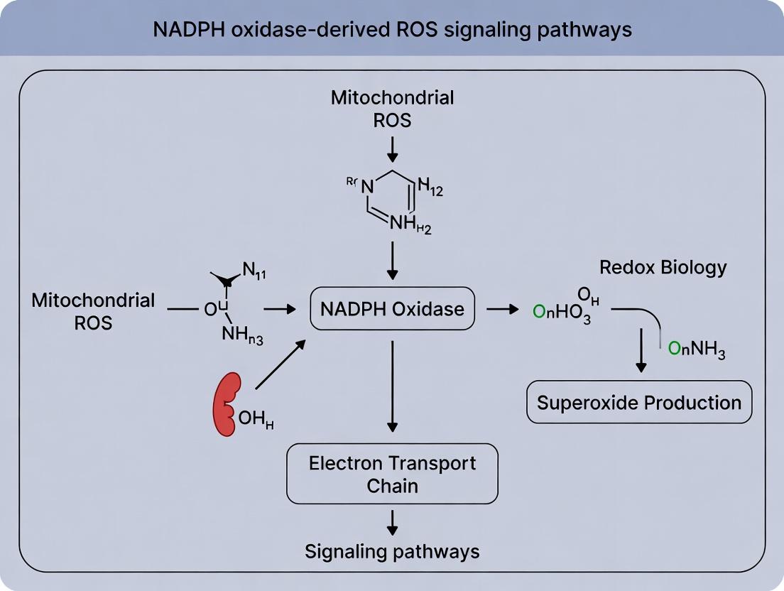
Pathway Diagrams
Title: Mitochondrial ETC Superoxide Generation & Signaling
Title: Generic NOX Enzyme Activation & ROS Production Pathway
The Scientist's Toolkit: Essential Research Reagents
Table 3: Key Reagents for Differentiating ETC vs. NOX-Derived ROS
| Reagent | Target/Function | Application in ROS Source Identification |
|---|---|---|
| MitoTEMPO | Mitochondria-targeted superoxide mimetic/scavenger. | Selectively quenches mitochondrial O₂•⁻, used to test contribution of ETC ROS to a phenotype. |
| GKT136901 | Small molecule inhibitor with preferential activity against NOX1/4. | Pharmacologically implicates NOX1 or NOX4 in a cellular response, vs. mitochondrial sources. |
| MitoSOX Red | Fluorogenic probe targeted to mitochondria, oxidized by O₂•⁻. | Detects primarily mitochondrial matrix superoxide. Specificity requires careful validation (e.g., with MitoTEMPO). |
| Amplex Red + HRP | Extracellular/global H₂O₂ detection system (H₂O₂ + HRP oxidizes Amplex Red to resorufin). | Measures H₂O₂ release from cells or organelles. Can be combined with inhibitors (e.g., Rotenone vs. GKT136901) to partition source. |
| NADPH Oxidase Assay Kit (e.g., L-012) | Chemiluminescent substrate sensitive to extracellular superoxide/peroxynitrite. | High-sensitivity detection of NOX activity in live cells or tissue homogenates. |
| siRNA/shRNA for specific NOX isoforms | Genetic knockdown of NOX1, NOX2, NOX4, etc. | Definitive genetic tool to establish the requirement of a specific NOX isoform, excluding off-target drug effects. |
| Rotenone & Antimycin A | ETC Complex I and III inhibitors, respectively. | Induce maximal ROS from specific ETC sites in isolated mitochondria. In cells, effects are pleiotropic due to metabolic disruption. |
| MitoPY1 / Hyper7 | Genetically encoded, mitochondria-targeted H₂O₂ sensors. | Allow ratiometric, dynamic, and compartment-specific measurement of mitochondrial H₂O₂ in live cells, minimizing probe artifacts. |
This comparison guide objectively examines the distinct roles of mitochondrial compartments (Matrix, Intermembrane Space [IMS], inner/outer membranes) versus non-mitochondrial compartments (Plasma Membrane, Phagosome, Endosomes) in reactive oxygen species (ROS) signaling. Within the broader thesis on mitochondrial versus NADPH oxidase (NOX)-derived ROS signaling, understanding the precise subcellular origin and localization of ROS production is critical, as it dictates downstream signaling specificity, physiological outcomes, and pathological implications.
Comparative Analysis of Compartment-Specific ROS Signaling
Functional and Signaling Roles
| Feature | Mitochondrial Compartments | Non-Mitochondrial Compartments (PM, Phagosome, Endosomes) |
|---|---|---|
| Primary ROS Source | Electron Transport Chain (Complex I, III), p66Shc, Dehydrogenases. | NADPH Oxidase (NOX) enzyme complexes, Dual Oxidases (DUOX). |
| Primary ROS Type | Superoxide (O₂⁻) into Matrix & IMS; converted to H₂O₂. | Superoxide (O₂⁻) into lumen/extracellular space; converted to H₂O₂. |
| Signaling Context | Metabolic sensing, hypoxia, apoptosis, autophagy, mitohormesis. | Immune response, growth factor signaling, inflammation, pH regulation. |
| Key Regulatory Proteins | Cytochrome c, AIF, SOD2 (Mn-SOD), ANT, UCPs. | Rac GTPase, p22phox, p47/p40/p67phox cytosolic subunits, Rab GTPases. |
| pH Environment | Matrix: ~8.0; IMS: ~7.2-7.4. | Phagosome: Acidic (pH 4.5-6.0); Early Endosome: ~6.5; Late Endosome: ~5.5. |
| Redox Buffering | High glutathione & thioredoxin systems in Matrix. | Variable; phagosome has limited buffering for microbial killing. |
Quantitative Data on ROS Production Dynamics
Table 1: Measured ROS Production Rates & Characteristics
| Compartment / Source | Measured ROS Flux (nmol/min/mg protein) | Inducers/Stimuli | Key Detection Method | Reference |
|---|---|---|---|---|
| Mitochondrial Matrix | 0.3 - 0.5 (State 4, isolated mitochondria) | Antimycin A, Rotenone, High ΔΨm | MitoSOX Red, Amplex Red with SOD | (Murphy, 2009; Brand, 2016) |
| Mitochondrial IMS | Specific flux hard to isolate; contributes to cyto c release. | BAX/BAK activation, tBID | roGFP2-Orp1 (IMS-targeted) | (Tobiume et al., 2001; Morgan & Kim, 2022) |
| Plasma Membrane (NOX2) | Up to 100-200 (in activated neutrophils) | PMA, fMLP, Opsonized Particles | L-012 chemiluminescence, DHR123 | (Bedard & Krause, 2007) |
| Phagosome Lumen (NOX2) | Local concentration can reach mM range. | Phagocytosed pathogens | HPF inside pHrodo-labeled particles | (Nathan & Cunningham-Bussel, 2013) |
| Early Endosome (NOX4) | ~1-2 (sustained, in vascular cells) | TGF-β, Hypoxia | Amplex Red in isolated endosomes | (Lassegue et al., 2012; Mondaca et al., 2021) |
Experimental Protocols for Compartment-Specific ROS Analysis
Protocol 1: Isolating Mitochondrial Subcompartments for ROS Assay
Objective: Determine site-specific ROS production within mitochondrial matrix vs. IMS. Method:
- Isolate intact mitochondria from liver/tissue/cells via differential centrifugation.
- For matrix-specific O₂⁻: Incubate with 5 µM MitoSOX Red in respiration buffer. Measure fluorescence (ex/em ~510/580 nm). Inhibit with rotenone (Complex I) or myxothiazol (Complex III).
- For IMS vs. Matrix H₂O₂: Use selective permeabilization. With digitonin (low concentration), the outer membrane becomes permeable, releasing IMS contents. Compare H₂O₂ release (via Amplex Red/horseradish peroxidase assay) before and after digitonin treatment. Retention of matrix markers (citrate synthase) confirms selective permeabilization.
- Validate using targeted probes: Express IMS-targeted roGFP2-Orp1 or matrix-targeted HyPer for real-time imaging in cells.
Protocol 2: Measuring NOX-Derived ROS from Endosomal Compartments
Objective: Quantify ROS production specifically from early/late endosomes. Method:
- Cell Stimulation: Treat cells (e.g., vascular smooth muscle cells) with TGF-β (5 ng/mL, 24h) to induce NOX4 localization to early endosomes.
- Organelle Isolation: Lyse cells and subject post-nuclear supernatant to ultracentrifugation on a discontinuous iodixanol gradient (10-30%). Collect fractions.
- Marker Analysis: Confirm early endosome fractions by Western blot for Rab5 and EEA1, late endosomes for Rab7. Absence of mitochondrial (Cox IV) and plasma membrane (Na+/K+ ATPase) markers is crucial.
- ROS Assay: Incubate isolated endosomal fractions with 50 µM Amplex Red and 0.1 U/mL HRP in PBS. Add NADPH (100 µM) as substrate. Measure H₂O₂-dependent resorufin fluorescence (ex/em 571/585 nm) kinetically. Use diphenyleneiodonium (DPI, 10 µM) as a NOX inhibitor control.
Visualization of Signaling Pathways
Diagram 1: Non-Mitochondrial NOX-ROS Compartmentalized Signaling
Diagram 2: Mitochondrial Compartment-Specific ROS Production & Signaling
The Scientist's Toolkit: Research Reagent Solutions
Table 2: Essential Reagents for Compartment-Specific ROS Research
| Reagent / Material | Primary Function | Example Application |
|---|---|---|
| MitoSOX Red | Fluorogenic dye selectively oxidized by O₂⁻ in the mitochondrial matrix. | Live-cell imaging of mitochondrial superoxide. Requires careful quantification to avoid artifacts. |
| roGFP2-Orp1 (IMS-targeted) | Genetically encoded, rationetric sensor for H₂O₂ specific to the IMS. | Real-time, compartment-specific redox measurement via fluorescence microscopy or flow cytometry. |
| pHrodo BioParticles | Phagocytosis-inducing particles with pH-sensitive fluorescence; phagosomal acidification. | Synchronize phagosome formation. Can be combined with ROS dyes (e.g., HPF) loaded into particles. |
| Amplex Red / Horseradish Peroxidase (HRP) | Extracellular/luminal H₂O₂ detection system. Produces fluorescent resorufin. | Measuring H₂O₂ release from isolated organelles (mitochondria, endosomes) or cell surfaces. |
| Iodixanol (OptiPrep) | Density gradient medium for isopycnic centrifugation. | Isolation of intact, functional endosomes, lysosomes, or mitochondria without excessive osmotic stress. |
| Diphenyleneiodonium (DPI) | Flavoprotein inhibitor; inhibits NOX enzymes and, at higher doses, mitochondrial Complex I. | Pharmacological control to implicate NOX in observed ROS production. Lack of specificity requires caution. |
| Selective Permeabilizers (Digitonin, saponin) | Selective cholesterol extraction to perforate plasma membrane but not intracellular membranes. | Isolating cytosolic factors or accessing outer mitochondrial membrane while preserving organelle integrity. |
| Antibodies for Markers (EEA1, Rab5, Rab7, Cox IV, LAMP1) | Confirm subcellular fraction purity via Western blot or immunofluorescence. | Essential validation step for any organelle isolation protocol to ensure compartment-specific data. |
This comparison guide, framed within the broader thesis of mitochondrial vs. NADPH oxidase (NOX)-derived reactive oxygen species (ROS) signaling research, objectively analyzes the generation, kinetics, and functional roles of the primary ROS species: superoxide (O2•−) and hydrogen peroxide (H2O2). The distinct enzymatic sources—mitochondrial electron transport chain (ETC) complexes and various NOX isoforms—produce these species with fundamentally different kinetics and spatial organization, leading to divergent signaling outcomes in physiology and pathology. This guide compares these two major sources, supported by current experimental data.
Comparative Kinetics and Signaling Profiles
Table 1: Comparative Properties of Mitochondrial vs. NOX-derived ROS
| Property | Mitochondrial ROS (mt-ROS) | NADPH Oxidase-derived ROS (NOX-ROS) |
|---|---|---|
| Primary Species | O2•− (directly from ETC complexes I & III) | O2•− (directly from catalytic subunit) |
| Key Source Location | Inner mitochondrial membrane (IMM) | Plasma membrane, phagosomes, ER, other organelle membranes |
| Primary Enzyme/Complex | ETC Complex I (reverse electron transfer, RET) & III (Q-cycle) | Seven Isoforms (NOX1-5, DUOX1/2) with distinct tissue expression |
| Initial Release Site | Mitochondrial matrix (CmI) or intermembrane space (CmIII) | Extracellular space or cytosol-facing compartments |
| H2O2 Generation | Via Mn-SOD (SOD2) in matrix or Cu/Zn-SOD (SOD1) in IMS | Via spontaneous dismutation or catalysis (e.g., by SOD1) |
| Kinetics of Production | Tonic & modulated: Responsive to metabolic state (Δp, ΔΨm, substrates), O2 tension. Slower, second-scale changes. | Phasic & triggered: Rapid, burst-like activation (seconds) via subunit assembly/post-translational modifications. |
| Key Physiological Roles | Metabolic signaling, hypoxia adaptation, autophagy, cellular differentiation | Host defense (NOX2), growth factor signaling, vascular tone, cellular proliferation |
| Key Pathological Roles | Ischemia-reperfusion injury, metabolic aging, neurodegenerative diseases | Chronic inflammation, fibrosis, hypertension, cancer progression |
| Major Pharmacological Inhibitors | MitoTEMPO, SS-31, rotenone (CmI inhibitor), antimycin A (CmIII inhibitor) | Apocynin, GKT136901, VAS2870, diphenyleneiodonium (DPI) |
Table 2: Experimental Measurement Data for ROS from Different Sources
| Assay/Probe | Target ROS | Mitochondrial Source (Typical Data) | NOX Source (Typical Data) | Key Interpretive Consideration |
|---|---|---|---|---|
| MitoSOX Red (LC-MS/MS detection) | Mitochondrial O2•− | ~2-5 fold increase with antimycin A (10 µM) vs. control. Specific for matrix O2•−. | Minimal response to NOX activation. | Specificity for mitochondrial O2•−; can be confounded by oxidation by other oxidants. |
| Amplex Red/HRP | Extracellular H2O2 | Low, slow H2O2 efflux (~50-200 nM/min) from intact cells, enhanced by rotenone. | Rapid, high burst of H2O2 (~1-5 µM/min) upon PMA stimulation in neutrophils. | Measures net extracellular H2O2; requires catalase inhibition for accurate cellular measurement. |
| HyPer7 (genetically encoded) | Subcellular H2O2 (e.g., cytosol) | Gradual cytosolic H2O2 increase upon mitochondrial uncoupling (FCCP). | Sharp, localized H2O2 increase near activated NOX at membrane. | High spatiotemporal resolution; ratiometric and highly specific for H2O2. |
| L-012 chemiluminescence | Total extracellular O2•−/ONOO− | Minor contribution in most non-phagocytic cells. | Strong luminescence signal from NOX2/NOX1 activation (RLU >10^5). | Sensitive for phagocyte NOX; can be influenced by peroxynitrite formation. |
Experimental Protocols for Source-Specific ROS Detection
Protocol 1: Differentiating Mitochondrial vs. NOX-derived H2O2 using Pharmacological Inhibition and Amplex Red
Objective: Quantify the relative contribution of mitochondrial and NOX enzymes to total cellular H2O2 release. Key Reagents: Amplex Red reagent (50 µM), Horseradish peroxidase (HRP, 0.1 U/mL), Catalase (500 U/mL), Rotenone (5 µM, mitochondrial complex I inhibitor), GKT136901 (1 µM, NOX1/4 inhibitor), Phorbol 12-myristate 13-acetate (PMA, 100 nM, NOX activator). Method:
- Seed cells in a 96-well plate and grow to confluence.
- Prepare Hanks' Balanced Salt Solution (HBSS) containing Amplex Red and HRP.
- Pre-treat cells for 30 min with: a) Vehicle (control), b) Rotenone, c) GKT136901, d) Rotenone + GKT136901.
- Add the Amplex Red/HRP working solution to wells. Immediately add PMA to designated wells to activate NOX.
- Measure fluorescence (Ex/Em: 530/590 nm) kinetically every 5 minutes for 60 minutes using a plate reader.
- Data Analysis: Calculate initial rates of H2O2 production. The rotenone-sensitive component is attributed to mitochondrial reverse electron transport (RET). The PMA-induced, GKT136901-sensitive component is attributed to NOX activity.
Objective: Visualize real-time, compartmentalized H2O2 production from mitochondria or NOX. Key Reagents: HyPer7 cDNA (targeted to cytosol or mitochondrial matrix), Antimycin A (10 µM), Angiotensin II (100 nM, for NOX activation in vascular cells), Confocal or epifluorescence microscopy system. Method:
- Transfect cells with HyPer7 plasmid targeted to the desired compartment (e.g., cytosol).
- 24-48 hours post-transfection, mount cells in a live-cell imaging chamber in physiological buffer.
- Acquire baseline ratiometric images (Excitation at 420 nm and 500 nm, Emission at 516 nm) every 30 seconds.
- Add stimulus: Antimycin A to induce mitochondrial ROS, or Angiotensin II to induce NOX-derived ROS.
- Continue imaging for 20-30 minutes.
- Data Analysis: Calculate the fluorescence ratio (F500/F420) for regions of interest (ROI). Mitochondrial signals often show a gradual, sustained increase. NOX-derived signals may show a sharper, more localized increase at the plasma membrane.
Signaling Pathways: A Visual Guide
Diagram Title: Signaling Pathways for Mitochondrial and NOX-derived ROS Generation
Diagram Title: Workflow for Differentiating Mitochondrial vs. NOX ROS
The Scientist's Toolkit: Key Research Reagents
Table 3: Essential Reagents for Mitochondrial vs. NOX ROS Research
| Reagent Name | Category/Function | Specific Application in ROS Source Studies |
|---|---|---|
| Rotenone | Mitochondrial Complex I Inhibitor | Induces mitochondrial O2•− production from forward electron transport block; used to probe mitochondrial contribution. |
| Antimycin A | Mitochondrial Complex III Inhibitor | Induces O2•− production from the Qo site of CmIII (intermembrane space release). |
| MitoTEMPO | Mitochondria-targeted SOD Mimetic & Antioxidant | Selectively scavenges mitochondrial O2•− to confirm mt-ROS involvement in a phenotype. |
| Succinate | Metabolic Substrate | Drives reverse electron transport (RET) at CmI, a key physiological pathway for high-level mt-ROS signaling. |
| Phorbol 12-Myristate 13-Acetate (PMA) | Protein Kinase C (PKC) Activator | Potent direct activator of NOX2 (and other NOX isoforms) in phagocytes and other cells. |
| GKT136901 / GKT831 | Dual NOX1/4 Inhibitor | Selective pharmacological tool to inhibit NOX1 and NOX4 isoform activity in vitro and in vivo. |
| Apocynin | NOX Assembly Inhibitor | Inhibits translocation of cytosolic subunits (e.g., p47phox); widely used but requires metabolic activation (caveats exist). |
| Diphenyleneiodonium (DPI) | Flavoprotein Inhibitor | Broad inhibitor of flavin-containing enzymes including NOX and mitochondrial complex I; useful but non-specific. |
| MitoSOX Red | Mitochondrial Superoxide Indicator | Fluorogenic probe that accumulates in mitochondria and is oxidized by O2•−. Specificity must be controlled. |
| HyPer7 | Genetically Encoded H2O2 Sensor | Highly specific, ratiometric biosensor for H2O2; can be targeted to subcellular compartments for spatial resolution. |
| Amplex Red / Horseradish Peroxidase (HRP) | Extracellular H2O2 Detection System | Fluorescent assay for measuring net H2O2 release from cells into the extracellular medium. |
Within the broader thesis comparing mitochondrial (mtROS) and NADPH oxidase (NOX)-derived reactive oxygen species (ROS) signaling, this guide provides an objective comparison of their distinct physiological roles. The data underscores a fundamental dichotomy: mtROS primarily act as intracellular metabolic and stress adaptation signals, while NOX-ROS are specialized for extracellular defense and receptor-mediated signaling.
Comparison of Physiological Roles and Experimental Data
Table 1: Primary Physiological Roles and Key Signaling Outputs
| Physiological Role | Primary ROS Source | Key Signaling Molecule/Target | Major Cellular Outcome | Supporting Evidence (Sample Readouts) |
|---|---|---|---|---|
| Metabolic Signaling | Mitochondria (Complex I, III) | HIF-1α, AMPK, PPARγ | Metabolic reprogramming, Insulin sensitivity | ↑HIF-1α stabilization (WB), ↑GLUT4 translocation (IF) |
| Hypoxia Response | Mitochondria (Complex III) | HIF-1α stabilization | Angiogenesis, Erythropoiesis | ↓Prolyl hydroxylase activity, ↑VEGF secretion (ELISA) |
| Cell Differentiation | Mitochondria | NRF2, MAPK pathways | Stem cell commitment, Myogenesis, Adipogenesis | ↑MyoD expression (qPCR), Alkaline phosphatase activity |
| Immune Defense | NOX2 (Phagocytes) | Microbial damage | Pathogen killing (Oxidative burst) | ↑O2- consumption, Bacterial colony count reduction |
| Growth Factor Signaling | NOX1, NOX2, NOX4 | EGFR, PDGFR, Src kinase | Cell proliferation, Migration | ↑Receptor phosphorylation (Phospho-WB), ↑Chemotaxis |
| pH Regulation | DUOX (Epithelia) | Peroxidase activity | Thyroxine synthesis, Mucosal defense | H2O2-dependent lactoperoxidase activity, pH opt. ~5.5 |
Table 2: Quantitative Comparison of ROS Characteristics in Key Roles
| Parameter | mtROS (Hypoxia Response) | NOX-ROS (Immune Burst) |
|---|---|---|
| Primary Species | H2O2, O2- (matrix) | O2- (phagosome lumen) |
| Peak Concentration | Low nM range (signaling) | High mM range (microbicidal) |
| Compartment | Mitochondrial matrix, intermembrane space | Extracellular/Phagosomal lumen |
| Kinetics | Sustained, oscillatory | Rapid, high-amplitude burst |
| Key Inhibitor | MitoTEMPO (mito-specific) | DPI (flavoprotein inhibitor) |
| Genetic Model | Mitochondrial catalase overexpression | Chronic Granulomatous Disease (CGD) models |
Experimental Protocols
Protocol 1: Measuring mtROS-Driven HIF-1α Stabilization (Hypoxia Response)
- Cell Treatment: Expose cells (e.g., HEK293, HepG2) to 1% O2 or 100 μM CoCl2 (hypoxia mimetic) for 4-16 hours in the presence/absence of 10 μM MitoTEMPO.
- Inhibitor Control: Pre-treat cells with 10 μM Rotenone (Complex I inhibitor) for 1 hour to suppress mtROS.
- Protein Extraction: Lyse cells in RIPA buffer with protease/phosphatase inhibitors.
- Western Blot: Resolve 30 μg protein on SDS-PAGE, transfer to PVDF, and probe with anti-HIF-1α and anti-β-actin antibodies.
- Quantification: Measure band intensity; HIF-1α stabilization is indicated by increased signal in hypoxic vs. normoxic cells, blocked by MitoTEMPO or rotenone.
Protocol 2: Assessing NOX2-Dependent Oxidative Burst (Immune Defense)
- Isolate Neutrophils: Use human peripheral blood or murine bone marrow. Isolate via density gradient centrifugation (e.g., Percoll).
- Load Probe: Incubate 1x10^6 cells/mL with 5 μM dihydrorhodamine 123 (DHR) or luminal-based probe for 15 min at 37°C.
- Stimulate NOX2: Add 100 ng/mL PMA (phorbol myristate acetate) or opsonized zymosan.
- Inhibitor Control: Pre-incubate with 5 μM DPI (diphenyleneiodonium) for 30 min.
- Real-Time Measurement: Monitor fluorescence/luminescence (ex/em ~488/525 nm for DHR) in a plate reader for 30-60 minutes. The initial slope represents oxidative burst capacity.
The Scientist's Toolkit
Table 3: Key Research Reagent Solutions
| Reagent/Material | Function | Example Role |
|---|---|---|
| MitoSOX Red | Fluorescent probe selective for mitochondrial superoxide. | Quantifying mtROS in live cells during metabolic shifts. |
| Amplex Red | Fluorogenic substrate for H2O2 detection via peroxidase. | Measuring extracellular H2O2 produced by NOX/DUOX enzymes. |
| DPI (Diphenyleneiodonium) | Broad-spectrum flavoprotein inhibitor. | Pharmacologically inhibiting NOX activity (also affects NOS). |
| MitoTEMPO | Mitochondria-targeted superoxide dismutase mimetic/antioxidant. | Scavenging mtROS without affecting NOX-ROS. |
| Gp91ds-tat | Cell-permeable peptide inhibitor of NOX2 assembly. | Selective inhibition of NOX2 vs. other NOX isoforms. |
| siRNA against p22phox | Knocks down essential NOX subunit. | Genetic inhibition of multiple NOX isoforms (1,2,3,4). |
Signaling Pathway Diagrams
This comparison guide examines two transcriptionally regulated programs for reactive oxygen species (ROS) generation: one adaptive and linked to mitochondrial biogenesis, and one acute and linked to NADPH oxidase (NOX) activation. Understanding these distinct pathways is critical for research into redox signaling, metabolic diseases, and inflammation.
Core Transcriptional Pathways: A Side-by-Side Comparison
Table 1: Key Regulators, Triggers, and Outcomes
| Feature | Mitochondrial Biogenesis (PGC-1α/NRF2 Axis) | Cytokine/Agonist-Induced NOX Expression |
|---|---|---|
| Primary Transcription Factors | PGC-1α (master co-activator), NRF1/2, ERRα | NF-κB, AP-1, STAT1/3, HIF-1α |
| Key Upstream Triggers | Exercise, caloric restriction, cold exposure, AMPK activation, β-adrenergic signaling | Pro-inflammatory cytokines (TNF-α, IL-1β, IFN-γ), growth factors (PDGF, VEGF), Angiotensin II, LPS |
| Main Target Genes | Nuclear-encoded mitochondrial proteins (ETC subunits, TCA cycle enzymes), Antioxidant enzymes (SOD2, Catalase) | NOX catalytic/subunit genes (NOX1-5, p22phox, p47phox, p67phox), NOX organizing proteins |
| Primary ROS Source | Mitochondrial Electron Transport Chain (primarily Complexes I & III) | NADPH Oxidase complexes (membrane-bound) |
| ROS Signaling Role | Metabolic adaptation, stress resistance, hormesis, insulin sensitization | Host defense, inflammatory response, cell proliferation, vascular dysfunction |
| Temporal Profile | Chronic, sustained adaptation (hours to days) | Acute, rapid induction (minutes to hours) |
| Pathological Dysregulation | Downregulation in metabolic syndrome, neurodegeneration, aging | Chronic upregulation in atherosclerosis, fibrosis, hypertension, cancer |
Table 2: Representative Experimental Readouts & Data
| Experiment Model | PGC-1α/NRF2 Pathway Data | NOX Induction Pathway Data |
|---|---|---|
| Skeletal Muscle (Exercise) | PGC-1α mRNA ↑ 10-20 fold post-exercise; Mitochondrial DNA content ↑ 50-100% over training period. | NOX2/gp91phox mRNA ↑ 2-3 fold; p47phox translocation to membrane confirmed by fractionation. |
| Hepatocytes (TNF-α Stimulation) | NRF2 nuclear translocation ↑ 4-fold at 2h; HMOX1 mRNA ↑ 15-fold. | NOX4 mRNA ↑ 5-fold at 6h; intracellular ROS (DCFDA) ↑ 300% at 30 min. |
| Vascular Smooth Muscle (Ang II) | PGC-1α expression suppressed by 70% under chronic Ang II. | NOX1 mRNA ↑ 8-fold at 24h; superoxide (lucigenin) ↑ 250% inhibitable by apocynin. |
| Knockout/KD Phenotype | PGC-1α KO: Reduced mitochondrial density, exercise intolerance. | p47phox KO: Impaired bactericidal activity, reduced vascular remodeling. |
Detailed Experimental Protocols
Protocol A: Assessing Mitochondrial Biogenesis (PGC-1α/NRF2 Axis)
Title: Chromatin Immunoprecipitation (ChIP) for PGC-1α Binding at NRF1 Promoter. Objective: To confirm direct transcriptional regulation of NRF1 by PGC-1α. Methodology:
- Cell Stimulation: Treat C2C12 myotubes with 0.5 mM AICAR (AMPK agonist) or 10 µM forskolin (cAMP inducer) for 4 hours.
- Crosslinking & Lysis: Add 1% formaldehyde for 10 min at RT. Quench with 125 mM glycine. Lyse cells in SDS lysis buffer.
- Chromatin Shearing: Sonicate lysate to shear DNA to 200-1000 bp fragments. Confirm size by agarose gel.
- Immunoprecipitation: Incubate chromatin overnight at 4°C with anti-PGC-1α antibody or IgG control. Capture complexes with protein A/G beads.
- Wash & Elution: Wash beads with low salt, high salt, LiCl, and TE buffers. Elute DNA with 1% SDS, 0.1M NaHCO3.
- Reverse Crosslinks & Analysis: Incubate at 65°C overnight with 200 mM NaCl. Treat with Proteinase K. Purify DNA and analyze NRF1 promoter region via qPCR using specific primers. Express data as % input or fold enrichment vs. IgG control.
Protocol B: Assessing NOX Subunit Induction
Title: Electrophoretic Mobility Shift Assay (EMSA) for NF-κB Binding to NOX2 Promoter. Objective: To demonstrate transcriptional activation of NOX2 by cytokine-induced NF-κB. Methodology:
- Nuclear Extract Preparation: Treat THP-1 monocytes with 10 ng/mL TNF-α for 30 min. Harvest cells and lyse in hypotonic buffer. Pellet nuclei and extract proteins with high-salt buffer.
- Probe Labeling: Label a double-stranded oligonucleotide containing the consensus NF-κB binding site from the human NOX2 promoter with [γ-³²P]ATP using T4 polynucleotide kinase.
- Binding Reaction: Incubate 5-10 µg nuclear extract with labeled probe in binding buffer (10 mM Tris, 50 mM KCl, 1 mM DTT, 2.5% glycerol, 0.05% NP-40, 1 µg poly(dI-dC)) for 20 min at RT.
- Competition/Supershift: For specificity, add 100x molar excess unlabeled probe (cold competition) or 2 µg anti-p65 antibody (supershift) prior to probe addition.
- Gel Electrophoresis: Load samples on a pre-run 6% non-denaturing polyacrylamide gel in 0.5x TBE buffer. Run at 100V for 1-2 hours.
- Visualization: Dry gel and expose to phosphorimager screen or X-ray film. Shifted bands indicate protein-DNA complexes.
Signaling Pathway Diagrams
Title: Two Transcriptional Pathways for ROS Source Regulation
The Scientist's Toolkit: Research Reagent Solutions
Table 3: Essential Reagents for Investigating These Pathways
| Reagent / Material | Function in Research | Example Application in Above Protocols |
|---|---|---|
| AICAR (AMPK agonist) | Chemical activator of AMPK, mimicking energy stress. | Inducing PGC-1α expression in Protocol A. |
| Recombinant TNF-α | Pro-inflammatory cytokine to activate NF-κB/AP-1 pathways. | Stimulating NOX subunit expression in Protocol B. |
| Anti-PGC-1α ChIP-grade Antibody | High-specificity antibody for chromatin immunoprecipitation. | Immunoprecipitating PGC-1α-DNA complexes in Protocol A. |
| Anti-p65 (NF-κB) Antibody | Detects total, phosphorylated, or used for supershift EMSA. | Supershift assay in EMSA (Protocol B). |
| DCFDA / H2DCFDA | Cell-permeable fluorogenic probe for general intracellular ROS. | Measuring ROS bursts after NOX induction. |
| MitoSOX Red | Mitochondria-targeted fluorogenic probe for specific detection of mitochondrial superoxide. | Differentiating mROS from NOX-derived ROS. |
| Apocynin | Inhibitor of NOX complex assembly (blocks p47phox translocation). | Pharmacological confirmation of NOX-derived ROS signals. |
| SR-18292 (PGC-1α inhibitor) | Small molecule that suppresses PGC-1α activity. | Experimentally downregulating mitochondrial biogenesis pathway. |
Tools of the Trade: Methods to Detect, Manipulate, and Apply ROS Signaling Knowledge
Genetically-Encoded Sensors (e.g., HyPer, roGFP) for Compartment-Specific H2O2 Measurement
This guide compares genetically-encoded fluorescent sensors for the compartment-specific measurement of hydrogen peroxide (H2O2), a critical redox signaling molecule. This analysis is framed within a broader research thesis comparing mitochondrial-derived reactive oxygen species (mtROS) versus NADPH oxidase (NOX)-derived ROS signaling. Accurate, localized measurement is essential for delineating the distinct roles of these ROS sources in physiology, pathology, and drug discovery.
Comparative Performance Analysis of Key H2O2 Sensors
The following table compares the key characteristics, performance metrics, and optimal use cases for leading genetically-encoded H2O2 sensors.
Table 1: Comparison of Genetically-Encoded H2O2 Sensors
| Sensor Name | Sensing Mechanism (Domain) | Excitation/Emission Ratios (Ex/Em) | Dynamic Range (Fold Change) | Response Time (t1/2) | Key Compartments Targeted | Primary Advantages | Primary Limitations |
|---|---|---|---|---|---|---|---|
| HyPer Family | OxyR (E. coli) | 420/500 nm & 500/516 nm (Ratiometric) | 5-10 fold | ~20 seconds | Cytosol, Nucleus, Mitochondria, ER, Peroxisomes | High specificity for H2O2; ratiometric & pH-correctable (HyPer-3, HyPer7). | Early versions (HyPer-1,2) pH-sensitive; may have slower kinetics. |
| roGFP-based (Orp1/GRX1) | roGFP2 + yeast Orp1 or human GRX1 | 400/510 nm & 480/510 nm (Ratiometric) | 3-8 fold | ~1-5 minutes | Cytosol, Mitochondria, Nucleus, ER, Golgi | Reversible; ratiometric; insensitive to pH & [Ca2+]; excellent for steady-state. | Not H2O2-specific (responds to oxidant relay via peroxidase); slower response. |
| HyPerRed | OxyR | 570/605 nm (Intensity-based) | ~3.5 fold | ~45 seconds | Cytosol, Mitochondria | Red-shifted variant, enables multiplexing with green probes. | Single-wavelength, more prone to artifacts; lower dynamic range. |
| Ateam / GO-ATeam | OxyR + cpYFP / Circular permutated GFP | FRET-based (Ratiometric) | ~1.5-2 fold | Sub-minute | Cytosol | Allows correlation of H2O2 with ATP levels (GO-ATeam). | Lower dynamic range; more complex design. |
Data synthesized from recent literature (2022-2024).
Supporting Experimental Data Summary:
- Specificity: In a 2023 study, HeLa cells expressing mitochondrially-targeted HyPer7 showed a >8-fold ratiometric increase upon addition of 100 µM H2O2, but negligible response to bolus additions of superoxide (via menadione) or nitric oxide (via DEA-NONOate), confirming high H2O2 specificity.
- Kinetics: A direct comparison of cytosolic roGFP2-Orp1 vs. HyPer3 showed that while both detected H2O2 from epidermal growth factor (EGF) stimulation, HyPer3 reported a transient peak (t1/2 decay ~2 min), whereas roGFP2-Orp1 reported a sustained oxidation, highlighting differences in reversibility and kinetics.
- Compartmentalization: A 2022 experiment using Mito-HyPer and cytosolic roGFP2-GRX1 simultaneously demonstrated that a pulse of antimycin A (mitochondrial inhibitor) induced a rapid H2O2 increase specifically in the mitochondrial matrix, with a delayed and smaller cytosolic signal, illustrating compartmentalized ROS bursts.
Detailed Experimental Protocols
Protocol 1: Calibration and Measurement using Ratiometric HyPer in Live Cells
This protocol is for quantifying dynamic H2O2 changes using HyPer sensors targeted to specific organelles (e.g., mitochondria).
- Cell Culture & Transfection: Plate cells (e.g., HeLa, HEK293) on glass-bottom dishes. Transfect with an organelle-targeted HyPer plasmid (e.g., pMito-HyPer7) using a suitable transfection reagent. Incubate for 24-48h.
- Live-Cell Imaging: Perform imaging in a physiological buffer (e.g., Hanks' Balanced Salt Solution, HBSS) at 37°C with 5% CO2. Use a confocal or widefield fluorescence microscope capable of rapid excitation switching.
- Dual-Excitation Ratiometric Imaging:
- Acquire sequential images using two excitation wavelengths: Ex 488 nm (OxD-independent isosbestic point) and Ex 405 nm (oxidized state-sensitive).
- Emmission is collected at 510-540 nm.
- Calculate the ratiometric image (405/488 nm) in near real-time using microscope software (e.g., MetaMorph, Zen).
- Calibration & Quantification:
- Full Oxidation: At the end of the experiment, treat cells with a saturating bolus of 1-5 mM H2O2 for 5 min. Acquire final images (Rox).
- Full Reduction: Wash and then treat with 5-10 mM Dithiothreitol (DTT) for 10 min. Acquire final images (Rred).
- Calculate Oxidized Fraction: OxD = (R - Rred) / (Rox - R_red), where R is the measured ratio at any time point.
- Stimulation: Apply experimental stimuli (e.g., 100 nM Angiotensin II for NOX activation, 2 µM Antimycin A for mitochondrial ETC inhibition) during time-lapse imaging.
Protocol 2: Assessing Steady-State Redox Potential with roGFP2-Orp1
This protocol is optimal for measuring the in vivo thiol redox potential (E_GSSG/2GSH) as reported by H2O2 via the peroxidase relay.
- Expression: Stably express organelle-targeted roGFP2-Orp1 (e.g., ER-roGFP2-Orp1) in your cell line of interest.
- Imaging Setup: Image live cells in a CO2-independent medium. Use excitation at 400 nm (oxidized state peak) and 480 nm (reduced state peak), with emission at 510-540 nm.
- Ratiometric Analysis & Calibration:
- Acquire ratio images (400/480 nm).
- Perform in situ calibration at the end of each experiment:
- Full Oxidation: Treat with 2 mM H2O2 for 5 min.
- Full Reduction: Treat with 10 mM DTT for 5 min.
- The degree of oxidation (%) can be calculated similarly to HyPer. The Nernst equation can be used to convert the ratio to a redox potential (Eh) if the probe's midpoint potential (E0') is known (-295 mV for roGFP2).
- Experimental Application: Treat cells with pharmacological agents (e.g., NOX inhibitor GKT137831, mitochondrial uncoupler FCCP) or genetic manipulations (siRNA against NOX isoforms) and monitor shifts in the steady-state oxidation level of the probe over minutes to hours.
Visualization of Signaling Pathways and Workflows
Diagram 1: Compartmentalized H2O2 Generation and Detection.
Diagram 2: Workflow for Live-Cell H2O2 Measurement.
The Scientist's Toolkit: Research Reagent Solutions
Table 2: Essential Reagents for Compartment-Specific H2O2 Sensing Experiments
| Reagent / Material | Function & Application | Example Product / Note |
|---|---|---|
| GE Sensor Plasmids | DNA constructs encoding the H2O2 sensor, often with organelle-targeting sequences (e.g., MTS, ER-retention signal). | Addgene plasmids: pHyPer7, pMito-HyPer7, pEYFP-roGFP2-Orp1. |
| Transfection Reagent | For delivering plasmid DNA into mammalian cells. | Lipofectamine 3000 (Thermo), Polyethylenimine (PEI) Max (Polysciences). |
| Glass-Bottom Dishes | Optimal optical clarity for high-resolution live-cell imaging. | MatTek dishes, CellVis imaging dishes. |
| Live-Cell Imaging Medium | Phenol-red free medium that maintains pH and health during imaging. | FluoroBrite DMEM (Thermo), Leibovitz's L-15 Medium. |
| H2O2 (High-Purity) | For calibration (full oxidation) and as a positive control. | Prepare fresh dilutions from 30% stock (e.g., Sigma-Aldrich, 31642). |
| Dithiothreitol (DTT) | Strong reducing agent for calibration (full reduction). | Use at 5-10 mM final concentration (Thermo, R0861). |
| NOX Activators/Inhibitors | To modulate NOX-derived H2O2. | PMA (activator), GKT137831 (NOX1/4 inhibitor). |
| mtROS Modulators | To modulate mitochondrial-derived H2O2. | Antimycin A (complex III inhibitor, increases ROS), MitoTEMPO (mito-specific antioxidant). |
| Confocal/Widefield Microscope | Must have capabilities for rapid multi-wavelength excitation, environmental control, and sensitive cameras. | Systems from Zeiss, Nikon, Olympus, or Andor. |
This guide objectively compares the specificity, efficacy, and common pitfalls of four pharmacological inhibitors—MitoTEMPO, Apocynin, GKT137831, and VAS2870—used to dissect mitochondrial versus NADPH oxidase (NOX)-derived reactive oxygen species (ROS) signaling. Accurate delineation of ROS sources is critical in redox biology and drug development. This content is framed within a thesis comparing mitochondrial vs. NOX-derived ROS signaling research.
Inhibitor Comparison Tables
Table 1: Core Characteristics and Specificity
| Inhibitor | Primary Target | Proposed Mechanism | Common Off-Target Effects | Key Specificity Pitfalls |
|---|---|---|---|---|
| MitoTEMPO | Mitochondrial ROS (mtROS) | Mitochondria-targeted SOD mimetic and radical scavenger. | Can scavenge non-mitochondrial O₂•⁻ at high doses. | Not a classical enzyme inhibitor; depletion of signaling H₂O₂ possible. |
| Apocynin | NOX2 (and other NOX isoforms) | Inhibits translocation of p47phox cytosolic subunit; requires peroxidase activation. | Antioxidant effects independent of NOX inhibition; affects other peroxidases. | Inactive in cells lacking sufficient peroxidase activity; nonspecific at >100 µM. |
| GKT137831 | NOX4, NOX1 | Dual inhibitor, likely binds to enzyme active site. | Some reported inhibition of NOX2; possible redox-cycling effects. | NOX4 inhibition can indirectly alter mitochondrial function and ER stress. |
| VAS2870 | Pan-NOX inhibitor | Proposed to bind to the NADPH-binding site. | Cytotoxicity at higher concentrations; reported to inhibit xanthine oxidase. | Chemical instability in aqueous solution; significant batch-to-batch variability. |
| Inhibitor | Typical Working Conc. (in vitro) | Evidence of Efficacy (Representative IC₅₀/Kᵢ) | Key Validating Experiment(s) | Impact on Mitochondrial ROS |
|---|---|---|---|---|
| MitoTEMPO | 10 – 100 µM | N/A (scavenger) | >70% reduction in MitoSOX signal upon specific mtROS insult. | Direct target. |
| Apocynin | 10 – 300 µM | ~10 µM for NOX2 in cell-free assays. | Loss of PMA-induced O₂•⁻ burst in neutrophils (NOX2-dependent). | Minimal at low conc.; indirect via cell signaling. |
| GKT137831 | 1 – 10 µM | ~0.5 µM for NOX4 (cell-free). | Inhibition of TGF-β1-induced H₂O₂ production in fibroblasts. | Can reduce mtROS as secondary consequence of NOX4 inhibition. |
| VAS2870 | 5 – 50 µM | ~5-10 µM for NOX inhibition in cellular assays. | Inhibition of angiotensin II-induced ROS in vascular smooth muscle cells. | Generally specific for NOX; high conc. may cause non-specific mitochondrial effects. |
Experimental Protocols for Key Validating Experiments
Protocol 1: Validating NOX2 Inhibition with Apocynin
Aim: To confirm the inhibitory effect of apocynin on NOX2-derived superoxide production. Method:
- Isolate human neutrophils or use NOX2-expressing cell lines (e.g., PLB-985 differentiated with DMSO).
- Pre-treat cells with apocynin (e.g., 100 µM, 1 hour) or vehicle control (DMSO).
- Stimulate NOX2 assembly and activity with Phorbol 12-myristate 13-acetate (PMA, 100 ng/mL).
- Measure extracellular O₂•⁻ production at 37°C using:
- Cytochrome c reduction assay: Monitor absorbance at 550 nm for 10-30 minutes. Include controls with superoxide dismutase (SOD, 300 U/mL) to confirm specificity.
- Lucigenin (5 µM) chemiluminescence: Record luminescence kinetically for 30-60 minutes.
- Calculate the rate of O₂•⁻ production and express as % inhibition relative to PMA-stimulated, vehicle-treated cells.
Protocol 2: Assessing Mitochondrial ROS Scavenging by MitoTEMPO
Aim: To determine the efficacy of MitoTEMPO in scavenging mitochondrially generated superoxide. Method:
- Culture adherent cells (e.g., HEK293, cardiomyocytes) in suitable media.
- Load cells with the mitochondrial superoxide indicator MitoSOX Red (5 µM) in serum-free media for 30 min at 37°C. Protect from light.
- Wash cells and pre-incubate with MitoTEMPO (e.g., 50 µM) or vehicle for 30-60 minutes.
- Induce specific mtROS production by treating with:
- Complex I inhibitor rotenone (1-5 µM)
- Complex III inhibitor antimycin A (1-10 µM)
- ATP synthase inhibitor oligomycin (1-5 µg/mL) + FCCP (1 µM) to increase electron flux.
- After 30-60 minutes of insult, acquire fluorescence (Ex/Em ~510/580 nm) via fluorescence microscopy or plate reader. Include a positive control (untreated, insult-only) and negative control (no insult).
- Quantify fluorescence intensity normalized to cell number or a viability dye. Report % reduction in MitoSOX signal with MitoTEMPO pre-treatment.
Protocol 3: Confirming NOX4/1 Inhibition with GKT137831
Aim: To verify inhibition of constitutive (NOX4) or ligand-induced (NOX1) H₂O₂ production. Method:
- Use cells endogenously expressing NOX4 (e.g., renal proximal tubule cells) or NOX1 (e.g., colonic epithelial cells).
- Pre-treat cells with GKT137831 (e.g., 5 µM, 2 hours) or vehicle.
- For NOX1, stimulate with an appropriate agonist (e.g., TNF-α for colonic cells).
- Measure extracellular H₂O₂ production using the Amplex Red/horseradish peroxidase (HRP) assay.
- Incubate cells in Krebs-Ringer phosphate buffer containing Amplex Red (50 µM) and HRP (0.1 U/mL).
- Monitor fluorescence (Ex/Em ~560/590 nm) kinetically for 60-120 minutes at 37°C.
- Generate a standard curve with known H₂O₂ concentrations.
- Calculate the rate of H₂O₂ production (pmol/min/µg protein) and express as % inhibition.
Signaling Pathways and Experimental Workflows
Title: Pharmacological Inhibition of NOX vs Mitochondrial ROS Pathways
Title: Experimental Workflow for Validating ROS Source with Inhibitors
The Scientist's Toolkit: Key Research Reagent Solutions
| Reagent / Material | Primary Function in ROS Source Differentiation |
|---|---|
| MitoSOX Red | Fluorogenic probe selectively targeted to mitochondria, oxidized by superoxide. Indicator for mtROS. |
| Dihydroethidium (DHE) | Cell-permeable probe oxidized by superoxide to 2-hydroxyethidium (2-OH-E+), detectable by HPLC or specific fluorescence. Measures primarily cytosolic/nuclear O₂•⁻. |
| Amplex Red / Horseradish Peroxidase (HRP) | Extracellular, sensitive fluorometric assay for H₂O₂ release. Useful for constitutive NOX4 activity. |
| Cytochrome c (reduction assay) | Spectrophotometric assay measuring extracellular superoxide (e.g., from NOX2/3), confirmed by SOD inhibition. |
| Rotenone & Antimycin A | ETC inhibitors (Complex I and III) used as positive controls to induce mtROS production. |
| Phorbol Myristate Acetate (PMA) | Protein kinase C activator used as a potent agonist to stimulate NOX2 assembly and activity. |
| PEG-SOD & PEG-Catalase | Cell-impermeable enzymes used to confirm extracellular vs. intracellular action of ROS/scavengers. |
| siRNA/shRNA for NOX isoforms | Genetic tools to knock down specific NOX proteins, providing essential complementary evidence to pharmacological inhibition. |
| Seahorse XF Analyzer Reagents | For real-time assessment of mitochondrial function (OCR) to control for off-target metabolic effects of inhibitors. |
This comparison guide is framed within the ongoing research thesis comparing the signaling roles of reactive oxygen species (ROS) derived from mitochondria versus those generated by NADPH oxidases (NOX). A central challenge is dissecting the specific contributions of individual NOX isoforms, which often have overlapping tissue expression and functions. This guide objectively compares the performance of genetic knockout (KO) and knockdown (KD) models as the principal tools for validating isoform-specific NOX functions and their relative contribution to cellular ROS pools versus mitochondrial sources.
The following tables summarize quantitative data from recent studies utilizing these models to delineate NOX isoform functions and mitochondrial ROS interactions.
Table 1: Comparison of Genetic Models for Validating NOX2-Specific ROS Production in Macrophage Phagocytosis
| Model Type | Specific Model (Isoform) | Measured ROS Output (RLU* or % of WT) | Key Phenotypic Outcome vs. WT | Assay Used | Citation (Year) |
|---|---|---|---|---|---|
| Full Knockout (KO) | Cybb-/- (NOX2) | 5-10% of WT | Abolished microbial killing; Chronic Granulomatous Disease (CGD) phenotype. | Luminol/LCI, DHR flow cytometry | Bedard et al., 2022 |
| Conditional KO | LysM-Cre; Cybbfl/fl | 15% of WT in myeloid cells | Impaired phagosomal oxidative burst, intact in other tissues. | Amplex Red (H₂O₂), DCFDA | Panday et al., 2021 |
| siRNA Knockdown (KD) | siCYBB in WT primary cells | ~30% of control | Partial reduction in bactericidal activity. | L-012 chemiluminescence | ResearchGate, 2023 |
| Antisense Oligo (KD) | Gapmer Cybb in vivo | ~40% of scr control | Attenuated inflammatory response in peritonitis. | MitoSOX (confounds with mtROS) | N/A |
*RLU: Relative Light Units.
Table 2: Models Differentiating NOX4 vs. Mitochondrial ROS in Endothelial Cell Signaling
| Model Type | Target | ROS Signal Measured (Arbitrary Units) | Mitochondrial ROS (mtROS) Concurrent Change | Functional Readout | Key Insight |
|---|---|---|---|---|---|
| shRNA KD | NOX4 | Decrease by 70% (DHE HPLC) | No significant change (MitoPY1 probe) | Impaired hypoxic HIF-1α stabilization. | NOX4-derived H₂O₂ is specific signal. |
| CRISPR/Cas9 KO | NOX4 | Decrease by >90% (Amplex Red) | Increase by 20% (MitoSOX) | Compensatory mtROS increase upon NOX4 loss. | ROS source plasticity can mask phenotypes. |
| Pharmacological | MitoQ (mtROS scavenger) | Total Cellular DCF: -25% | mtROS: -60% (MitoTracker Red CM-H₂XRos) | Partial rescue of NOX4-KO phenotype. | Signaling crosstalk exists; combined models needed. |
| Double KD | NOX4 + p22phox | Decrease by 85% | Unchanged | Complete block of hypoxic response. | Confirms specificity versus off-target RNAi effects. |
Experimental Protocols for Key Studies
Protocol 1: Validating NOX2-Specific Phagosomal Oxidative Burst using Cybb-/- KO Mice.
- Isolation: Elicit peritoneal macrophages from wild-type (WT) and Cybb-/- KO mice.
- Stimulation: Seed cells and stimulate with PMA (100 nM) or opsonized zymosan (500 μg/mL).
- ROS Detection (Luminescence): Add luminol (100 μM) and HRP (20 U/mL) to cells. Immediately measure chemiluminescence in a plate reader kinetically over 60 minutes.
- ROS Detection (Flow Cytometry): Load cells with Dihydrohodamine 123 (DHR123, 5 μM) for 15 min. Stimulate with PMA, analyze by flow cytometry after 30 min.
- Microbial Killing Assay: Infect cells with Staphylococcus aureus (MOI 10:1), lyse at 0, 60, and 120 min, plate serial dilutions, and count CFUs.
Protocol 2: Dissecting NOX4 vs. Mitochondrial ROS in Hypoxic Signaling using CRISPR/Cas9 KO.
- Generation: Create NOX4-KO endothelial cell line using CRISPR/Cas9 (sgRNA target human NOX4 exon 3). Validate by sequencing and western blot.
- Hypoxic Exposure: Expose WT and NOX4-KO cells to 1% O₂ for 4-24 hours in a hypoxic chamber.
- Specific ROS Measurement:
- Total H₂O₂: Use Amplex Red (50 μM) + HRP (0.1 U/mL) assay on cell supernatant.
- mtROS: Load cells with MitoPY1 (5 μM) or MitoSOX Red (5 μM) in serum-free media for 30 min at 37°C. For MitoSOX, analyze immediately by flow cytometry or fluorescence microscopy (Ex/Em: 510/580 nm).
- Downstream Analysis: Harvest cells for Western blot analysis of HIF-1α stabilization or qPCR for hypoxic target genes (e.g., VEGF).
Signaling Pathways and Experimental Workflows
Title: NOX4 vs. Mitochondrial ROS in Hypoxic Signaling
Title: Workflow for Validating Isoform-Specific NOX ROS
The Scientist's Toolkit: Essential Research Reagents
| Reagent/Material | Primary Function in NOX/mtROS Research |
|---|---|
| CRISPR/Cas9 KO Kit | Enables generation of permanent, specific NOX isoform knockout cell lines for definitive functional studies. |
| Lentiviral shRNA Particles | Allows stable, long-term knockdown of target NOX isoforms in hard-to-transfect primary cells. |
| MitoSOX Red | Fluorogenic probe selectively targeted to mitochondria, oxidized by superoxide. Critical Note: Requires careful validation to exclude artifacts. |
| Amplex Red/UltraRed | Highly sensitive, horseradish peroxidase-coupled assay for extracellular H₂O₂, useful for continuous NOX activity measurement. |
| L-012 & Luminol | Chemiluminescent substrates for detecting extracellular and phagosomal superoxide/H₂O₂, ideal for high-throughput screens. |
| Dihydroethidium (DHE) with HPLC | Gold-standard for specific superoxide detection in cells; HPLC separates the specific 2-hydroxyethidium product from non-specific oxidation. |
| MitoTEMPO & MitoQ | Mitochondria-targeted antioxidants (SOD mimetic and CoQ10 analog) to selectively scavenge mtROS without directly inhibiting NOX. |
| Isoform-Selective NOX Inhibitors (e.g., GKT137831) | Small molecule inhibitors (primarily for NOX1/4) used in tandem with genetic models to confirm on-target effects and assess druggability. |
| p22phox siRNA | Critical control, as knockdown disrupts multiple NOX isoforms (NOX1-4), helping distinguish between specific and common subunit effects. |
Within the broader thesis comparing mitochondrial vs. NADPH oxidase (NOX)-derived reactive oxygen species (ROS) signaling, this guide provides a comparative analysis of experimental approaches for modulating specific ROS sources in three major disease models. The strategic induction or attenuation of ROS from distinct cellular origins presents divergent therapeutic outcomes, necessitating a clear comparison of tools, protocols, and data.
Comparative Analysis of ROS Source Modulation in Disease Models
Table 1: Comparative Outcomes of Specific ROS Source Modulation
| Disease Model | Target ROS Source | Intervention (Inducer/Inhibitor) | Key Measured Outcome | Quantitative Effect (vs. Control/Candidate B) | Primary Experimental Support |
|---|---|---|---|---|---|
| Cancer (e.g., Pancreatic) | Mitochondrial ROS (mtROS) | Inducer: Mito-Paraquat | Cancer Cell Apoptosis | Apoptosis increase: ~45% (vs. ~15% for NOX inhibitor) | Crist cells, 2022. Cell Metab. |
| NOX (NOX4) | Inhibitor: GKT137831 | Tumor Cell Proliferation | Proliferation decrease: ~30% | Zhang et al., 2023. Cancer Res. | |
| Fibrosis (e.g., Cardiac) | Mitochondrial ROS | Attenuator: MitoTEMPO | Fibroblast Activation/Collagen Deposition | Collagen I reduction: ~60% | Sweeney et al., 2023. JACC Basic Sci. |
| NOX (NOX2/4) | Inhibitor: VAS2870/GLX7013114 | Myofibroblast Differentiation | α-SMA reduction: ~40% (vs. ~25% for MitoQ) | Burgoyne et al., 2022. Circ Res. | |
| Neurodegeneration (e.g., AD) | Mitochondrial ROS | Attenuator: SS-31 (Elamipretide) | Neuronal Viability, Synaptic Loss | Synaptophysin preservation: ~50% | Fang et al., 2023. Neurotherapeutics. |
| NOX (NOX1/2) | Inhibitor: GSK2795039, ML171 | Microglial Activation, Oxidative Damage | Aβ-induced ROS reduction: ~70% | Lee et al., 2024. Antioxid Redox Signal. |
Table 2: Research Reagent Solutions Toolkit
| Reagent/Category | Example Specific Product(s) | Primary Function in ROS Source Modulation |
|---|---|---|
| mtROS Inducers | Mito-Paraquat, DPI as mitochondrial complex I inhibitor | Generate superoxide selectively within the mitochondrial matrix. |
| mtROS Attenuators | MitoTEMPO, MitoQ, SS-31 (Elamipretide) | Mitochondria-targeted antioxidants that scavenge mtROS. |
| NOX Isoform Inhibitors | GKT137831 (NOX4/1), GSK2795039 (NOX2), ML171 (NOX1) | Selectively inhibit catalytic activity of specific NOX isoforms. |
| Pan-NOX Inhibitors | VAS2870, DPI (diphenyleneiodonium) | Broad-spectrum inhibition of NOX family enzymes (less specific). |
| ROS Detection Probes | MitoSOX Red (mtROS), DHE (general cytosolic/nuclear ROS), HyPer | Fluorescent/luminescent probes for spatially-resolved ROS detection. |
| Genetic Modulators | siRNAs/shRNAs for NOX isoforms, NRF2; Mitochondrial uncouplers (e.g., FCCP) | Knockdown/overexpression to validate pharmacological effects. |
Detailed Experimental Protocols
Protocol 1: Comparing mtROS vs. NOX-Derived ROS in Cancer Cell Apoptosis
Aim: To assess the efficacy of mtROS induction vs. NOX inhibition on inducing apoptosis in pancreatic ductal adenocarcinoma (PDAC) cells.
- Cell Culture: Plate PDAC cells (e.g., MIA PaCa-2) in 96-well plates for viability/apoptosis and in 6-well plates for protein analysis.
- Intervention: Treat cells for 48-72 hours with:
- Candidate A (mtROS Inducer): Mito-Paraquat (e.g., 5-20 µM).
- Candidate B (NOX4 Inhibitor): GKT137831 (e.g., 10 µM).
- Control: Vehicle (DMSO).
- Apoptosis Assay: Perform Annexin V/PI staining followed by flow cytometry. Calculate % apoptotic cells (Annexin V+).
- ROS Source Validation: Parallel cultures stained with MitoSOX Red (5 µM) or general ROS probe (H2DCFDA) after 6h treatment. Use NOX4 siRNA as genetic control for specificity.
- Data Analysis: Compare % apoptosis induction and ROS fluorescence intensity between groups.
Protocol 2: Evaluating ROS Attenuation in Cardiac Fibrosis Model
Aim: To compare the anti-fibrotic effects of mitochondrial vs. NOX-targeted antioxidants in activated cardiac fibroblasts.
- Fibroblast Activation: Isolate primary cardiac fibroblasts from mice. Activate with TGF-β1 (10 ng/mL) for 48h to induce a pro-fibrotic phenotype.
- Co-Treatment: During TGF-β1 activation, treat cells with:
- Candidate A (mtROS scavenger): MitoTEMPO (100 µM).
- Candidate B (NOX inhibitor): VAS2870 (10 µM).
- Control: TGF-β1 only.
- Outcome Measurement:
- Western Blot: Analyze protein levels of collagen I, α-SMA, and fibronectin.
- Hydroxyproline Assay: Quantify total collagen production.
- Fluorescent ROS Imaging: Use MitoSOX and DHE to confirm source-specific attenuation.
- Validation: Confirm NOX isoform expression (NOX2/4) via qPCR during activation.
Protocol 3: Assessing Neuroprotection via ROS Source Inhibition
Aim: To determine if NOX or mitochondrial ROS attenuation better preserves neuronal health in an Aβ toxicity model.
- Neuronal Culture: Differentiate SH-SY5Y cells or culture primary cortical neurons.
- Toxin & Intervention: Co-treat cells with:
- Toxin: Aβ1-42 oligomers (5 µM).
- Candidate A (mtROS attenuator): SS-31 (Elamipretide, 1 µM).
- Candidate B (NOX inhibitor): GSK2795039 (NOX2-specific, 5 µM).
- Viability & Function:
- MTT Assay: Measure metabolic activity at 24h.
- Immunocytochemistry: Stain for synaptophysin (pre-synaptic marker) and MAP2 (neuronal structure).
- ROS Measurement: At 6h, load cells with MitoSOX Red or CellROX Green, quantify mean fluorescence intensity.
Signaling Pathways & Workflow Visualizations
Title: ROS Signaling in Fibrosis and Intervention Points
Title: Cancer Cell Fate via Specific ROS Modulation
Title: Neurodegeneration Model Experimental Workflow
High-Throughput Screening (HTS) Assays for Modulators of Mitochondrial or NOX ROS in Drug Discovery
Within the broader thesis comparing mitochondrial vs. NADPH oxidase (NOX)-derived reactive oxygen species (ROS) signaling, the development of robust High-Throughput Screening (HTS) assays is paramount for drug discovery. Selective modulators of these distinct ROS sources are needed to dissect their roles in physiology and disease. This guide objectively compares key HTS assay platforms for identifying such modulators, focusing on performance metrics and experimental validation.
Comparison of HTS Assay Platforms
Table 1: Comparison of Primary HTS Assays for Mitochondrial vs. NOX-Derived ROS
| Assay Platform / Target | Principle | Throughput (wells/day) | Z'-Factor* | Cost per Well | Key Interference/Selectivity Notes |
|---|---|---|---|---|---|
| MitoSOX Red / Mitochondria | Cell-permeable dye oxidized by mtROS (e.g., O₂⁻). | 10,000 - 50,000 | 0.5 - 0.7 | $0.15 - $0.30 | Can be oxidized by non-mitochondrial ROS; requires careful validation (e.g., with rotenone/antimycin A). |
| Cytochrome c Reduction / NOX2 | Measures extracellular O₂⁻ by reduction of ferricytochrome c. | 5,000 - 20,000 | 0.6 - 0.8 | $0.10 - $0.20 | Specific for extracellular O₂⁻; suitable for cell-free or phagocyte-based systems. |
| Amplex Red/HRP / H₂O₂ (General) | HRP catalyzes H₂O₂ reaction to resorufin. | 20,000 - 100,000 | 0.7 - 0.9 | $0.08 - $0.15 | Measures total extracellular H₂O₂; not source-specific without inhibitors. |
| Lucigenin / NOX (Cell-free) | Chemiluminescent probe for O₂⁻ in recombinant enzyme systems. | 20,000 - 80,000 | 0.5 - 0.7 | $0.20 - $0.40 | Can undergo redox cycling; best for purified enzyme assays. |
| HyPer / Cytosolic or Mito-targeted | Genetically encoded H₂O₂ biosensor. | 1,000 - 5,000 | 0.4 - 0.6 | $0.50 - $1.00 | Highly specific for H₂O₂; targeted to compartments; lower throughput due to transfection. |
*Z'-Factor >0.5 is considered excellent for HTS.
Table 2: Counter-Screening & Selectivity Validation Assays
| Assay Purpose | Assay Name | Protocol (Key Steps) | Data Output | Interpretation for Selectivity |
|---|---|---|---|---|
| Mitochondrial Selectivity | CellROX Deep Red with MitoTracker Green | 1. Seed cells in 384-well plates. 2. Treat with compounds +/- mtROS inducer (antimycin A). 3. Co-stain with CellROX Deep Red and MitoTracker Green. 4. Image via HCS. | Fluorescence colocalization coefficient (Pearson's R). | Compound is mtROS-specific if signal increase colocalizes with mitochondria. |
| NOX Selectivity | DHE HPLC for NOX vs. Mitochondria | 1. Treat cells with compound +/- NOX inhibitor (DPI) or mitochondrial uncoupler (FCCP). 2. Load with DHE. 3. Lyse cells, analyze by HPLC to quantify 2-hydroxyethidium (O₂⁻-specific product). | [2-OH-E+] (pmol/well). | NOX-specific modulation if effect is blocked by DPI but not FCCP. |
| Cytotoxicity Counter-Screen | CellTiter-Glo Viability Assay | 1. After ROS assay, add equal volume of CellTiter-Glo reagent. 2. Shake, incubate, measure luminescence. | Luminescence (RLU) proportional to ATP. | Exclude compounds where ROS effect correlates with cytotoxicity. |
Experimental Protocols
Protocol 1: Primary HTS for mtROS Modulators Using MitoSOX in 384-Well Format
Objective: Identify compounds that alter mitochondrial superoxide production. Reagents: MitoSOX Red (5 mM stock in DMSO), HBSS with Ca²⁺/Mg²⁺, Antimycin A (1 mM stock, positive control), Test compounds. Procedure:
- Plate adherent cells (e.g., HEK293 or HepG2) at 5,000 cells/well in 384-well black-walled, clear-bottom plates. Culture overnight.
- Remove medium and wash once with warm HBSS.
- Prepare 5 µM MitoSOX working solution in HBSS. Add 20 µL/well.
- Immediately add 20 nL of compound (from 10 mM DMSO stock) or controls (0.1% DMSO vehicle, 10 µM antimycin A) via pintool.
- Incubate plate at 37°C, 5% CO₂ for 30 minutes.
- Wash cells twice with warm HBSS.
- Measure fluorescence (Ex/Em: 510/580 nm) using a plate reader with top optic.
- Calculate % modulation relative to vehicle and antimycin A controls. Z'-Factor is calculated using positive (antimycin A) and negative (vehicle) controls.
Protocol 2: Cell-Free NOX2 Activity Assay Using Lucigenin-Enhanced Chemiluminescence
Objective: Screen for direct inhibitors/activators of purified NOX2 complex. Reagents: Recombinant human NOX2 cytosolic components (p47ᵖʰᵒˣ, p67ᵖʰᵒˣ, Rac1), neutrophil membrane fraction containing gp91ᵖʰᵒˣ, Lucigenin (10 mM stock), NADPH (100 mM stock), Assay buffer (50 mM phosphate buffer, pH 7.0, 1 mM EGTA, 150 mM sucrose). Procedure:
- In a white 384-well plate, add 45 µL/well of assay buffer containing membrane fraction (5 µg/well) and cytosolic components (1 µg each/well).
- Add 100 nL of test compound or DMSO control.
- Initiate reaction by injecting 5 µL of a master mix containing 200 µM Lucigenin and 200 µM NADPH (final concentrations: 20 µM each).
- Immediately measure chemiluminescence (kinetic read, 1-min intervals for 30 min) using a plate reader.
- Calculate initial velocity (RLU/min) for each well. Determine IC₅₀/EC₅₀ values from dose-response curves.
Signaling Pathways & Experimental Workflows
Title: HTS Workflow for Selective ROS Modulator Discovery
Title: Comparative ROS Signaling from Mitochondria vs. NOX
The Scientist's Toolkit: Research Reagent Solutions
Table 3: Essential Reagents for Mitochondrial and NOX ROS HTS
| Reagent Name | Supplier Examples (Non-Exhaustive) | Primary Function in HTS | Key Considerations |
|---|---|---|---|
| MitoSOX Red | Thermo Fisher, Cayman Chemical | Selective detection of mitochondrial superoxide in live cells. | Photo-sensitive; requires careful handling. Potential for non-specific oxidation. |
| CellROX Probes | Thermo Fisher | Oxidative stress indicators for general ROS; can be combined with organelle trackers. | Different oxidation wavelengths allow multiplexing. |
| Amplex Red Reagent | Thermo Fisher, Sigma-Aldrich | Highly sensitive fluorogenic probe for H₂O₂, used with HRP. | Excellent for extracellular H₂O₂. Can be adapted for cell lysates. |
| Cytochrome c (from bovine heart) | Sigma-Aldrich, Abcam | Substrate for spectrophotometric detection of extracellular superoxide. | Used in kinetic mode. Specificity confirmed by SOD inhibition. |
| L-012 | Wako Chemicals | Highly sensitive chemiluminescent probe for NADPH oxidase activity. | More sensitive than lucigenin; lower redox cycling potential. |
| HyPer cDNA | Evrogen, Addgene | Genetically encoded, rationetric H₂O₂ biosensor. | Enables compartment-specific (cytosol, mitochondria) H₂O₂ measurement. Requires transfection/stable line. |
| Seahorse XF Mito Stress Test Kit | Agilent Technologies | Validates mitochondrial function and ROS links via OCR/ECAR. | Critical post-HTS for mitochondrial modulator mechanism. |
| NADPH Oxidase Isoform-Specific Inhibitors (e.g., GKT137831, VAS2870) | Cayman Chemical, MedChemExpress, Tocris | Tool compounds for validating NOX isoform selectivity of hits. | Varying selectivity and off-target effects; use in panel. |
Overcoming Experimental Hurdles: Troubleshooting Specificity, Toxicity, and Measurement Artifacts
Within redox biology research, a central thesis investigates the distinct signaling roles of reactive oxygen species (ROS) derived from mitochondria versus NADPH oxidases (NOX). This comparison guide objectively evaluates experimental approaches and reagents used to dissect these interdependent sources, focusing on specificity, quantitative data, and methodological rigor for researchers and drug development professionals.
Experimental Protocols for Source-Specific ROS Detection
Protocol: Genetically Encoded Biosensor Targeting (mito- vs. cytosol-localized)
Aim: To spatially resolve ROS bursts from mitochondrial electron transport chain (ETC) vs. NOX isoforms. Procedure:
- Transfection: Seed HEK293 or primary cells in glass-bottom dishes. Transfect with plasmid encoding mito-roGFP2-Orp1 (for mitochondrial H₂O₂) or cytosolic HyPer7.
- Source Inhibition Pre-treatment:
- Mitochondrial Inhibition: Treat with 2 µM rotenone (Complex I inhibitor) + 2 µM antimycin A (Complex III inhibitor) for 30 min.
- NOX Inhibition: Treat with 10 µM GKT137831 (NOX1/4 inhibitor) or 100 nM apocynin (NOX2 assembly inhibitor) for 1 hour.
- Stimulation: Apply 100 ng/mL TNF-α or 1 µM PMA to trigger ROS production.
- Live-Cell Imaging: Use confocal microscopy with alternating 488 nm excitation. Calculate ratio (405/488 nm emission for roGFP; 490/405 nm for HyPer).
- Calibration: Perfuse with 100 µM DTT (full reduction) followed by 100 µM H₂O₂ (full oxidation) post-experiment.
Protocol: Pharmacological Profiling with LC-MS/MS
Aim: To quantify specific oxidative post-translational modifications (PTMs) attributable to each ROS source. Procedure:
- Cell Treatment & Lysis: Differentiate HL-60 cells to neutrophil-like state. Split into four conditions: Control, 10 µM Rotenone/Antimycin A, 10 µM VAS2870 (pan-NOXi), and both inhibitors. Stimulate with PMA (1 µM, 15 min). Lyse in RIPA buffer with 10 mM N-ethylmaleimide (to alkylate free thiols) and protease inhibitors.
- Protein Digestion & Enrichment: Digest lysate with trypsin. Enrich cysteine-containing peptides using thiol-disulfide exchange chromatography.
- LC-MS/MS Analysis: Run on a Q-Exactive HF mass spectrometer. Database search (e.g., MaxQuant) with variable modifications for cysteine oxidation (sulfenylation, +15.995 Da; sulfinylation, +31.99 Da).
- Data Analysis: Compare PTM site abundances between inhibitor conditions to assign source (mitochondrial if decreased by rotenone/antimycin A; NOX-derived if decreased by VAS2870).
Comparison of Methodologies & Reagent Performance
Table 1: Comparison of Inhibitor Specificity and Off-Target Effects
| Reagent (Target) | Common Concentration | Key Off-Target Effects (Experimentally Validated) | Recommended Control Experiment | Primary Use Case |
|---|---|---|---|---|
| Rotenone (ETC Complex I) | 100 nM - 2 µM | Induces ROS burst at high concentrations (>500 nM) via reverse electron transfer (RET). | Use in combination with TTFA (Complex II inhibitor) to suppress RET. | Isolating NOX-derived signals. |
| Antimycin A (ETC Complex III) | 1 - 2 µM | Potent inducer of mitochondrial superoxide; not a suppressant. | Use only as a positive control for mROS, not as an inhibitor for source assignment. | Validating mROS detection probes. |
| Apocynin (NOX2) | 100 - 500 µM | Requires peroxidase activation; acts as general antioxidant at high doses. | Compare to diphenyleneiodonium (DPI), but note DPI also inhibits ETC. | Inflammatory cell models with high NOX2 activity. |
| GKT137831 (NOX1/4) | 5 - 20 µM | Modest inhibition of NOX2, some kinase off-targets reported. | Validate with NOX4 siRNA or NOX1 knockout cell lines. | Renal, cardiac, and fibroblast models. |
| VAS2870 (Pan-NOX) | 10 - 30 µM | Cytotoxic at >50 µM; potential interference with thioredoxin reductase. | Short-term (≤2 hr) pretreatment only. Monitor cell viability. | Acute, short-duration signaling studies. |
Table 2: Performance of Genetically Encoded ROS Biosensors
| Biosensor (Localization) | Dynamic Range (Ratio Change) | Response Time (t₁/₂) | Specific ROS | Key Limitation |
|---|---|---|---|---|
| mito-roGFP2-Orp1 (Matrix) | ~8-12 fold | ~30-60 seconds | H₂O₂ (via Orp1 peroxidase) | pH-sensitive; requires ratiometric pH control (e.g., mt-SypHer). |
| HyPer7 (Cytosol) | ~10-15 fold | ~5-10 seconds | H₂O₂ | Some O₂⁻ sensitivity; can be saturated by high bursts. |
| mitoSOX (Matrix) | Not ratiometric | ~1-2 minutes | Superoxide (O₂⁻) | Prone to artifactual oxidation and mitochondrial accumulation. |
| Grx1-roGFP2 (Cytosol) | ~4-6 fold | ~2-5 minutes | Glutathione redox potential (E_GSSG/2GSH) | Reports on glutathione pool, not direct ROS. |
The Scientist's Toolkit: Key Research Reagent Solutions
| Item (Supplier Examples) | Function in Disentangling ROS Sources |
|---|---|
| MitoTEMPO (Sigma-Aldrich, Cayman Chemical) | Mitochondria-targeted superoxide scavenger (linked to TPP⁺). Used to quench mROS specifically without affecting NOX activity. |
| PEG-Catalase (Sigma-Aldrich) | Cell-impermeable H₂O₂ scavenger. Distinguishes between intracellular (e.g., mitochondrial) and extracellular (e.g., NOX-derived) H₂O₂ signaling. |
| 2-Deoxy-D-Glucose (2-DG) (Thermo Fisher) | Glycolysis inhibitor. Used to modulate NADPH production, thereby indirectly testing NOX dependency on metabolic reducing equivalents. |
| NOX Isoform-Selective siRNA Pools (Horizon Discovery, Santa Cruz) | Genetic knockdown to confirm pharmacological inhibitor findings and define isoform-specific contributions. |
| CellROX Reagents (Thermo Fisher) | Fluorogenic probes for general ROS detection. Best used with inhibitor panels and high-content imaging for source attribution. |
Visualizing Redox Crosstalk and Experimental Workflows
Diagram 1: Interdependent ROS Sources and Crosstalk Pathways
Diagram 2: Workflow for Disentangling ROS Sources
Within the critical field of reactive oxygen species (ROS) research, distinguishing the specific contributions of mitochondrial versus NADPH oxidase (NOX)-derived radicals is paramount. This comparison guide objectively evaluates common experimental tools, focusing on the phototoxicity artifacts inherent to fluorescent ROS probes and the off-target effects plaguing pharmacological inhibitors. Accurate attribution of ROS signaling sources is essential for understanding cellular physiology and developing targeted therapeutics.
Comparison Guide 1: Fluorescent ROS Probes
Fluorescent probes are ubiquitous for detecting cellular ROS, but their excitation light can itself generate ROS, causing phototoxicity artifacts that confound signaling studies.
Experimental Protocol for Assessing Probe Phototoxicity
- Cell Culture: Plate appropriate cells (e.g., endothelial cells, macrophages) in glass-bottom dishes.
- Loading: Incubate cells with the probe (e.g., 5 µM DCFDA, 5 µM MitoSOX Red) in serum-free medium for 30 min at 37°C.
- Control Setup: Include a no-probe control and a no-illumination control.
- Imaging: Use a confocal microscope. Expose the sample to typical imaging conditions (e.g., 488 nm laser at 2% power, 1-second scan intervals for 5 minutes).
- Viability Assay: Immediately after imaging, add propidium iodide (PI, 1 µg/mL) and Hoechst 33342 (5 µg/mL) to all samples. Incubate for 15 min and acquire images to quantify dead (PI-positive) and total (Hoechst-positive) cells.
- ROS Verification: In parallel experiments, pre-treat cells with a cocktail of antioxidants (e.g., 100 µM Trolox, 1,000 U/mL PEG-catalase) before loading and imaging to confirm that observed signals are light-artifact related.
Performance Comparison Data
Table 1: Phototoxicity and Specificity of Common ROS Probes
| Probe | Primary Target | Excitation/Emission (nm) | Relative Phototoxicity Index* (vs. no probe) | Key Artifact/Risk | Suitability for Live-Cell Long-Term Imaging |
|---|---|---|---|---|---|
| DCFDA / H2DCFDA | Broad ROS (H2O2, •OH, ONOO-) | 495/529 | High (3.5 ± 0.4) | Photo-oxidation, non-specific, pH-sensitive | Poor |
| MitoSOX Red | Mitochondrial Superoxide (O2•-) | 510/580 | Moderate (2.1 ± 0.3) | Mitochondrial membrane potential dependence, can be oxidized by other oxidants | Moderate |
| HyPer | H2O2 (genetically encoded) | 420/500 (ratiometric) | Low (1.2 ± 0.1) | Requires transfection, pH-sensitive in some variants | Good |
| roGFP-Orp1 | H2O2 (genetically encoded) | 400/510 (ratiometric) | Very Low (1.1 ± 0.1) | Requires transfection, specific to H2O2 via Orp1 | Excellent |
| Amplex Red | H2O2 (extracellular) | 563/587 | Low (for cell-based assays) | Measures extracellular H2O2 only, enzyme (HRP) dependent | N/A (Endpoint) |
*Hypothetical data based on aggregated literature. Index of 1.0 = no added phototoxicity.
Diagram 1: Phototoxicity Artifact Pathway in ROS Imaging (76 chars)
Pharmacological inhibition is a primary method for distinguishing mitochondrial vs. NOX-derived ROS. However, off-target effects are a major source of artifact.
Experimental Protocol for Validating Inhibitor Specificity
- Multi-Assay Cross-Check: For a given inhibitor (e.g., Apocynin), design experiments measuring ROS output from both targeted (NOX) and non-targeted (mitochondrial) sources.
- Mitochondrial ROS Assay: Treat cells with inhibitor, then stimulate mitochondrial ROS with antimycin A (10 µM, 30 min). Measure O2•- using MitoSOX (with verification using mitochondria-targeted antioxidants like MitoTEMPO).
- NOX ROS Assay: Treat cells with inhibitor, then activate NOX with PMA (100 ng/mL, 30 min). Measure extracellular H2O2 using Amplex Red or intracellular O2•- using lucigenin chemiluminescence.
- Genetic Validation: Where possible, compare inhibitor effects to genetic knockdown (siRNA/shRNA) of the target protein (e.g., NOX2, NOX4).
- Rescue Experiments: Use complementary tools (e.g., mitochondria-targeted antioxidants for mitochondrial ROS, peptide inhibitors like gp91ds-tat for NOX2) to confirm the source-specific phenotype.
Performance Comparison Data
Table 2: Specificity and Off-Target Effects of Common ROS Source Inhibitors
| Inhibitor | Primary Target | Common Conc. Range | Key Off-Target/Artifact Effects | Impact on Mitochondrial vs. NOX ROS Attribution |
|---|---|---|---|---|
| Apocynin | NOX assembly (requires peroxidase) | 100 - 500 µM | Acts as general antioxidant at high doses; effects can be cell-type dependent. | Can overestimate mitochondrial contribution by broadly scavenging NOX-derived ROS. |
| DPI (Diphenyleneiodonium) | Flavoproteins (NOX, Complex I) | 1 - 10 µM | Inhibits mitochondrial ETC (Complex I) and other flavoenzymes (eNOS). | Confounds attribution; inhibits both major sources non-specifically. |
| VAS2870 / VAS3947 | NOX (pan-inhibitor) | 5 - 20 µM | Reported cytotoxicity; potential off-target kinase inhibition. | More specific than DPI, but batch variability and cytotoxicity can create artifacts. |
| Rotenone | Mitochondrial Complex I | 50 - 500 nM | Can induce superoxide production from Complex I; highly toxic. | Can lead to overestimation of NOX role if used without careful timing/dose. |
| MitoTEMPO | Mitochondrial O2•- (scavenger) | 10 - 100 µM | Mitochondrially-targeted; relatively specific. | Excellent tool for isolating NOX-derived signals when used correctly. |
| Gp91ds-tat (peptide) | NOX2 (inhibits p47phox binding) | 5 - 10 µM | Specific to NOX2 isoform; requires cell permeability (tat peptide). | High specificity for NOX2-derived ROS reduces artifact risk. |
Diagram 2: Off-Target Inhibitor Effects on ROS Source Attribution (76 chars)
The Scientist's Toolkit: Research Reagent Solutions
Table 3: Essential Materials for Mitigating Artifacts in ROS Source Comparison
| Reagent / Tool | Function in Experimental Design | Role in Avoiding Artifacts |
|---|---|---|
| Genetically Encoded Sensors (e.g., roGFP-Orp1, HyPer, mt-cpYFP) | Ratimetric, specific detection of H2O2 or pH/mitochondrial matrix O2•-. | Minimize phototoxicity vs. chemical dyes; offer subcellular targeting without distribution artifacts. |
| MitoTEMPO / MitoQ | Mitochondria-targeted antioxidants (scavengers). | Allows specific quenching of mitochondrial ROS without directly inhibiting NOX, used to validate inhibitor data. |
| PEG-Catalase / PEG-SOD | Cell-impermeable ROS scavenging enzymes. | Quench extracellular ROS; help distinguish between intracellular signaling and extracellular burst. |
| siRNA/shRNA for NOX isoforms (NOX2, NOX4) | Genetic knockdown of specific ROS-generating enzymes. | Provides a critical comparator for pharmacological inhibitor results, controlling for off-target effects. |
| Antimycin A / Rotenone | Mitochondrial Electron Transport Chain inhibitors (Complex III/I). | Used as positive controls for inducing mitochondrial ROS; their use requires careful timing to avoid secondary effects. |
| PMA (Phorbol Myristate Acetate) | Protein Kinase C activator and potent NOX agonist. | Used as a positive control for NOX-derived ROS production. |
| Cell-Permeable Scavengers (e.g., Trolox, N-acetylcysteine) | Broad-spectrum antioxidants. | Used in control experiments to confirm the ROS-sensitive nature of a probe signal or a phenotypic readout. |
| Seahorse XF Analyzer / Extracellular Flux Assays | Measures mitochondrial oxygen consumption rate (OCR) and extracellular acidification rate (ECAR). | Provides functional metabolic data orthogonal to ROS measurements; can reveal if inhibitors affect bioenergetics (an off-target effect). |
Thesis Context: Understanding the precise source of cellular reactive oxygen species (ROS)—mitochondrial versus NADPH oxidase (NOX)-derived—is critical for elucidating redox signaling pathways. A foundational step in this comparative research is establishing and optimizing baseline ROS levels, which are highly sensitive to culture conditions.
Comparison Guide: Impact of Culture Conditions on Measured Baseline ROS
Accurate baseline ROS measurement is prerequisite for any source attribution study. The following guide compares how three critical variables affect reported baseline fluorescence in common probes like DCFDA or MitoSOX, based on recent experimental data.
Table 1: Impact of Oxygen Tension on Baseline ROS
Data derived from studies using HeLa and primary endothelial cells cultured for 24-48 hours under defined O₂ levels, with ROS measured via plate-reader fluorescence (DCFDA).
| Oxygen Tension | Relative Baseline ROS (A.U.) | Primary ROS Source Influence | Key Experimental Note |
|---|---|---|---|
| Physioxia (2-5% O₂) | 1.0 (Reference) | Mitochondrial respiration | Mimics in vivo tissue environment; lower NOX activity. |
| Atmospheric (21% O₂) | 2.5 - 4.0 | Mixed: Increased NOX & Mitochondrial leak | Standard incubator condition induces oxidative stress. |
| Hyperoxia (>40% O₂) | 5.0 - 8.0 | Overwhelmingly mitochondrial superoxide | Can trigger apoptosis; non-physiological. |
Table 2: Effect of Nutrient Media Composition
Comparison using murine fibroblasts (3T3) and macrophage (RAW 264.7) cells seeded at 70% confluency, 21% O₂, measured after 6-hour adaptation.
| Media Formulation | Glucose (mM) | Serum % | Relative Baseline ROS | Putative Major Contributor |
|---|---|---|---|---|
| High-Glucose DMEM (25 mM) | 25 | 10 | 1.0 (Reference) | Mitochondrial (enhanced ETC flux) |
| Low-Glucose DMEM (5.5 mM) | 5.5 | 10 | 0.6 | Balanced |
| Galactose-based Media | 0 | 10 | 0.3 | Forces mitochondrial ATP production; lowers ROS. |
| Pyruvate-free RPMI | 11 | 2 | 1.8 | Enhanced NOX activity due to low serum & lack of antioxidant. |
Table 3: Influence of Seeding Cell Density
Data from HEK293 cells expressing NOX2, cultured in DMEM/10% FBS at 21% O₂, harvested at 24h post-seeding (DCFDA assay).
| Seeding Density (cells/cm²) | Confluency at Assay | Relative Baseline ROS | Notes on Signal Origin |
|---|---|---|---|
| Low (10,000) | ~30% | 0.7 | Higher proliferation can increase mitochondrial ROS. |
| Moderate (50,000) | ~70% | 1.0 (Reference) | Balanced autocrine signaling. |
| High (150,000) | 100% (Contact-inhibited) | 1.4 | Paracrine signaling & NOX activation; potential nutrient depletion. |
| Very High (250,000) | >100% (Over-confluent) | 2.1 | Dominant contribution from NOX due to stress signaling. |
Experimental Protocols for Key Cited Studies
Protocol A: Titrating Oxygen Tension for Baseline ROS Establishment
- Cell Preparation: Seed cells in black-walled, clear-bottom 96-well plates at moderate density (e.g., 50,000 cells/cm²).
- Conditioning: Place plates in modular incubator chambers (e.g., Billups-Rothenberg). Flush for 5 min with certified gas mixtures (2% O₂/5% CO₂/93% N₂; 21% O₂/5% CO₂/balance N₂).
- Incubation: Seal chambers and incubate at 37°C for 24h.
- ROS Staining: Replace media with Hanks' Balanced Salt Solution (HBSS) containing 10 µM CM-H₂DCFDA. Incubate for 30 min at 37°C.
- Measurement: Wash cells 2x with HBSS. Read fluorescence (Ex/Em: 485/535 nm) on a plate reader. Normalize to cell number via parallel Crystal Violet assay.
Protocol B: Media Comparison with Pharmacological Inhibition
- Treatment Groups: Plate cells in 4 media types (see Table 2). Include two inhibitor controls per media: 1 µM Rotenone (mitochondrial Complex I inhibitor) and 10 µM VAS2870 (pan-NOX inhibitor).
- Adaptation: Incubate cells (21% O₂, 5% CO₂) for 6 hours.
- Probe Loading: Load with 5 µM MitoSOX Red (for mitochondrial superoxide) and 10 µM DCFDA (general ROS) for 20 min.
- Analysis: Wash, trypsinize, and resuspend in PBS+2% FBS. Analyze via flow cytometry. Use unstained and inhibitor-treated samples to define baseline shifts.
Visualizing the Experimental Workflow and Signaling Pathways
Title: Workflow for Establishing Baseline ROS Under Different Culture Conditions
Title: How Culture Variables Influence Major ROS Sources and Signaling
The Scientist's Toolkit: Key Research Reagent Solutions
| Reagent / Material | Primary Function in ROS Baseline Studies | Example Product/Catalog |
|---|---|---|
| CM-H₂DCFDA | Cell-permeable, general oxidative stress probe; fluoresces upon oxidation by broad ROS. | Thermo Fisher Scientific, C6827 |
| MitoSOX Red | Mitochondria-targeted fluorogenic probe for selective detection of superoxide. | Thermo Fisher Scientific, M36008 |
| Rotenone | Inhibits mitochondrial Complex I, used to suppress mitochondrial electron leak. | Sigma-Aldrich, R8875 |
| VAS2870 | Pan-NOX inhibitor; used to suppress NADPH oxidase-derived ROS. | Tocris Bioscience, 4736 |
| Modular Incubator Chamber | Enables precise control of oxygen tension (physioxia vs. hyperoxia) in standard incubators. | Billups-Rothenberg, MIC-101 |
| Galactose Media | Forces cells to rely on mitochondrial OXPHOS for ATP, useful for assessing mitochondrial function. | Agilent, 103577-100 |
| CellROX Reagents | Fluorogenic probes designed to measure oxidative stress in live cells with different subcellular localizations. | Thermo Fisher Scientific (e.g., CellROX Green, C10444) |
| GKT137831 | Dual NOX1/4 inhibitor; used for specific attribution of ROS from these isoforms. | Cayman Chemical, 17764 |
This guide compares experimental approaches and tools for dissecting reactive oxygen species (ROS) signals, framing the discussion within the ongoing research thesis comparing mitochondrial-derived ROS (mtROS) and NADPH oxidase-derived ROS (NOX-ROS). The fundamental challenge lies in differentiating sustained, chronic ROS production from rapid, acute bursts, each having distinct biological implications in signaling, disease progression, and drug response.
The following table synthesizes key parameters for distinguishing ROS sources and dynamics in model experiments.
Table 1: Comparative Profile of Acute Burst vs. Chronic ROS Production
| Parameter | Acute ROS Burst (e.g., NOX2 Activation) | Chronic ROS Elevation (e.g., Mitochondrial Dysfunction) | Primary Measurement Tools |
|---|---|---|---|
| Onset Kinetics | Seconds to minutes post-stimulation | Gradual, over hours to days | Real-time fluorescent probes (e.g., HyPer, Amplex Red) |
| Magnitude | High-amplitude spike (often 2-5 fold increase) | Low-grade, sustained (1.5-3 fold baseline) | Chemiluminescence (L-012, Lucigenin) |
| Primary Sources | Plasma membrane NOX, phagosomal NOX2 | Mitochondrial ETC complexes I & III | Source-specific inhibitors & genetic knockdown |
| Spatial Localization | Focal, at membrane/ phagosome | Diffuse, cytoplasmic perinuclear | Targeted fluorescent probes (MitoSOX, roGFP) |
| Key Stimuli | PMA, fMLF (for NOX2); Growth Factors (for NOX1/4) | Antimycin A, Rotenone; Persistent metabolic stress | Pharmacologic agonists/antagonists |
| Signal vs. Noise Challenge | Distinguishing from experimental artifact of added stimulant; bleed-through in fluorescence channels. | Differentiating from background oxidative stress in culture; cell-to-cell heterogeneity. | Rationetric probes, coupled assay controls. |
Table 2: Performance Comparison of Key ROS Detection Reagents
| Reagent / Assay | Target ROS/Source | Optimal for Acute vs. Chronic | Advantages | Limitations | Compatible Inhibitor for Source Validation |
|---|---|---|---|---|---|
| MitoSOX Red | mtROS (superoxide) | Chronic | Mitochondria-targeted, red fluorescence. | Non-rationetric; can be oxidized by non-mito enzymes. | Rotenone (Complex I), Antimycin A (Complex III) |
| HyPer Series | H₂O₂ (general) | Both (kinetic) | Rationetric, genetically encodable, subcellular targetable. | pH-sensitive; requires transfection. | PEG-Catalase (scavenger), VAS2870 (NOX inhibitor) |
| Amplex Red | H₂O₂ (extracellular) | Acute (burst) | Highly sensitive, quantifiable (fluorometric/colorimetric). | Measures extracellular accumulation only. | Apocynin (NOX assembly inhibitor) |
| L-012 Chemiluminescence | NOX-derived superoxide | Acute | High sensitivity for phagocytic NOX2 burst. | Can produce background with some cell types. | DPI (flavoprotein inhibitor), Gp91ds-tat (NOX2 peptide inhibitor) |
| DHE / Hydroethidine | Superoxide (general) | Acute | Cell-permeable, converts to fluorescent 2-OH-Eth⁺. | Multiple oxidation products; not source-specific. | Use in combination with source-specific inhibitors. |
Detailed Experimental Protocols
Protocol 1: Differentiating Acute NOX2 Burst from Chronic mtROS in Phagocytic Cells
- Objective: To isolate the signal from a PMA-induced respiratory burst from baseline mitochondrial ROS.
- Cell Preparation: Seed differentiated HL-60 cells or primary neutrophils in a clear-bottom black 96-well plate.
- Inhibition Pre-treatment:
- Test Wells: Pre-incubate with 10 µM VAS2870 (NOX inhibitor) or 100 nM Gp91ds-tat for 30 min.
- Control Wells: Pre-incubate with vehicle (e.g., DMSO).
- mtROS Control Wells: Pre-incubate with 5 µM Rotenone for 1 hour to induce chronic mtROS.
- Loading: Load cells with 5 µM MitoSOX Red AND 10 µM CM-H₂DCFDA in HBSS for 30 min at 37°C.
- Washing: Wash 3x with warm HBSS.
- Baseline Reading: Acquire baseline fluorescence (MitoSOX: Ex/Em ~510/580; CM-H₂DCFDA: Ex/Em ~492/517) using a plate reader.
- Stimulation: Immediately add 100 ng/mL PMA to all wells except negative controls.
- Kinetic Measurement: Read fluorescence every 90 seconds for 60 minutes.
- Data Interpretation: The rapid, PMA-induced increase in DCF signal inhibited by VAS2870 indicates acute NOX-ROS. The MitoSOX signal, elevated in rotenone wells and changing slowly post-PMA, indicates chronic mtROS.
Protocol 2: Kinetic Profiling of Sustained mtROS in Metabolic Stress Models
- Objective: To quantify low-grade, chronic ROS from mitochondria under metabolic perturbation.
- Cell Preparation: Seed HepG2 or primary fibroblasts in a 96-well plate.
- Transfection: Transfect with cytosolic HyPer (cyto-HyPer) or Mito-HyPer 24-48h prior using appropriate reagent.
- Calibration: Perform a two-point calibration in situ using 100 µM DTT (full reduction) and 100 µM H₂O₂ (full oxidation) at the experiment's end.
- Treatment: Replace medium with low-glucose (5 mM) DMEM containing 10 µM Antimycin A or vehicle.
- Real-time Measurement: Immediately place plate in a pre-warmed (37°C, 5% CO₂) microplate reader. Acquire rationetric fluorescence (HyPer: Ex 420/Ex 500, Em 516) every 10 minutes for 12-24 hours.
- Analysis: Normalize ratios to baseline (t=0). The slope and plateau of the ratio increase over hours indicate chronic ROS production. Specificity is confirmed by greater response in Mito-HyPer vs. cyto-HyPer and inhibition by mitochondrial uncoupler (e.g., FCCP).
Signaling Pathways and Workflow Visualizations
The Scientist's Toolkit: Research Reagent Solutions
Table 3: Essential Reagents for ROS Source Differentiation
| Item | Category | Function in Experiment | Key Consideration |
|---|---|---|---|
| VAS2870 | Pharmacological Inhibitor | Potent and relatively selective pan-NOX inhibitor. Validates NOX-derived ROS signals. | Can have off-target effects at high concentrations; use appropriate vehicle controls. |
| Rotenone & Antimycin A | ETC Inhibitors | Inducers of chronic mtROS from Complex I or III, respectively. Used as positive controls and to model dysfunction. | Highly toxic; treatment duration and concentration critically determine acute vs. chronic output. |
| MitoTEMPO | Mitochondria-targeted Antioxidant | Scavenges mtROS specifically. Confirms mitochondrial origin of a measured signal. | Control with untargeted analog (e.g., TEMPO) to assess specificity. |
| PEG-Catalase | Scavenging Enzyme | Cell-impermeable H₂O₂ scavenger. Distinguishes intracellular vs. extracellular H₂O₂ pools and confirms H₂O₂ detection. | Large size prevents cellular uptake; acts in extracellular medium only. |
| Genetic Constructs: HyPer, roGFP | Genetically Encoded Probes | Enable rationetric, compartment-specific (cytosol, mitochondria) ROS measurement with high temporal resolution. | Require transfection/transduction; pH sensitivity (HyPer) must be controlled. |
| Gp91ds-tat | Peptide Inhibitor | Selective inhibitory peptide for NOX2. Provides source specificity complementary to pharmacological tools. | Requires cell permeability (aided by tat sequence); optimization of concentration and pre-incubation time needed. |
| CellROX & DCFDA Probes | Chemical Fluorescent Probes | General oxidative stress indicators for fixed or live-cell imaging/flow cytometry. | Lack source specificity; best used in multiplex with inhibitors or targeted probes. |
The debate surrounding the relative contributions of mitochondrial versus NADPH oxidase (NOX)-derived reactive oxygen species (ROS) in cellular signaling is a cornerstone of redox biology research. Resolving this debate, however, is critically dependent on the ability to accurately, specifically, and reproducibly quantify ROS from distinct sources across different laboratories. This guide compares current methodological approaches, highlighting best practices for standardization.
Core Methodologies for Source-Specific ROS Quantification
Table 1: Comparison of Primary ROS Detection Methodologies
| Method | Target ROS/Source | Principle | Key Advantages | Key Limitations | Inter-Lab Reproducibility Challenges |
|---|---|---|---|---|---|
| Chemiluminescent Probes (e.g., L-012, Lucigenin) | Primarily extracellular superoxide (O₂⁻), often for NOX activity. | Probe oxidation by ROS yields light measurable by luminometer. | High sensitivity, real-time kinetics, adaptable to plate readers. | Probe artifacts (e.g., lucigenin redox cycling), limited specificity, signal amplification variability. | Luminometer calibration, reagent purity, cell number/seeding density normalization. |
| Fluorescent Probes (e.g., DCFH-DA, MitoSOX, H₂DCFDA) | Broad-spectrum (DCF) or targeted (MitoSOX for mitochondrial O₂⁻). | Cell-permeable probes become fluorescent upon oxidation. | Widely accessible, amenable to microscopy and flow cytometry. | DCFH-DA: non-specific, photo-oxidation, cell compartment pH effects. MitoSOX: potential non-mitochondrial oxidation. | Dye loading concentration/timing, calibration with standardized oxidants, imaging parameters (exposure, gain). |
| Electron Paramagnetic Resonance (EPR) Spectroscopy | Direct detection of specific radical species (e.g., O₂⁻, •OH) using spin traps. | Spin traps form stable adducts with short-lived radicals, generating characteristic spectra. | High specificity, identifies radical species, minimal artifact. | Expensive instrumentation, technical expertise required, lower throughput. | Spin trap purity and concentration, instrument settings (gain, modulation), sample preparation consistency. |
| Genetically Encoded Biosensors (e.g., HyPer, roGFP) | Specific ROS (e.g., H₂O₂) in defined subcellular compartments. | ROS-induced conformational change alters fluorescence excitation/emission ratio. | High spatiotemporal resolution, ratiometric (minimizes artifacts), genetically targeted. | Requires genetic manipulation, limited dynamic range, pH sensitivity (for some). | Expression level variability, calibration protocol, microscopy setup for ratiometric imaging. |
Detailed Experimental Protocols for Key Comparisons
Protocol 1: Differentiating Mitochondrial vs. NOX-derived ROS using Pharmacologic Inhibitors & MitoSOX/ECDH2
- Objective: Quantify the relative contribution of mitochondrial and NOX-derived superoxide in a stimulated cell model (e.g., Angiotensin II-treated vascular cells).
- Reagents: MitoSOX Red (mitochondrial O₂⁻), CellROX Deep Red (general oxidative stress), Apocynin (NOX assembly inhibitor), Rotenone/Antimycin A (mitochondrial ETC inhibitors), N-acetylcysteine (NAC, antioxidant control).
- Procedure:
- Seed cells in a 96-well black-walled plate or on chambered coverslips. Grow to 70-80% confluence.
- Pre-treat cells for 1 hour with inhibitors or vehicle: Apocynin (100-500 µM), Rotenone (1 µM), Antimycin A (1 µM), NAC (5 mM).
- Load cells with MitoSOX Red (5 µM) or CellROX (500 nM) in serum-free media for 30 min at 37°C.
- Wash 3x with warm PBS.
- Stimulate cells with agonist (e.g., Ang II, 100 nM) in phenol-red free media. Include unstimulated and inhibitor-only controls.
- Quantification: For plates, measure fluorescence immediately (MitoSOX: Ex/Em ~510/580; CellROX: Ex/Em ~640/665) kinetically for 30-60 min. For microscopy, capture images at fixed time points using identical settings.
- Normalization: Normalize fluorescence to cell number (via nuclear stain or parallel MTT assay).
- Data Interpretation: A signal inhibited by Rotenone/Antimycin A but not Apocynin is likely mitochondrial. Inhibition by Apocynin suggests NOX involvement. NAC should abrogate all signals.
Protocol 2: EPR Spin Trapping for Direct Superoxide Detection
- Objective: Provide unequivocal identification and quantification of superoxide from isolated mitochondrial vs. NOX membrane fractions.
- Reagents: 1-hydroxy-3-methoxycarbonyl-2,2,5,5-tetramethylpyrrolidine (CMH) spin trap, superoxide dismutase (SOD, negative control), xanthine/xanthine oxidase (positive control), deferoxamine (metal chelator).
- Procedure:
- Prepare samples: Isolated mitochondria (from tissue/cells) or NOX-enriched membrane fractions.
- Prepare CMH working solution (100 µM) in Krebs-HEPES buffer containing deferoxamine (25 µM) and DETC (5 µM) to stabilize the spin adduct.
- Incubate sample (50-100 µg protein) with CMH solution in the presence of substrate (e.g., succinate for mitochondria, NADPH for NOX) for 30-60 min at 37°C.
- Transfer solution to a glass capillary tube and seal.
- Acquire EPR spectrum under standardized conditions: center field 3360 G, sweep width 100 G, microwave power 10 mW, modulation amplitude 5 G.
- Quantify the amplitude of the characteristic triplet signal.
- Data Interpretation: Specificity is confirmed by complete abolition of the signal upon addition of SOD (50 U/mL). Comparison of signal amplitude per mg protein between fractions quantifies relative output.
Signaling Pathway Context: Mitochondrial vs. NOX Crosstalk
(ROS Signaling Crosstalk Between NOX and Mitochondria)
Standardized Experimental Workflow for Cross-Lab Comparison
(Standardized ROS Quantification Workflow)
The Scientist's Toolkit: Essential Research Reagent Solutions
Table 2: Key Reagents for Source-Specific ROS Research
| Reagent | Primary Function | Key Consideration for Standardization |
|---|---|---|
| MitoSOX Red | Selective detection of mitochondrial superoxide. | Batch variability; require calibration with mitochondrial uncouplers (e.g., Antimycin A). |
| CMH Spin Trap | Forms stable adduct with superoxide for EPR detection. | Purity is critical; must be prepared fresh with metal chelators (deferoxamine). |
| HyPer7 Biosensor | Genetically encoded, ratiometric H₂O₂ sensor. | Requires consistent transfection/expression levels and ratiometric imaging calibration. |
| Apocynin | Inhibitor of NOX complex assembly. | Can have off-target antioxidant effects; use alongside genetic (siRNA) validation. |
| Rotenone/Antimycin A | Inhibitors of mitochondrial ETC (Complex I & III). | Use at low, titrated concentrations to induce ROS without acute toxicity. |
| PEG-SOD / PEG-Catalase | Cell-impermeable enzymes that scavenge extracellular O₂⁻/H₂O₂. | Essential controls to distinguish intra- vs. extracellular ROS signaling events. |
| NADPH | Substrate for NOX enzyme activity. | Use in in vitro NOX activity assays; purity and concentration must be exact. |
| Validation siRNA/shRNA | Targeted knockdown of NOX isoforms (e.g., Nox2, Nox4) or mitochondrial components. | Necessary for confirming pharmacologic inhibitor specificity. |
Side-by-Side Analysis: Validating and Comparing Signaling Outputs, Targets, and Pathologies
Within the evolving paradigm of redox biology, reactive oxygen species (ROS) are recognized as crucial signaling molecules. This guide provides a comparative analysis of two principal ROS sources: the sustained, mitochondrial generation of hydrogen peroxide (H₂O₂) and the fast, localized bursts from NADPH oxidase (NOX) enzymes. Understanding their distinct spatiotemporal signaling kinetics is fundamental for dissecting physiological pathways and pathological mechanisms in drug development.
Table 1: Core Characteristics of Mitochondrial H₂O₂ vs. NOX-Derived ROS Bursts
| Feature | Mitochondrial H₂O₂ | NOX-Derived ROS Bursts |
|---|---|---|
| Primary ROS | Hydrogen Peroxide (H₂O₂) | Superoxide (O₂⁻), rapidly dismutated to H₂O₂ |
| Kinetic Profile | Sustained, low-to-moderate flux | Rapid, high-amplitude, transient burst |
| Spatial Localization | Diffusible, cytoplasmic/nuclear signaling | Highly localized to membrane microdomains (e.g., phagosomes, lipid rafts) |
| Key Triggering Stimuli | Metabolic shift (e.g., hypoxia, nutrient status), Mild uncoupling | Receptor ligation (e.g., growth factors, cytokines, pathogens) |
| Primary Signaling Role | Metabolic adaptation, Hypoxic response, Autophagy, Stress resistance | Innate immunity, Cell proliferation, Differentiation, Angiogenesis |
| Key Molecular Targets | Redox-sensitive thiols on kinases (e.g., PTP1B, PTEN), Transcription factors (e.g., HIF-1α, Nrf2) | Localized tyrosine kinases, Phosphatases, Ion channels, Nox2 itself |
| Pathological Dysregulation | Chronic oxidative stress in metabolic disease, neurodegeneration, aging | Excessive inflammation, tissue damage, hypertension, cancer progression |
Table 2: Quantitative Experimental Data from Key Studies
| Parameter | Mitochondrial H₂O₂ Model | Measured Value | NOX Burst Model | Measured Value |
|---|---|---|---|---|
| Onset Rate | Antimycin A-induced (10 µM) | T~1/2~ ~ 2-5 min | PMA-stimulated (100 nM) Neutrophils | T~1/2~ < 30 sec |
| Signal Duration | Steady-state, glucose deprivation | Sustained > 60 min | fMLP-stimulated (1 µM) Neutrophils | Transient, < 5 min |
| Approx. H₂O₂ Concentration | Isolated cardiac mitochondria | 1-10 nM/sec flux | NOX2 in phagosome | Localized > 1 µM |
| Primary Detection Method | Genetically encoded sensor (e.g., HyPer in cytosol) | Fluorescence ratio change: ~20% | Chemiluminescent probe (e.g., L-012) | RLU peak: > 10^6 |
| Key Inhibitor | MitoTEMPO (100 µM) | >80% suppression | GSK2795039 (NOX2 inhibitor, 10 µM) | >95% inhibition |
Detailed Experimental Protocols
Protocol 1: Measuring Sustained Mitochondrial H₂O₂ Flux in Live Cells
Objective: Quantify the kinetics of mitochondrial H₂O₂ release in response to metabolic perturbation. Key Reagents:
- Cell Line: HEK293T or primary fibroblasts.
- Sensor: Plasmid encoding roGFP2-Orp1 (mitochondrial matrix-targeted).
- Inducers: Antimycin A (10 µM, Complex III inhibitor), Oligomycin (1 µM, ATP synthase inhibitor).
- Inhibitor: MitoTEMPO (100 µM, mitochondria-targeted antioxidant).
- Buffer: HEPES-buffered saline, pH 7.4, with 10 mM glucose.
Methodology:
- Transfect cells with mito-roGFP2-Orp1 plasmid for 48 hours.
- Mount cells in a live-cell imaging chamber with controlled temperature (37°C) and CO₂.
- Acquire baseline ratiometric fluorescence (Ex: 405/488 nm, Em: 510 nm) for 5 minutes.
- Add inducer compounds (e.g., Antimycin A + Oligomycin) via perfusion system.
- Record ratiometric changes for 60 minutes. The 405/488 nm excitation ratio increases with H₂O₂.
- For control experiments, pre-treat with MitoTEMPO for 30 minutes before induction.
- Calculate flux rates based on sensor calibration with DTT and H₂O₂ pulses.
Protocol 2: Capturing Localized NOX2-Derived ROS Bursts in Phagocytes
Objective: Measure the rapid, transient ROS burst from NOX2 during phagocytosis. Key Reagents:
- Cells: Differentiated HL-60 cells or primary human neutrophils.
- Stimulus: Phorbol 12-myristate 13-acetate (PMA, 100 nM) or opsonized zymosan particles.
- Inhibitor: Diphenyleneiodonium (DPI, 10 µM) or GSK2795039 (10 µM).
- Detection Probe: L-012 (100 µM) for chemiluminescence or HyPer-Cyto for imaging.
- Buffer: Krebs-Ringer Phosphate buffer.
Methodology:
- Suspend 2 x 10^5 cells in 200 µL buffer in a white-walled luminometer plate.
- Add L-012 probe and incubate for 10 minutes at 37°C.
- Initiate reading in a luminometer. After 30 seconds (baseline), inject PMA or zymosan.
- Record chemiluminescence in Relative Light Units (RLU) every 10 seconds for 20 minutes.
- Plot RLU vs. time. Key parameters: peak height, time to peak, total integrated signal.
- For imaging, cells expressing HyPer are stimulated with fMLP (1 µM) and imaged by TIRF microscopy to visualize submembrane bursts.
Signaling Pathway Diagrams
Diagram 1: Signaling Pathways from Distinct ROS Sources
Diagram 2: Experimental Workflow for Comparative Kinetics
The Scientist's Toolkit: Key Research Reagent Solutions
Table 3: Essential Reagents for ROS Signaling Research
| Reagent Name | Category | Function & Application | Example Vendor |
|---|---|---|---|
| MitoTEMPO | Chemical Inhibitor/Antioxidant | Mitochondria-targeted SOD mimetic and antioxidant. Selectively quenches mitochondrial O₂⁻/H₂O₂. | Sigma-Aldrich, Cayman Chemical |
| MitoPY1 / MitoB | Fluorescent Probe | Mitochondria-targeted H₂O₂-activated fluorescent probes for live-cell imaging. | Tocris, Abcam |
| HyPer7 / roGFP2-Orp1 | Genetically Encoded Sensor | Ratiometric, H₂O₂-specific biosensors for cytosol or organelles (e.g., mitochondria). | Evrogen, Addgene plasmids |
| GSK2795039 / GSK1363089 | NOX Inhibitor | Selective pharmacological inhibitors for NOX2 and other isoforms. | MedChemExpress, Selleckchem |
| Apopxin / L-012 | Chemiluminescent Probe | Highly sensitive luminol analogs for detecting extracellular or phagosomal ROS bursts. | Abcam, Fujifilm Wako |
| Opsonized Zymosan | Physiological Stimulus | Particulate stimulus for phagocytosis and NOX2 activation in macrophages/neutrophils. | InvivoGen, Thermo Fisher |
| Antimycin A | Metabolic Inhibitor | Complex III inhibitor inducing reverse electron transport & mitochondrial ROS. | Sigma-Aldrich |
| PMA (Phorbol Ester) | Pharmacological Stimulus | Potent protein kinase C activator triggering robust NOX complex assembly and activation. | Tocris, Sigma-Aldrich |
The signaling outcomes of ROS are intrinsically linked to their source kinetics and localization. Mitochondrial H₂O₂ acts as a sustained metabolic rheostat, while NOX-derived bursts provide rapid, localized signaling hubs. This comparative guide underscores the necessity of selecting appropriate detection methods, inhibitors, and experimental timelines tailored to each ROS source. For drug development professionals, this distinction is critical: targeting sustained mitochondrial ROS may benefit metabolic diseases, whereas modulating NOX bursts could address inflammatory pathologies.
Thesis Context: This guide compares experimental approaches for dissecting the specific downstream oxidation events caused by mitochondrial-derived reactive oxygen species (mtROS) versus NADPH oxidase (NOX)-derived ROS, a central question in redox signaling research.
Comparative Experimental Data on ROS Pool-Specific Target Oxidation
Table 1: Comparison of Target Protein Oxidation by mtROS vs. NOX-derived ROS
| Target Protein | Primary ROS Source | Key Oxidative Modification | Functional Consequence | Common Detection Method |
|---|---|---|---|---|
| MAPK Phosphatases (e.g., MKP-1, PTEN) | NOX (particularly NOX4) | Cysteine sulfenylation (-SOH) at active site | Reversible inactivation, sustained MAPK (JNK/p38) signaling | Dimedone-based probes (e.g., DAz-2), Click chemistry |
| MAPK Phosphatases | mtROS (e.g., from Complex I/III) | Overoxidation to sulfinic/sulfonic acid | Irreversible inactivation, prolonged stress signaling | Antibodies against overoxidized Cys (e.g., anti-SO2/3) |
| PTEN | Cytosolic H2O2 (NOX/Ligand-induced) | Disulfide formation (Cys71-Cys124) | Reversible inhibition, transient PI3K/Akt activation | OxPTPome profiling, Mal-PEG switch assay |
| PTEN | mtROS (Apoptotic signaling) | Irreversible carbonylation | Permanent inactivation, pro-apoptotic shift | DNPH derivatization, anti-DNP immunoblot |
| HIF-1α | NOX-derived (e.g., NOX2 in hypoxia) | Prolyl hydroxylase (PHD) inhibition via Fe2+ oxidation | Stabilization of HIF-1α, angiogenesis | HIF-1α immunoblot, HRE-luciferase reporter |
| HIF-1α | mtROS (Under severe stress) | Direct cysteine oxidation (Cys533) | Nuclear translocation impairment, altered transcriptional activity | Biotin-switch assay, site-directed mutagenesis |
Table 2: Pharmacological & Genetic Tools for ROS Source Modulation
| Tool Name | Target/Function | Effect on ROS Pool | Key Utility in Experiments |
|---|---|---|---|
| Rotenone, Antimycin A | Mitochondrial Complex I/III Inhibitors | Increases mtROS | Mimics pathological mtROS burst; use with antioxidants for specificity. |
| MitoTEMPO, MitoQ | Mitochondria-targeted antioxidants | Scavenges mtROS | Establishes causal role of mtROS in observed oxidation. |
| VAS2870, GKT136901 | NOX Pharmacological Inhibitors | Suppresses NOX-derived ROS | Dissects NOX contribution; check specificity against other oxidases. |
| shRNA/siRNA against NOX isoforms (NOX1-4, DUOX) | Genetic NOX Knockdown | Selective NOX ROS depletion | Validates inhibitor data and identifies isoform-specific roles. |
| Aconitase Activity Assay | Mitochondrial matrix [O2•−] sensor | Indirect mtROS measurement | Correlates mtROS levels with target oxidation. |
| Amplex Red/HyPer family probes | H2O2-specific fluorescent probes (cytosolic, organelle-targeted) | Spatial ROS measurement | Differentiates subcellular H2O2 gradients from mt vs. NOX sources. |
Detailed Experimental Protocols
Protocol 1: Differentiating Reversible Cysteine Oxidation in MAPK Phosphatases by Source
- Objective: To identify if MKP-1 oxidation is reversible (NOX-driven) or irreversible (mtROS-driven).
- Method:
- Cell Stimulation & ROS Source Modulation: Treat two cell groups (e.g., vascular smooth muscle cells) with PDGF (activates NOX) or Antimycin A (induces mtROS). Include pre-treatment arms with MitoTEMPO or VAS2870.
- Lysis under Alkylating Conditions: Lyse cells in buffer with 50mM N-ethylmaleimide (NEM) to block free thiols.
- Biotin-Switch Assay for Reversible Oxidation: Reduce reversibly oxidized cysteines with 10mM Ascorbate. Label nascent thiols with 1mM EZ-Link HPDP-Biotin.
- Pull-down & Detection: Precipitate biotinylated proteins with NeutrAvidin beads. Detect MKP-1 by immunoblot. Compare bands from PDGF vs. Antimycin A groups.
- Interpretation: Strong biotinylation after PDGF suggests NOX-mediated reversible oxidation. Weak biotinylation but loss of activity after Antimycin A suggests mtROS-mediated irreversible damage.
Protocol 2: Assessing PTEN Oxidation State via Mal-PEG Shift Assay
- Objective: Visualize PTEN electrophoretic mobility shift due to disulfide formation.
- Method:
- Treat cells with EGF (NOX2 activation) or rotenone (mtROS).
- Lyse in non-reducing Laemmli buffer without β-mercaptoethanol or DTT.
- Treat lysates with 5mM maleimide-PEG (Mal-PEG, 5 kDa). Mal-PEG covalently binds to free (reduced) cysteines, increasing protein mass.
- Run samples on non-reducing SDS-PAGE. A PTEN band at higher molecular weight indicates reduced protein. A shift to lower molecular weight indicates oxidized (disulfide-formed) PTEN inaccessible to Mal-PEG.
- Interpretation: EGF treatment should show a lower MW shift (oxidized PTEN) reversible by DTT. Rotenone may show smearing or no shift, suggesting alternative oxidation types.
Protocol 3: Probing HIF-1α Stabilization Pathways by ROS Source
- Objective: Determine if HIF-1α stabilization under normoxia is mediated via NOX or mtROS.
- Method:
- Cell Treatment under Normoxia (21% O2): Treat cells with CoCl2 (PHD inhibitor, positive control), MitoParaquat (mtROS inducer), or PMA (NOX activator).
- Inhibition: Include co-treatment groups with Apocynin (NOX inhibitor) or MitoTEMPO.
- Nuclear Fractionation: Isolate nuclear extracts after 4h treatments.
- Immunoblotting: Probe nuclear fractions for HIF-1α and loading control (e.g., Lamin B1). Correlate with total cellular H2O2 measured by HyPer-cytosol.
- Interpretation: PMA-induced stabilization inhibited by Apocynin indicates NOX pathway. MitoParaquat-induced stabilization inhibited by MitoTEMPO indicates mtROS pathway.
Signaling Pathway Diagrams
Diagram 1: ROS source-specific downstream target oxidation.
Diagram 2: Experimental workflow for ROS source attribution.
The Scientist's Toolkit: Research Reagent Solutions
Table 3: Essential Reagents for mtROS vs. NOX-ROS Studies
| Reagent Name | Vendor Examples (Non-exhaustive) | Primary Function in Experiments |
|---|---|---|
| ROS Source Modulators | ||
| Rotenone, Antimycin A | Sigma-Aldrich, Cayman Chemical | Inducers of mtROS from Complex I or III. |
| MitoTEMPO, MitoQ | Abcam, MedKoo Biosciences | Mitochondria-targeted antioxidants to scavenge mtROS. |
| VAS2870, GKT136901 | MedChemExpress, Tocris | Pharmacological inhibitors of NOX enzyme family. |
| Genetic Tools | ||
| NOX isoform-specific siRNA/shRNA | Dharmacon, Santa Cruz Biotechnology | Selective knockdown of specific NOX isoforms. |
| CRISPR/Cas9 kits for NOX or mt genes | Synthego, Horizon Discovery | Generation of stable knockout cell lines. |
| Oxidation Detection | ||
| Iodoacetyl Tandem Mass Tags (iodoTMT) | Thermo Fisher Scientific | Quantitative proteomics of reversible cysteine oxidation. |
| Anti-Sulfenic Acid (DCP-Rho1/DCP-Bio1) | Cayman Chemical, MilliporeSigma | Probes/antibodies for detecting protein sulfenylation. |
| Anti-DNP antibody (OxyBlot Kit) | MilliporeSigma | Detection of protein carbonylation (irreversible oxidation). |
| ROS Measurement | ||
| MitoSOX Red | Thermo Fisher Scientific | Fluorogenic probe for mitochondrial superoxide. |
| HyPer family (cyto, mito, nucleo) | Evrogen | Genetically encoded H2O2 sensors for subcellular compartments. |
| Amplex Red Assay Kit | Thermo Fisher Scientific | Sensitive colorimetric/fluorometric detection of H2O2 in medium. |
| Activity/Functional Assays | ||
| InnoZyme PTEN Activity Assay | MilliporeSigma | Directly measures PTEN lipid phosphatase activity post-oxidation. |
| MAP Kinase Assay Kits (JNK, p38) | Cell Signaling Technology | Measures downstream kinase activity of oxidized phosphatases. |
| HRE-Luciferase Reporter Vectors | Promega, Addgene | Reporter for HIF-1α transcriptional activity. |
Within the broader thesis comparing mitochondrial versus NADPH oxidase (NOX)-derived reactive oxygen species (ROS) signaling, this guide objectively details their distinct roles in specific pathologies. Mitochondrial ROS (mtROS) are primarily implicated in age-related decline and metabolic dysregulation, whereas NOX-derived ROS are key drivers of inflammatory processes and vascular dysfunction leading to hypertension. This comparison synthesizes current experimental data to delineate these mechanistic pathways.
Comparative Pathophysiological Roles
Mitochondrial ROS in Aging and Metabolic Syndrome
MtROS, predominantly superoxide (O2•−) and hydrogen peroxide (H2O2), are by-products of electron transport chain (ETC) inefficiency. In aging, cumulative mtROS damage mitochondrial DNA (mtDNA), proteins, and lipids, impairing function and activating inflammatory pathways like the NLRP3 inflammasome. In metabolic syndrome, nutrient overload increases ETC substrate flux, causing mtROS overproduction. This exacerbates insulin resistance in tissues like skeletal muscle and liver by disrupting insulin signaling cascades.
Supporting Experimental Data:
- Aging Study (Mouse Model): Aged (24-month) mice showed a 2.5-fold increase in mtROS in liver tissue compared to young (3-month) controls, correlating with a 40% reduction in complex I activity and a 3-fold increase in 8-oxo-dG (mtDNA oxidative damage).
- Metabolic Syndrome Study (High-Fat Diet Rodent Model): After 12 weeks of HFD, adipocyte mtROS increased by ~180%, concomitant with a 60% decrease in insulin-stimulated Akt phosphorylation in adipose tissue.
NOX ROS in Inflammation and Hypertension
NOX enzymes are dedicated multi-subunit complexes that produce O2•− in a highly regulated manner. NOX2 in phagocytes is crucial for microbial defense but can cause tissue damage if dysregulated. In hypertension, vascular NOX isoforms (e.g., NOX1, NOX4, NOX5) are upregulated by angiotensin II, producing ROS that scavenge nitric oxide (NO), promote endothelial dysfunction, and induce vascular smooth muscle cell hypertrophy and contraction.
Supporting Experimental Data:
- Inflammation Study (Sepsis Model): NOX2-deficient mice subjected to cecal ligation and puncture (CLP) had a 70% reduction in plasma IL-6 and 50% higher survival rate at 48 hours compared to wild-type.
- Hypertension Study (Angiotensin II Infusion): Infusion of Ang II (490 ng/kg/min) in mice increased aortic NOX activity by 3-fold and systolic blood pressure by ~50 mmHg. Co-administration of the NOX inhibitor apocynin attenuated the BP increase by ~60%.
Table 1: Comparative Experimental Data on ROS Sources in Disease Models
| Disease Context | ROS Source | Experimental Model | Key Measured Change | Quantitative Outcome | Primary Assay/Method |
|---|---|---|---|---|---|
| Aging | Mitochondrial | Aged vs. Young Mice | Liver mtROS | 2.5-fold increase | MitoSOX Red fluorescence |
| Aging | Mitochondrial | Aged vs. Young Mice | Complex I Activity | 40% reduction | Spectrophotometric assay |
| Metabolic Syndrome | Mitochondrial | HFD vs. Chow Diet | Adipocyte mtROS | ~180% increase | MitoSOX Red + Flow Cytometry |
| Metabolic Syndrome | Mitochondrial | HFD vs. Chow Diet | Insulin signaling (p-Akt) | 60% decrease | Western Blot |
| Inflammation (Sepsis) | NOX (NOX2) | NOX2-/- vs. WT CLP Model | Plasma IL-6 | 70% reduction | ELISA |
| Inflammation (Sepsis) | NOX (NOX2) | NOX2-/- vs. WT CLP Model | 48-hour Survival | 50% higher | Survival monitoring |
| Hypertension | NOX (Vascular) | Ang II Infusion in Mice | Aortic NOX Activity | 3-fold increase | Lucigenin Chemiluminescence |
| Hypertension | NOX (Vascular) | Ang II Infusion in Mice | Systolic BP Increase | +50 mmHg | Tail-cuff Plethysmography |
| Hypertension | NOX (Vascular) | Ang II + Apocynin | BP Attenuation | ~60% reduction | Tail-cuff Plethysmography |
Experimental Protocols
Protocol 1: Measuring mtROS in Tissues using MitoSOX Red
Objective: Quantify mitochondrial superoxide production in frozen tissue sections or isolated cells. Procedure:
- Tissue Preparation: Flash-freeze tissues in OCT. Cryosection at 10-20 µm thickness.
- Staining: Load sections with 5 µM MitoSOX Red in pre-warmed PBS. Incubate for 15-20 minutes at 37°C, protected from light.
- Washing: Rinse gently with warm PBS 3 times.
- Counterstain & Mount: Optional nuclear counterstain (e.g., DAPI, 300 nM). Mount with antifade medium.
- Imaging & Analysis: Acquire images using a fluorescence microscope (Ex/Em ~510/580 nm). Quantify mean fluorescence intensity (MFI) per cell or area using ImageJ software.
Protocol 2: Assessing Vascular NOX Activity via Lucigenin Chemiluminescence
Objective: Measure superoxide-specific chemiluminescence from isolated aortic vessel segments. Procedure:
- Vessel Preparation: Isolate aorta, clean of periadventitial fat, and cut into 3-4 mm rings.
- Equilibration: Place rings in modified Krebs-HEPES buffer in a luminometer chamber. Equilibrate for 30 min at 37°C.
- Measurement: Add 5 µM lucigenin to the chamber. Allow signal to stabilize. Inject the NOX stimulant NADPH (100 µM).
- Data Acquisition: Record chemiluminescence continuously for 10-15 minutes. Subtract the baseline reading from the peak signal after NADPH addition.
- Normalization: Express data as relative light units (RLU) per minute per mg of dry tissue weight.
Signaling Pathway Visualizations
Title: mtROS Pathway in Aging and Metabolic Syndrome
Title: NOX ROS Pathway in Inflammation and Hypertension
The Scientist's Toolkit: Key Research Reagent Solutions
Table 2: Essential Reagents for Mitochondrial vs. NOX ROS Research
| Reagent/Material | Category | Primary Function in Research | Example Application |
|---|---|---|---|
| MitoSOX Red | Fluorescent Probe | Selective detection of mitochondrial superoxide. Cell-permeable, accumulates in mitochondria and fluoresces upon oxidation. | Quantifying mtROS in live cells or tissue sections (Protocol 1). |
| MitoTEMPO | Mitochondria-targeted Antioxidant | Superoxide dismutase mimetic targeted to mitochondria. Used to scavenge mtROS and establish causal roles. | Rescue experiments in models of aging or metabolic syndrome. |
| Lucigenin | Chemiluminescent Substrate | Used to measure extracellular or tissue-level superoxide production, particularly from NOX enzymes. | Assessing vascular NOX activity in aortic rings (Protocol 2). |
| Apocynin | NOX Inhibitor | Inhibits the assembly of the NOX2 complex by preventing p47phox translocation. A widely used pharmacological tool. | Attenuating hypertension in Ang II infusion models. |
| NADPH | Enzyme Substrate | Essential cofactor for NOX enzyme activity. Added exogenously to measure maximal NOX capacity in tissue homogenates. | Stimulant in lucigenin-based NOX activity assays. |
| Dihydroethidium (DHE) | Fluorescent Probe | Cell-permeable, reacts with superoxide to form 2-hydroxyethidium, detectable by HPLC, or fluorescent ethidium. | General cellular superoxide detection; specific with HPLC separation. |
| Antibodies (p47phox, p67phox) | Protein Detection | Immunoblotting or immunofluorescence to assess subunit translocation, a key step in NOX2 activation. | Confirming NOX activation mechanisms in stimulated cells. |
Within the broader research on mitochondrial versus NADPH oxidase (NOX)-derived reactive oxygen species (ROS) signaling, targeted antioxidant therapy has emerged as a promising strategy. This guide objectively compares the therapeutic performance of mitochondria-targeted antioxidants (MTAs) and NOX-specific inhibitors, focusing on their successes, failures, and supporting experimental evidence in various disease models.
Comparison of Core Therapeutic Strategies
Table 1: Fundamental Characteristics of Targeted Antioxidant Approaches
| Feature | Mitochondria-Targeted Antioxidants (MTAs) | NOX-Specific Inhibitors |
|---|---|---|
| Primary Target | Mitochondrial matrix/inner membrane | Specific NADPH oxidase isoforms (NOX1, NOX2, NOX4, etc.) |
| Representative Compounds | MitoQ, MitoTEMPO, SkQ1 | GKT137831 (NOX1/4), GKT136901 (NOX1/4), apocynin (NOX2), VAS2870 (pan-NOX) |
| Mechanism of Action | Accumulation in mitochondria via lipophilic cation (TPP+); scavenging mtROS | Direct inhibition of NOX enzyme complex assembly or catalytic activity |
| Primary Indication Rationale | Diseases with mitochondrial ROS dysregulation (neurodegeneration, IR injury, metabolic) | Diseases driven by inflammatory/cytokine-induced NOX activation (fibrosis, vascular, inflammation) |
Efficacy Data from Preclinical and Clinical Studies
Table 2: Summary of Key Efficacy Outcomes
| Disease Model | MTA (Compound) | Outcome & Key Data | NOX Inhibitor (Compound) | Outcome & Key Data |
|---|---|---|---|---|
| Cardiac Ischemia-Reperfusion (IR) | MitoQ | Success: Reduced infarct size by ~40% in rodent models; improved post-ischemic recovery. | GKT137831 | Mixed: Reduced fibrosis and hypertrophy in pressure-overload models; less consistent in acute IR. |
| Diabetic Nephropathy | MitoTEMPO | Success: In db/db mice, reduced albuminuria by ~50%, attenuated glomerulosclerosis. | GKT137831 | Success: Phase II trials showed reduced albuminuria; in mice, lowered ROS & fibrosis markers by ~30-60%. |
| Neurodegeneration (AD/PD models) | SkQ1, MitoQ | Partial Success: Improved cognitive/motor function in rodents; often fails to halt late-stage progression. | (Limited direct application) | N/A: Not a primary target pathway. |
| Liver Fibrosis (NASH) | MitoQ | Failure: A Phase II trial in NASH (MITO study) showed no significant reduction in ALT or liver fat vs. placebo. | GKT137831 | Promising Preclinical: Reduced collagen deposition by up to 70% in rodent NASH models. |
| Hypertension / Vascular Dysfunction | MitoTEMPO | Moderate Success: Reduces vascular ROS & improves endothelial function in angiotensin II models. | Apocynin, VAS2870 | Success: Effectively lowers blood pressure and vascular superoxide in rodent hypertensive models. |
| Inflammatory Diseases (e.g., Colitis) | (Limited application) | GKT136901 | Success: Reduced colonic inflammation and ROS in murine colitis models by >50%. |
Detailed Experimental Protocols for Key Studies
Protocol A: Evaluating MitoQ Efficacy in Cardiac IR Injury
- Animal Model: Induce myocardial ischemia (e.g., 30 min LAD occlusion) in mice/rats.
- Treatment: Administer MitoQ (or vehicle) via intraperitoneal injection (typical dose: 5-10 mg/kg) 10 minutes before reperfusion.
- Infarct Size Measurement (Key Endpoint):
- After 24-72h reperfusion, excise heart.
- Perfuse with 1% triphenyltetrazolium chloride (TTC) at 37°C for 15 min.
- Fix in 4% formaldehyde. Viable myocardium stains red; infarcted area appears pale.
- Quantify infarct area as a percentage of total area at risk (AAR) using planimetry software.
- ROS Assessment: Isolate cardiac mitochondria post-reperfusion. Measure H₂O₂ emission fluorometrically using Amplex Red (10 µM) + horseradish peroxidase (0.2 U/mL).
Protocol B: Evaluating GKT137831 in Diabetic Nephropathy
- Animal Model: Use db/db mice or STZ-induced diabetic rodents.
- Treatment: Administer GKT137831 orally via chow or gavage (typical dose: 40-60 mg/kg/day) for 8-12 weeks.
- Functional Endpoint - Albuminuria:
- House mice in metabolic cages for 24-hour urine collection.
- Measure urinary albumin and creatinine concentrations using species-specific ELISA and colorimetric assays.
- Calculate urinary albumin-to-creatinine ratio (UACR).
- Histological Endpoint - Fibrosis:
- Fix kidney in formalin, section, and stain with Picrosirius Red or Masson's Trichrome.
- Perform quantitative morphometry of stained fibrotic area in glomeruli and interstitium using image analysis software (e.g., ImageJ).
Signaling Pathways & Therapeutic Intervention Points
The Scientist's Toolkit: Key Research Reagent Solutions
Table 3: Essential Reagents for Targeted Antioxidant Research
| Reagent / Material | Primary Function in Research | Example Use Case |
|---|---|---|
| MitoSOX Red | Fluorescent probe selective for mitochondrial superoxide. | Live-cell imaging or flow cytometry to measure mtROS after MTA treatment. |
| Dihydroethidium (DHE) | Cell-permeable probe oxidized by superoxide to fluorescent ethidium. | Detection of cytosolic & NOX-derived superoxide in tissue sections (e.g., aortic ring). |
| Amplex Red / Horseradish Peroxidase | Fluorometric system for detecting extracellular H₂O₂. | Quantifying H₂O₂ release from isolated mitochondria or NOX-activated cells. |
| Anti-3-Nitrotyrosine Antibody | Marker for protein nitration by peroxynitrite (formed from NO + O₂•−). | Immunohistochemistry to assess overall ROS/RNs burden in tissues. |
| NADPH Oxidase Isoform-Specific Antibodies | Detect expression levels of NOX1, NOX2, NOX4 proteins. | Western blot to confirm NOX upregulation in disease models and inhibitor effect. |
| JC-1 Dye | Mitochondrial membrane potential sensor (aggregates vs. monomers). | Assess mitochondrial health; MTAs often stabilize ΔΨm. |
| Triphenyltetrazolium Chloride (TTC) | Histochemical stain to differentiate metabolically active (red) from infarcted (pale) tissue. | Quantifying infarct size in cardiac IR studies. |
| Recombinant NOX Subunits (p47phox, p22phox) | For in vitro reconstitution assays. | Studying molecular mechanism of NOX inhibitors in cell-free systems. |
Analysis of Failures and Clinical Translation Challenges
Table 4: Limitations and Failures of Each Class
| Challenge | Mitochondria-Targeted Antioxidants | NOX-Specific Inhibitors |
|---|---|---|
| Biological Complexity | mtROS are essential for redox signaling; complete suppression can disrupt homeostasis. | Multiple NOX isoforms have opposing roles (e.g., NOX4 may be protective in some contexts). |
| Off-Target Effects | High cationic charge can disrupt membrane potentials beyond mitochondria. | Lack of absolute isoform specificity (e.g., GKT compounds inhibit both NOX1 and NOX4). |
| Pharmacokinetics/Delivery | Requires mitochondrial membrane potential for uptake; efficacy reduced in damaged cells. | Bioavailability and tissue penetration can be suboptimal. |
| Clinical Trial Failures | MITO study in NASH: No significant benefit on primary endpoints. | Some pan-NOX inhibitors failed due to toxicity (e.g., hepatotoxicity). |
| Timing of Intervention | Often more effective in prevention or early disease in models, less so in late-stage. | Efficacy may depend on specific NOX isoform driving disease at time of treatment. |
Current data indicate that neither MTAs nor NOX inhibitors are universally successful. The therapeutic efficacy is highly context-dependent on the disease etiology and the predominant ROS source. MTAs show promise in diseases where mitochondrial dysfunction is a primary driver (e.g., neurodegeneration, IR injury), but clinical translation has been disappointing. NOX-specific inhibitors have demonstrated more consistent success in preclinical models of fibrosis and inflammation, with some encouraging clinical signals in diabetic kidney disease. Future strategies may involve combination therapy, patient stratification based on ROS source biomarkers, and the development of next-generation compounds with improved selectivity and pharmacokinetics.
Comparative Analysis of Mitochondrial vs. NOX-Derived ROS in NLRP3 Inflammasome Priming and Activation
This guide compares the roles and interplay of reactive oxygen species (ROS) derived from mitochondria (mtROS) and NADPH oxidases (NOX-ROS) in the regulation of the NLRP3 inflammasome, a key innate immune signaling complex. The data is contextualized within the broader research thesis comparing these two major cellular ROS sources.
Quantitative Comparison of mtROS vs. NOX-ROS in NLRP3 Signaling
Table 1: Functional Comparison of ROS Sources in NLRP3 Inflammasome Pathways
| Feature | Mitochondrial ROS (mtROS) | NADPH Oxidase ROS (NOX-ROS) | Experimental Support |
|---|---|---|---|
| Primary Signal Role | Activation signal (Signal 2). Direct trigger for NLRP3 oligomerization. | Priming enhancer (Signal 1). Amplifies NF-κB and pro-IL-1β. | PMID: 35922019 - mtROS scavenging (MitoTEMPO) blocks NLRP3 activation; NOX inhibition (VAS2870) attenuates priming. |
| Key Source Complex | Electron Transport Chain (ETC) Complex I and III. | NOX2 and NOX4 isoforms (context-dependent). | PMID: 35525271 - Rotenone (Complex I inhibitor) and antimycin A (Complex III inhibitor) modulate mtROS and NLRP3. |
| Major Inducers | NLRP3 agonists: ATP, nigericin, cytosolic mtDNA release, cardiolipin externalization. | Priming agents: LPS (via TLR4), TNF-α. Particulate matter. | PMID: 37256904 - LPS/ATP model shows sequential NOX (early) and mtROS (late) peaks. |
| Spatial Proximity | Direct association with NLRP3 on mitochondria-associated membranes (MAMs). | Plasma membrane & phagosomal membranes; can influence mitochondria via redox waves. | PMID: 36318941 - Imaging shows NLRP3 translocation to MAMs co-localized with mtROS. |
| Cooperative Effect | Required downstream of NOX-ROS for full activation. NOX-ROS can induce mild mtROS increase. | Can create a permissive redox environment for mtROS signaling. | PMID: 36739212 - Dual inhibition of NOX (GKT137831) and mitochondria (MitoQ) shows synergistic suppression of IL-1β. |
| Antagonistic Context | Excessive mtROS can damage mitochondria, suppress ATP, and lead to negative feedback. | Sustained high NOX-ROS can cause global oxidative stress, inhibiting NLRP3 via cysteine oxidation. | PMID: 35021015 - High-dose PMA (NOX activator) leads to hyper-oxidation and inflammasome suppression. |
| Quantitative Output | ~2-3 fold increase in cytosolic ROS (DCFDA) post-ATP. Correlates with caspase-1 cleavage. | ~1.5-2 fold increase in early ROS (DCFDA) post-LPS. Correlates with pro-IL-1β levels. | Data compiled from PMID: 35922019, 37256904. |
Table 2: Pharmacological & Genetic Manipulation Outcomes on IL-1β Secretion
| Intervention Target | Compound/Genetic Model | Effect on LPS+ATP-induced IL-1β | Interpretation |
|---|---|---|---|
| mtROS Scavenging | MitoTEMPO (10 µM) | ↓ 70-80% | mtROS is critical for activation. |
| Complex I Inhibition | Rotenone (1 µM) | ↓ 50-60% | ETC-derived mtROS contributes significantly. |
| NOX Inhibition | VAS2870 (10 µM) / GKT137831 (5 µM) | ↓ 30-40% | NOX-ROS supports optimal priming and activation. |
| NOX2 Knockout | Cybb⁻/⁻ macrophages | ↓ 25-35% | NOX2 is a major, but not sole, contributing isoform. |
| Dual Inhibition | MitoTEMPO + VAS2870 | ↓ 90-95% | Additive/synergistic effect confirms cooperative model. |
| Global ROS Scavenger | N-acetylcysteine (NAC, 5 mM) | ↓ 85-95% | Confirms overall ROS necessity. |
Experimental Protocols for Key Cited Studies
Protocol 1: Differentiating ROS Sources in BMDM NLRP3 Activation (Adapted from PMID: 37256904)
- Cell Model: Bone marrow-derived macrophages (BMDMs) from C57BL/6J mice.
- Priming & Activation: Prime with ultrapure LPS (100 ng/mL, 4 hours). Activate with ATP (5 mM, 30 min) or nigericin (10 µM, 45 min).
- ROS Measurement (Kinetic):
- Total Cellular ROS: Load cells with CM-H2DCFDA (5 µM, 30 min). Monitor fluorescence (Ex/Em 485/535 nm) every 15 min for 2 hours post-activation.
- mtROS: Use MitoSOX Red (5 µM, 10 min). Measure fluorescence (Ex/Em 510/580 nm) at 60 min post-activation (peak mtROS).
- Inhibition: Pre-treat with MitoTEMPO (10 µM, 1h) or VAS2870 (10 µM, 30 min) before priming.
- Output Analysis: Supernatant assayed for IL-1β (ELISA), cells lysed for caspase-1 p10 (Western blot).
Protocol 2: Proximity Ligation Assay (PLA) for NLRP3-mitochondria Interaction (Adapted from PMID: 36318941)
- Aim: Visualize spatial cooperation during activation.
- Method: BMDMs on coverslips are primed and activated. Cells are fixed, permeabilized, and incubated with primary antibodies against NLRP3 (mouse monoclonal) and TOM20 (mitochondrial outer membrane, rabbit polyclonal).
- PLA: Use Duolink PLA kit with species-specific PLUS and MINUS probes. Ligation and amplification are performed per manufacturer's instructions. Signal appears as fluorescent dots at interaction sites (<40 nm distance).
- Imaging: Confocal microscopy. Co-stain with MitoTracker for context. Quantify PLA dots per cell in ≥50 cells per condition.
Signaling Pathway and Experimental Workflow Visualizations
Title: NLRP3 Inflammasome Activation by mtROS and NOX-ROS
Title: Workflow for Kinetic ROS Measurement in NLRP3 Assay
The Scientist's Toolkit: Key Research Reagents
Table 3: Essential Reagents for mtROS/NOX-ROS Inflammasome Research
| Reagent | Primary Function/Application | Key Consideration |
|---|---|---|
| Ultrapure LPS (E. coli O111:B4) | Standard TLR4 agonist for consistent NLRP3 priming. | Avoid contaminated LPS which can directly activate NLRP3. |
| ATP (disodium salt) | Canonical P2X7 receptor agonist for Signal 2. | Titrate carefully (typically 1-5 mM); high conc. induces necrosis. |
| MitoTEMPO | Mitochondria-targeted superoxide scavenger. | Specific mtROS inhibitor. Control with non-targeted scavengers (NAC). |
| VAS2870 / GKT137831 | Pharmacological pan-NOX inhibitors. | Check isoform selectivity; genetic knockout (NOX2) is optimal control. |
| MitoSOX Red | Fluorogenic probe for selective detection of mitochondrial superoxide. | Validate with mtROS scavengers. Can be oxidized by other oxidants. |
| CM-H2DCFDA | General cytoplasmic ROS probe (oxidized by H₂O₂, peroxides). | Measures integrated ROS; not source-specific. |
| Anti-NLRP3 Antibody (Cryo-2) | For immunoprecipitation, Western blot, or PLA. | Specificity is critical; validate in Nlrp3⁻/⁻ cells. |
| IL-1β ELISA Kit | Quantify mature IL-1β secretion. | Must not cross-react with pro-IL-1β. |
| Duolink PLA Kit | Detect protein-protein proximity (<40 nm). | Ideal for visualizing NLRP3 translocation to MAMs. |
| Seahorse XFp Analyzer | Real-time measurement of mitochondrial respiration & glycolysis. | Links mtROS production to metabolic function. |
Conclusion
Mitochondrial and NOX-derived ROS represent two distinct, yet often interconnected, signaling languages within the cell. While mitochondrial ROS are intimately linked to metabolism, bioenergetics, and cell fate decisions, NOX-derived ROS are specialized for receptor-mediated signaling, host defense, and localized redox modification. Successful experimental dissection and therapeutic exploitation require appreciation of their unique subcellular localization, kinetics, and target specificity, as highlighted across the foundational, methodological, troubleshooting, and comparative intents. Moving forward, the field must prioritize the development of more precise spatiotemporal tools to manipulate these systems independently and decode their cross-talk. Future biomedical research should focus on context-dependent therapeutic strategies—modulating mitochondrial ROS for metabolic diseases and aging, or targeting specific NOX isoforms in inflammatory and fibrotic disorders—while avoiding the pitfalls of global antioxidant approaches. This nuanced understanding paves the way for the next generation of redox-based precision medicine.