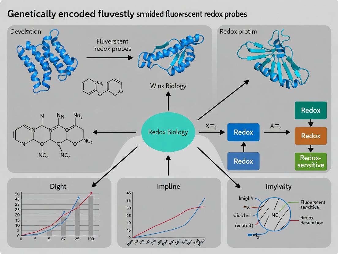
Illuminating Cellular Stress: The Development and Application of Genetically Encoded Fluorescent Redox Probes
This article provides a comprehensive overview for researchers and drug development professionals on the rapidly advancing field of genetically encoded fluorescent redox probes.
Illuminating Cellular Stress: The Development and Application of Genetically Encoded Fluorescent Redox Probes
Abstract
This article provides a comprehensive overview for researchers and drug development professionals on the rapidly advancing field of genetically encoded fluorescent redox probes. We first establish the foundational principles, detailing the biology of redox signaling and the molecular engineering of redox-sensitive fluorescent proteins (roGFPs, HyPer, rxYFPs). The methodological section explores practical applications in cell culture, organoids, and in vivo models for studying oxidative stress in diseases like cancer and neurodegeneration. We address common troubleshooting issues, including probe specificity, photostability, and calibration. Finally, we present a comparative analysis of current probe families, their validation strategies, and emerging benchmarks. The conclusion synthesizes key advancements and future clinical translation opportunities.
From Redox Biology to Biosensors: The Foundation of Genetically Encoded Fluorescent Probes
Genetically encoded fluorescent redox probes (GERPs) represent a transformative technology for real-time, compartment-specific monitoring of cellular redox states. Their development is central to a thesis focused on elucidating the spatiotemporal dynamics of redox signaling and oxidative stress in vivo. This application note provides the foundational redox biology, key quantitative metrics, and essential protocols for validating and utilizing such probes within the complex landscape of reactive oxygen/nitrogen species (ROS/RNS) and the major antioxidant systems: the glutathione (GSH/GSSG) and thioredoxin (Trx) systems.
Table 1: Key Cellular Redox Couples and Their Parameters
| Redox Couple | Typical Ratio (Reduced/Oxidized) | Approximate Potential (Eh) in Cytosol | Major Cellular Compartment |
|---|---|---|---|
| GSH/GSSG | 30:1 to 100:1 | -260 mV to -200 mV | Cytosol, Nucleus, Mitochondria |
| Trx-(SH)2/Trx-S2 | High (>>1) | ≈ -280 mV | Cytosol, Nucleus |
| NADPH/NADP+ | ~100:1 | -400 mV (via enzyme systems) | Cytosol, Mitochondria |
| NADH/NAD+ | ~0.01 (cytosol) | -320 mV (mitochondrial matrix) | Mitochondria |
| Cysteine/Cystine | Variable | -250 mV to -150 mV | Extracellular, ER |
Table 2: Common ROS/RNS and Their Sources
| Species | Full Name | Primary Generation Sites/Enzymes | Approximate Half-Life |
|---|---|---|---|
| H2O2 | Hydrogen Peroxide | NOX, ETC, SOD, Oxidases | ~1 ms |
| O2•− | Superoxide Anion | NOX, ETC, XOR | ~1 μs |
| •OH | Hydroxyl Radical | Fenton reaction (Fe2+ + H2O2) | ~1 ns |
| NO• | Nitric Oxide | NOS isoforms (eNOS, iNOS, nNOS) | ~1-10 s |
| ONOO− | Peroxynitrite | NO• + O2•− | ~10 ms |
The Scientist's Toolkit: Essential Reagents & Materials
| Item/Category | Example Specifics | Function in Redox Research |
|---|---|---|
| Redox Probes | roGFP2, Grx1-roGFP2, HyPer, rxRFP1 | Genetically encoded sensors for H2O2 or GSH/GSSG ratio. |
| Chemical Inducers | Tert-Butyl Hydroperoxide (tBHP), Menadione, Antimycin A | Induce controlled oxidative stress (mitochondrial/cytosolic). |
| Redox Modulators | N-Acetylcysteine (NAC), Buthionine Sulfoximine (BSO), Auranofin | NAC boosts GSH; BSO inhibits GSH synthesis; Auranofin inhibits TrxR. |
| Detection Kits | GSH/GSSG-Glo Assay, NADP/NADPH Assay Kit (Colorimetric) | Quantify absolute levels of redox metabolites. |
| Critical Buffers | PBS without Ca2+/Mg2+, HEPES, Lysis buffer with NEM | N-ethylmaleimide (NEM) in lysis buffer alkylates and preserves thiol redox state. |
| Imaging Setup | Confocal/Fluorescence Microscope with time-lapse capability, appropriate filter sets (e.g., 405/488 nm for roGFP). | For live-cell, ratiometric imaging of GERPs. |
Experimental Protocols
Protocol 4.1: Live-Cell Ratiometric Imaging of GSH/GSSG Redox Potential using roGFP2-Grx1
Principle: The roGFP2 (redox-sensitive GFP) is fused to human glutaredoxin-1 (Grx1), catalyzing rapid, equilibration between the sensor and the GSH/GSSG pool. Excitation at 405 nm and 488 nm yields a ratiometric signal inversely proportional to glutathione redox potential (EGSH).
Materials:
- Cells expressing roGFP2-Grx1 (cytosolic or organelle-targeted).
- Live-cell imaging medium (e.g., FluoroBrite DMEM).
- Confocal or widefield fluorescence microscope capable of rapid excitation switching.
- 𝛽-Mercaptoethanol (DTT) and Diamide for calibration.
Procedure:
- Cell Preparation: Plate cells expressing the probe in an imaging chamber 24-48h prior. Achieve 70-80% confluency.
- Image Acquisition: a. Replace medium with pre-warmed imaging medium. b. Acquire sequential images at two excitation wavelengths (Ex405/Em525 and Ex488/Em525). Use minimal exposure to avoid phototoxicity. c. Perform time-series imaging for kinetic studies before/after treatment.
- In-situ Calibration (Post-Experiment): a. Treat cells with 10 mM DTT (full reduction) for 10 min, acquire images. b. Wash and treat with 5 mM Diamide (full oxidation) for 10 min, acquire images.
- Data Analysis: a. Calculate the background-subtracted 405/488 excitation ratio (R). b. Determine the fully reduced (Rred) and oxidized (Rox) ratios. c. Calculate the degree of oxidation: Oxidation Degree = (R - Rred) / (Rox - Rred). d. Convert to EGSH using the Nernst equation: Eh = E0 - (RT/nF) ln([GSH]2/[GSSG]). For roGFP2, E0 ≈ -280 mV.
Protocol 4.2: Biochemical Quantification of Total and Oxidized Glutathione
Principle: Thiol-scavenging reagent N-ethylmaleimide (NEM) traps reduced GSH during lysis. GSSG is selectively measured after derivatization of GSH. A luminescent-based assay (GSH/GSSG-Glo) is described.
Materials:
- GSH/GSSG-Glo Assay Kit.
- Phosphate-Buffered Saline (PBS).
- Lysis buffer with/without NEM.
- White-walled multiwell plates, luminometer.
Procedure:
- Sample Preparation (CRITICAL): a. For Total GSH (GSHT): Lyse cells directly in assay-compatible lysis buffer without NEM. b. For GSSG: Lyse cells in ice-cold PBS containing 10-40 mM NEM. Vortex and incubate on ice for 30-60 min to alkylate all GSH. Proceed with kit protocol.
- Assay Execution: a. Transfer lysate to a white plate. b. Add Luciferin-NT substrate and GSH S-Transferase from kit. Incubate (typically 30-60 min). c. Add Luciferin Detection Reagent. Incubate (typically 15 min). d. Measure luminescence.
- Calculation: a. Generate standard curves for GSH and GSSG. b. Calculate GSSG concentration from the "GSSG sample" reading. c. Calculate GSHT from the "Total GSH sample" reading. d. Reduced GSH = GSHT - (2 × GSSG). e. Redox Potential (Eh) can be calculated using the Nernst equation.
Within the broader thesis on genetically encoded fluorescent redox probes, roGFPs (redox-sensitive Green Fluorescent Proteins) represent a seminal advancement. They are engineered variants of GFP where two surface-exposed cysteine residues are introduced into the β-barrel structure, forming a redox-active disulfide bridge. The core principle of signal transduction lies in the reversible formation and reduction of this disulfide bond, which directly alters the protonation state of the chromophore, thereby shifting its excitation spectrum.
In the reduced state, the chromophore is predominantly deprotonated, favoring excitation at ~488 nm. Upon oxidation, strain from the disulfide bond favors the protonated form, shifting peak excitation to ~405 nm. Emission remains constant at ~510 nm. The ratiometric measurement of emission following 405 nm and 488 nm excitation provides a quantitative, internally calibrated readout of redox potential, independent of probe concentration and instrument variability.
Application Notes
Primary Applications:
- Compartment-Specific Redox Monitoring: Targeting roGFP to organelles (mitochondria, ER, peroxisomes, nucleus) via specific targeting sequences.
- Real-Time Redox Dynamics: Live-cell imaging of redox changes during processes like growth factor signaling, metabolic shifts, and apoptosis.
- Oxidative Stress Assessment: Quantifying the production and quenching of H₂O₂ and other reactive oxygen species (ROS).
- Drug Discovery & Development: Screening for compounds that modulate cellular redox states in diseases like cancer, neurodegeneration, and metabolic disorders.
Key roGFP Variants and Their Characteristics:
Table 1: Common roGFP Variants and Their Properties
| Variant | Redox Partner (Fusion) | Redox Potential (E⁰') | Dynamic Range (Rₒₓ/Rᵣₑd) | Key Application |
|---|---|---|---|---|
| roGFP1 | N/A | ~ -288 mV | ~ 5.0 | General redox sensing; slower kinetics. |
| roGFP2 | N/A | ~ -280 mV | ~ 8.5 | High dynamic range, most widely used. |
| roGFP2-Orp1 | Yeast Orp1 (GPx-like) | N/A | N/A | Specific, rapid detection of H₂O₂. |
| roGFP2-Grx1 | Human Grx1 | ~ -280 mV | N/A | Rapid equilibration with glutathione pool (GSH/GSSG). |
| roGFP-R12 | N/A | ~ -256 mV | ~ 3.8 | Brighter, optimized for plant systems. |
Data sourced from recent literature (2021-2024). Dynamic range is ratio of 405/488 nm excitation ratio in fully oxidized vs. fully reduced state.
Quantitative Data Interpretation: The measured ratio (R = I₅₁₀ₙₘ @ Ex₄₀₅ₙₘ / I₅₁₀ₙₘ @ Ex₄₈₈ₙₘ) is normalized to the fully reduced (Rᵣₑd) and fully oxidized (Rₒₓ) states obtained experimentally using DTT and H₂O₂/aldrithiol, respectively.
Table 2: Typical Normalization and Calculation Parameters
| Parameter | Typical Treatment | Purpose | Formula |
|---|---|---|---|
| Rᵣₑd | 10 mM DTT, 5-10 min | Define minimum ratio (100% reduced) | |
| Rₒₓ | 2-10 mM H₂O₂ or 2 mM Aldrithiol-2, 5-10 min | Define maximum ratio (100% oxidized) | |
| Degree of Oxidation | Quantifies redox state | OxD = (R - Rᵣₑd) / (Rₒₓ - Rᵣₑd) | |
| Apparent Redox Potential | Relates OxD to cellular GSH/GSSG | Eₕ = E⁰' - (RT/nF) * ln([GSH]²/[GSSG]) |
Where E⁰' is the standard potential of the probe, R is gas constant, T is temperature, n=2, F is Faraday's constant.
Detailed Protocols
Protocol 1: Live-Cell Ratiometric Imaging of roGFP2
Objective: To measure the glutathione redox potential (Eₕ) in the cytosol of adherent cells.
The Scientist's Toolkit: Key Reagents & Materials Table 3: Essential Research Reagent Solutions
| Item | Function & Specification |
|---|---|
| Plasmid: pCMV-roGFP2 | Mammalian expression vector for cytosolic roGFP2. |
| Cell Line: HeLa or HEK293T | Robust, easily transfected adherent cells. |
| Transfection Reagent: PEI or Lipofectamine 3000 | For plasmid delivery. |
| Imaging Buffer: Hanks' Balanced Salt Solution (HBSS), pH 7.4 | Physiological salt solution for live imaging. |
| Reducing Agent: 10 mM Dithiothreitol (DTT) in HBSS | Fully reduces roGFP2 (defines Rᵣₑd). |
| Oxidizing Agent: 2 mM Aldrithiol-2 (AT-2) in HBSS | Fully oxidizes roGFP2 (defines Rₒₓ). |
| Calibration Agent: 100 µM - 1 mM H₂O₂ in HBSS | For challenge experiments. |
| Microscope: Confocal or widefield fluorescence microscope | Equipped with 405 nm and 488 nm lasers/LEDs and a 510/20 nm emission filter. |
| Image Analysis Software: ImageJ/FIJI with RatioPlus plugin or Python/Matlab scripts | For ratio calculation and analysis. |
Methodology:
- Transfection: Seed cells in 35 mm glass-bottom dishes. At 60-70% confluency, transfect with 1-2 µg pCMV-roGFP2 using standard protocol.
- Imaging (24-48h post-transfection):
- Wash cells 2x with pre-warmed HBSS.
- Maintain temperature at 37°C with stage-top incubator.
- Acquire Baseline: Capture sequential images using 405 nm and 488 nm excitation (same exposure times, 510 nm emission).
- Calibration: Perfuse with 10 mM DTT, incubate 10 min, image. Wash 3x with HBSS. Perfuse with 2 mM AT-2, incubate 10 min, image.
- Experimental Challenge: After re-establishing baseline, perfuse with compound of interest (e.g., 200 µM H₂O₂) and image over time.
- Data Analysis:
- Generate ratio images (405nm/488nm) pixel-by-pixel for each time point.
- Define regions of interest (ROIs) for individual cells.
- Calculate OxD for each cell using the formula in Table 2, with Rᵣₑᵈ and Rₒₓ from the calibration step.
Protocol 2: In Vitro Characterization of a Novel roGFP Variant
Objective: To determine the standard redox potential (E⁰') and dynamic range of a purified roGFP protein.
Methodology:
- Protein Purification: Express His-tagged roGFP variant in E. coli and purify via Ni-NTA affinity chromatography.
- Spectrofluorometric Titration:
- Prepare 2 µM roGFP in 100 mM potassium phosphate buffer, pH 7.0, with 1 mM EDTA.
- Set up a titration series of redox buffers with defined [GSH]/[GSSG] ratios (e.g., from 100% reduced to 100% oxidized) using total glutathione of 10 mM.
- Incubate roGFP with each buffer for 2-4 hours at room temp under argon to reach equilibrium.
- Measure fluorescence emission at 510 nm following excitation at 405 nm and 488 nm.
- Calculation:
- Plot the measured ratio (405/488) against the calculated Eₕ of each buffer solution (using Nernst equation for glutathione).
- Fit data to a Nernst equation modified for the probe:
Ratio = (Rᵣₑᵈ + Rₒₓ * 10^(n(Eₕ-E⁰')/59.1)) / (1 + 10^(n(Eₕ-E⁰')/59.1))at 25°C, n=2. - The midpoint of the sigmoidal fit is the E⁰' of the roGFP variant.
Signaling Pathway & Experimental Visualization
Title: roGFP Signal Transduction Pathway from Redox Chemistry to Light
Title: Live-Cell roGFP Imaging & Calibration Workflow
Application Notes
Historical Progression of Redox Sensing Tools
The development of redox probes has evolved through distinct technological phases, each addressing limitations of prior methods. Early synthetic dyes provided foundational insights but suffered from poor specificity, cellular toxicity, and irreversible reactions. The advent of protein-based sensors, notably using Green Fluorescent Protein (GFP), enabled genetic encoding and subcellular targeting but initially lacked dynamic range and redox specificity. Modern genetically encoded indicators leverage sophisticated design principles—circular permutation, FRET pairs, and single fluorescent protein-based ratiometric designs—to provide reversible, specific, and quantitative measurements of cellular redox states (e.g., glutathione redox potential [E_GSH], H₂O₂, NADPH/NADP⁺ ratios). Their integration into drug development pipelines allows for high-throughput screening of compounds affecting oxidative stress pathways, a key factor in neurodegenerative diseases, cancer, and metabolic disorders.
Key Design Principles and Applications
Current probes are classified by target and mechanism:
- Thiol-based Redox Potential Probes (e.g., roGFP, Grx1-roGFP2): These probes incorporate redox-active disulfide bonds into GFP, causing reversible fluorescence excitation ratio shifts (400/490 nm) upon reduction/oxidation. Grx1-roGFP2 is specifically coupled to the glutathione pool, reporting E_GSH.
- Reactive Oxygen Species (ROS) Probes (e.g., HyPer, rxYFP): HyPer uses a circularly permuted YFP (cpYFP) inserted into the microbial peroxide sensor OxyR, producing a ratiometric (420/500 nm excitation) response to H₂O₂.
- NADPH/NADP⁺ Probes (e.g., iNAP, Apollo-NADP⁺): These utilize ligand-binding domains (e.g., Rex from B. subtilis) fused to fluorescent proteins, changing FRET efficiency upon NADPH binding.
Their primary application in drug development is in phenotypic screening for antioxidant or pro-oxidant therapeutics and in validating target engagement for pathways regulating redox homeostasis.
Experimental Protocols
Protocol 1: Live-Cell Ratiometric Imaging of Glutathione Redox Potential (E_GSH) Using Grx1-roGFP2
Objective: To measure compartment-specific glutathione redox potential in cultured mammalian cells. Principle: Grx1-roGFP2 is a genetically encoded, rationetric probe whose excitation spectrum shifts reversibly upon redox changes. The glutaredoxin-1 (Grx1) domain specifically equilibrates the probe with the glutathione redox couple.
Materials & Reagents:
- Cells expressing Grx1-roGFP2 (targeted to cytosol, mitochondria, or ER).
- Imaging medium (e.g., FluoroBrite DMEM, without phenol red).
- Calibration Solutions:
- Solution A (Fully Oxidized): 10 mM H₂O₂ in imaging medium.
- Solution B (Fully Reduced): 10 mM Dithiothreitol (DTT) in imaging medium.
- Confocal or widefield fluorescence microscope with a 400-500 nm excitation tunable source or appropriate filter sets (e.g., 400/10 nm and 490/10 nm excitation, 525/40 nm emission).
Procedure:
- Cell Preparation: Seed cells on imaging-compatible dishes. Transfect or transduce with Grx1-roGFP2 construct 24-48 hours prior to imaging.
- Image Acquisition:
- Wash cells twice with pre-warmed imaging medium.
- Acquire two sequential excitation images: first at 400 nm (I₄₀₀), then at 490 nm (I₄₉₀), using a consistent emission window (~525 nm).
- Maintain constant exposure time, gain, and laser power between channels and samples.
- In-Situ Calibration (Critical for Quantitative E_GSH):
- After baseline imaging, replace medium with Solution A (H₂O₂). Incubate for 5-10 minutes. Acquire I₄₀₀ and I₄₉₀ images.
- Wash cells thoroughly with imaging medium.
- Replace medium with Solution B (DTT). Incubate for 5-10 minutes. Acquire I₄₀₀ and I₄₉₀ images.
- Data Analysis:
- Calculate the ratio R = I₄₀₀ / I₄₉₀ for each pixel/cell at each condition.
- Determine Rₒₓ (mean ratio under H₂O₂), Rᵣₑₔ (mean ratio under DTT).
- The degree of oxidation (OxD) of the probe = (R - Rᵣₑₔ) / (Rₒₓ - Rᵣₑₔ).
- Calculate EGSH using the Nernst equation: EGSH = E⁰' (Grx1-roGFP2) - (RT/nF) * ln[(1 - OxD)/OxD], where E⁰' for Grx1-roGFP2 is typically -280 mV at pH 7.0, 30°C.
- Report values as mean E_GSH ± SEM.
Protocol 2: High-Throughput Screening of Redox-Modulating Compounds Using HyPer
Objective: To screen a compound library for modulators of intracellular H₂O₂ levels. Principle: HyPer exhibits a H₂O₂-dependent increase in the 500/420 nm excitation ratio.
Materials & Reagents:
- Stable cell line expressing cytosol-targeted HyPer.
- Compound library in 384-well format.
- Cell-dispensing instrument.
- Fluorescent plate reader capable of dual-excitation ratiometric measurement (ex: 490 nm & 405 nm, em: 535 nm) or an automated imaging system.
- Positive controls: 100 µM H₂O₂ (oxidant), 10 mM DTT (reductant).
Procedure:
- Plate Preparation:
- Dispense 50 nL of each compound (or DMSO control) into black-walled, clear-bottom 384-well assay plates.
- Trypsinize and resuspend HyPer-expressing cells in growth medium. Dispense 5,000 cells in 50 µL medium per well.
- Incubate plates at 37°C, 5% CO₂ for 16-24 hours.
- Assay Execution:
- Carefully remove 30 µL of medium from each well and replace with 30 µL of fresh, pre-warmed FluoroBrite imaging medium.
- Load plate into the reader/imager. Equilibrate to 37°C for 15 min.
- Acquire a pre-read of the 490/405 nm excitation ratio (emission 535 nm).
- Optional Kinetic Mode: Acquire ratio reads every 5 minutes for 60-120 minutes post-addition of a sub-lethal stressor (e.g., 50 µM menadione) to identify compounds that alter the rate of H₂O₂ generation.
- Data Processing:
- Calculate the fold-change in fluorescence ratio for each well relative to the DMSO-treated control wells.
- Set hit thresholds (e.g., compounds causing a ratio change > 3 standard deviations from the plate mean). Confirm hits in dose-response and secondary assays.
Table 1: Characteristics of Representative Genetically Encoded Redox Probes
| Probe Name | Target | Dynamic Range (ΔR/R) | Response Time (t₁/₂) | Key Applications | Reference (Example) |
|---|---|---|---|---|---|
| roGFP1 | General thiol redox | ~5.0 | ~5 min | ER redox status | (Hanson et al., 2004) |
| Grx1-roGFP2 | Glutathione (E_GSH) | ~6.0 | <1 min | Cytosolic/mitochondrial GSH | (Gutscher et al., 2008) |
| HyPer-3 | H₂O₂ | ~8.0 (ex ratio) | ~1 min | Real-time H₂O₂ dynamics | (Bilan et al., 2013) |
| iNAP1 | NADPH/NADP⁺ | ~2.5 (FRET ratio) | Seconds | Pentose phosphate pathway flux | (Zhao et al., 2015) |
| Apollo-NADP⁺ | NADP⁺ | ~4.0 (intensity) | Seconds | Oxidative stress response | (Cameron et al., 2016) |
Table 2: Comparison of Redox Probe Generations
| Feature | Synthetic Dyes (e.g., DCFH-DA) | Protein-Based (e.g., roGFP) | 2nd-Gen GEIs (e.g., HyPer, iNAP) |
|---|---|---|---|
| Specificity | Low (multiple ROS) | Moderate (redox couples) | High (specific molecules) |
| Reversibility | Irreversible | Reversible | Reversible |
| Quantification | Semi-quantitative | Quantitative (rationetric) | Highly quantitative |
| Subcellular Targeting | Difficult | Precise (genetic encoding) | Precise (genetic encoding) |
| Toxicity/Photobleaching | High (photooxidation) | Low | Low |
| HTS Compatibility | Moderate | High | High |
Visualizations
Title: Historical Progression of Redox Probes
Title: Mechanism of a H₂O₂ Sensor
Title: Experimental Workflow for roGFP-based Assays
The Scientist's Toolkit: Research Reagent Solutions
| Item | Function & Rationale |
|---|---|
| Grx1-roGFP2 Plasmid (Addgene #64995) | The foundational genetically encoded plasmid for measuring glutathione redox potential (E_GSH). Targetable to different organelles. |
| HyPer-3 Plasmid (Addgene #42131) | A high-performance, ratiometric H₂O₂ sensor with improved dynamic range and photostability for dynamic imaging. |
| FluoroBrite DMEM | Phenol red-free, low-fluorescence imaging medium essential for reducing background in live-cell fluorescence experiments. |
| CellRox Deep Red Reagent | A synthetic, fluorogenic dye for complementary detection of general cellular oxidative stress, often used to validate GEI findings. |
| Cytation or ImageXpress Micro | Automated live-cell imaging systems enabling kinetic, multi-well plate ratiometric imaging for high-throughput screening applications. |
| N-Acetyl Cysteine (NAC) | A cell-permeable antioxidant precursor (increases glutathione), used as a standard negative control (reducing agent) in redox assays. |
| Menadione (Vitamin K3) | A redox-cycling compound that generates superoxide and H₂O₂, used as a standard positive control (oxidant stressor) in assay validation. |
The development of genetically encoded fluorescent redox probes represents a critical advancement for monitoring cellular redox dynamics in real time. A core strategy in this field involves engineering key structural motifs—specifically, disulfide bonds and conformation-sensitive elements—into fluorescent proteins (FPs). This approach transforms FPs from passive markers into active sensors of oxidation-reduction potential (Eh). The broader thesis of this research is to create a toolkit of robust, ratiometric, and target-specific fluorescent redox probes for applications in oxidative stress research, drug discovery, and metabolic disease modeling.
Application Notes: Structural Principles & Probe Design
Disulfide Bond Engineering
The introduction of a redox-active disulfide bond into the β-barrel structure of an FP (e.g., roGFP, rxYFP) creates a sensor whose fluorescence properties change upon reduction/oxidation. The key is positioning the cysteine pair to form a disulfide without destabilizing the chromophore.
Key Considerations:
- FP Scaffold Selection: GFP, YFP, and cpGFP variants are preferred for their brightness and stability.
- Cysteine Pair Geometry: The engineered cysteines must be positioned to allow reversible disulfide formation, inducing a measurable conformational shift. Typical sites are on adjacent β-strands.
- Linker Design: For fusion constructs targeting specific cellular compartments (e.g., Grx1-roGFP2), flexible linkers (e.g., (GGGGS)n) are used to tether the FP to a redox-active enzyme, facilitating rapid, specific equilibration with the target pool.
Engineering Conformational Coupling
Disulfide formation induces a subtle conformational change that is transduced to the chromophore environment. Common mechanisms include:
- Changes in pKa: Altering the protonation state of the chromophore (e.g., in roGFP).
- H-Bond Network Perturbation: Affecting chromophore stability and fluorescence efficiency.
- Direct Steric Effects: Modifying the chromophore's planarity or surrounding cavity.
Table 1: Characteristics of Representative Engineered Redox Fluorescent Proteins
| Probe Name | FP Scaffold | Redox-Active Motif | Excitation/Emission Maxima (nm) Ox/Red | Midpoint Potential (mV, pH 7.0) | Dynamic Range (ΔR/R) | Primary Application |
|---|---|---|---|---|---|---|
| roGFP1 | GFP (S65T) | Disulfide (S147C, Q204C) | 490/510 | -291 | ~5.0 (Ex Ratio) | General cytosolic redox |
| roGFP2 | GFP (S65T, S147C, Q204C) | Disulfide + Stabilizing Mutations | 400,490/510 | -280 | ~6.0 (Ex Ratio) | Improved brightness & stability |
| rxYFP | cpYFP (Venus) | Disulfide (S147C, Q204C) | 515/527 | -261 | ~2.5 (Intensity) | Glutathione redox potential |
| HyPer | cpYFP | Fusion to OxyR-RD (Regulatory Domain) | 420,500/516 | -280 (H2O2-specific) | ~5.0 (Ex Ratio) | Specific H2O2 detection |
| Grx1-roGFP2 | roGFP2 | Fusion to Human Glutaredoxin 1 | 400,490/510 | ~-233 | ~6.0 (Ex Ratio) | Glutathione redox potential (Grx1-coupled) |
Table 2: Performance Metrics in Live-Cell Imaging
| Probe | Response Time (t1/2, Oxidation) | Photostability (T1/2, s) | pH Sensitivity | Recommended Calibration Method |
|---|---|---|---|---|
| roGFP2 | ~5-10 min (DTT to H2O2) | High (>300) | Moderate (pKa~6.0) | In situ with DTT & H2O2/AT |
| rxYFP | ~1-2 min (Grx1-coupled) | Moderate (~150) | High (cpYFP scaffold) | In situ with defined GSH/GSSG buffers |
| HyPer-3 | <1 min (H2O2 addition) | Moderate (~120) | Low (optimized) | In situ with H2O2 & DTT |
| Grx1-roGFP2 | ~1-2 min | High (>300) | Moderate (pKa~6.0) | In situ with DTT & Diamide |
Experimental Protocols
Protocol:In SituCalibration of roGFP-based Probes in Adherent Cells
Purpose: To convert ratiometric fluorescence measurements into absolute redox potential (Eh) values. Materials: See "Scientist's Toolkit" (Section 6).
Procedure:
- Transfection & Plating: Seed cells in an imaging-compatible dish (e.g., µ-Slide). Transfect with the probe plasmid using standard methods (e.g., lipofection). Incubate for 24-48h.
- Imaging Setup: Use a confocal or widefield microscope with capability for sequential excitation at 405 nm and 488 nm. Collect emission between 500-540 nm. Maintain temperature at 37°C with 5% CO2.
- Baseline Measurement: Acquire images at both excitation wavelengths for 3-5 fields of view to establish the baseline ratio (R = I405/I488).
- Full Oxidation: Gently replace medium with pre-warmed imaging buffer containing 5 mM H2O2 and 50 µM aldrithiol (AT, thiol oxidizer). Incubate for 5-10 min until the ratio stabilizes. Acquire images. This defines R_ox.
- Full Reduction: Replace medium with imaging buffer containing 10 mM DTT (reducing agent). Incubate for 5-10 min until ratio stabilizes. Acquire images. This defines R_red.
- Calculation:
- Compute the degree of oxidation (OxD) for each pixel/time point: OxD = (R - Rred) / (Rox - R_red)
- Calculate the redox potential (Eh): Eh = E0 + (RT/nF) * ln(OxD / (1 - OxD))
- Where E0 is the probe's midpoint potential (e.g., -280 mV for roGFP2), R is gas constant, T is temperature, n=2, F is Faraday's constant.
- Data Analysis: Use image analysis software (e.g., ImageJ/Fiji, CellProfiler) to segment cells and calculate average Eh values per cell or compartment.
Protocol: Molecular Cloning for a New Redox FP Fusion (e.g., Target Protein-roGFP2)
Purpose: To create a genetically encoded probe targeted to a specific organelle or protein complex. Procedure:
- Vector Design: Using a mammalian expression vector (e.g., pCDNA3.1), clone the coding sequence for the target protein (e.g., a mitochondrial targeting sequence) upstream of the roGFP2 sequence.
- Linker Insertion: Insert a DNA sequence encoding a flexible peptide linker (e.g., (GGGGS)2) between the target protein and roGFP2 via PCR overlap extension or Gibson Assembly. This minimizes steric interference.
- Restriction Digestion & Ligation: Digest the vector and insert fragments with appropriate restriction enzymes (e.g., BamHI, XhoI). Purify fragments and ligate using T4 DNA Ligase.
- Transformation & Verification: Transform ligation mix into competent E. coli (DH5α). Screen colonies by colony PCR and verify the final construct by Sanger sequencing, paying special attention to the linker and fusion junction.
- Functional Validation: Transfect the construct into cells and verify correct subcellular localization via fluorescence microscopy. Perform a redox titration (as in Protocol 4.1) to confirm the probe's dynamic range remains intact.
Diagrams
Design Logic for Redox FP Probes
Live-Cell Redox Probe Calibration Workflow
The Scientist's Toolkit
Table 3: Essential Research Reagent Solutions for Redox FP Experiments
| Item | Function & Specification | Example Product/Catalog # |
|---|---|---|
| roGFP2 Plasmid | Genetically encoded sensor for general redox potential. | Addgene #64945 (pLPCX-roGFP2) |
| Grx1-roGFP2 Plasmid | Sensor equilibrated with the glutathione pool via glutaredoxin. | Addgene #64955 |
| HyPer-7 Plasmid | Ultrasensitive, specific probe for hydrogen peroxide (H2O2). | Addgene #135668 |
| Live-Cell Imaging Medium | Phenol-red free medium for fluorescence imaging, with stable pH. | Gibco FluoroBrite DMEM |
| Thiol Oxidizer (Aldrithiol-2) | Membrane-permeable oxidant (Diamide alternative) for full probe oxidation. | Sigma-Aldrich, D134803 |
| Reducing Agent (DTT) | Strong reductant to fully reduce probe disulfide bonds. | Thermo Scientific, R0861 |
| H₂O₂ Solution (Cell Culture Grade) | Physiological oxidant for calibration and stimulus experiments. | Sigma-Aldrich, H1009 |
| Cloning Kit (Gibson Assembly) | For constructing novel FP fusions with high efficiency. | NEB HiFi DNA Assembly Master Mix |
| Competent E. coli (Cloning Strain) | For plasmid propagation and storage. | NEB 5-alpha F'Iq |
| Transfection Reagent (Lipid-based) | For efficient delivery of plasmid DNA into mammalian cells. | Lipofectamine 3000 |
| Glass-Bottom Imaging Dishes | Optically clear dishes for high-resolution microscopy. | MatTek, P35G-1.5-14-C |
The development of genetically encoded fluorescent redox probes represents a cornerstone in modern cell biology and oxidative stress research. This field has evolved from simple pH indicators to sophisticated, rationetric probes that specifically monitor discrete redox couples within living cells. The probes discussed herein—roGFPs, HyPer, rxYFP, and Grx1-roGFP2—are pivotal tools that enable real-time, compartment-specific measurement of redox dynamics, moving beyond destructive, population-averaged assays. Their integration into a broader thesis on probe development highlights the iterative design philosophy: moving from general redox sensitivity (roGFP) to specific peroxide sensing (HyPer) and finally to precise glutathione redox potential reporting (Grx1-roGFP2), each generation improving specificity, kinetics, and dynamic range.
Probe Families: Mechanisms & Applications
roGFPs (Redox-sensitive Green Fluorescent Proteins)
Mechanism: roGFPs are engineered by introducing two surface-exposed cysteine residues capable of forming a reversible disulfide bond. Oxidation induces a conformational change that alters the chromophore's protonation state, leading to a decrease in excitation at ~400 nm and an increase at ~490 nm. The ratio of emissions (510 nm) from these two excitations provides a rationetric, quantitative measure of redox state. Primary Application: General cytosolic and organellar (e.g., mitochondrial, ER) thiol redox potential (Eh).
HyPer
Mechanism: HyPer is a circularly permuted YFP (cpYFP) inserted into the regulatory domain of the bacterial hydrogen peroxide-sensing protein, OxyR. H2O2 oxidizes specific cysteines in OxyR, causing a conformational change that alters cpYFP fluorescence. It is excitation-rationetric (Ex420/Ex500). Primary Application: Specific, real-time detection of hydrogen peroxide dynamics in living cells.
rxYFP (Redox-sensitive Yellow Fluorescent Protein)
Mechanism: Similar to roGFP, rxYFP contains a disulfide-forming dithiol pair. Reduction increases fluorescence intensity, while oxidation quenches it. It is typically used in non-rationetric, intensity-based mode but can be calibrated. Primary Application: Monitoring the thioredoxin pathway and general redox changes.
Grx1-roGFP2
Mechanism: This is a fusion protein where human glutaredoxin-1 (Grx1) is linked to roGFP2. Grx1 catalyzes the reversible glutathionylation of roGFP2, effectively equilibrating the probe with the glutathione (GSH/GSSG) redox couple. This enables highly specific measurement of the glutathione redox potential (EGSSG/2GSH). Primary Application: Compartment-specific reporting of the glutathione redox buffer system, the primary cellular redox buffer.
Table 1: Key Characteristics of Major Redox Probes
| Probe Family | Redox Couple Reported | Dynamic Range (Ratio Ox/Red) | Excitation (nm) / Emission (nm) | Response Time | Primary Compartment |
|---|---|---|---|---|---|
| roGFP2 | General thiol/disulfide | ~6.0 – 8.0 | 400/490 → 510 | Seconds to minutes | Cytosol, Mitochondria, ER |
| HyPer-3 | H2O2 | ~4.0 – 5.0 | 420/500 → 516 | < 1 minute | Cytosol, Nucleus, Mitochondria |
| rxYFP | Thioredoxin/General | Intensity-based | 514 → 527 | Minutes | Cytosol, Secretory Pathway |
| Grx1-roGFP2 | GSH/GSSG | ~6.0 | 400/490 → 510 | Minutes | Cytosol, Mitochondria, Nucleus |
Table 2: Typical Calibration Values in Mammalian Cells
| Probe | Approx. Eh at pH 7.2 (mV) | Fully Reduced Ratio | Fully Oxidized Ratio | Key Reference (Example) |
|---|---|---|---|---|
| roGFP2 (Cytosol) | -320 to -300 | ~0.3 – 0.4 | ~2.5 – 3.0 | Dooley et al., 2004 |
| HyPer-3 (Cytosol) | [Reports nM H2O2] | ~0.5 – 0.7 | ~2.5 – 3.5 | Bilan et al., 2013 |
| Grx1-roGFP2 (Cytosol) | -310 to -290 | ~0.4 | ~2.4 | Gutscher et al., 2008 |
Detailed Experimental Protocols
Protocol 1: Live-Cell Rationetric Imaging of roGFP2 or Grx1-roGFP2
Objective: To measure the glutathione redox potential in adherent HeLa cells. Materials: See "The Scientist's Toolkit" below. Procedure:
- Cell Preparation: Seed HeLa cells stably expressing mitochondrially-targeted Grx1-roGFP2 (mito-Grx1-roGFP2) on glass-bottom dishes. Culture for 24-48h to 70% confluency.
- Imaging Medium: Prior to imaging, replace growth medium with pre-warmed (37°C), colorless HEPES-buffered imaging medium (e.g., HBSS with 20 mM HEPES, pH 7.4).
- Microscope Setup: Use a confocal or widefield fluorescence microscope equipped with a 40x oil objective, environmental chamber (37°C, 5% CO2), and appropriate filter sets.
- Dual-Excitation Rationetric Imaging:
- Acquire two sequential images using rapid switching between two excitation wavelengths: 405 nm (protonated, oxidized state-sensitive) and 488 nm (deprotonated, reduced state-sensitive).
- Use a standard GFP emission filter (510/20 nm).
- Keep laser power and detector gain constant for all experiments.
- Calibration (In-situ):
- After baseline imaging, apply 10 mM DTT (dithiothreitol) in imaging medium to fully reduce the probe. Image every 2 minutes until ratio stabilizes (~10-15 min). This gives Rmin.
- Wash cells 3x with fresh medium.
- Apply 100 µM - 1 mM Diamide (thiol oxidant) to fully oxidize the probe. Image until stable (~10-15 min). This gives Rmax.
- (Optional) Apply 50 µM Aldrithiol (2,2'-dithiodipyridine) to validate Grx1-catalyzed equilibration.
- Data Analysis:
- Calculate the 405/488 nm excitation ratio (R) for each pixel/time point.
- Normalize the ratio: Oxidation Degree = (R - Rmin) / (Rmax - Rmin).
- Convert to redox potential (Eh) using Nernst equation: Eh = E0 - (RT/nF) ln (Red/Ox), where E0 for roGFP2 is ~ -280 mV at pH 7.2.
Protocol 2: Kinetic Measurement of H2O2Flux Using HyPer
Objective: To monitor acute hydrogen peroxide generation upon growth factor stimulation. Materials: See "The Scientist's Toolkit." Procedure:
- Cell Transfection: Transiently transfect HEK293 cells with cytosolically-targeted HyPer-3 using a suitable transfection reagent. Image 24-48 hours post-transfection.
- Imaging Setup: As in Protocol 1, but configure for HyPer: Ex 420/500 nm, Em 516 nm.
- Baseline Acquisition: Acquire ratio images (420/500 nm) every 30 seconds for 5 minutes to establish a stable baseline.
- Stimulation: At t=0, add 100 ng/mL Epidermal Growth Factor (EGF) or 100 µM Histamine directly to the imaging dish to stimulate endogenous NADPH oxidase (NOX) activity.
- Kinetic Imaging: Continue acquiring ratio images every 30 seconds for 20-30 minutes.
- Calibration: Perform in-situ calibration using 1-5 mM DTT (full reduction) and 100-500 µM H2O2 (full oxidation). Note: HyPer is pH-sensitive; control experiments with pH probes like pHluorin are recommended.
- Analysis: Plot the normalized 420/500 nm excitation ratio over time. The rate and amplitude of increase reflect H2O2 production.
Signaling Pathway & Experimental Workflow Diagrams
Diagram 1: HyPer Reports H2O2 in Cell Signaling
Diagram 2: Redox Imaging Workflow
The Scientist's Toolkit: Key Research Reagent Solutions
Table 3: Essential Materials for Redox Probe Experiments
| Item / Reagent | Function & Application | Example Vendor/Cat. No. (Representative) |
|---|---|---|
| Plasmid: pLPC-mito-Grx1-roGFP2 | Mammalian expression vector for mitochondrial glutathione redox potential sensing. | Addgene, #64985 |
| Plasmid: pHyPer-3-cytosol | Mammalian expression vector for cytosolic H2O2 sensing. | Evrogen, #FP941 |
| Lipofectamine 3000 | High-efficiency transfection reagent for plasmid delivery into mammalian cells. | Thermo Fisher, #L3000015 |
| Glass-Bottom Culture Dishes (35 mm) | High-quality optical surface for high-resolution live-cell imaging. | MatTek, #P35G-1.5-14-C |
| DTT (Dithiothreitol) | Strong reducing agent for in-situ probe calibration (Rmin). | Sigma-Aldrich, #D0632 |
| Diamide | Thiol-oxidizing agent for in-situ probe calibration (Rmax). | Sigma-Aldrich, #D3648 |
| Hydrogen Peroxide (H2O2) | Direct oxidant for HyPer calibration and oxidative challenge experiments. | Sigma-Aldrich, #H1009 |
| HEPES-Buffered Imaging Medium | Phenol-red free, CO2-independent medium for stable pH during imaging. | Thermo Fisher, #A1458801 |
| Attofluor Cell Chamber | Microscope stage-mounted chamber for controlled environment (temp, CO2). | Thermo Fisher, #A7816 |
Practical Guide: Deploying Redox Probes in Live-Cell Imaging and Disease Research
Redox signaling is a fundamental cellular process, with molecules like hydrogen peroxide (H₂O₂), glutathione (GSH), and nicotinamide adenine dinucleotide phosphate (NADPH) playing distinct yet interconnected roles. Genetically encoded fluorescent probes are indispensable tools for visualizing these species with high spatiotemporal resolution in living cells and organisms. This guide provides a structured approach for selecting and applying the optimal probe for your specific target, framed within ongoing research to develop next-generation probes with enhanced specificity and dynamic range.
The selection process begins with understanding the core design and specificity of available probes. The following table summarizes key characteristics.
Table 1: Key Characteristics of Genetically Encoded Redox Probes
| Target | Probe Name(s) | Core Sensing Domain | Excitation/Emission Peaks (nm) | Dynamic Range (Fold-Change) | Primary Specificity & Notes |
|---|---|---|---|---|---|
| H₂O₂ | HyPer, HyPer7, roGFP2-Orp1 | OxyR (E. coli), roGFP | ~420/500 & ~500/516 (rationetric) | 5-10 (HyPer7) | Highly specific for H₂O₂ over other ROS. pH-sensitive (except pH-stable variants). |
| GSH/GSSG | roGFP2-Grx1, Grx1-roGFP2, GRX1-P | roGFP fused to human glutaredoxin-1 | ~400/510 & ~480/510 (rationetric) | 5-8 | Reports the glutathione redox potential (EGSH); reversible. |
| NADPH | iNAP, Peredox, RexYFP | Rex (B. subtilis Tpx) domain fused to cpFP | ~420/480 & ~500/540 (iNAP) | ~4-5 | Reports NADPH:NADP⁺ ratio. Peredox reports free cytosolic NADH:NAD⁺ ratio. |
| General Oxidant | roGFP2, rxYFP | roGFP, rxYFP | Rationetric as above | 3-6 | Sensitive to various oxidants (H₂O₂, peroxynitrite) via dithiol/disulfide. |
Title: Decision Workflow for Selecting a Redox Probe
Detailed Experimental Protocols
Protocol 3.1: Live-Cell Rationetric Imaging of H₂O₂ with HyPer7
Objective: To measure dynamic changes in cytosolic H₂O₂ levels in response to a stimulus. Materials: See "The Scientist's Toolkit" below. Procedure:
- Cell Culture & Transfection: Seed HeLa or HEK293 cells in a glass-bottom dish. Transfect with a plasmid encoding cytosolic HyPer7 (e.g., pcDNA3-HyPer7) using a standard transfection reagent. Incubate for 24-48h.
- Microscope Setup: Use a widefield or confocal fluorescence microscope equipped with suitable filters. Configure two excitation channels: Ex1 ~400-420 nm (peak for reduced state) and Ex2 ~480-500 nm (peak for oxidized state). Use a single emission band ~510-540 nm.
- Calibration & Imaging:
- Acquire baseline images in both excitation channels in live-cell imaging medium.
- Add a bolus of fresh H₂O₂ (100-500 µM final) to fully oxidize the probe. Acquire images after signal stabilizes (~5-10 min).
- Wash cells and add a strong reducing agent (e.g., 5 mM DTT) to fully reduce the probe. Acquire final images.
- Data Analysis: For each cell and time point, calculate the ratio R = Fluorescence(Ex500)/Fluorescence(Ex420). Normalize data to the minimum (DTT, Rmin) and maximum (H₂O₂, Rmax) ratios from the calibration step. Plot normalized ratio (R - Rmin)/(Rmax - Rmin) over time.
Protocol 3.2: Measuring Glutathione Redox Potential (EGSH) with roGFP2-Grx1
Objective: To determine the steady-state glutathione redox potential in the mitochondrial matrix. Materials: See toolkit. Procedure:
- Expression: Stably express pLPC-mito-roGFP2-Grx1 in your cell line of interest. Verify mitochondrial localization via co-staining with MitoTracker.
- Live-Cell Imaging: Image cells in a physiological buffer. Acquire rationetric images as for HyPer (Ex405 and Ex488, Em510). Maintain cells at 37°C with 5% CO₂.
- In Situ Calibration (Critical): At the end of each experiment, perform a two-point calibration on the same cells:
- Add 10 mM DTT (in the presence of 5 µM rotenone and antimycin A to inhibit metabolism) for 20 min to get the fully reduced signal (Rred).
- Wash and add 100 µM aldrithiol (2,2'-dithiodipyridine) or 5 mM diamide for 20 min to get the fully oxidized signal (Rox).
- Calculation of EGSH: The degree of oxidation (OxD) = (R - Rred)/(Rox - Rred). Calculate EGSH using the Nernst equation: EGSH = E⁰ - (RT/nF)ln([GSH]²/[GSSG]). For roGFP2, E⁰' is -280 mV at pH 7.0. Use OxD to solve for [GSH]²/[GSSG] and derive EGSH, assuming a total glutathione pool ([GSH]+2[GSSG]) of ~1-10 mM (measure independently).
Title: roGFP2-Grx1 Sensing Mechanism for Glutathione Redox State
Protocol 3.3: Monitoring NADPH Dynamics with iNAP
Objective: To monitor changes in the NADPH:NADP⁺ ratio in the cytosol during metabolic perturbation. Materials: See toolkit. Procedure:
- Transfection & Preparation: Transfect cells with iNAP expression plasmid. 24h later, trypsinize and resuspend cells in imaging buffer for plate reader or flow cytometry analysis. For microscopy, use glass-bottom dishes.
- Rationetric Measurement:
- Microscopy/Plate Reader: Acquire fluorescence using Ex415/Em480 (NADPH-bound peak) and Ex500/Em540 (NADP⁺-bound peak). Calculate the emission ratio R = F540/F480.
- Flow Cytometry: Use lasers at 405 nm and 488 nm, with emission filters 450/50 and 585/42. Calculate the ratio of signals from the 585 nm channel over the 450 nm channel.
- Calibration: For approximate quantification, treat cells at the end of the experiment with 10 µM rotenone/antimycin A (to minimize NADPH, get Rmin) and 100 µM tert-butyl hydrogen peroxide (to maximize NADPH via antioxidant response, get Rmax). Normalize ratios as in Protocol 3.1.
- Interpretation: An increased normalized ratio indicates a more reduced state (higher NADPH:NADP⁺), while a decreased ratio indicates a more oxidized state.
The Scientist's Toolkit: Research Reagent Solutions
Table 2: Essential Materials for Redox Probe Experiments
| Reagent/Material | Function/Description | Example Vendor/Catalog |
|---|---|---|
| HyPer7 Plasmid | Genetically encoded, highly sensitive H₂O₂ probe. | Addgene #153492 |
| roGFP2-Grx1 Plasmid | Probe for glutathione redox potential. | Addgene #64995 (mito-targeted) |
| iNAP Plasmid | Genetically encoded indicator for NADPH:NADP⁺. | N/A (available from developer labs) |
| Glass-Bottom Dishes | High-quality imaging for live cells. | MatTek P35G-1.5-14-C |
| Fluorescence Microscope | Capable of rationetric imaging with fast filter switching. | Systems from Nikon, Zeiss, Olympus |
| H₂O₂, 30% Stock | For calibration and experimental challenge. | Sigma-Aldrich H1009 |
| Dithiothreitol (DTT) | Strong reducing agent for probe calibration. | Thermo Fisher 20291 |
| Aldrithiol-2 (2,2'-DTDP) | Thiol-oxidizing agent for GSSG calibration. | Sigma-Aldrich 143049 |
| Live-Cell Imaging Buffer | Phenol-free medium maintaining physiology. | Gibco FluoroBrite DMEM |
| Transfection Reagent | For plasmid delivery into mammalian cells. | Mirus Bio TransIT-LT1 |
| MitoTracker Deep Red | Validates mitochondrial probe localization. | Thermo Fisher M22426 |
Data Interpretation and Key Considerations
Table 3: Troubleshooting and Probe Cross-Talk
| Issue | Possible Cause | Solution |
|---|---|---|
| Low signal-to-noise ratio | Poor expression, photobleaching. | Optimize transfection; reduce exposure time; use brighter probe variant (e.g., HyPer7 over HyPer). |
| Unexpected ratio changes | pH fluctuations, spectral cross-talk. | Co-express a pH probe (e.g., SypHer) as control; verify filter sets are optimal. |
| Slow or no response | Probe saturated, incorrect localization. | Perform in-situ calibration; verify targeting sequence (e.g., correct organelle). |
| Apparent H₂O₂ signal with GSH probe | Severe oxidative stress oxidizing roGFP2 directly. | Use probes in tandem; employ specific pharmacological inhibitors (e.g., catalase for H₂O₂). |
The development of genetically encoded probes is an active field, with current research focusing on minimizing cross-talk, expanding the color palette for multiplexing, and improving brightness and photostability. The choice of probe must be validated with appropriate controls and calibration within your specific experimental system to yield quantitative, biologically meaningful insights into redox biology.
In the development and validation of genetically encoded fluorescent redox probes (e.g., roGFP2, HyPer), selecting the appropriate delivery method is paramount for introducing the DNA encoding the probe into target cells or organisms. The choice impacts expression level, uniformity, cell type specificity, and long-term stability, which are critical for accurate measurement of intracellular redox potentials (e.g., glutathione redox potential, H₂O₂ dynamics). Transfection is ideal for rapid, transient screening in cell lines. Viral transduction, particularly with lentivirus or adeno-associated virus (AAV), enables efficient and stable delivery into hard-to-transfect cells (e.g., primary neurons) and in vivo applications. Transgenic model generation creates stable, heritable lines for systemic, reproducible study of redox biology in a whole-organism context. This document provides application notes and protocols framed within a thesis on novel redox probe development.
Detailed Protocols
Transfection of Adherent Cells with Polyethylenimine (PEI) for Probe Expression Screening
Aim: Transient expression of a novel roGFP2-iLid construct in HEK293T cells for initial functionality assessment.
Materials:
- HEK293T cells
- Plasmid DNA encoding the redox probe (e.g., pMAX-roGFP2-iLid), purified, endotoxin-free
- Linear 25 kDa PEI (1 mg/mL in water, pH 7.0)
- Opti-MEM Reduced Serum Medium
- Appropriate cell culture medium and supplements
Procedure:
- Seed HEK293T cells in a 24-well plate at 1.5 x 10⁵ cells/well in complete medium. Incubate overnight to reach ~70-80% confluency.
- For each well, prepare two separate solutions in Opti-MEM:
- Solution A: Dilute 0.5 µg of plasmid DNA in 50 µL Opti-MEM.
- Solution B: Dilute 1.5 µL of PEI solution (1 mg/mL) in 50 µL Opti-MEM (3:1 PEI:DNA ratio).
- Combine Solution A and B, mix gently, and incubate at room temperature for 15-20 minutes to allow complex formation.
- Add the 100 µL DNA-PEI complex dropwise to the cell medium. Gently swirl the plate.
- Incubate cells at 37°C, 5% CO₂ for 24-48 hours.
- Replace medium 6 hours post-transfection to reduce toxicity.
- Analyze probe expression and redox responsiveness via live-cell fluorescence microscopy 24-48 hours post-transfection.
Lentiviral Transduction of Primary Cortical Neurons for Stable Probe Expression
Aim: Generate stable expression of HyPer-7 in primary mouse cortical neurons for long-term study of synaptic H₂O₂ flux.
Materials:
- Primary cortical neurons from E16-E18 mouse embryos
- Lentiviral transfer plasmid (e.g., pLV-HyPer-7-PGK), packaging plasmids (psPAX2, pMD2.G)
- HEK293FT cells for virus production
- Poly-D-lysine coated cultureware
- Neurobasal/B-27 medium
- Polybrene (hexadimethrine bromide, 4-8 µg/mL final concentration)
- Ultracentrifuge and appropriate tubes
Procedure: Part A: Lentivirus Production (HEK293FT cells)
- In a 10cm dish of 70% confluent HEK293FT cells, co-transfect with 10 µg transfer plasmid, 7.5 µg psPAX2, and 2.5 µg pMD2.G using PEI method (scale up from 2.1).
- Replace medium 6-8 hours post-transfection with fresh complete medium.
- Collect viral supernatant at 48 and 72 hours post-transfection. Pool supernatants and filter through a 0.45 µm PES filter.
- Concentrate virus by ultracentrifugation at 70,000 x g for 2 hours at 4°C. Resuscentrifuge pellet in cold Neurobasal medium (100-200 µL), aliquot, and store at -80°C. Titer determination (TU/mL) is essential.
Part B: Transduction of Primary Neurons
- Plate primary cortical neurons on poly-D-lysine coated coverslips in 24-well plates.
- At DIV 3-5, add concentrated lentivirus (MOI ~5-10) and Polybrene (4 µg/mL final) directly to the culture medium.
- After 24 hours, replace with fresh Neurobasal/B-27 medium.
- Allow 5-7 days for robust probe expression before imaging experiments.
Generation of a Transgenic Mouse Line via Pronuclear Injection
Aim: Create a germline transgenic mouse expressing a cytosolic roGFP2-Orp1 probe under the CAG ubiquitous promoter.
Materials:
- Purified, linearized transgenic construct (CAG-roGFP2-Orp1-pA).
- Fertilized mouse zygotes (C57BL/6J background)
- Microinjection apparatus
- Pseudopregnant female mice (e.g., ICR strain)
Procedure:
- Construct Preparation: Purify the transgenic fragment free of vector backbone. Dilute in microinjection buffer (Tris-EDTA) to 1-2 ng/µL.
- Microinjection: Using a micromanipulator, inject ~1-2 pL of the DNA solution into the pronucleus of each fertilized zygote.
- Embryo Transfer: Surgically transfer 20-30 viable injected zygotes into the oviduct of each pseudopregnant female mouse on day 0.5 of pseudopregnancy.
- Genotyping: At birth, take tail biopsies from potential founder (F0) pups. Screen by PCR and Southern blot for integration of the transgene.
- Founder Expansion: Breed positive F0 founders to wild-type C57BL/6J mice to establish independent lines. Characterize expression patterns and levels in F1 offspring. Select a line with stable, Mendelian inheritance and desired expression profile for redox studies.
Data Presentation & Comparison
Table 1: Comparative Analysis of DNA Delivery Methods for Redox Probe Expression
| Parameter | Chemical Transfection (PEI/Lipid) | Viral Transduction (Lentivirus) | Transgenic Model Generation (Pronuclear Injection) |
|---|---|---|---|
| Primary Use Case | Rapid, transient screening in immortalized cell lines. | Stable expression in hard-to-transfect cells (primary, neurons) & in vivo local delivery. | Creation of heritable, whole-organism models for systemic study. |
| Typical Efficiency (in susceptible cells) | 70-95% (HEK293) | >90% (with sufficient MOI) | 10-30% of pups born are transgenic founders. |
| Expression Onset | 6-24 hours | 48-72 hours (immediate post-transduction) + time for integration/expression. | From embryonic stages, constitutive in founders. |
| Expression Duration | Transient (3-7 days, episomal) | Stable (integrated into genome). | Stable & Heritable (germline integration). |
| Titer/Amount Used | 0.5-2 µg DNA/well (24-well) | Multiplicity of Infection (MOI) 5-10. | 1-2 ng/µL per zygote injection. |
| Key Advantages | Fast, inexpensive, high-throughput. | High efficiency in diverse cells, stable expression. | Reproducible, organism-level context, enables breeding studies. |
| Key Limitations | Cytotoxicity, variable efficiency, cell-type restricted, transient. | Biosafety constraints, size limit for cargo (~8 kb for lentivirus). | Technically demanding, time-intensive (months), potential insertional effects. |
| Optimal for Redox Probe Development Phase | Initial in vitro validation of probe function and dynamic range. | Advanced in vitro & acute in vivo studies (e.g., brain region-specific). | Chronic/longitudinal in vivo studies of redox signaling in development, aging, or disease. |
The Scientist's Toolkit: Research Reagent Solutions
Table 2: Essential Materials for Redox Probe Delivery Experiments
| Item | Function & Application Note |
|---|---|
| Linear PEI (25 kDa) | Cationic polymer for transient transfection; cost-effective for high-throughput screening of probe plasmids. |
| Lipofectamine 3000 | Proprietary lipid-based transfection reagent; often provides high efficiency and low toxicity in many cell lines. |
| Lentiviral Packaging Mix (2nd/3rd Gen) | Split-genome plasmids (gag/pol, rev, vsv-g) for producing replication-incompetent lentivirus safely. Essential for neuronal transduction. |
| Adeno-Associated Virus (AAV) serotype 9 | For efficient in vivo transduction with low immunogenicity. Serotype dictates tropism (e.g., AAV9 for broad CNS delivery). |
| Polybrene | Cationic polymer that enhances viral transduction efficiency by neutralizing charge repulsion between virus and cell membrane. |
| Puromycin Dihydrochloride | Selection antibiotic for stable cell line generation post-transduction/transfection when plasmid contains a puromycin resistance gene. |
| Crispr-Cas9 reagents (sgRNA, Cas9) | For targeted knock-in of redox probe sequences at specific genomic loci (e.g., safe-harbor locus), an advanced alternative to random transgenic integration. |
| In vivo-jetPEI | A specialized PEI formulation designed for safe and efficient local or systemic in vivo DNA delivery in animal models. |
Visualizations
Diagram 1: Decision Workflow for Selecting a Probe Delivery Method
Diagram 2: PEI Transfection Protocol for Probe Screening
Application Notes: Genetically Encoded Fluorescent Redox Probes
The development and application of genetically encoded fluorescent redox probes (e.g., roGFPs, HyPer, Mrx1) require precise live-cell imaging methodologies to quantify dynamic changes in cellular redox states, such as glutathione redox potential (EGSSG/2GSH) or H2O2 levels. The choice of imaging setup is critical for balancing spatial/temporal resolution, throughput, and physiological relevance. Ratiometric imaging provides robust, quantitative data independent of probe concentration and optical path length. Confocal microscopy enables high-resolution, subcellular compartment-specific measurements (e.g., mitochondrial matrix vs. cytosol). Plate reader assays facilitate high-throughput screening of redox perturbations in drug discovery.
Key Quantitative Comparison of Imaging Modalities Table 1: Comparison of Live-Cell Imaging Setups for Redox Probe Analysis
| Parameter | Widefield Ratiometric | Confocal Microscopy | Microplate Reader |
|---|---|---|---|
| Primary Use Case | Kinetics in single cells/regions | High-resolution subcellular imaging | High-throughput population averaging |
| Spatial Resolution | ~200-300 nm (lateral) | ~180-250 nm (lateral), ~500-700 nm (axial) | No spatial resolution (whole well) |
| Temporal Resolution | High (ms-s) | Moderate to High (s) | Low to Moderate (minutes) |
| Throughput | Low (few fields/experiment) | Low (few cells/field) | High (96/384/1536-well plates) |
| Key Advantage | Quantitative, minimizes artifacts | Optical sectioning, 3D localization | Statistical power, compound screening |
| Typical Probe Examples | roGFP2, Grx1-roGFP2 | mito-roGFP, HyPer-7 | Cytosolic roGFP, Orp1-roGFP |
| Excitation Scheme | Dual-ex (e.g., 405/488 nm), single-em | Sequential line scanning | Bottom-read dual excitation |
Detailed Experimental Protocols
Protocol 1: Ratiometric Imaging of roGFP2 in Adherent Cells Using a Widefield Microscope
Objective: To measure dynamic changes in cytosolic glutathione redox potential.
Materials & Reagents:
- Cells stably expressing cytosolic roGFP2.
- Imaging medium (e.g., phenol red-free HBSS with 10 mM HEPES).
- Positive controls: 2 mM DTT (reducing agent), 100 µM Diamide (oxidizing agent).
- 35 mm glass-bottom dish or µ-Slide.
- Widefield fluorescence microscope equipped with a 40x/60x oil objective, stable light source (Xenon or LED), and high-sensitivity camera (sCMOS/EMCCD). Filter sets: Ex 387/11 & 474/28, Em 525/48.
Procedure:
- Preparation: Plate cells 24-48h prior. Replace medium with imaging medium 30 min before experiment.
- Microscope Setup: Maintain environmental control at 37°C, 5% CO2. Set acquisition software for sequential dual-excitation ratiometric imaging.
- Acquisition Parameters: Use minimal exposure times to avoid phototoxicity (e.g., 50-100 ms). Acquire 405 nm and 488 nm excitation images every 30-60 seconds.
- Calibration: At experiment end, perfuse with 2 mM DTT (fully reduced state), then 100 µM Diamide (fully oxidized state). Acquire image pairs for each condition.
- Data Analysis:
- Generate ratio images (R = I405/I488).
- Calculate the degree of oxidation (OxD): OxD = (R - Rred) / (Rox - Rred), where Rred and Rox are ratios under DTT and Diamide, respectively.
- Convert OxD to redox potential using the Nernst equation: E = E0 - (RT/nF)ln((1-OxD)/OxD), where E0 for roGFP2 is ~ -280 mV.
Protocol 2: Confocal Microscopy for Mitochondrial Redox State with mito-roGFP
Objective: To assess compartment-specific redox changes with high spatial fidelity.
Materials & Reagents:
- Cells expressing mito-roGFP (targeted via COX8A or MLS signal).
- Live-cell imaging medium.
- Confocal microscope (e.g., spinning disk or point scanner) with 405 nm and 488 nm laser lines and a 60x/63x oil immersion objective (NA ≥ 1.4).
- Environmental chamber.
Procedure:
- Sample Preparation: Plate cells on high-performance cover glass. Transfer to imaging chamber.
- Microscope Configuration: Use sequential line scanning mode to eliminate crosstalk. Set pinhole to 1 Airy unit for optimal sectioning.
- Region of Interest (ROI) Definition: Draw ROIs around individual mitochondria and cytosolic regions for background subtraction.
- Time-Series Acquisition: Acquire image pairs at desired intervals. Laser power must be minimized using AOTF or AOBS controls.
- Quantification: Extract mean intensity per ROI for each channel. Calculate background-subtracted 405/488 ratio per mitochondrion over time. Calibrate with DTT/Diamide in situ if possible.
Protocol 3: High-Throughput Redox Screening Using a Plate Reader
Objective: To screen a compound library for modulators of cellular H2O2 levels using HyPer-expressing cells.
Materials & Reagents:
- Cells stably expressing cytosolic HyPer in a 96-well or 384-well black-walled, clear-bottom microplate.
- Assay buffer (HBSS with HEPES).
- Test compound library.
- Positive controls: 100 µM H2O2 (oxidant), 10 mM DTT (reductant).
- Fluorescent plate reader capable of dual-excitation ratiometric measurements (e.g., Ex 400-420 nm & 480-500 nm, Em 510-540 nm).
Procedure:
- Plate Preparation: Seed cells at optimal density (e.g., 20,000/well in 96-well plate) 24h prior. On day of assay, replace medium with 100 µL assay buffer.
- Reader Setup: Set temperature to 37°C. Configure kinetic cycle: dual-excitation read, wait, repeat every 2-5 minutes for 60-120 minutes.
- Compound Addition: After 3 baseline reads, add 25 µL of 5x compound or control using an injector or manual pipetting. Include vehicle controls.
- Data Processing:
- Calculate ratio (R = F420/F480) for each well at each time point.
- Normalize data: ΔR/R0 = (R - Rbaseline)/Rbaseline.
- Determine Z'-factor for assay quality: Z' = 1 - [3(σp + σn) / |μp - μn|], using H2O2 as positive (p) and vehicle as negative (n) control.
Visualizations
Title: Logic for Selecting Live-Cell Redox Imaging Modalities
Title: Ratiometric roGFP Redox Sensing Principle and Workflow
The Scientist's Toolkit
Table 2: Essential Research Reagent Solutions for Live-Cell Redox Imaging
| Item | Function & Rationale | Example Product/Catalog |
|---|---|---|
| Genetically Encoded Probe | Specific sensor for redox couple (e.g., H2O2, GSH/GSSG). Enables non-invasive, compartment-targeted measurement. | pLVX-roGFP2-Orp1, Addgene #64995; HyPer-7, Evrogen #FP941. |
| Phenol Red-Free Medium | Imaging medium without autofluorescent components that interfere with probe signal. | Gibco Hanks' Balanced Salt Solution (HBSS), no phenol red. |
| Validated Redox Modulators | Essential for in situ probe calibration and positive/negative controls. | DTT (reducing agent), Diamide (thiol oxidizer), H2O2. |
| Glass-Bottom Culture Vessels | Provide optimal optical clarity and high NA objective compatibility for microscopy. | MatTek dishes, Ibidi µ-Slides. |
| Environmental Control System | Maintains 37°C, 5% CO2, and humidity for physiological cell health during imaging. | Tokai Hit stage top incubator. |
| High-Sensitivity Camera | Essential for detecting low-light fluorescence with high signal-to-noise ratio, especially for ratiometric quantitation. | Hamamatsu Orca-Fusion sCMOS. |
| Dual-Excitation Filter Sets | For precise, separate excitation of probe's two redox-sensitive states. | Chroma 59022x (for roGFP: Ex 387/11, 474/28; Em 525/48). |
| Analysis Software | Enables image ratioing, background subtraction, ROI tracking, and kinetic analysis. | Fiji/ImageJ with Ratio Plus plugin, or commercial software (MetaMorph, ZEN). |
The development and application of genetically encoded fluorescent redox probes represent a cornerstone in modern redox biology. This research, central to our broader thesis, focuses on engineering probes for high-fidelity, compartment-specific measurements. The mitochondrion, a primary site of reactive oxygen species (ROS) production and a hub of redox signaling, is a critical target. Real-time mapping of its redox dynamics—specifically the glutathione redox potential (EGSSG/2GSH) and H22 flux—is essential for unraveling metabolic regulation, oxidative stress responses, and the mechanisms of redox-active therapeutics. This application note details protocols for using leading genetically encoded probes to visualize these parameters in live cells.
Quantitative Probe Comparison
The table below summarizes key performance metrics for the primary genetically encoded probes used in mitochondrial redox mapping.
Table 1: Characteristics of Key Mitochondrial-Targeted Redox Probes
| Probe Name | Target Analyte | Excitation/Emission (nm) | Dynamic Range (in vitro) | Response Time (t50) | Key Reference (Recent) |
|---|---|---|---|---|---|
| Grx1-roGFP2 | EGSSG/2GSH | 400, 490 / 510 | -320 mV to -280 mV | < 5 minutes | (Gutscher et al., 2008; Morgan et al., 2011) |
| mito-roGFP2-Orp1 | H2O2 (via Orp1) | 400, 490 / 510 | ~1-100 µM H2O2 | ~1-2 minutes | (Gutscher et al., 2009) |
| HyPer7-mito | H2O2 | 490 / 516, 527 | ~5 nM – 1 µM H2O2 | ~20 seconds | (Pak et al., 2020) |
| Mrx1-roGFP2 | EGSSG/2GSH (Mycothiol) | 400, 490 / 510 | Specific to mycothiol | Minutes | (Bhaskar et al., 2014) |
Detailed Experimental Protocols
Protocol 1: Real-Time Imaging of Mitochondrial Glutathione Redox Potential using Grx1-roGFP2
Objective: To measure the real-time dynamics of the mitochondrial matrix EGSSG/2GSH in live mammalian cells.
Materials:
- Cells expressing mito-Grx1-roGFP2 (e.g., HeLa, MEFs).
- Live-cell imaging medium (e.g., FluoroBrite DMEM, no phenol red).
- Confocal or widefield fluorescence microscope with rapid wavelength switching capability.
- 10 mM Dithiothreitol (DTT) in PBS (fully reducing control).
- 10 mM Diamide in PBS (fully oxidizing control).
- Pharmacological agents of interest (e.g., Antimycin A, Paraquat).
Procedure:
- Cell Preparation: Plate cells on glass-bottom dishes 24-48h before imaging. Transfect or transduce with a plasmid encoding mito-Grx1-roGFP2.
- Microscope Setup: Set up time-lapse imaging with sequential excitation at 405 nm and 488 nm. Collect emission between 500-540 nm. Use a 40x or 60x oil-immersion objective.
- Rationetric Calibration (In-situ):
- Acquire a baseline image series (5-10 time points).
- Perfuse with 10 mM DTT for 5 min, acquire images (fully reduced state, Rmin).
- Wash with imaging medium.
- Perfuse with 10 mM Diamide for 5 min, acquire images (fully oxidized state, Rmax).
- Experimental Measurement:
- After establishing a stable baseline, add the experimental stimulus (e.g., 1 µM Antimycin A, 100 µM Paraquat).
- Continue time-lapse acquisition for the desired duration (typically 30-60 min).
- Data Analysis:
- Calculate the 405/488 nm excitation ratio (R) for each time point and region of interest (ROI) over individual mitochondria.
- Normalize the ratio using the formula: OxD = (R – Rmin) / (Rmax – R). The degree of oxidation (OxD) ranges from 0 (fully reduced) to 1 (fully oxidized).
- Convert OxD to EGSSG/2GSH using the Nernst equation (E = E0 – (RT/nF)ln([GSH]2/[GSSG])), where E0 for roGFP2 is -280 mV.
Protocol 2: Quantifying Mitochondrial H2O2Flux using HyPer7
Objective: To detect rapid, sub-micromolar changes in mitochondrial matrix H2O2 concentration.
Materials:
- Cells expressing HyPer7-mito.
- Live-cell imaging medium.
- Microscope as above, but with capability for ratiometric emission measurement.
- 100 µM H2O2 stock in buffer (for calibration).
- 10 mM DTT (for full reduction).
- Note: HyPer7 is pH-stable; pH controls are optional.
Procedure:
- Cell Preparation: As in Protocol 1, but using HyPer7-mito expression construct.
- Microscope Setup: Use single 490 nm excitation. Acquire emission simultaneously or sequentially in two channels: 500-530 nm (Emax of reduced state) and 530-560 nm (Emax of oxidized state).
- Calibration:
- Acquire baseline.
- Perfuse with 10 mM DTT to establish the fully reduced ratio (Rred).
- Wash and perfuse with a saturating dose of H2O2 (e.g., 100 µM) to establish the fully oxidized ratio (Rox).
- Experimental Measurement:
- Stimulate cells with a metabolic modulator (e.g., 1 µM Rotenone) or a drug candidate.
- Acquire time-lapse data at high temporal resolution (e.g., every 10-30 seconds).
- Data Analysis:
- Calculate the emission ratio (500-530 nm / 530-560 nm) for each time point.
- Normalize the ratio: Normalized Ratio = (R – Rred) / (Rox – Rred).
- Convert to [H2O2] using an appropriate calibration curve derived from known H2O2 concentrations.
Visualization of Pathways and Workflows
Diagram Title: Workflow for Real-Time Redox Imaging
Diagram Title: Mitochondrial Redox Signaling & Probe Sensing
The Scientist's Toolkit: Research Reagent Solutions
Table 2: Essential Reagents for Mitochondrial Redox Imaging
| Reagent / Material | Function in Experiment | Key Consideration |
|---|---|---|
| Genetically Encoded Probe Plasmids (e.g., mito-Grx1-roGFP2, mito-HyPer7) | Engineered biosensor for specific redox couples; mitochondrial targeting ensures compartment-specific measurement. | Choose based on analyte (H2O2 vs. GSH), sensitivity, and response kinetics. |
| Live-Cell Imaging Medium (Phenol Red-Free) | Maintains cell viability during imaging while minimizing background fluorescence autofluorescence. | Must contain necessary energy sources (e.g., glucose, glutamine) and buffers (e.g., HEPES). |
| Chemical Redox Titrants (DTT & Diamide) | Used for in-situ calibration of roGFP-based probes to define Rmin and Rmax. | High purity is essential. Aliquot and store frozen. Use at defined concentrations (e.g., 10 mM). |
| Mitochondrial Modulators (e.g., Antimycin A, Rotenone, CCCP) | Pharmacological tools to perturb electron transport chain function, inducing defined redox shifts. | Titrate concentration carefully to achieve desired effect without inducing acute cell death. |
| Transfection Reagent (e.g., Lipofectamine, PEI) | For delivering plasmid DNA encoding the redox probe into mammalian cells. | Optimization of DNA:reagent ratio is critical for high expression with minimal toxicity. |
| Glass-Bottom Culture Dishes | Provides optimal optical clarity for high-resolution live-cell microscopy. | Must be sterile and compatible with the microscope stage incubator (if used). |
Application Notes: Genetically Encoded Probes in Disease Models
Genetically encoded fluorescent redox probes (e.g., roGFP, HyPer, Grx1-roGFP2) have become indispensable for real-time, subcellular resolution monitoring of oxidative stress in living systems. Their integration into disease models allows precise interrogation of redox dysregulation, a hallmark of diverse pathologies. The following application notes detail their use in three key areas.
Cancer: Tumor progression is characterized by elevated but controlled ROS, driving proliferation and survival. Probes like roGFP2-Orp1 (for H₂O₂) reveal heterogeneous redox states within tumors, often showing a more oxidized environment in invasive fronts compared to the core. This heterogeneity can predict metastatic potential and resistance to therapies.
Neurodegeneration: Models of Alzheimer's (e.g., APP/PS1 mice) and Parkinson's disease (α-synuclein overexpression) show chronic oxidative stress in neurons. Targeted expression of roGFP to the mitochondrial matrix (mito-roGFP) or cytosol quantifies glutathione redox potential (E_GSH), demonstrating progressive oxidation that precedes cell death, linking redox failure to protein aggregation.
Metabolic Disorders: In models of type 2 diabetes (e.g., db/db mice) or non-alcoholic fatty liver disease (NAFLD), probes like Grx1-roGFP2 (for glutathione redox state) uncover tissue-specific stress. Hepatocytes show a pronounced oxidized shift, correlating with insulin resistance and inflammation, while adipose tissue exhibits distinct redox dynamics during lipotoxicity.
Quantitative Data Summary:
Table 1: Redox Probe Measurements in Representative Disease Models
| Disease Model (Cell/Organelle) | Probe Used | Parameter Measured | Typical Observation (vs. Control) | Key Implication |
|---|---|---|---|---|
| Breast Cancer Cell (MCF-7, Cytosol) | roGFP2-Orp1 | H₂O₂ Dynamics | 2.5-3.5 fold increase upon EGF stimulation | ROS as signaling molecules in oncogenic pathways. |
| Alzheimer's Model Neuron (Mitochondria) | mito-roGFP | E_GSH | +15 to +20 mV shift (more oxidized) | Mitochondrial redox dysfunction precedes Aβ plaque formation. |
| db/db Mouse Liver (Hepatocyte Cytosol) | Grx1-roGFP2 | % Oxidation (GSSG/GSH) | Increase from ~10% to ~35% oxidation | Strong link between hepatic oxidative stress and systemic insulin resistance. |
| Parkinson's Model (SH-SY5Y Cytosol) | HyPer-7 | [H₂O₂] | Sustained elevation of 50-100 nM | Connects α-synuclein toxicity to peroxide accumulation. |
Experimental Protocols
Protocol 1: Lentiviral Transduction for Stable roGFP2 Expression in a 3D Tumor Spheroid Model. Objective: To generate stable cancer cell lines expressing cytosolic roGFP2 for confocal rationetric imaging of redox states in tumor spheroids. Materials: HEK293T packaging cells, target cancer cells (e.g., MDA-MB-231), lentiviral vector (e.g., pLVX-roGFP2), packaging plasmids (psPAX2, pMD2.G), polybrene (8 µg/mL), DMEM/FBS, Matrigel. Procedure:
- Virus Production: Co-transfect HEK293T cells with pLVX-roGFP2, psPAX2, and pMD2.G using a standard transfection reagent (e.g., PEI). Change medium after 6-8 hours.
- Harvesting: Collect virus-containing supernatant at 48 and 72 hours post-transfection. Filter through a 0.45 µm filter, aliquot, and store at -80°C.
- Transduction: Plate target cells at 50% confluence. Add viral supernatant supplemented with 8 µg/mL polybrene. Centrifuge at 800 x g for 30 min (spinoculation). Replace medium after 24 hours.
- Selection: Begin puromycin selection (e.g., 2 µg/mL) 48 hours post-transduction for 5-7 days to obtain a stable polyclonal population.
- Spheroid Formation & Imaging: Seed 5,000 stable cells/well in a 96-well ultra-low attachment plate. Allow spheroids to form over 72 hours. Embed in Matrigel. Image using a confocal microscope with sequential excitation at 405 nm and 488 nm; collect emission at 510 nm. Calculate the 405/488 ratio pixel-by-pixel after background subtraction.
Protocol 2: Assessing Mitochondrial Redox Stress in Primary Hippocampal Neurons from AD Model Mice. Objective: To measure the glutathione redox potential (E_GSH) in neuronal mitochondria using AAV-delivered mito-roGFP. Materials: Primary hippocampal neurons from postnatal day 0-1 wild-type and APP/PS1 pups, AAV9-mito-roGFP2, neurobasal/B27 medium, poly-D-lysine coated imaging dishes, confocal microscope, 10 mM DTT (reducing control), 100 µM Diamide (oxidizing control). Procedure:
- Neuron Culture & Transduction: Dissociate hippocampi and plate neurons at 150,000 cells/cm² on coated dishes. At DIV 3, transduce with AAV9-mito-roGFP2 at an MOI of 50,000.
- Imaging at DIV 14-21: Wash neurons with pre-warmed Hanks' Balanced Salt Solution (HBSS). Perform live-cell imaging in HBSS at 37°C.
- Rationetric Imaging: Acquire images at excitations 405 nm and 488 nm (emission 510 nm). Ensure minimal laser power to avoid phototoxicity.
- In-situ Calibration: After baseline imaging, perfuse with 10 mM DTT (full reduction), then 100 µM Diamide (full oxidation). Image after 5 min incubation for each.
- Data Analysis: Calculate the 405/488 ratio (R). Determine the degree of oxidation (OxD%) using the formula: OxD% = (R - Rred) / (Rox - Rred) * 100, where Rred and Rox are ratios under DTT and Diamide, respectively. Convert to EGSH using the Nernst equation.
Visualizations
Title: Workflow for Redox Probing in Disease Models
Title: Pro-Tumorigenic ROS Signaling Pathways in Cancer
The Scientist's Toolkit
Table 2: Essential Research Reagent Solutions for Redox Probing
| Reagent / Material | Function / Application | Key Consideration |
|---|---|---|
| roGFP2 (or variants) | Genetically encoded, rationetric probe sensitive to glutathione redox couple (E_GSH). | Requires dual-excitation imaging; can be targeted to organelles (e.g., mito-roGFP). |
| HyPer-7 | Genetically encoded, rationetric probe specifically sensitive to H₂O₂. | Highly dynamic range; pH-sensitive, requires control with SypHer. |
| AAV9-mito-roGFP | Adeno-associated virus serotype 9 for efficient neuronal transduction with mitochondrial targeting. | High transduction efficiency in neurons in vitro and in vivo; low immunogenicity. |
| Lentiviral pLVX Vectors | For stable integration and expression of redox probes in dividing cells (e.g., cancer lines). | Enables creation of stable polyclonal or monoclonal cell lines. |
| DTT (Dithiothreitol) | Strong reducing agent used for in-situ calibration of roGFP probes (defines R_min). | Must be used fresh; can affect cellular physiology at high (10 mM) concentrations. |
| Diamide | Thiol-oxidizing agent used for in-situ calibration of roGFP probes (defines R_max). | Fast-acting; can induce acute oxidative stress. |
| CellRox / MitoSOX | Chemical fluorescent dyes for general ROS or mitochondrial superoxide detection. | Useful for validation but prone to artifacts; not rationetric. |
| Polybrene | Cationic polymer used to enhance viral transduction efficiency. | Can be toxic; optimal concentration (e.g., 8 µg/mL) must be determined per cell type. |
| Matrigel | Basement membrane matrix for 3D cell culture and spheroid embedding. | Preserves tumor microenvironment and polarity for physiologically relevant imaging. |
Solving Common Challenges: Optimization, Pitfalls, and Best Practices for Redox Imaging
The development and application of genetically encoded fluorescent redox probes (GERPs), such as roGFP2, rxYFP, and HyPer, represent a cornerstone of modern redox biology research within our broader thesis on probe development. These probes enable real-time, compartment-specific monitoring of cellular redox states. However, their accurate quantitative interpretation in situ is fundamentally dependent on robust calibration protocols. This document details standardized application notes and protocols for the critical in situ calibration of GERPs using the redox agents dithiothreitol (DTT) and hydrogen peroxide (H₂O₂). This calibration is essential for converting ratiometric fluorescence readings into precise thermodynamic metrics, such as oxidation percentage or redox potential (E_h), thereby ensuring data fidelity for researchers and drug development professionals.
The Calibration Imperative in GERP Research
GERPs function by incorporating redox-sensitive cysteine pairs into fluorescent protein scaffolds, leading to reversible, oxidation-state-dependent shifts in excitation or emission spectra. The measured fluorescence ratio (e.g., 405nm/488nm for roGFP2) is a relative value. Without in situ calibration, this ratio is susceptible to artifacts from variable expression levels, pH fluctuations, photobleaching, and cell-type-specific biochemical environments. The use of DTT (a strong reductant) and H₂O₂ (an oxidant) to define the fully reduced (Rmin) and fully oxidized (Rmax) ratio limits within the actual experimental system allows for the normalization of data and calculation of the probe oxidation state, enabling direct comparison across experiments, cell types, and laboratories.
Research Reagent Solutions & Essential Materials
| Reagent/Material | Function & Rationale |
|---|---|
| Genetically Encoded Redox Probe (e.g., roGFP2-Orp1, Grx1-roGFP2, HyPer) | The sensor protein, typically targeted to specific cellular compartments (cytosol, mitochondria, ER). |
| Dithiothreitol (DTT), 100-500 mM stock | Strong reducing agent. Applied to define the R_min (fully reduced) state of the probe. Must be prepared fresh in buffer. |
| Hydrogen Peroxide (H₂O₂), 100-500 mM stock | Oxidizing agent. Applied to define the R_max (fully oxidized) state. Concentration must be verified spectrophotometrically (ε₂₄₀ = 43.6 M⁻¹cm⁻¹). |
| Diamide, 100-200 mM stock | Thiol-oxidizing agent. Alternative to H₂O₂ for some probes; useful for faster oxidation kinetics. |
| 2-Mercaptoethanol, 1M stock | Alternative reducing agent for post-experiment quenching of extracellular H₂O₂. |
| Catalase, 2000-5000 U/mL stock | Enzyme used to rapidly quench extracellular H₂O₂ after R_max determination, preventing prolonged oxidative stress. |
| Buffered Imaging Medium (e.g., HEPES-buffered HBSS, pH 7.4) | Physiologically relevant, serum-free medium for live-cell imaging, maintaining stable pH without CO₂ control. |
| Proper Imaging Chamber (e.g., Lab-Tek chambered coverslips) | For maintaining cell viability and allowing fluid exchange during live-cell calibration. |
| Confocal or Widefield Fluorescence Microscope | Equipped with appropriate excitation lasers/filters and a sensitive detector (e.g., PMT, sCMOS camera) for ratiometric imaging. |
Data compiled from recent literature (2022-2024) and empirical validation.
Table 1: Typical Calibration Values and Conditions for Selected GERPs
| Probe | Target Redox Couple | Recommended Calibration Concentrations | Approximate Dynamic Range (Rmin to Rmax) | Notes |
|---|---|---|---|---|
| roGFP2 | Glutathione (GSH/GSSG) via equilibration | 10 mM DTT, 1-5 mM H₂O₂ | Ratio change: ~5- to 8-fold (405/488) | Requires glutaredoxin (Grx) for equilibration with GSH pool. Grx1-roGFP2 fusion is standard. |
| roGFP2-Orp1 | H₂O₂ (via Orp1) | 10 mM DTT, 0.1-1 mM H₂O₂ | Ratio change: ~3- to 5-fold | Direct, specific response to H₂O₂; fast kinetics. |
| rxYFP | Thioredoxin (Trx1/2) | 5-10 mM DTT, 5-10 mM Diamide | Ratio change: ~1.5- to 2.5-fold (ex 500/420) | Calibrate with diamide; less responsive to H₂O₂. |
| HyPer (e.g., HyPer-3) | H₂O₂ | 5-10 mM DTT, 0.01-0.1 mM H₂O₂ | Ratio change: ~4- to 6-fold (488/405) | pH-sensitive; requires parallel pH control experiments. |
| mito-CP | H₂O₂ (in mitochondria) | 10 mM DTT, 0.5-2 mM H₂O₂ | Fluorescence intensity increase | Intensity-based probe; rationetric versions available. |
Detailed Experimental Protocols
Protocol 1:In SituCalibration for roGFP2-based Probes in Adherent Cells
Objective: To determine Rmin and Rmax for calculating probe oxidation percentage: %Ox = (Rmeasured - Rmin) / (Rmax - Rmin) * 100.
Materials:
- Cells expressing the roGFP2 probe (e.g., Grx1-roGFP2) in imaging chamber.
- Pre-warmed (37°C) HEPES-buffered imaging medium.
- 100 mM DTT stock in imaging medium (fresh).
- 200 mM H₂O₂ stock in imaging medium (verify concentration).
- 5,000 U/mL Catalase stock in imaging medium.
- Fluorescence microscope with 405 nm and 488 nm excitation capability.
Procedure:
- Baseline Recording: Wash cells twice with imaging medium. Acquire a time-series of ratiometric images (405ex/488ex, with 510-530 nm emission) every 30-60 seconds for 3-5 minutes to establish a stable baseline ratio (R_baseline).
- Define Rmax (Full Oxidation): a. Gently add H₂O₂ to the chamber to a final concentration of 1-2 mM (empirically determined for your cell type). b. Continue imaging until the fluorescence ratio stabilizes at a new maximum (typically 5-15 minutes). Record this as Rmax. c. Quench: Add catalase to a final concentration of ~200 U/mL to degrade extracellular H₂O₂. Image for 2-3 minutes.
- Define Rmin (Full Reduction): a. Gently add DTT to the chamber to a final concentration of 10 mM. b. Continue imaging until the fluorescence ratio stabilizes at a new minimum (typically 5-10 minutes). Record this as Rmin.
- Post-Calibration (Optional): Re-add a low dose of H₂O₂ to confirm reversibility and ratio return towards R_max.
- Data Analysis: For each cell/ROI, plot the ratio over time. Use the plateaus from steps 2b and 3b to define Rmax and Rmin. Calculate %Oxidation for the baseline and all subsequent experimental time points.
Protocol 2: Single-Point Ratiometric Calibration for High-Throughput Assays
Objective: A streamlined protocol for microplate readers, defining Rmin and Rmax in separate wells for endpoint assays.
Materials:
- Cells expressing GERP in a 96-well black-walled, clear-bottom plate.
- Imaging medium.
- 100 mM DTT, 500 mM H₂O₂, 1M 2-Mercaptoethanol stocks.
- Microplate reader capable of dual-excitation ratiometric measurements.
Procedure:
- Plate Setup: Seed cells into at least three replicate wells per condition: Baseline, Rmax, and Rmin.
- Treatment: a. Baseline wells: Replace medium with fresh imaging medium. b. Rmax wells: Replace medium with imaging medium containing a saturating H₂O₂ concentration (e.g., 5-10 mM for roGFP2). Incubate 15-20 min at 37°C. c. Rmin wells: Replace medium with imaging medium containing 10-20 mM DTT. Incubate 15-20 min at 37°C.
- Quenching: For R_max wells, carefully add 2-mercaptoethanol (final ~20 mM) to quench H₂O₂ immediately before reading to protect the plate reader optics.
- Measurement: Read the plate using the appropriate excitation/emission pairs for your probe. Acquire both excitation channels.
- Analysis: Calculate the average ratio for Rmax and Rmin wells. Use these population averages to normalize the ratios from the experimental (Baseline and treated) wells.
Visualization of Pathways and Workflows
Title: In Situ Calibration Experimental Workflow
Title: Redox Signaling & Probe Calibration Logic
Within the broader research on developing genetically encoded fluorescent redox probes, artifact avoidance is paramount for data integrity. Key challenges include probe sensitivity to non-target physiological variables like pH, irreversible photobleaching during imaging, and expression level variability leading to erroneous signal interpretation. This Application Note details protocols and considerations to mitigate these artifacts, ensuring robust measurements of cellular redox states.
Table 1: Common Artifacts in Fluorescent Redox Probe Imaging
| Artifact Type | Primary Cause | Effect on Signal | Typical Error Range |
|---|---|---|---|
| pH Sensitivity | Protonation of fluorophore at low pH | False increase (e.g., roGFP) or decrease in fluorescence ratio | ΔpH 0.5 can mimic 10-40% redox change |
| Photobleaching | Irreversible fluorophore damage under illumination | Signal loss, altered excitation/emission ratios | Up to 50% intensity loss per imaging session |
| Expression Level Variability | Non-uniform promoter activity or copy number effects | Intensity differences misattributed to redox state | CV can exceed 30% in isogenic populations |
| Maturation Inefficiency | Incomplete chromophore formation at 37°C or in anaerobic conditions | Reduced effective probe concentration | Maturation yields range from 60-95% |
| Cytosolic Sequestration | Misfolding or aggregation of probe | Reduced accessibility to target redox couples | Can cause >50% signal attenuation |
Table 2: pH Robustness of Common Redox Probes
| Probe | Redox Sensor | pKa of Fluorescence | Recommended pH Buffer Range | pH Correction Method |
|---|---|---|---|---|
| roGFP1 | Glutaredoxin | ~6.0 (for excitation peaks) | 7.0 - 8.5 | Co-imaging with pH-insensitive RFP |
| roGFP2 | Glutaredoxin | ~6.0 (for excitation peaks) | 7.0 - 8.5 | Rationetric calibration buffers |
| rxRFP1 | RoGFP-RFP hybrid | ~4.5 (RFP moiety) | 6.5 - 8.5 | Built-in RFP reference channel |
| Grx1-roGFP2 | Glutathione redox potential | ~6.0 | 7.0 - 8.0 | In situ titration with buffers |
Detailed Experimental Protocols
Protocol 1: Assessing and Correcting for pH Sensitivity
Objective: To determine the pH dependency of the redox probe and establish a correction protocol.
- Cell Preparation: Seed cells expressing the redox probe in a 96-well glass-bottom plate.
- pH Calibration Buffers: Prepare 1X PBS buffers with pH ranging from 6.0 to 8.5 in 0.5 increments, using 10 µM nigericin and 10 µM monensin to equilibrate intracellular and extracellular pH.
- Imaging: Acquire rationetric images (e.g., 405/488 nm excitation for roGFP) at each buffer pH. Include a pH-insensitive fluorescent protein (e.g., mCherry) as control if possible.
- Data Analysis: Plot the fluorescence ratio vs. pH. Use this standard curve to correct experimental redox ratios. For in vivo correction, co-express a pH-insensitive reference fluorophore and normalize signals.
Protocol 2: Quantifying and Minimizing Photobleaching
Objective: To characterize photobleaching kinetics and establish safe imaging parameters.
- Bleaching Rate Assay: Image a control sample expressing the probe at high intensity using standard acquisition settings. Take 50 consecutive images at 10-second intervals.
- Quantification: Plot fluorescence intensity (both channels for rationetric probes) vs. time. Fit to a single exponential decay to calculate the time constant (τ) for bleaching.
- Safe Imaging Setup: Adjust imaging parameters so that total light exposure during an experiment causes <10% intensity loss. Utilize:
- Minimum necessary excitation intensity.
- Fastest acceptable camera readout.
- Hardware-based neutral density filters.
- Environmental control (glucose/O₂) to minimize oxidative stress during imaging.
Protocol 3: Standardizing Expression Levels
Objective: To ensure uniform probe expression and correct for concentration-dependent artifacts.
- Clonal Selection: Generate stable cell lines via FACS sorting for low, medium, and high expression levels based on fluorescence intensity.
- Functional Titration: Treat each clonal population with 10 mM DTT (full reduction) and 1 mM H₂O₂ (full oxidation). Calculate the dynamic range (Rₘₐₓ/Rₘᵢₙ). Discard clones where dynamic range correlates with expression level.
- Normalization in Experiments: For transient transfection, always include a constitutively expressed, redox-and pH-insensitive fluorescent protein (e.g., sfGFP under a strong promoter) in a separate channel. Use its intensity to normalize the redox probe signal to expression level.
Signaling Pathways and Workflows
Diagram Title: Workflow for Redox Probe Artifact Mitigation
Diagram Title: Artifact Interference in Redox Signaling
The Scientist's Toolkit: Research Reagent Solutions
Table 3: Essential Reagents for Artifact-Free Redox Imaging
| Reagent/Material | Function/Application | Key Consideration |
|---|---|---|
| roGFP2-Orp1/GRX1-roGFP2 | Specific probe for H₂O₂ or glutathione redox potential. | Choose based on target; verify subcellular targeting sequence. |
| pH-Calibration Kit (e.g., Invitrogen) | Contains high-K⁺ buffers with ionophores for in situ pH calibration. | Essential for establishing pH-ratio standard curve. |
| Nigericin & Monensin | K⁺/H⁺ ionophores to clamp intracellular pH to extracellular buffer pH. | Use at 10 µM in calibration buffers. |
| CellMask Deep Red | Non-permeant, far-red fluorescent stain for cytosol normalization. | Masks background and corrects for cell volume/expression. |
| DTT (Dithiothreitol) | Strong reducing agent for establishing Rₘᵢₙ (fully reduced probe state). | Use at 10-50 mM for 5-10 min. |
| Diamide or H₂O₂ | Thiol oxidant for establishing Rₘₐₓ (fully oxidized probe state). | Titrate concentration (e.g., 1-5 mM diamide) to avoid cytotoxicity. |
| Tet Systems (Doxycycline-inducible) | Allows controlled, tunable expression of the redox probe. | Minimizes expression level variability and maturation issues. |
| Antifade Reagents (e.g., Oxyrase, Trolox) | Reduces photobleaching and ROS generation during live imaging. | Critical for long-term time-lapse experiments. |
| Poly-D-Lysine or Fibronectin | Enhances cell adherence for stable imaging over time. | Reduces focal plane drift, a source of intensity artifact. |
Within the development of genetically encoded fluorescent redox probes (GERPs), achieving a high signal-to-noise ratio (SNR) is paramount for accurate, sensitive detection of cellular redox states. This optimization is a multi-parametric challenge, hinging on the precise control of probe expression levels, subcellular localization, and temporal dynamics. This Application Note provides detailed protocols and analyses for systematically optimizing three critical levers: promoter strength, targeting sequences, and expression time, to maximize the SNR for GERPs such as roGFP, rxYFP, and H2O2-specific probes like HyPer.
Quantitative Analysis of Optimization Parameters
Table 1: Promoter Characteristics for Mammalian Cell Expression
| Promoter | Relative Strength | Noise (CV) | Induction Factor | Best Use Case for Redox Probes |
|---|---|---|---|---|
| CMV | 1.00 (Reference) | High (~30%) | Constitutive | General cytosol, high expression screens |
| EF1α | 0.8 - 0.9 | Low (~15%) | Constitutive | Stable, uniform expression; long-term imaging |
| CAG | 1.2 - 1.5 | Medium (~22%) | Constitutive | Very high expression in difficult-to-transfect cells |
| TRE (Tet-On) | 0.01 - 1.0* | Low with tight system | Doxycycline (10-1000x) | Tunable expression; avoiding probe toxicity |
| UBC | 0.4 - 0.6 | Very Low (~10%) | Constitutive | Low, consistent background for sensitive detection |
| SV40 | 0.5 - 0.7 | Medium (~20%) | Constitutive | Moderate expression in various cell lines |
*Inducible range depends on transactivator and response element configuration.
Table 2: Targeting Sequences and Their Impact on SNR
| Targeting Sequence | Localization | Signal Compartmentalization | Common Noise Sources | SNR Optimization Tip |
|---|---|---|---|---|
| None (Cytosolic) | Cytosol | Low | Cytosolic autofluorescence, global redox changes | Use low-strength promoter (e.g., UBC) |
| MLS (Mitochondria) | Matrix | High | Mitochondrial membrane potential dyes | Couple with COX8A or SOD2 signal sequence |
| KDEL (ER) | Endoplasmic Reticulum | High | High [Ca2+], protein misfolding | Use ER-optimized redox probe variant (e.g., roGFP1-iE) |
| NLS (Nucleus) | Nucleus | High | Nuclear stains, DNA-binding dyes | Ensure probe is inert to chromatin |
| PTS1 (Peroxisome) | Peroxisomal Matrix | Very High | Low abundance of organelle | Use bright probe (e.g., roGFP2) with strong promoter |
| Gap1 (Plasma Membrane) | Inner Leaflet | High | Membrane dyes, endocytosis | Fuse with inert transmembrane domain |
Table 3: Expression Time vs. SNR for Common Probes
| Probe | Optimal Expression Window (Post-Transfection) | SNR Peak (Hours) | Notes on Toxicity/Artifact |
|---|---|---|---|
| roGFP2-Orp1 | 24 - 48 hours | 36 hours | Overexpression >72h can buffer cellular H2O2 |
| Grx1-roGFP2 | 24 - 72 hours | 48 hours | Stable; longer expression maintains high SNR |
| HyPer-3 | 12 - 36 hours | 24 hours | Prone to pH artifacts; shorter expression recommended |
| rxRFP1 | 48 - 96 hours | 72 hours | Maturation time longer; requires later imaging |
| iNAP1 | 24 - 48 hours | 36 hours | NADP+/NADPH balance can be perturbed after 72h |
Detailed Experimental Protocols
Protocol 1: Systematic Promoter Screening for SNR
Objective: To quantitatively compare the SNR of a GERP (e.g., roGFP2-Orp1) driven by different promoters in a live-cell imaging setup.
Materials:
- HEK293T or HeLa cells
- Plasmid constructs: roGFP2-Orp1 cloned into vectors with CMV, EF1α, CAG, and TRE promoters.
- Fluorescence plate reader or confocal microscope with 405nm and 488nm excitation lasers.
- Analysis software (ImageJ/Fiji, GraphPad Prism).
Procedure:
- Seed cells in a black-walled, clear-bottom 96-well plate at 10,000 cells/well 24 hours before transfection.
- Transfect cells using a consistent lipid-based method (e.g., Lipofectamine 3000). For each promoter construct, transfer a minimum of 6 replicate wells. Include an empty vector control.
- For inducible (TRE) system: Co-transfect with a transactivator (rtTA) plasmid. Add doxycycline (e.g., 100 ng/mL) 12 hours post-transfection to induce expression.
- Image cells at 24, 36, 48, and 72 hours post-transfection.
- Acquire ratiometric images: Excite at 405nm and 488nm, collect emission at 510-540nm.
- Acquire a brightfield image for cell segmentation.
- Process data: Calculate the 405/488 emission ratio for each cell. For each well, define Signal as the mean ratio of the population. Define Noise as the standard deviation of the ratio across the population. Calculate SNR = Mean / SD.
- Plot results: Graph SNR vs. Time for each promoter construct. Identify the promoter and time point yielding the highest SNR.
Protocol 2: Validating Subcellular Targeting and Compartment-Specific Calibration
Objective: To confirm correct localization of a targeted GERP and perform an in situ calibration for accurate redox potential estimation.
Materials:
- Cells expressing targeted GERP (e.g., roGFP2-MLS for mitochondria).
- Confocal microscope with high-resolution capability.
- Calibration reagents: 10mM DTT (reducing agent), 1mM Diamide (oxidizing agent), 50µM Antimycin A (mitochondrial ROS inducer), 1µM Rotenone.
- Organelle-specific dyes (e.g., MitoTracker Deep Red for validation).
Procedure:
- Validation Imaging: 36 hours post-transfection, incubate cells with organelle-specific dye (e.g., 50nM MitoTracker Deep Red, 30 min). Acquire high-resolution z-stacks of the GERP and the marker dye. Perform colocalization analysis (calculate Pearson's or Manders' coefficients).
- In Situ Calibration: a. Establish Baseline: Acquire ratiometric (405/488) images of the expressing cells in imaging medium. b. Fully Reduce: Replace medium with medium containing 10mM DTT and 50µM Antimycin A (to inhibit mitochondrial re-oxidation). Incubate for 10-15 min and acquire images. c. Fully Oxidize: Wash cells and replace medium with medium containing 1mM Diamide and 50µM Antimycin A. Incubate for 10-15 min and acquire images. d. Optional Compartment-Specific Perturbation: To validate mitochondrial targeting, treat with 1µM Rotenone and image over 30 minutes to observe probe oxidation.
- Data Analysis: For each cell, plot the 405/488 ratio over time. Calculate the fully reduced (Rmin) and fully oxidized (Rmax) ratio values. The redox potential can be estimated using the Nernst equation. A successful targeting experiment will show a rapid and specific response to compartment-specific perturbations (e.g., Rotenone).
Protocol 3: Kinetic Profiling of Expression Time for SNR
Objective: To determine the optimal expression time window that maximizes SNR before the onset of probe toxicity or artifact.
Materials:
- Cells stably expressing the GERP under a promoter of choice (from Protocol 1 results).
- Real-time live-cell imaging system (incubated microscope).
- Oxidative stress inducer: e.g., 100µM H2O2.
Procedure:
- Seed stable cells into a 96-well imaging plate.
- Initiate time-lapse imaging starting at 12 hours post-seeding. Program the system to acquire ratiometric (405/488) images every 4 hours for 96 hours.
- At the 36-hour time point, introduce a bolus of H2O2 (final 100µM) to half of the wells. Continue imaging.
- Analysis: For each time point, calculate the mean basal ratio (Signal) and its standard deviation (Noise) for control wells to derive SNR. For H2O2-treated wells, calculate the dynamic range (ΔRatio = Max ratio post-stimulation - Basal ratio).
- Identify Optimal Window: The optimal expression time is defined as the period where: a) Basal SNR is highest, and b) The dynamic range in response to H2O2 is maximal, indicating full probe functionality without cellular buffering.
Visualization of Key Concepts
Diagram 1: Optimization Parameters for GERP SNR
Title: Three Levers for Optimizing Redox Probe Signal-to-Noise
Diagram 2: Experimental Workflow for SNR Optimization
Title: Stepwise Protocol for SNR Optimization of Redox Probes
The Scientist's Toolkit
Table 4: Essential Research Reagent Solutions
| Item | Function in GERP SNR Optimization | Example Product/Details |
|---|---|---|
| Low-Autofluorescence Medium | Minimizes background noise in live-cell imaging, crucial for ratiometric measurements. | Phenol-red free DMEM/F-12, supplemented with 10% dialyzed FBS. |
| Tight Inducible System | Allows precise control of expression level and timing to avoid probe overload. | Tet-On 3G or iCap system for minimal leak and high induction. |
| Organelle-Specific Chemical Inducers | Validates targeting and function of localized probes. | Antimycin A/Rotenone (mito), Thapsigargin (ER), AA6-017 (peroxisomes). |
| Cellular Redox Modulators (Calibration) | For in situ determination of probe dynamic range (Rmin/Rmax). | DTT (10mM, reducing), Diamide (1-2mM, oxidizing). Use with inhibitors (e.g., Antimycin A) for compartments. |
| Validated Organelle Markers | Confirms correct subcellular targeting of engineered probes. | MitoTracker Deep Red, ER-Tracker Blue-White DPX, H2B-mCherry (nucleus). |
| Lipid-Based Transfection Reagent (Low Toxicity) | Ensures high transfection efficiency without inducing acute oxidative stress. | Lipofectamine 3000 or PEI MAX, optimized for minimal cytotoxicity. |
| Ratiometric Imaging Calibration Slides | Validates microscope laser stability and detector linearity over time. | Slides with stable fluorescent dyes (e.g., TetraSpeck beads). |
| Live-Cell Imaging-Compatible Plates | Provides optimal optical clarity and cell health for long-term experiments. | Black-walled, clear-bottom µ-plates (e.g., CellCarrier-96 Ultra). |
| Analysis Software with Ratiometric Tools | Enables batch processing of dual-excitation images and population analysis. | ImageJ/Fiji with Ratio Plus plugin or commercial software (MetaMorph, Harmony). |
Within the broader thesis on developing genetically encoded fluorescent redox probes (e.g., roGFP, HyPer), a critical application is their integration into multiplexed live-cell imaging. This allows for the simultaneous monitoring of redox dynamics alongside other cellular parameters like Ca²⁺ signaling, pH, mitochondrial membrane potential, or specific protein localization. The principal challenges arise from the spectral overlap of fluorophores, the need to maintain probe functionality, and the minimization of phototoxicity during extended imaging. This application note provides updated protocols and considerations for successful multiplexing, based on current best practices.
Key Considerations for Multiplex Design
Spectral Separation: The excitation/emission spectra of all probes must be carefully analyzed. Redox probes like roGFP2 (ex~400/490 nm, em~510 nm) can conflict with many blue/green-emitting probes. Newer red-shifted redox probes (e.g., rxRFP) offer better separation. Probe Crosstalk: Ensure the physiological parameter measured by one probe (e.g., pH sensitivity of some roGFP variants) does not interfere with another. Hardware Requirements: A microscope with capable light sources (lasers or LEDs) and sensitive, spectral detection (e.g., spectral detectors or filter-based systems with narrow bandpass filters) is essential.
Current Multiplexing Strategies & Quantitative Data
The table below summarizes viable combinations for multiplexing with common genetically encoded redox probes, based on recent literature and product notes.
Table 1: Compatible Multiplexing Pairings with Genetically Encoded Redox Probes
| Redox Probe | Target Parameter | Compatible Reporter/Dye | Target Parameter | Excitation (nm) | Emission (nm) | Key Consideration |
|---|---|---|---|---|---|---|
| roGFP2-Orp1 | H₂O₂ (Golgi) | R-GECO1 | Ca²⁺ | 557 | 575 | Use 405-ex for roGFP ratiometry; 488-ex minimally excites R-GECO. |
| rxRFP1 | General Thiol Redox | GCaMP6f | Ca²⁺ | 490 | 515 | rxRFP (ex 583/em 608) offers excellent spectral separation from green probes. |
| HyPer7 | H₂O₂ | MitoTracker Deep Red | Mitochondrial Mass | 644 | 665 | HyPer7's GFP-based signal is spectrally distinct from far-red dyes. |
| Grx1-roGFP2 | Glutathione Redox | SNARF-5F | pH | 490, 540 | 640 | SNARF emits in red; use isosbestic point for pH-independent rationing. |
| roGFP2 | Glutathione Redox | tdTomato | Protein Localization | 554 | 581 | tdTomato is bright and photostable; minimal bleed-through into roGFP channel. |
| roGFP2 | Glutathione Redox | DAPI | Nucleus | 358 | 461 | Fixed-cell only. DAPI is UV-excited; no spectral conflict with roGFP imaging. |
Detailed Experimental Protocol: Simultaneous Imaging of Cytosolic H₂O₂ and Mitochondrial Ca²⁺
This protocol describes live-cell multiplexing using the red-shifted redox probe rxRFP1 and the green mitochondrial calcium probe mito-GCaMP6f.
A. Materials & Reagents
Table 2: Research Reagent Solutions Toolkit
| Item | Function/Description |
|---|---|
| HEK293T or HeLa Cells | Standard mammalian cell line models. |
| Poly-D-Lysine | Coats coverslips for improved cell adhesion. |
| Lipofectamine 3000 | Transfection reagent for plasmid delivery. |
| Plasmid: pCMV-rxRFP1 | Encodes cytosolic red redox sensor. |
| Plasmid: pCMV-mito-GCaMP6f | Encodes mitochondria-targeted green Ca²⁺ sensor. |
| FluoroBrite DMEM | Low-fluorescence imaging medium, phenol red-free. |
| H₂O₂ (e.g., 100 µM) | Positive control for redox perturbation. |
| Histamine (e.g., 100 µM) | Agonist to induce Ca²⁺ release. |
| Antimycin A (e.g., 1 µM) | Mitochondrial stressor to induce redox change. |
| Confocal Microscope | Equipped with 488 nm and 561 nm laser lines, and suitable emission filters. |
B. Protocol Steps
Cell Seeding & Transfection:
- Seed HeLa cells onto poly-D-lysine-coated 35 mm glass-bottom dishes 24 hours prior to transfection.
- At ~70% confluency, co-transfect with 1 µg of pCMV-rxRFP1 and 1 µg of pCMV-mito-GCaMP6f using Lipofectamine 3000 according to the manufacturer's protocol.
- Incubate cells for 24-48 hours to allow for sufficient probe expression.
Microscope Setup:
- Use a confocal microscope with environmental control (37°C, 5% CO₂).
- Configure sequential scanning mode to minimize crosstalk:
- Channel 1 (GCaMP6f): 488 nm laser excitation; emission collection: 500-550 nm bandpass filter.
- Channel 2 (rxRFP1): 561 nm laser excitation; emission collection: 580-620 nm bandpass filter.
- Set laser powers and detector gains using single-transfected controls to ensure zero bleed-through.
- Use a 60x oil immersion objective.
Image Acquisition:
- Replace culture medium with pre-warmed FluoroBrite DMEM.
- Locate a field of view with healthy, co-expressing cells.
- Acquire a time-series baseline (e.g., 5 minutes at 30-second intervals).
- Add Stimulant: Carefully add 100 µM histamine to the dish and mix gently. Continue acquisition for 10-15 minutes to capture Ca²⁺ transients.
- Add Redox Perturbation: Subsequently, add 1 µM Antimycin A to induce mitochondrial ROS production. Continue acquisition for an additional 20-30 minutes.
Data Analysis:
- For rxRFP1 (Redox): Calculate the ratio of fluorescence intensity with 561 nm excitation (oxidized state is more fluorescent). Normalize to the baseline ratio (R/R₀).
- For mito-GCaMP6f (Ca²⁺): Calculate fluorescence intensity (F) in the green channel. Normalize to baseline (F/F₀).
- Plot normalized ratios/intensities over time to correlate mitochondrial Ca²⁺ fluxes with subsequent cytosolic redox changes.
Signaling Pathway & Experimental Workflow Diagrams
Diagram Title: Multiplexing Experimental Workflow for Redox and Calcium Imaging
Diagram Title: Signaling Cascade Linking Calcium to Redox Balance
Application Notes
Within the ongoing development of genetically encoded fluorescent redox probes (GERPs), three primary failure modes dominate: poor cellular expression, dim fluorescent signal, and unresponsiveness to redox stimuli. These issues are critical bottlenecks in research aimed at quantifying compartment-specific redox dynamics for drug discovery and fundamental biology. The following notes contextualize these problems within the probe development pipeline.
Poor Expression: Often rooted in codon bias, improper subcellular targeting sequences, or protein misfolding. Low expression levels preclude accurate measurement, especially in primary cells or sensitive models. This directly impacts the thesis aim of generating robust, universally expressible probes.
Dim Signal: A dim probe compromises signal-to-noise ratio and temporal resolution. Causes include inefficient chromophore maturation (particularly for GFP-derived sensors), suboptimal fluorescence resonance energy transfer (FRET) efficiency in rationetric probes, or quenching due to the local environment. This challenges the core thesis requirement for high-fidelity, real-time redox monitoring.
Unresponsive Probes: The most critical failure, where fluorescence remains static despite redox changes. This indicates a flawed design of the redox-sensing domain (e.g., rxYFP, roGFP, HaloTag-based), where cysteine disulfide bridges are improperly positioned, have incorrect redox potentials, or are kinetically sluggish. This negates the fundamental purpose of the probe within the broader research.
Protocols for Diagnosis and Validation
Protocol 1: Quantitative Assessment of Probe Expression and Localization
Objective: To quantify cellular expression levels and verify correct subcellular targeting. Materials: Cells transfected with GERP construct, fixation reagents, primary antibody against probe tag (e.g., anti-GFP), fluorescent secondary antibody, DAPI, confocal microscope, flow cytometer. Method:
- Fixation: 48h post-transfection, fix cells with 4% paraformaldehyde for 15 min.
- Immunostaining: Permeabilize with 0.1% Triton X-100, block with 5% BSA, incubate with primary antibody (1:1000) overnight at 4°C, then with secondary antibody (1:500) for 1h at RT. Counterstain with DAPI.
- Imaging & Analysis: Acquire confocal images using identical settings for all samples. Quantify co-localization with organelle markers (e.g., MitoTracker) using Pearson's correlation coefficient. Perform flow cytometry on live transfected cells to measure expression distribution (GFP channel).
- Troubleshooting: If expression is poor, consider codon optimization or using a stronger/inducible promoter. If localization is incorrect, re-validate targeting sequence.
Protocol 2: In Vitro Characterization of Fluorescent Properties and Redox Response
Objective: To measure key photophysical properties and the dynamic range of the purified probe. Materials: Purified recombinant GERP protein, spectrophotometer, fluorometer, redox buffers (DTT for reduction, H₂O₂ or diamide for oxidation). Method:
- Spectroscopy: Record absorbance spectrum (350-550 nm) and fluorescence emission spectrum (excitation at relevant peaks, e.g., 400nm and 480nm for roGFP).
- Quantum Yield (QY) Determination: Calculate relative QY using a standard (e.g., fluorescein in 0.1 M NaOH, QY=0.92). Compare integrated fluorescence intensity.
- Redox Titration: Incubate purified probe (1 µM) in a series of buffered redox potentials (e.g., from -300 mV to -150 mV) using defined glutathione redox couples (GSH:GSSG). Measure fluorescence intensities at excitation peaks. Fit data to Nernst equation to determine midpoint potential (E₀).
- Kinetics: Rapidly mix oxidized probe with excess DTT and record fluorescence change over time to assess responsiveness.
- Troubleshooting: Dim signal may require optimization of chromophore maturation during protein expression. Unresponsive probes with aberrant E₀ require re-engineering of the sensing domain.
Table 1: Key Photophysical and Redox Properties for Common GERPs
| Probe Name | Excitation Peaks (nm) | Emission Peak (nm) | Midpoint Potential (E₀, mV) | Dynamic Range (Rmax/Rmin) |
|---|---|---|---|---|
| roGFP2 | 400, 490 | 510 | -280 | ~6-8 |
| rxYFP | 490, 514 | 527 | -261 | ~2.5 |
| Grx1-roGFP2 | 400, 490 | 510 | -233 (for Grx1 couple) | ~6-8 |
| HyPer | 420, 500 | 516 | N/A (H₂O₂-specific) | ~5-6 (ratio) |
Protocol 3: In Cellulo Calibration and Responsiveness Validation
Objective: To verify probe functionality in the live cellular environment. Materials: Live cells expressing GERP, live-cell imaging setup, calibration reagents (DTT, H₂O₂, diamide, aldrithiol), ionophores (e.g., CCCP for mitochondrial probes). Method:
- Live-Cell Imaging: Plate cells on glass-bottom dishes. Image using dual-excitation ratio imaging (e.g., 405/488 nm ex for roGFP).
- Maximum/Minimum Ratio Determination: Perfuse cells with 10 mM DTT (for full reduction) followed by 1-5 mM H₂O₂ or diamide (for full oxidation). Record fluorescence at both channels. Calculate R = F(ex1)/F(ex2). Determine Rmax (oxidized) and Rmin (reduced).
- Specificity Test: Challenge probe with compartment-specific stimuli (e.g., mitochondrial uncoupler CCCP for matrix-targeted probe).
- Troubleshooting: If the dynamic range in cells is low, check for improper maturation or probe buffering by endogenous systems. Use compartment-specific positive controls.
Table 2: Common Calibrants and Treatments for In Cellulo Validation
| Treatment | Concentration | Purpose | Target Compartment | Incubation Time |
|---|---|---|---|---|
| Dithiothreitol (DTT) | 10 mM | Full chemical reduction of probe | Global | 5-10 min |
| Hydrogen Peroxide (H₂O₂) | 1-5 mM | Full chemical oxidation of probe | Global | 5-10 min |
| Aldrithiol-2 (AT-2) | 200 µM | Thiol-specific oxidant | Cytosol/Nucleus | 10-15 min |
| Carbonyl cyanide m-chlorophenyl hydrazone (CCCP) | 10-50 µM | Depolarizes mitochondria, induces oxidation | Mitochondria | 5-10 min |
| Butyl Hydroperoxide (tBHP) | 100-200 µM | Membrane-permeable organic peroxide | Multiple | 10-20 min |
Visualizations
Diagram Title: GERP Failure Mode Troubleshooting Flowchart
Diagram Title: In Vitro Redox Titration Protocol Steps
The Scientist's Toolkit: Key Research Reagent Solutions
Table 3: Essential Materials for GERP Development and Troubleshooting
| Item | Function/Benefit | Example/Note |
|---|---|---|
| Codon-Optimized Gene Synthesis | Ensures high expression in target host organisms (e.g., mammalian, yeast). Critical for solving poor expression. | Services from IDT, GenScript, Twist Bioscience. |
| Inducible/Strong Promoter Vectors | Allows control over expression levels to balance signal and toxicity. | Tetracycline-inducible (Tet-On), CMV, EF1α promoters. |
| Organelle-Specific Targeting Sequences | Directs probe to precise subcellular compartments for localized measurements. | MLS (Mitochondria), KDEL (ER), NLS (Nucleus). |
| Defined Glutathione Redox Buffers | Essential for in vitro determination of probe midpoint potential (E₀). | Prepared from precise ratios of GSH and GSSG with glutathione reductase. |
| Membrane-Permeable Redox Modulators | For in cellulo calibration and challenge experiments. | DTT (reducer), H₂O₂ (oxidizer), AT-2 (thiol oxidizer). |
| Live-Cell Imaging-Optimized Media | Maintains cell health during prolonged imaging without autofluorescence. | Phenol-red free media with stable glutamine. |
| Dual-Wavelength Rationetric Imaging Setup | Enables quantitative measurement independent of probe concentration. | Microscope with fast wavelength switching (e.g., Lambda DG-4). |
| FRET-Accepting Chromophores (For FRET Probes) | Partner fluorophore for constructing rationetric redox probes. | cpYFP, mCherry, T-Sapphire. |
Benchmarking Performance: A Comparative Analysis of Modern Redox Probes and Validation Strategies
This application note, framed within ongoing research on the development of genetically encoded fluorescent redox probes, provides a comparative analysis of three leading indicators: roGFP2 (for glutathione redox potential, EGSSG/2GSH), HyPer7 (for H2O2), and rxYFP (for the glutathione redox state). Understanding their distinct dynamic ranges, reaction kinetics, and molecular specificities is critical for selecting the optimal probe for specific experimental designs in redox biology and drug discovery.
Table 1: Core Performance Characteristics of roGFP2, HyPer7, and rxYFP
| Parameter | roGFP2 | HyPer7 | rxYFP |
|---|---|---|---|
| Redox Target | Glutathione redox potential (EGSSG/2GSH) | Hydrogen peroxide (H2O2) | Glutathione redox state (Grx1-catalyzed equilibrium) |
| Dynamic Range (ΔR/Rmax) | ~6-10 (ratioometric) | ~12-15 (ratioometric) | ~5-7 (rationetric) |
| Excitation Peaks (nm) | 400 nm (reduced) / 490 nm (oxidized) | 420 nm (reduced) / 500 nm (oxidized) | 420 nm (reduced) / 500 nm (oxidized) |
| Emission Peak (nm) | ~510-525 | ~516 | ~525 |
| Apparent Midpoint Potential (E0') | -280 to -270 mV (pH 7.0) | N/A (H2O2 sensor) | -261 mV (when fused to human Grx1) |
| Response Time (t1/2) | ~Seconds (via glutaredoxin) | ~<30 seconds (oxidation); ~5-10 min (reduction) | ~Minutes (Grx1-dependent equilibrium) |
| pH Sensitivity | Moderate; requires control with pH probes | High; requires control with SypHer or pHRed | Moderate |
| Primary Application | Compartment-specific EGSH measurement | Real-time, specific H2O2 dynamics | Glutathione redox state in cytosol/nucleus |
Table 2: Recommended Experimental Conditions & Considerations
| Consideration | roGFP2 | HyPer7 | rxYFP |
|---|---|---|---|
| Optimal Expression System | Mammalian cells, yeast, plants (with organelle targeting) | Mammalian cells, in vivo models | Mammalian cells, bacteria |
| Critical Control Experiment | Co-expression with redox-insensitive reference (e.g., roGFP2-C170S) | Co-expression with pH sensor (e.g., SypHer) | Expression of Grx1-fusion construct is essential |
| Key Interferent | Thiol-reactive agents, pH shifts | pH changes, other peroxides (low specificity vs. H2O2) | pH shifts, alterations in Grx1 expression/activity |
| Calibration Method | In situ with DTT (reduce) and Diamide/AT (oxidize) | In situ with bolus H2O2 (oxidize) and DTT (reduce) | In situ with DTT and Diamide |
Experimental Protocols
Protocol 1: General Live-Cell Rationetric Imaging for roGFP2, HyPer7, and rxYFP
This protocol outlines the standard setup for live-cell imaging of all three ratio-based probes.
Materials: See "The Scientist's Toolkit" below. Procedure:
- Cell Culture & Transfection: Seed cells (e.g., HeLa, HEK293) on high-quality glass-bottom dishes. At 50-70% confluency, transfect with the plasmid encoding the probe (e.g., roGFP2-Orp1, HyPer7-cyto, rxYFP-Grx1) using a suitable transfection reagent. Incubate for 24-48 hours.
- Microscope Setup: Use a widefield epifluorescence, confocal, or spinning disk microscope equipped with a stable temperature (37°C) and CO2 (5%) incubation system. For rationetric imaging, configure excitation light sources/filters: 400/10 nm and 490/10 nm for roGFP2; 420/20 nm and 500/20 nm for HyPer7/rxYFP. Use a single emission filter (e.g., 525/30 nm). Use a 40x or 60x oil-immersion objective.
- Image Acquisition: Select cells expressing moderate probe levels. Acquire image pairs (Ex400/Ex490 or Ex420/Ex500) sequentially with minimal delay (<1 sec). Use minimal exposure times to avoid phototoxicity and probe photobleaching. Acquire time series at intervals appropriate for the experiment (e.g., every 30 seconds).
- Image Analysis: For each time point, create a ratio image (Reduced-ex/ Oxidized-ex) using image analysis software (e.g., ImageJ/FIJI, MetaMorph). Define a region of interest (ROI) for individual cells. Plot the mean ratio within each ROI over time. Normalize ratios to initial baseline (R/R0) or calibrate to full scale (see Protocol 2).
Protocol 2:In SituCalibration for roGFP2 and rxYFP
Determines the fully reduced (Rmin) and oxidized (Rmax) ratio values for quantitative potential estimation.
Procedure:
- Perform live-cell imaging as in Protocol 1 to establish a baseline ratio.
- Full Oxidation: Replace medium with imaging buffer containing 5-10 mM Diamide or 2-5 mM Aldrithiol-2 (AT-2). Acquire ratio images every minute until the ratio stabilizes (~5-10 min). This gives Rmax.
- Wash: Gently wash cells 2-3 times with fresh imaging buffer.
- Full Reduction: Replace medium with imaging buffer containing 10-20 mM Dithiothreitol (DTT). Acquire ratio images until stabilization (~5-10 min). This gives Rmin.
- Calculation: The degree of oxidation (OxD) is calculated as: OxD = (R - Rmin) / (Rmax - Rmin). The glutathione potential (EGSH) can be estimated using the Nernst equation: EGSH = E0 - (RT/2F)*ln((1-OxD)/OxD), where E0 is the probe's midpoint potential.
Protocol 3: H2O2Titration and Specificity Check for HyPer7
Characterizes the dynamic range and confirms H2O2 specificity of HyPer7.
Procedure:
- Perform baseline imaging of HyPer7-expressing cells in HEPES-buffered saline.
- H2O2 Response: Add a low, physiologically relevant concentration of H2O2 (e.g., 5-20 µM) directly to the dish while imaging. Monitor the rapid increase in the Ex500/Ex420 ratio.
- Titration: In separate cell samples, apply increasing concentrations of H2O2 (1-200 µM) and record the plateau ratio value for each dose to generate a calibration curve.
- Specificity Control (Critical): Repeat the experiment in cells co-expressing HyPer7 and a pH sensor (e.g., SypHer). Apply the same H2O2 bolus. The true HyPer7 signal is the ratio change not mirrored by the pH sensor, as H2O2 addition can sometimes alter cytosolic pH.
Visualizations
Title: Redox Probe Signaling Pathways & Specificity
Title: Experimental Workflow for Redox Probes
The Scientist's Toolkit: Essential Research Reagents & Materials
| Item | Function & Application |
|---|---|
| Plasmids (Addgene): pLAS2.roGFP2-Orp1, pHyPer7-cyto, pEYFP-rxYFP-Grx1 | Source of genetically encoded probes for transfection. |
| Glass-bottom Dishes (e.g., µ-Dish 35mm) | Optimal for high-resolution live-cell microscopy. |
| Transfection Reagent (e.g., PEI, Lipofectamine 3000) | For delivering plasmid DNA into mammalian cells. |
| HEPES-buffered Imaging Saline (e.g., HBSS + 10mM HEPES) | Maintains pH during imaging without CO2 control. |
| Dithiothreitol (DTT), 1M Stock | Strong reducing agent for in situ calibration (Rmin). |
| Diamide (Azodicarboxylic acid bis(Dimethylamide)), 100mM Stock | Thiol-oxidizing agent for in situ calibration (Rmax). |
| Hydrogen Peroxide (H2O2), 30% Stock | Primary stimulus for HyPer7; dilute freshly for use. |
| pH Control Probe (e.g., SypHer plasmid) | Essential control for HyPer7 experiments to dissect pH artifacts. |
| Microscope with Rationetric Capability | System with fast switching excitation and sensitive camera (EM-CCD/sCMOS). |
| Image Analysis Software (FIJI/ImageJ + RatioPlus plugin) | For processing time-series and calculating ratio images. |
Thesis Context: This document details application notes and protocols for the validation of genetically encoded fluorescent redox probes, which are essential tools for dynamic, compartment-specific measurement of cellular redox states in living cells and organisms.
Application Notes & Protocols
Correlative Biochemistry for Probe Calibration
Purpose: To establish a quantitative relationship between the fluorescent signal (e.g., emission ratio) of the redox probe and the defined biochemical redox potential (Eh) of the cellular compartment.
Protocol: In Vitro Calibration of roGFP2-Orp1 (Peroxiredoxin-Based H₂O₂ Sensor)
- Protein Purification: Express and purify the recombinant probe protein (e.g., roGFP2-Orp1) using a His-tag system.
- Buffer Preparation: Prepare calibration buffers (pH 7.2) with defined redox potentials using mixtures of reduced (DTTred) and oxidized (DTTox) dithiothreitol. Use the Nernst equation and a standard potential (E°') of -330 mV for DTT at pH 7.0 to calculate required ratios for a range of -320 mV to -200 mV.
- Spectrofluorometry: Incubate 2 µg of purified probe in 2 mL of each calibration buffer for 1 hour at room temperature, protected from light.
- Measurement: Record excitation spectra (400-490 nm) while monitoring emission at 510 nm. Alternatively, measure fluorescence intensities at two excitation wavelengths (e.g., 400 nm and 488 nm).
- Data Analysis: Calculate the fluorescence emission ratio (R = F₄₈₈/F₄₀₀). Plot R against the calculated Eh. Fit the data to the Nernst equation: Eh = E°' - (59.1/n)*log([Red]/[Ox]) at 25°C, where n=2 for the dithiol/disulfide couple. The oxidation degree = (R - Rred)/(Rox - R_red).
Table 1: Example In Vitro Calibration Data for roGFP2
| Redox Potential (Eh, mV) | DTTox:DTTred Ratio | Excitation Ratio (488/400 nm) | Oxidation Degree |
|---|---|---|---|
| -320 | 0.01:1 | 0.15 ± 0.02 | 0.05 |
| -300 | 0.04:1 | 0.35 ± 0.03 | 0.25 |
| -280 | 0.17:1 | 0.65 ± 0.04 | 0.55 |
| -260 | 0.70:1 | 0.90 ± 0.03 | 0.80 |
| -240 | 2.93:1 | 1.05 ± 0.02 | 0.95 |
CRISPR-Cas9 Knockout Validation of Probe Specificity
Purpose: To confirm the biological specificity and minimal off-target interactions of the probe by eliminating putative sensing or competing pathways.
Protocol: Validating H₂O₂ Probe Specificity Using CRISPR-Knockout of Peroxiredoxin 2 (Prdx2)
- Design: Design sgRNAs targeting early exons of the human PRDX2 gene. Include a non-targeting control sgRNA.
- Transfection: Co-transfect HEK293T cells expressing the roGFP2-Orp1 probe with a plasmid expressing Cas9 and the PRDX2-targeting sgRNA (or control) using a lipid-based method.
- Selection & Cloning: Apply appropriate antibiotic selection (e.g., puromycin) for 48 hours. Subsequently, single cells are sorted by FACS into 96-well plates to generate monoclonal lines.
- Validation of Knockout:
- Genomic DNA PCR: Amplify the target region and sequence to confirm indels.
- Western Blot: Use anti-Prdx2 antibodies to confirm protein ablation.
- Functional Assay: Treat control and Prdx2-KO cells with bolus H₂O₂ (10-100 µM) or physiological stimuli (e.g., EGF, 100 ng/mL). Image the roGFP2-Orp1 ratiometric signal over time using live-cell microscopy. In Prdx2-KO cells, the amplitude and kinetics of the probe response should be significantly altered, confirming Prdx2 as the primary in vivo transducer of H₂O₂ to the probe.
Pharmacological Challenge for Dynamic Validation
Purpose: To test probe functionality and specificity in living cells under controlled oxidative or reductive challenges.
Protocol: Pharmacological Perturbation of the Glutathione Redox Couple for Grx1-roGFP2
- Cell Culture & Imaging: Seed cells expressing the glutathione redox potential probe Grx1-roGFP2 in an imaging chamber. Acquire baseline ratiometric images (Ex 405/488 nm, Em 510 nm).
- Oxidative Challenge: Add dithiodipyridine (DTDP, 100 µM), a thiol-oxidizing agent that directly depletes glutathione. Monitor the ratio increase for 15-30 minutes.
- Reductive Challenge: Wash cells and apply N-acetylcysteine (NAC, 5 mM), a precursor of glutathione synthesis, or the specific glutathione reductase inhibitor carmustine (BCNU, 100 µM). Monitor the ratio decrease or increase, respectively.
- Specificity Control: Pre-treat cells with buthionine sulfoximine (BSO, 1 mM, 24 hours), an inhibitor of glutathione synthesis. This should blunt or abolish the probe's response to DTDP and NAC, confirming its specificity for the glutathione pool.
Table 2: Expected Probe Responses to Pharmacological Challenges
| Probe | Challenge Agent (Concentration) | Expected Ratiometric Response | Mechanistic Interpretation |
|---|---|---|---|
| Grx1-roGFP2 | DTDP (100 µM) | Ratio Increase | Glutathione oxidation/depletion |
| Grx1-roGFP2 | NAC (5 mM) | Ratio Decrease | Enhanced reductive capacity via glutathione synthesis |
| Grx1-roGFP2 | BCNU (100 µM) | Ratio Increase | Inhibition of glutathione reduction |
| roGFP2-Orp1 | H₂O₂ (50 µM) | Rapid Ratio Increase | Direct Prdx-mediated probe oxidation |
| roGFP2-Orp1 | DTT (1 mM) | Ratio Decrease | Direct chemical reduction of the probe |
The Scientist's Toolkit: Research Reagent Solutions
Table 3: Essential Materials for Redox Probe Validation
| Item | Function & Application |
|---|---|
| roGFP2 (or rxYFP) Plasmid | Genetically encoded basis for rationetric sensing; contains engineered dithiol/disulfide pair. |
| Targeting Fusion Constructs (e.g., Orp1, Grx1) | Confers molecular specificity to H₂O₂ or glutathione redox potential (Eh). |
| Dithiothreitol (DTT) (Reduced & Oxidized) | Defined redox buffer system for in vitro probe calibration. |
| Lipofectamine 3000 / JetPEI | Transfection reagents for delivering probe plasmids and CRISPR components. |
| LentiCRISPRv2 or similar plasmid | All-in-one vector for stable expression of Cas9 and sgRNA. |
| Anti-Prdx2 / Anti-Glutathione Antibodies | Validation of knockout models via Western blot. |
| Dithiodipyridine (DTDP) | Cell-permeable thiol-specific oxidant for challenging glutathione pools. |
| Buthionine Sulfoximine (BSO) | Irreversible inhibitor of γ-glutamylcysteine synthetase, depletes cellular glutathione. |
| N-Acetylcysteine (NAC) | Cell-permeable cysteine pro-drug, boosts glutathione synthesis. |
| Carmustine (BCNU) | Glutathione reductase inhibitor, shifts glutathione pool to oxidized state. |
| Fluorescence Plate Reader / Live-Cell Microscope | Equipped with appropriate filters (Ex 400/490, Em 510 nm) for rationetric imaging. |
Diagrams
Title: H₂O₂ Sensing Pathway & CRISPR Validation
Title: Redox Probe Validation Workflow
Title: Logic of Pharmacological Challenges
Application Notes
This application note details the performance characteristics and implementation strategies of genetically encoded fluorescent redox probes targeted to specific subcellular compartments. Their development is central to a broader thesis on elucidating compartmentalized redox signaling and stress in health and disease, a critical frontier for drug discovery.
Cytosolic Probes (e.g., roGFP2, rxRFP1): Serve as the baseline for cellular redox state. They are sensitive to rapid, global changes but lack information on organelle-specific events. Ideal for initial screening of redox perturbations by xenobiotics.
Mitochondrial-Targeted Probes (e.g., mito-roGFP2, Grx1-roGFP2): Crucial for assessing the redox environment of the powerhouse of the cell, a major source of reactive oxygen species (ROS) and a key target in metabolic diseases, neurodegeneration, and cancer. Probes fused to mitochondrial targeting sequences (e.g., COX VIII) provide resolution of matrix glutathione redox potential (EGSSG/2GSH).
Endoplasmic Reticulum-Targeted Probes (e.g., eroGFP): The ER maintains an oxidative folding environment. Probes here, often fused to calreticulin or KDEL signals, monitor disulfide bond formation status and ER stress, a pathway heavily implicated in protein misfolding diseases and metabolic disorders.
Nuclear-Targeted Probes (e.g., nuc-roGFP): Enable investigation of redox-regulated transcription and DNA damage repair. Targeting via an NLS (nuclear localization signal) allows assessment of how nuclear processes are influenced by compartment-specific redox shifts.
Table 1: Key Performance Characteristics of Representative Compartment-Specific Redox Probes
| Probe Name | Target Compartment | Redox Sensor | Key Readout | Dynamic Range (Oxidation Ratio) | Responsiveness (t1/2) | Primary Utility |
|---|---|---|---|---|---|---|
| roGFP2 | Cytosol | roGFP2 | EGSSG/2GSH | ~6-8 (in vitro) | <1 min | Global cellular redox state |
| mito-roGFP2-Grx1 | Mitochondrial Matrix | roGFP2 fused to Grx1 | EGSSG/2GSH | ~5-7 (in vivo) | ~1-2 min | Mitochondrial glutathione redox potential |
| eroGFP | ER Lumen | roGFP1 | Thiol-disulfide equilibrium | ~4-5 | Seconds (to DTT) | ER oxidative protein folding capacity |
| nuc-roGFP2 | Nucleus | roGFP2 | EGSSG/2GSH | Similar to cytosolic | <1 min | Nuclear glutathione redox state |
| rxRFP1 | Cytosol | rxRFP1 | Glutathione redox state | ~3.5 | ~0.1 sec | Rapid, ratiometric glutathione sensing |
Table 2: Recommended Excitation/Emission for Ratiometric roGFP-Based Probes
| Probe | Reduced State Peak Ex (nm) | Oxidized State Peak Ex (nm) | Emission (nm) | Standard Dichroic/Filter Set |
|---|---|---|---|---|
| roGFP2, mito-roGFP2, nuc-roGFP2 | 400 | 490 | 510 | 400/490/510 nm (ratiometric) |
| eroGFP | 400 | 490 | 510 | 400/490/510 nm (ratiometric) |
Experimental Protocols
Protocol 1: Transient Transfection and Live-Cell Ratiometric Imaging of Compartment-Specific Redox Probes
Objective: To measure compartment-specific glutathione redox potential (EGSSG/2GSH) in live cells using ratiometric roGFP2-based probes.
I. The Scientist's Toolkit: Key Reagents & Materials
| Item | Function/Description |
|---|---|
| HEK293T or HeLa Cells | Common mammalian cell lines with good transfection efficiency. |
| Plasmid DNA | pCMV-mito-roGFP2-Grx1, pCMV-eroGFP, pCMV-roGFP2, pCMV-nuc-roGFP2. |
| Transfection Reagent | Polyethylenimine (PEI) or Lipofectamine 3000 for plasmid delivery. |
| Phenol Red-Free Imaging Medium | Minimizes background fluorescence for sensitive live-cell imaging. |
| Dithiothreitol (DTT, 10mM) | Strong reducing agent for probe calibration (full reduction). |
| Diamide (10mM) | Thiol-oxidizing agent for probe calibration (full oxidation). |
| Confocal or Widefield Fluorescence Microscope | Equipped with stable light source and capable of rapid excitation wavelength switching (405 nm and 488 nm lasers/filters). |
| Matlab or ImageJ (FIJI) with RatioPlus Plugin | Software for ratiometric image calculation and analysis. |
II. Procedure:
- Cell Seeding & Transfection: Seed cells onto poly-D-lysine coated 35mm glass-bottom dishes 24h prior. At 70-80% confluence, transfert with 1-2 µg of the desired probe plasmid using the manufacturer's protocol. Incubate for 24-48h to allow expression.
- Microscope Setup: Equilibrate the microscope environmental chamber to 37°C and 5% CO2. Configure acquisition software for sequential dual-excitation imaging: Ex 405 nm / Em 510 nm and Ex 488 nm / Em 510 nm. Use minimal exposure times to avoid phototoxicity.
- Image Acquisition: Replace culture medium with pre-warmed phenol red-free imaging medium. Locate cells expressing the probe at moderate levels. Acquire a baseline time-series (e.g., 1 image pair every 30 sec for 5 min).
- In-Situ Calibration (Critical):
- After baseline, gently add DTT to the dish for a final concentration of 5-10 mM. Acquire images until the 405/488 ratio stabilizes at its minimum (fully reduced state, Rmin).
- Wash cells 2x with imaging medium.
- Add diamide for a final concentration of 1-2 mM. Acquire images until the ratio stabilizes at its maximum (fully oxidized state, Rmax).
- Data Analysis:
- For each cell and time point, calculate the background-subtracted fluorescence intensity (I405 and I488) from the region of interest (ROI).
- Compute the ratiometric value R = I405 / I488.
- Normalize the ratio: Normalized Oxidation = (R - Rmin) / (Rmax - Rmin). This yields a value between 0 (fully reduced) and 1 (fully oxidized).
- For EGSSG/2GSH calculation, use the Nernst equation with the probe's standard potential (E0 for roGFP2 is ~ -280 mV).
Protocol 2: Validating Subcellular Targeting via Co-Localization
Objective: To confirm correct probe localization using organelle-specific dyes.
Procedure:
- Transfert cells with the probe (e.g., mito-roGFP2) as in Protocol 1.
- Staining:
- Mitochondria: Incubate with 100-200 nM MitoTracker Deep Red (Ex/Em ~644/665 nm) for 30 min. Wash.
- ER: Incubate with 1 µM ER-Tracker Red (Ex/Em ~587/615 nm) for 30 min. Wash.
- Nucleus: Stain with 1 µg/mL Hoechst 33342 (Ex/Em ~350/461 nm) for 10 min.
- Image Acquisition: Acquire multi-channel images: probe channel (510 nm emission) and the respective organelle dye channel.
- Analysis: Calculate the Manders' Overlap Coefficient (MOC) or Pearson's Correlation Coefficient (PCC) between the probe and marker channels using FIJI's Coloc 2 plugin. A coefficient >0.8 indicates strong co-localization.
Visualizations
Title: Development & Application Workflow for Targeted Redox Probes
Title: Ratiometric Imaging & Data Processing Pipeline
Title: Compartmentalized Redox Signaling in Drug Response
The development of genetically encoded fluorescent redox probes is a cornerstone of modern redox biology, enabling real-time, compartment-specific monitoring of cellular redox states. The latest frontier in this field is the creation of Near-Infrared (NIR) redox sensors and their ultrasensitive variants. These probes address critical limitations of earlier GFP-based sensors, such as phototoxicity, autofluorescence, and poor tissue penetration.
NIR redox sensors primarily exploit the unique properties of bacterial phytochrome-derived proteins or engineered infrared fluorescent proteins (IFPs). They are typically paired with redox-sensitive domains (e.g., roGFP, rxYFP) or utilize direct redox-coupled chromophore states. The shift to the NIR window (650-900 nm) allows for deeper tissue imaging, reduced scattering, and multiplexing with visible-light probes. Ultrasensitive variants are achieved through strategies like circular permutation, fine-tuning of redox potential, and incorporation of multiple sensing domains, achieving unprecedented dynamic ranges and specificity for key redox couples like NAD+/NADH, NADP+/NADPH, glutathione (GSH/GSSG), and H₂O₂.
Key Applications:
- In Vivo Tumor Metabolism Imaging: Tracking redox heterogeneity in deep tissues in animal models.
- Neurodegenerative Disease Research: Monitoring oxidative stress in neuronal models and brain slices.
- Drug Discovery: High-throughput screening for compounds that modulate cellular redox state in complex 3D models.
- Metabolic Flux Analysis: Simultaneous monitoring of redox couples and other metabolic parameters via multiplexing.
Table 1: Comparison of Representative Next-Gen NIR Redox Probes
| Probe Name | Redox Target | Excitation/Emission Max (nm) | Dynamic Range (ΔR/R) | Response Time (t₁/₂) | Key Advantage | Reference (Example) |
|---|---|---|---|---|---|---|
| iNap Sensors (e.g., iNap1) | NAD+ / NADH | 588 / 609 | ~8.0 (in vitro) | < 1 min | High specificity for free NADH; Ratiometric. | Zhao et al., Cell Metab, 2015 |
| Frex / SoNar variants | NAD(H) / NADP(H) | 420 / 510 (Fret) | ~10-20 | Seconds | Ultrasensitive to NADH/NAD+ ratio. | Zhao et al., Cell Metab, 2016 |
| NIR-Grx1-roGFP2 | GSH / GSSG (via Grx1) | 400, 490 / 510 | ~6.0 (rationetric) | Minutes | NIR-excitable, deep-tissue GSH sensing. | MCE et al., 2023 Catalog |
| Cys-SH sensors (cpIFP) | Protein Thiol Oxidation | 690 / 713 | ~1.5-2.0 | Seconds to Minutes | Direct, reversible monitoring of cysteine oxidation. | Yu et al., Nat Methods, 2014 |
| Apollo-NADP+ | NADP+ / NADPH | 400, 500 / 515 | ~4.0 (rationetric) | < 2 min | Specific for NADPH over NADH. | Bilan et al., Antioxid Redox Signal, 2018 |
| Hyper sensors (e.g., HyPer7) | H₂O₂ | 490 / 520 (oxidized) | ~8.0 (rationetric) | < 10 sec | Ultrasensitive, fast H₂O₂ detection. | Pak et al., Nat Commun, 2020 |
Detailed Experimental Protocols
Protocol 1: Live-Cell Ratiometric Imaging of Glutathione Redox Potential Using NIR-Grx1-roGFP2
Objective: To quantify the GSH/GSSG redox potential in the cytosol of adherent cells.
Materials: See "The Scientist's Toolkit" below.
Procedure:
- Cell Seeding & Transfection: Seed HeLa cells in a glass-bottom 35 mm dish. At 60-70% confluence, transfert with the plasmid encoding NIR-Grx1-roGFP2 using a suitable transfection reagent (e.g., PEI, Lipofectamine 3000). Incubate for 24-48 hours.
- Preparation of Imaging Buffer: Prepare a HEPES-buffered saline solution (HBSS) containing 20 mM HEPES (pH 7.4), 120 mM NaCl, 5 mM KCl, 1 mM CaCl₂, 1 mM MgCl₂, and 10 mM glucose.
- Microscope Setup: Use a confocal or widefield fluorescence microscope equipped with stable 405 nm and 488 nm laser lines (for roGFP excitation) and a sensitive NIR-capable PMT or camera. Set emission collection between 500-550 nm.
- Calibration Imaging (In-situ):
- Replace culture media with imaging buffer.
- Acquire a baseline ratiometric image (I₄₈₈ / I₄₀₅).
- Treat cells with 10 mM Dithiothreitol (DTT) in buffer for 10 min to fully reduce the probe. Acquire the "Rmin" image.
- Wash cells twice with fresh buffer.
- Treat cells with 10 mM Diamide (oxidizing agent) for 10 min to fully oxidize the probe. Acquire the "Rmax" image.
- Experimental Imaging: For experimental samples, acquire dual-excitation images under treatment conditions (e.g., H₂O₂, drugs). Maintain identical acquisition settings throughout.
- Data Analysis:
- Calculate the normalized redox index (OxD) for each pixel/cell: OxD = (R - Rmin) / (Rmax - Rmin).
- Convert OxD to redox potential (E) using the Nernst equation: E = E₀ - (RT/nF) * ln([GSH]²/[GSSG]), where E₀ is the standard potential for the probe (determined from calibration).
Protocol 2: Quantifying NADH/NAD+ Dynamics in 3D Spheroids Using iNap Sensors
Objective: To image metabolic heterogeneity in a tumor spheroid model.
Procedure:
- Spheroid Generation: Use the hanging drop or ultra-low attachment plate method to form spheroids from cells stably expressing iNap1 (NADH sensor).
- Imaging Setup: Use a two-photon or light-sheet microscope equipped with a tunable Ti:Sapphire laser (set to 750-800 nm for efficient iNap excitation) or a dedicated 561 nm laser line. Collect emission at 580-620 nm.
- Ratiometric Calibration: As iNap is a single-excitation sensor, perform a in-situ calibration using metabolic modulators:
- Max Oxidation (Fmin): Treat spheroids with 10 µM Carbonyl cyanide 4-(trifluoromethoxy)phenylhydrazone (FCCP, uncoupler) for 30 min.
- Max Reduction (Fmax): Treat spheroids with 50 mM 2-Deoxy-D-glucose and 5 µM Rotenone/Antimycin A for 60 min.
- Normalize fluorescence intensity (F) as: Normalized F = (F - Fmin) / (Fmax - Fmin).
- Time-Lapse Experiment: Embed the spheroid in low-melt agarose and perfuse with buffer. Acquire time-lapse images before and after treatment with a drug (e.g., 100 nM Oligomycin). Monitor changes in normalized fluorescence intensity.
- Analysis: Segment the spheroid into core, intermediate, and periphery regions. Plot the kinetics and steady-state NADH levels in each region to assess metabolic zonation.
Visualization Diagrams
Diagram Title: NIR Redox Probe Signaling Logic
Diagram Title: Live-Cell Redox Imaging Workflow
The Scientist's Toolkit: Research Reagent Solutions
Table 2: Essential Materials for NIR Redox Probe Experiments
| Item | Function & Rationale | Example Product/Source |
|---|---|---|
| NIR Redox Probe Plasmids | Genetically encoded DNA constructs for expression in cells. Crucial for targeting specific cellular compartments. | Addgene (e.g., #73369 for iNap1, #134964 for Hyper7), MCE HY-P8478 (NIR-Grx1-roGFP2). |
| Low-Autofluorescence Imaging Medium | Provides nutrients and pH stability during live imaging without interfering with NIR signals. | Gibco FluoroBrite DMEM, Leibovitz's L-15 Medium. |
| Redox Modulators for Calibration | Chemicals to define the minimum (Rmin/Fmin) and maximum (Rmax/Fmax) probe response in situ. | Dithiothreitol (DTT, reducing agent), Diamide (oxidizing agent). |
| Metabolic Inhibitors/Modulators | Tools to perturb specific pathways and validate probe specificity (e.g., for NADH sensors). | Rotenone (Complex I inhibitor), Oligomycin (ATP synthase inhibitor), FCCP (mitochondrial uncoupler). |
| Transfection Reagent (for Adherent Cells) | For plasmid delivery in hard-to-transfect or primary cells. | Lipofectamine 3000, JetPRIME, FuGENE HD. |
| Lentiviral Packaging System | For creating stable, long-term expressing cell lines, especially in spheroids or in vivo. | psPAX2 & pMD2.G plasmids, Lenti-X Concentrator. |
| Glass-Bottom Culture Dishes | Optically clear substrate for high-resolution microscopy. | MatTek dishes, Cellvis dishes. |
| Ultra-Low Attachment (ULA) Plates | For reliable formation of 3D spheroids or organoids. | Corning Spheroid Microplates. |
| Recombinant Glutaredoxin (Grx1) | Required for equilibration-based probes (e.g., roGFP-Grx1) to specifically report on GSH/GSSG. | Sigma-Aldrich, recombinant human Grx1. |
| NIR-Optimized Mounting Medium | For preserving fluorescence in fixed samples imaged in the NIR range. | ProLong Diamond Antifade Mountant. |
Within the development of genetically encoded fluorescent redox probes, selecting a "gold standard" is not a one-size-fits-all process. The optimal probe is defined by its congruence with the specific biological question, model system, and experimental parameters. This guide provides application notes and protocols for systematic probe selection and rigorous validation, framed within redox biology research.
Comparative Probe Characteristics
Selection begins with understanding the key photophysical and biochemical parameters of available probes. The table below summarizes core metrics for several commonly used and next-generation redox probes.
Table 1: Comparative Analysis of Genetically Encoded Redox Probes
| Probe Name | Redox Target (Sensing Domain) | Excitation/Emission (nm) | Dynamic Range (ΔF/F) | Response Time (t1/2) | Key Interferences/Notes |
|---|---|---|---|---|---|
| roGFP1 | Glutathione redox potential (hGrx1) | 400, 490 / 510 | ~5.0 (in vitro) | Seconds to minutes | pH stable (ratiometric); requires glutaredoxin expression. |
| roGFP2 | Glutathione redox potential (hGrx1) | 400, 490 / 510 | ~6.5 (in vitro) | Seconds to minutes | Improved brightness & dynamic range vs. roGFP1. |
| roGFP1-Orp1 | H₂O₂ (Orp1 yeast peroxidase) | 400, 490 / 510 | ~4.0 | ~1-2 minutes | Specific for H₂O₂; ratiometric. |
| HyPer | H₂O₂ (OxyR bacterial protein) | 420, 500 / 516 | ~5.0-8.0 | <1 minute | pH-sensitive; dual-excitation ratiometric. |
| HyPer7 | H₂O₂ (OxyR) | 490 / 516 | ~10.0 | <20 seconds | Improved brightness, reduced pH sensitivity vs. HyPer. |
| Grx1-roGFP2 | Glutathione redox potential (Fused hGrx1) | 400, 490 / 510 | ~6.0 | Seconds | Direct fusion simplifies expression & coupling. |
| rxRFP1 | Glutathione redox potential | 580 / 610 | ~3.0 | Minutes | Ratiometric, red-shifted; useful for multiplexing. |
Decision Framework & Experimental Validation Protocol
The following workflow outlines the logical process for probe selection and the key validation experiments required.
Title: Probe Selection and Validation Workflow
Detailed Validation Protocols
Protocol 1: In Vitro Characterization of Purified Probe Protein
Objective: Determine the intrinsic photophysical and redox properties of the probe independent of cellular context. Materials:
- Purified probe protein (e.g., roGFP2, HyPer7).
- Spectrofluorometer with thermostatic control.
- Calibration buffers: Defined redox buffers (e.g., 10 mM DTT, 10 mM H₂O₂, or GSH/GSSG mixes) in appropriate physiological buffer (e.g., PBS, pH 7.4). Procedure:
- Dilute purified probe to a low μM concentration in a non-reducing buffer.
- For rationetric probes (roGFP, HyPer): Acquire full excitation spectra (e.g., 350-500 nm) while monitoring emission at 510-520 nm. Alternatively, record fluorescence intensities at the two specified excitation peaks (e.g., 400 nm and 490 nm).
- Add aliquots of redox buffer to achieve desired potential. Incubate until signal stabilizes (5-15 min).
- Plot the ratiometric value (e.g., F400/F490) against the log of the redox buffer potential or oxidant concentration. Fit the data with a Nernst or sigmoidal equation to determine the dynamic range and midpoint potential (E₀).
- For kinetics, rapidly mix probe with oxidizing/reducing agent in a stopped-flow or rapid-mixing attachment and monitor fluorescence change over time.
Protocol 2: In Cellulo Calibration and Dynamic Range Assessment
Objective: Measure the practical operating range and responsiveness of the probe expressed in your target cells. Materials:
- Stable or transiently transfected cell line.
- Live-cell imaging setup with appropriate filters.
- Permeabilizing agents (e.g., digitonin) or specific pharmacological agents: DTT (strong reductant), H₂O₂ (oxidant), Diamide (thiol oxidizer), Aldrithiol (specific oxidant). Procedure:
- Seed cells expressing the probe in an imaging chamber.
- Acquire baseline ratiometric images (e.g., roGFP: Ex400/Em510 and Ex490/Em510).
- For glutathione probes (roGFP): Treat cells sequentially with:
- 10 mM DTT (in imaging buffer) to fully reduce the probe. Incubate 5-10 min, image.
- Wash, then treat with 2-5 mM Diamide or 1-5 mM H₂O₂ to fully oxidize the probe. Incubate, image.
- Calculate the cellular dynamic range as (Roxidized - Rreduced) / R_reduced.
- For H₂O₂ probes (HyPer, roGFP-Orp1): Treat cells with a titration of H₂O₂ (1-1000 μM). Establish a dose-response curve and determine the half-maximal effective concentration (EC₅₀) in your cellular system.
Protocol 3: Specificity and Crosstalk Validation
Objective: Confirm the probe responds specifically to its intended analyte and rule out major artifacts. Materials:
- Probe-expressing cells.
- Specific agonists/antagonists: e.g., PEG-Catalase (H₂O₂ scavenger), Thioredoxin Reductase inhibitor (Auranofin), Glutathione synthesis inhibitor (BSO).
- pH-sensitive probe (e.g., pHluorin) as control. Procedure:
- pH Sensitivity Test: Co-express or co-load a pH indicator. Induce cytosolic pH changes (e.g., using NH₄Cl pulse). Monitor if the redox probe signal changes correlatively with pH. True redox probes should be pH-insensitive (e.g., roGFP2) or have characterized pH profiles.
- Scavenger/Inhibitor Controls: Pre-treat cells with PEG-Catalase (500-1000 U/mL) for 30 min before applying H₂O₂. A specific H₂O₂ probe should show attenuated or abolished response.
- Pathway Specificity: For glutathione probes, deplete cellular glutathione with BSO (100 μM, 24h). The probe's response to certain oxidative stimuli may be altered, confirming coupling to the glutathione pool.
The Scientist's Toolkit: Research Reagent Solutions
Table 2: Essential Reagents for Redox Probe Validation
| Reagent | Category | Function in Validation |
|---|---|---|
| Dithiothreitol (DTT) | Strong reductant | Fully reduces disulfide bonds in probes; defines minimum ratio (R_min) in calibration. |
| Diamide | Thiol-specific oxidant | Chemically oxidizes glutathione and probe thiols; defines maximum ratio (R_max) for glutathione probes. |
| Hydrogen Peroxide (H₂O₂) | Physiological oxidant | Primary analyte for H₂O₂ probes; used for dose-response and dynamic range testing. |
| PEG-Catalase | H₂O₂ scavenger | Validates H₂O₂ specificity by quenching extracellular and intracellular H₂O₂. |
| L-Buthionine-sulfoximine (BSO) | GSH synthesis inhibitor | Depletes cellular glutathione pool; tests coupling of roGFP probes to glutathione redox system. |
| Auranofin | Thioredoxin Reductase Inhibitor | Perturbs the Thioredoxin system; tests probe specificity against complementary redox pathways. |
| Digitonin | Permeabilizing agent | Gently permeabilizes plasma membrane for controlled access of calibration buffers to cytosolic probes. |
| N-Ethylmaleimide (NEM) | Thiol alkylating agent | Traps probe in its current redox state during cell lysis for downstream biochemical analysis. |
Conclusion
The development of genetically encoded fluorescent redox probes has fundamentally transformed our ability to visualize and quantify redox physiology in living systems. From foundational roGFPs to the latest ultra-sensitive and specific variants, these tools provide unprecedented spatiotemporal resolution of oxidative stress and signaling. Key takeaways include the necessity of careful probe selection based on redox couple specificity, rigorous in-situ calibration, and awareness of potential artifacts. Methodologically, these probes are now indispensable for studying redox biology in health, disease models, and drug discovery. Looking forward, the field is moving towards more multiplexable, near-infrared probes for deeper tissue imaging and potential clinical translation, such as in vivo sensing of therapy-induced oxidative stress. The continued refinement and intelligent application of these molecular tools will undoubtedly illuminate new mechanisms in pathophysiology and accelerate the development of redox-modulating therapeutics.