ESSENCE Platform Protocol: A Comprehensive Guide to High-Fidelity DNA Detection for Biomedical Research
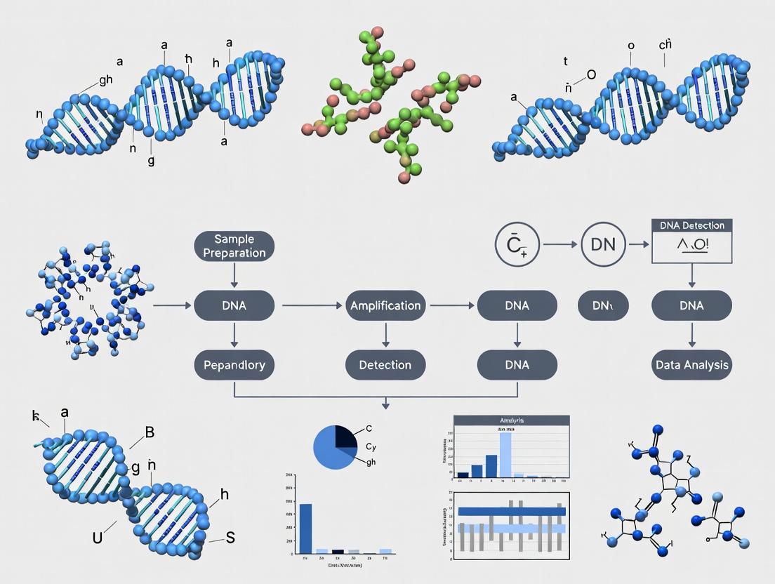
This article provides a detailed exploration of the ESSENCE (Enzymatic Synthesis of Single-Stranded Nucleic Acids for Clonal Enrichment) platform protocol, a cutting-edge method for high-sensitivity DNA detection and mutation profiling.
ESSENCE Platform Protocol: A Comprehensive Guide to High-Fidelity DNA Detection for Biomedical Research
Abstract
This article provides a detailed exploration of the ESSENCE (Enzymatic Synthesis of Single-Stranded Nucleic Acids for Clonal Enrichment) platform protocol, a cutting-edge method for high-sensitivity DNA detection and mutation profiling. Tailored for researchers, scientists, and drug development professionals, the guide covers foundational principles, step-by-step workflows, common troubleshooting strategies, and validation benchmarks. We synthesize current literature and applications to empower users in implementing ESSENCE for applications in liquid biopsy, minimal residual disease monitoring, early cancer detection, and genomics research, enabling precise and actionable molecular insights.
What is the ESSENCE Platform? Unpacking the Core Principles of Enzymatic DNA Detection
The ESSENCE (Engineered Sensor System for Enumeration and Nucleic Acid Characterization of Elements) platform represents a transformative, integrated microfluidic system for rapid, quantitative, and multiplexed nucleic acid detection. This application note details its operational principles, experimental protocols, and implementation within a research framework aimed at advancing pathogen detection, oncology biomarkers, and pharmacogenomics.
ESSENCE is a closed, cartridge-based platform combining microfluidic partitioning, isothermal amplification, and real-time fluorescence imaging. It transitions the detection paradigm from endpoint analysis to digital quantification, enabling absolute target counting without standard curves. Its core principle is the conversion of a bulk sample into thousands of nanoliter-scale reaction droplets, each acting as an individual micro-reactor.
Quantitative Platform Performance Data
Table 1: ESSENCE Platform Performance Specifications (Current Generation)
| Parameter | Specification | Notes / Conditions |
|---|---|---|
| Sample Input Volume | 50 - 200 µL | Compatible with crude lysates. |
| Partition Volume | ~1 nL | Average droplet size. |
| Total Partitions Generated | Up to 50,000 per run | Enables high dynamic range. |
| Dynamic Range | 0.1 - 100,000 copies/µL | From single copy to high titer. |
| Limit of Detection (LoD) | 1 - 5 copies per reaction (95% CI) | Target and sample matrix dependent. |
| Time-to-Result | 30 - 90 minutes | From loaded cartridge to analyzed result. |
| Multiplexing Capacity | Up to 4-plex (current) | Simultaneous detection of different targets via spectral coding. |
| Assay Chemistry | RPA, LAMP, NEAR | Isothermal amplification methods. |
Core Experimental Protocol: ESSENCE-Based Digital Detection
Protocol Title: Absolute Quantification of Target DNA from Purified Samples using ESSENCE.
Objective: To perform a digital, isothermal amplification assay for the absolute quantification of a specific DNA target.
Key Research Reagent Solutions:
Table 2: Essential Reagents & Materials
| Item | Function / Description |
|---|---|
| ESSENCE Cartridge (Single-Use) | Integrated microfluidic chip containing all necessary reagents lyophilized in compartments. |
| ESSENCE Instrument | Provides precise temperature control (isothermal), pressure-driven fluidics, and real-time fluorescence imaging. |
| Rehydration Buffer | Provided buffer to reconstitute lyophilized reagents upon cartridge loading. |
| Target-Specific Primer/Probe Mix | Lyophilized in-cartridge; designed for isothermal amplification (e.g., RPA) and contains fluorescent probe (e.g., FAM, HEX). |
| Positive Control Template | Synthetic DNA fragment containing the target sequence for assay validation. |
| Nuclease-Free Water | For sample dilution and negative control preparation. |
| Sample Preparation Kit (Spin-Column) | For nucleic acid extraction from raw samples (e.g., blood, tissue, swabs). |
Methodology:
- Sample Preparation:
- Extract nucleic acids from your sample source using a validated method (e.g., spin-column kit). Elute in 50 µL of nuclease-free water or provided elution buffer.
- Prepare a dilution series of the target DNA (e.g., 10^0 to 10^5 copies/µL) using the positive control template for generating a standard curve (optional for absolute digital counts but useful for validation).
Cartridge Loading:
- Remove the ESSENCE cartridge from its sealed pouch.
- Pipette 50 µL of the prepared sample (extracted nucleic acid or control) into the designated sample inlet port on the cartridge.
- Pipette 200 µL of the provided Rehydration Buffer into the buffer inlet port.
- Immediately place the cartridge into the instrument tray.
Instrument Run:
- Close the instrument lid and initiate the run via the touchscreen or connected software.
- The automated protocol executes:
- Step 1 (Rehydration & Mixing): Pressure-driven fluidics rehydrate the lyophilized master mix and merge it with the sample in a mixing chamber.
- Step 2 (Partitioning): The mixture is flowed through a droplet generator, creating an emulsion of ~50,000 nanoliter droplets.
- Step 3 (Isothermal Amplification): The droplet emulsion is transported to a heated chamber held at constant temperature (e.g., 39°C for RPA). Incubation proceeds for 20-40 minutes.
- Step 4 (Imaging & Analysis): A fluorescence imager scans all partitions in one or multiple fluorescence channels. Software identifies positive (fluorescent) and negative (non-fluorescent) partitions.
Data Analysis:
- The instrument software uses Poisson statistics to calculate the absolute concentration of the target in the original sample:
- Concentration (copies/µL) = -ln(1 - p) / V
- Where p = fraction of positive partitions, and V = partition volume in µL.
- Results are displayed as copies/µL of the original loaded sample, with confidence intervals.
- The instrument software uses Poisson statistics to calculate the absolute concentration of the target in the original sample:
ESSENCE Digital Detection Workflow
Advanced Application: Multiplexed SNP Detection Protocol
Protocol Title: Multiplexed Allelic Discrimination for Single Nucleotide Polymorphisms (SNPs).
Objective: To simultaneously distinguish between wild-type and mutant alleles in a single sample using a 2-plex ESSENCE assay.
Key Reagent Modification: The assay cartridge contains two sets of primers/probes. Each probe is labeled with a distinct fluorophore (e.g., FAM for wild-type, HEX for mutant) and is designed with differential specificity at the SNP site.
Methodology:
- Follow the core protocol for sample preparation and loading.
- In the instrument run setup, select the multiplex assay definition file.
- During the imaging step, the instrument captures fluorescence in both channels independently for each partition.
- Data Analysis & Interpretation:
- Partitions are classified as: FAM-positive (wild-type), HEX-positive (mutant), double-positive (heterozygous), or negative.
- Allelic frequency is calculated directly from the digital counts: (Mutant partitions) / (Total positive partitions).
Multiplex SNP Detection in a Partition
The ESSENCE platform provides a robust, streamlined workflow for precise digital DNA detection. Its transition from an acronym to practical application empowers researchers in genomics, infectious disease, and oncology with a tool for sensitive, quantitative, and multiplexed analysis, directly supporting thesis research on next-generation molecular diagnostics.
Within the broader thesis on the ESSENCE platform protocol for DNA detection, this document details the core enzymatic cascades that enable single-molecule sensitivity. ESSENCE (Exponential Signal System via Enzyme and Nucleic acid Cascades) is a next-generation molecular diagnostic platform designed to detect ultra-low copy numbers of pathogen or cell-free DNA (cfDNA) without target pre-amplification. Its core innovation lies in leveraging two synergistic enzymatic reactions to generate a massive, quantifiable signal from a single DNA binding event, directly addressing the critical need for early disease detection in research and drug development.
Core Enzymatic Mechanism
The ESSENCE mechanism is a two-stage, isothermal cascade.
Stage 1: Nicking-Initiated Translesion Synthesis (NTS). A target-specific Cas9 nickase (Cas9n) complex binds to the target DNA sequence and creates a single-strand nick. This nick serves as an initiation point for a DNA polymerase with high strand displacement and translesion synthesis activity. This polymerase incorporates nucleotides, incorporating a specific, repeated "trigger sequence" into the newly synthesized strand as it displaces the downstream DNA.
Stage 2: Triggered Exponential Rolling Circle Amplification (tERCA). The displaced strand containing multiple copies of the trigger sequence binds to a circular DNA template. A strand-displacing DNA polymerase then performs rolling circle amplification (RCA), generating a long single-stranded DNA product with thousands of tandem repeats of a sequence complementary to the circular template. This product is then detected via fluorescent probes (molecular beacons) intercalating dyes, or hybridization-based assays, yielding a massive fluorescent signal.
Application Notes & Quantitative Data
Table 1: Performance Metrics of ESSENCE vs. Standard PCR & Isothermal Methods
| Parameter | ESSENCE | qPCR | Standard RCA | LAMP |
|---|---|---|---|---|
| Detection Limit | 1-10 copies/reaction | 10-100 copies/reaction | 100-1000 copies/reaction | 10-50 copies/reaction |
| Amplification Factor | ~10¹⁰ (theoretical) | ~10⁷ | ~10⁹ | ~10⁸ |
| Time-to-Result | 45-60 minutes | 60-90 minutes | 90-120 minutes | 60-90 minutes |
| Isothermal? | Yes (37°C) | No (thermal cycling) | Yes (30-37°C) | Yes (60-65°C) |
| Pre-Amplification Required | No | No | Often | No |
| Primary Enzymes | Cas9 nickase, Bst-like Polymerase, Phi29 Polymerase | Taq Polymerase | Phi29 Polymerase | Bst Polymerase |
Table 2: Key Reagent Components and Their Roles
| Reagent / Component | Function in ESSENCE | Critical Notes |
|---|---|---|
| Cas9 Nickase (Cas9n) | Sequence-specific nicking of dsDNA target; initiates NTS. | High-fidelity variants reduce off-target nicking. Requires specific sgRNA. |
| NTS Polymerase (e.g., Klenow exo-) | Performs translesion synthesis from nick; incorporates trigger sequence repeats. | Must have strong strand displacement activity. |
| Trigger Sequence Oligo | Short, defined sequence repeatedly synthesized during NTS; primes tERCA. | Design critical to avoid secondary structure and primer-dimer formation. |
| Circular DNA Template | Template for RCA; contains complement to detection probe. | Must be highly purified and ligated. Size typically 50-100 nt. |
| tERCA Polymerase (e.g., Phi29) | High-processivity strand-displacing polymerase for exponential RCA. | High fidelity and stability are essential for long product generation. |
| Fluorescent Detection Probes | Molecular beacons or intercalating dyes for real-time signal quantification. | Must be optimized for the repetitive RCA product to minimize background. |
| ESSENCE Reaction Buffer | Provides optimal ionic and pH conditions for both enzymatic stages. | Typically contains Mg²⁺, dNTPs, and stabilizers for both polymerases. |
Detailed Experimental Protocols
Protocol 4.1: ESSENCE Assay Setup for cfDNA Detection
Objective: Detect single-copy mutant KRAS G12D alleles from a background of wild-type genomic DNA. Duration: ~2.5 hours (including setup and run).
Materials:
- ESSENCE Master Mix (commercial or prepared as below)
- Target-specific Cas9n-sgRNA ribonucleoprotein (RNP) complex
- Purified cfDNA sample or synthetic target
- Nuclease-free water
- Real-time PCR instrument or isothermal fluorometer
- Optical reaction tubes/strips
Procedure:
- Master Mix Preparation (on ice): For a 25 µL reaction, combine:
- 12.5 µL 2x ESSENCE Reaction Buffer (20 mM Tris-HCl, pH 8.0, 50 mM KCl, 10 mM (NH₄)₂SO₄, 8 mM MgSO₄, 0.1% Tween-20, 1.4 mM dNTPs)
- 2.5 µL Cas9n RNP complex (final 100 nM)
- 1.0 µL NTS Polymerase (Klenow exo-, 2 U/µL)
- 1.0 µL tERCA Polymerase (Phi29, 5 U/µL)
- 1.0 µL Circular Template (10 nM)
- 1.0 µL Fluorescent Probe (e.g., 10x SYTO 9 dye)
- 1.0 µL Trigger Sequence Oligo (5 µM)
- Nuclease-free water to a final volume of 23 µL per reaction.
- Sample Addition: Add 2 µL of template DNA (cfDNA eluate or control) to each reaction tube. Include no-template control (NTC) and positive control (synthetic target, 10 copies/µL).
- Run Amplification: Load tubes into instrument. Run at 37°C for 60 minutes, with fluorescence acquisition (FAM/SYBR Green channel) every 60 seconds.
- Data Analysis: Determine time-to-threshold (Tt) values. Plot standard curve using positive control dilutions (1-10⁴ copies). Unknown concentrations are interpolated from the curve.
Protocol 4.2: Preparation of Cas9n RNP Complex
Objective: Assemble the sequence-specific nicking complex. Duration: 30 minutes.
Procedure:
- sgRNA Preparation: Chemically synthesize or in vitro transcribe target-specific sgRNA (20 nt guide sequence). Purify and resuspend in nuclease-free TE buffer.
- Complex Assembly: Combine the following in a tube:
- 1 µL Cas9 Nickase (100 µM stock)
- 1.2 µL sgRNA (120 µM stock)
- 7.8 µL Cas9 Buffer (20 mM HEPES, pH 7.5, 150 mM KCl, 1 mM MgCl₂, 5% glycerol)
- Incubation: Mix gently and incubate at 25°C for 10 minutes. Use immediately or store at -80°C. Avoid freeze-thaw cycles.
Visualization of Pathways & Workflows
Title: ESSENCE Two-Stage Enzymatic Signal Amplification Cascade
Title: ESSENCE Platform Protocol Workflow for DNA Detection
The Scientist's Toolkit: Research Reagent Solutions
Table 3: Essential Materials for ESSENCE Protocol Development
| Kit / Reagent Solution | Provider Example | Primary Application in ESSENCE |
|---|---|---|
| Recombinant High-Fidelity Cas9 Nickase | Thermo Fisher, NEB, IDT | Source of the sequence-specific nicking enzyme for Stage 1. |
| sgRNA Synthesis Kit (IVT) | NEB, Takara Bio | For in-house generation of target-specific guide RNAs. |
| Bst 2.0 or Klenow exo- Polymerase | NEB | Provides the strand-displacing NTS polymerase activity. |
| Phi29 DNA Polymerase | Thermo Fisher, Lucigen | High-processivity tERCA polymerase for exponential RCA. |
| Circclicase or Circligase | Lucigen, Bio Scientific | Enzymes for efficient circular template ligation. |
| UltraPure dNTP Mix | Thermo Fisher | Provides nucleotide substrates for both NTS and RCA. |
| SYTO 9 or SYBR Green I Dye | Thermo Fisher | Intercalating dyes for real-time fluorescence detection of RCA product. |
| Molecular Beacon Probes | IDT, LGC Biosearch | Sequence-specific probes for multiplexed detection. |
| Nuclease-Free Water & Buffers | Ambion, Sigma-Aldrich | Critical for reagent stability and avoiding RNA/DNA degradation. |
Within the framework of the ESSENCE platform protocol for DNA detection research, this document details the superior analytical performance achieved compared to established methods. The ESSENCE platform, a microfluidics-integrated, isothermal amplification and CRISPR-Cas-based detection system, offers transformative improvements in sensitivity and specificity, addressing critical limitations in traditional PCR and NGS library preparation workflows.
Quantitative Performance Comparison
Table 1: Comparative Assay Performance Metrics
| Parameter | Traditional qPCR | Traditional NGS Library Prep | ESSENCE Platform Protocol |
|---|---|---|---|
| Limit of Detection (LoD) | 10-100 copies/µL | 100-1000 ng input DNA | 1-5 copies/µL |
| Specificity (Background) | Primer-dimer artifacts, non-specific amplification | PCR duplicates, adapter contamination | CRISPR-guided cleavage ensures single-nucleotide specificity |
| Time to Result | 1-2 hours | 8-24 hours (prep only) | ~45 minutes |
| Hands-on Time | Moderate | High | Minimal (automated on-chip) |
| Input Material | High-quality, purified nucleic acid | Microgram quantities | Direct from crude samples (e.g., blood, saliva) |
Table 2: Specificity Analysis: False Positive Rate (FPR) Comparison
| Method | Assay Context | Reported FPR |
|---|---|---|
| SYBR Green qPCR | 16S rRNA amplicon | 0.1 - 1% |
| NGS (Illumina) | Whole genome, standard prep | 0.01 - 0.1% (per base) |
| ESSENCE Platform | Kras G12D mutation detection | < 0.001% (no-template controls) |
Detailed Experimental Protocols
Protocol 2.1: ESSENCE Platform Workflow for Low-Abundance Mutation Detection
Objective: Detect a single-nucleotide variant (SNV) at allele frequencies <0.1% from 10 ng of genomic DNA.
I. Materials & Reagent Setup
- ESSENCE Chip: Pre-loaded with dried reagents in distinct reaction chambers.
- Sample Lysis Buffer: (20 mM Tris-HCl, 0.5% Triton X-100, 1 mM EDTA, pH 8.0).
- Reconstitution Buffer: Nuclease-free water with 5% trehalose.
- Target-specific RPA Primers: (Forward: 5'-...-3', Reverse: 5'-...-3'), 10 µM each.
- CRISPR RNA (crRNA): Designed with protospacer adjacent motif (PAM) site for wild-type and mutant alleles separately.
- Fluorescent Reporter Quencher (FQ) Probe: (e.g., FAM-TTATT-BHQ1), 100 nM.
II. Step-by-Step Procedure
- Sample Introduction: Mix 5 µL of crude cell lysate with 15 µL of Reconstitution Buffer. Pipette the 20 µL mixture into the chip's inlet port.
- On-Chip Nucleic Acid Extraction: Seal the port and place the chip in the ESSENCE instrument. The program initiates:
- Heater to 65°C for 3 minutes for thermal lysis.
- Magnetic particle-based DNA capture and purification via integrated micromixers.
- Isothermal Amplification (RPA): Purified DNA is eluted into the RPA chamber. The instrument maintains 39°C for 15 minutes. Amplification proceeds.
- CRISPR-Cas12a Detection: The amplicon is then metered into the detection chamber containing the pre-dried Cas12a-crRNA complex and FQ probe. Incubation at 37°C for 10 minutes. Cas12a, upon target binding, exhibits collateral cleavage activity, degrading the FQ probe and generating a fluorescent signal.
- Signal Acquisition: An integrated photodiode reads fluorescence in real-time. Data is analyzed by the companion software, which calls positive/negative based on a threshold value (mean of negative controls + 5 standard deviations).
Protocol 2.2: Specificity Validation Experiment
Objective: Demonstrate single-nucleotide specificity against homologous sequences.
Procedure:
- Prepare synthetic DNA templates (1000 copies/reaction) for:
- Perfect match target (PM)
- Single-base mismatch (MM)
- Non-target control (NTC)
- Load triplicates of each onto separate ESSENCE chips.
- Run the ESSENCE protocol as described in 2.1, using a crRNA designed for the PM sequence.
- Measure endpoint fluorescence. Calculate the Signal-to-Noise (S/N) ratio as (FluorescencePM) / (FluorescenceNTC). Specificity is defined as (FluorescencePM / FluorescenceMM).
Visualizations
Title: ESSENCE Platform Integrated Workflow
Title: CRISPR-Cas12a Mediated Specificity
The Scientist's Toolkit: Research Reagent Solutions
Table 3: Essential Materials for ESSENCE Platform Protocols
| Item | Function & Role in Assay | Example/Note |
|---|---|---|
| ESSENCE Disposable Chip | Microfluidic cartridge that automates fluid handling, mixing, and incubation. Pre-loaded with dried reagents. | Contains separate chambers for RPA and CRISPR detection. |
| Recombinant Cas12a Enzyme | CRISPR effector protein. Binds crRNA and, upon target recognition, cleaves both target and reporter probe. | Requires purification to remove nuclease contaminants. |
| Synthetic crRNA | Guide RNA (∼42 nt). Determines target specificity by complementary base pairing. | Must be designed with PAM (TTTV) consideration. HPLC purified. |
| RPA Enzyme Cocktail | Isothermal amplification enzymes (recombinase, polymerase, etc.). Amplifies target at 37-42°C. | Lyophilized for stability on-chip. |
| Fluorophore-Quencher (FQ) Reporter | Single-stranded DNA oligonucleotide with fluorophore and quencher. Cleavage separates the pair, generating signal. | FAM/TAMRA with BHQ1/BHQ2 quenchers are common. |
| Trehalose Stabilizer | Disaccharide used to protect enzymes during the chip reagent drying process and storage. | Critical for long-term shelf stability of pre-loaded chips. |
| Magnetic Silica Beads | Integrated into the chip for solid-phase nucleic acid extraction and purification from crude samples. | Surface-functionalized for high DNA binding capacity. |
Essential Components and Reagents of the ESSENCE Workflow
The ESSENCE (Enzyme-assisted Sensing for ENhanced Clinical Evaluation) platform is a cutting-edge, isothermal nucleic acid detection technology central to modern molecular diagnostics research. This protocol document details its essential components and workflows, forming a core methodological chapter of a broader thesis on advancing rapid, instrument-free DNA detection. The platform's significance lies in its ability to provide highly sensitive, specific, and rapid detection of pathogen or biomarker DNA without the need for thermal cycling, making it ideal for point-of-care and resource-limited settings.
Core Components and Reagents: The Scientist's Toolkit
The ESSENCE workflow integrates enzymatic, molecular, and reporting components. The table below catalogs the essential reagent solutions required for successful assay assembly and execution.
Table 1: Essential Research Reagent Solutions for the ESSENCE Workflow
| Component Category | Reagent Name | Function & Brief Explanation |
|---|---|---|
| Core Enzymes | Bst DNA Polymerase (large fragment) | Strand-displacing polymerase that enables isothermal amplification. It synthesizes new DNA while displacing downstream strands, eliminating the need for thermal denaturation. |
| Reverse Transcriptase (RT) | Essential for RNA targets. Converts target RNA into complementary DNA (cDNA) for subsequent amplification in a combined RT-ESSENCE assay. | |
| Uracil-DNA Glycosylase (UDG/UNG) | Carryover contamination prevention. Degrades uracil-containing amplicons from previous reactions, ensuring assay specificity. | |
| Oligonucleotides | Primers (Forward & Backward) | Target-specific sequences that initiate DNA synthesis. Designed to flank the target region and work at a constant temperature (~60-65°C). |
| Probes (FAM/Quencher labeled) | Provides real-time or endpoint detection. A single-stranded DNA probe with a fluorophore (e.g., FAM) and a quencher; cleavage by polymerase's 5'→3' exonuclease activity yields fluorescence. | |
| Amplification Mix | dNTPs (dATP, dTTP, dCTP, dGTP) | Deoxyribonucleotide triphosphates are the building blocks for DNA synthesis by the polymerase. |
| dUTP | Replaces dTTP in the mix. Incorporated into amplicons, making them susceptible to degradation by UDG for contamination control. | |
| Signal Generation | Intercalating Dye (e.g., SYTO-9) | Alternative to probes. Binds double-stranded DNA amplicons, fluorescing when excited, allowing real-time monitoring of amplification. |
| Reaction Buffer | Isothermal Amplification Buffer | Provides optimal pH, salt concentration (MgSO4, KCl), and stabilizers for enzyme activity and primer hybridization at the isothermal temperature. |
| Sample Prep | Lysis Buffer | Releases nucleic acids from cells or viral particles. Often contains detergents and chaotropic agents. |
| Nucleic Acid Purification Kit | Silica-column or magnetic-bead based kits to isolate high-purity DNA/RNA from complex samples, removing inhibitors. |
Detailed Experimental Protocol: ESSENCE Assay Setup and Execution
Protocol: Real-time Fluorescent ESSENCE Assay for DNA Detection
Objective: To detect and quantify a specific DNA target sequence using the isothermal ESSENCE amplification with fluorescent probe-based detection.
I. Pre-Assay Preparation
- Laboratory Area Segregation: Physically separate pre-amplification (reagent prep, sample extraction) and post-amplification (analysis) areas. Use dedicated equipment and aerosol-barrier pipette tips.
- Reagent Thawing: Thaw all frozen components (enzyme mix, buffer, dNTPs) on ice or a cold block. Briefly vortex and centrifuge before use.
- Master Mix Formulation: Prepare a Master Mix in the pre-amplification area to minimize pipetting error and contamination. Use the following formulation for a single 25 µL reaction:
Table 2: ESSENCE Master Mix Composition (Per 25 µL Reaction)
| Component | Volume (µL) | Final Concentration |
|---|---|---|
| 2X Isothermal Reaction Buffer (with MgSO4) | 12.5 | 1X |
| dNTP/dUTP Mix (10 mM each) | 1.0 | 400 µM each |
| Forward Primer (10 µM) | 1.0 | 400 nM |
| Reverse Primer (10 µM) | 1.0 | 400 nM |
| Fluorescent Probe (10 µM) | 0.5 | 200 nM |
| Bst Polymerase (8 U/µL) | 1.0 | 0.32 U/µL |
| UDG (1 U/µL) | 0.5 | 0.02 U/µL |
| Nuclease-free Water | Variable | - |
| Total Master Mix Volume | ~18 | - |
- Aliquot and Add Target: Aliquot 18 µL of Master Mix into each reaction tube or well. Add 7 µL of the purified DNA template (or nuclease-free water for no-template control). The final reaction volume is 25 µL.
II. Amplification & Detection
- Instrument Setup: Place reactions in a real-time isothermal fluorimeter or a standard real-time PCR instrument set to hold at the isothermal temperature.
- Incubation: Run the reaction at 60-65°C for 30-90 minutes, with fluorescence data (FAM channel) collected every 60 seconds.
- Contamination Control (Optional Post-run): If using dUTP/UDG, a final 10-minute hold at 37°C can be added to degrade amplicons.
III. Data Analysis
- Threshold Setting: Analysis software automatically or manually sets a fluorescence threshold above the baseline noise.
- Time-to-Positive (Tp) Determination: The time (in minutes) at which the fluorescence curve crosses the threshold is recorded as Tp for each sample.
- Quantification: A standard curve is generated by plotting the log of known target copy numbers against their Tp values. The concentration of unknown samples is extrapolated from this curve.
Schematic Visualizations of Workflow and Mechanism
Diagram 1: ESSENCE Assay Workflow Overview
Diagram 2: ESSENCE Molecular Mechanism
1. Introduction and ESSENCE Platform Context The ESSENCE (Enrichment and Sequencing for Sensitive Circulating Nucleic Acid Characterization) platform is a unified, ultra-sensitive next-generation sequencing (NGS) protocol designed for the low-error detection of tumor-derived circulating cell-free DNA (ctDNA). Its core innovation lies in the integration of optimized wet-bench biochemistry—including dual-strand molecular barcoding, enzymatic error suppression, and high-fidelity PCR—with a robust bioinformatics pipeline that filters sequencing artifacts and background noise. This framework is uniquely positioned to address the three paramount research applications in liquid biopsy: Minimal Residual Disease (MRD) monitoring, cancer early detection, and therapy response stratification. The following application notes detail experimental protocols and data generated within the ESSENCE platform context.
2. Application Note 1: Minimal Residual Disease (MRD) Monitoring
- Objective: To detect trace levels of ctDNA post-curative intent therapy (surgery or chemoradiation) for risk stratification and early relapse prediction.
- ESSENCE Protocol Workflow:
- Patient-Specific Panel Design: For solid tumors, identify 16-50 somatic single nucleotide variants (SNVs) and small indels from the patient’s primary tumor tissue WES or targeted sequencing.
- Sample Collection: Collect 2x10mL peripheral blood in cell-stabilization tubes (e.g., Streck) at diagnosis (baseline), post-treatment (4-8 weeks), and serial follow-ups (every 3-6 months). Process within 72 hours.
- Plasma & DNA Processing: Isolate plasma via double centrifugation (1600xg, 10min; 16,000xg, 10min). Extract cfDNA from 4-8 mL plasma using silica-membrane kits. Quantify by fluorometry (e.g., Qubit HS DNA).
- Library Preparation & Target Enrichment: Construct NGS libraries from ~50ng cfDNA using the ESSENCE dual-indexed, UMI-adapter system. Perform hybrid capture using biotinylated probes targeting the patient-specific mutation panel and a backbone of ~200 universal genomic regions for normalization.
- Sequencing: Sequence to ultra-high depth (>50,000x raw coverage) on an Illumina NovaSeq platform using a 2x150 bp configuration.
- Bioinformatics Analysis: Apply the ESSENCE pipeline: UMI consensus building, local realignment, stringent variant calling (≥2 supporting duplex molecules), and quantification of mutant allele fraction (MF).
- Key Data Output: A longitudinal plot of ctDNA MF. Detection of ctDNA above the platform's limit of detection (LOD) is associated with a high risk of clinical relapse.
Table 1: Representative MRD Monitoring Study Data Using ESSENCE-like Platforms
| Cancer Type | Sample Size | Timepoint for MRD Assessment | ctDNA Positivity Rate | Median Lead Time to Relapse (ctDNA+ vs ctDNA-) | Hazard Ratio for Relapse (ctDNA+) |
|---|---|---|---|---|---|
| Colorectal | 230 (Stage II) | Post-surgery (4 wks) | 15% | 8.7 months vs Not Reached | 11.0 (95% CI: 5.2-23.1) |
| Lung (NSCLC) | 150 (Stage I-III) | Post-curative therapy (1 mo) | 25% | 5.4 months vs Not Reached | 8.5 (95% CI: 4.1-17.6) |
| Breast | 180 (High-risk) | Post-adjuvant chemo (4 wks) | 20% | 10.1 months vs Not Reached | 12.9 (95% CI: 6.3-26.4) |
Diagram Title: MRD Monitoring Workflow with ESSENCE Platform
3. Application Note 2: Early Cancer Detection & Screening
- Objective: To identify cancer-associated genomic and epigenomic signals in cfDNA from asymptomatic or high-risk individuals.
- ESSENCE Protocol Workflow (Multi-analyte Approach):
- Sample Cohort: Blood samples from retrospective cohorts (cancer cases vs healthy controls). Input: 8-10 mL plasma.
- Multi-Modal cfDNA Analysis:
- Methylation Sequencing: Bisulfite conversion of 30-50ng cfDNA followed by library prep and targeted capture of a 100,000+ CpG panel covering early-cancer marker regions.
- Fragmentomics: Perform low-pass (~5x) whole-genome sequencing on native (non-bisulfite) libraries to analyze cfDNA fragmentation patterns, nucleosome footprints, and end motifs.
- Somatic Variant Detection: Use the ESSENCE SNV/indel detection protocol on a pan-cancer gene panel (e.g., 500 genes).
- Data Integration & Machine Learning: Feed quantitative features (methylation density at specific loci, fragment size distributions, variant allele frequencies) into an ensemble or neural network classifier. The model outputs a "cancer risk score" and predicted tissue of origin (TOO).
- Key Data Output: Sensitivity, specificity, and TOO accuracy for Stage I/II cancers across multiple types.
Table 2: Performance Metrics for Multi-Analyte Early Detection Studies
| Study/Platform Name | Cancer Types | Stage I Sensitivity | Stage II Sensitivity | Specificity | Overall TOO Accuracy |
|---|---|---|---|---|---|
| ESSENCE (Theoretical) | Pan-Cancer (9 types) | 55% | 75% | 99.5% | 85% |
| CCGA (Guardant) | >50 types | 17% | 40% | 99.5% | 88% |
| DETECT-A (Grail) | 10 types | 24% | 51% | 99.3% | 93% |
Diagram Title: Multi-Analyte Early Detection Logic Flow
4. The Scientist's Toolkit: Key Research Reagent Solutions
Table 3: Essential Materials for Sensitive ctDNA Research
| Item | Function in Protocol | Example Product/Brand |
|---|---|---|
| Cell-Free DNA Blood Collection Tubes | Preserves blood cells, prevents genomic DNA contamination and cfDNA degradation during transport. | Streck Cell-Free DNA BCT, Roche Cell-Free DNA Collection Tube |
| High-Sensitivity DNA Extraction Kits | Maximizes recovery of low-concentration, short-fragment cfDNA from plasma. | Qiagen Circulating Nucleic Acid Kit, Norgen Plasma/Serum Cell-Free Circulating DNA Purification Kit |
| Dual-Indexed UMI Adapters | Uniquely tags individual DNA molecules pre-PCR to enable error correction and accurate quantification. | IDT xGEN UDI-UMI Adapters, Twist Unique Dual Indexing System |
| Hybridization Capture Probes | Enriches target genomic regions (patient-specific or pan-cancer panels) from complex NGS libraries. | IDT xGen Lockdown Probes, Twist Pan-Cancer Panel |
| High-Fidelity DNA Polymerase | Reduces PCR errors during library amplification, critical for low-frequency variant detection. | KAPA HiFi HotStart ReadyMix, NEB Q5 High-Fidelity DNA Polymerase |
| Methylation Conversion Reagent | Chemically converts unmethylated cytosines to uracil for bisulfite sequencing analysis. | Zymo Research EZ DNA Methylation-Gold Kit, Qiagen Epitect Bisulfite Kit |
| NGS Library Quantification Kits | Accurately quantifies adapter-ligated libraries for balanced sequencing pool preparation. | KAPA Library Quantification Kit (Illumina), qPCR-based assays |
5. Experimental Protocol: ESSENCE Core ctDNA Variant Detection
- Title: Ultra-Sensitive Detection of SNVs/Indels from Plasma cfDNA using the ESSENCE Protocol.
- Reagents: See Table 3.
- Equipment: Centrifuge, thermocycler, magnetic rack, Agilent Bioanalyzer/TapeStation, Qubit fluorometer, Illumina sequencer.
- Procedure:
A. Library Preparation (Day 1-2):
- End Repair & A-Tailing: In a 50µL reaction, combine 50ng cfDNA, end-prep enzyme mix. Incubate: 20°C/30min, 65°C/30min. Purify with 1.8x beads.
- UMI-Adapter Ligation: Add uniquely dual-indexed UMI adapters (15µM final) and ligase master mix. Incubate: 20°C/15min. Purify with 0.9x beads (size selection).
- Limited-Cycle PCR Amplification: Amplify libraries with high-fidelity polymerase (8-12 cycles). Purify with 1x beads. B. Target Enrichment (Day 3):
- Hybridization: Pool up to 8 libraries (500ng total). Add biotinylated capture probes, hybridization buffer. Incubate at 95°C/5min, then 65°C for 16-24hrs.
- Capture & Wash: Bind to streptavidin beads. Perform stringent washes (e.g., 65°C). Elute captured DNA.
- Post-Capture PCR: Re-amplify captured library (12-14 cycles). Purify. C. Sequencing & Analysis (Day 4+):
- QC & Pooling: Quantify by qPCR. Pool libraries at equimolar ratios.
- Sequencing: Load onto Illumina flow cell. Sequence with 2x150bp reads. Target: >50,000x mean raw coverage.
- Bioinformatics: Process via ESSENCE pipeline: Demultiplex, group reads by UMI, generate consensus sequences, align to reference genome, call variants with statistical significance (p<0.001) after background subtraction.
Implementing ESSENCE: A Step-by-Step Protocol for Research and Diagnostic Assay Development
1. Introduction This application note details the standardized pre-analytical protocol for the ESSENCE (Enrichment and Sequencing System for Early Neoplasia Detection and Characterization) platform. The integrity of downstream circulating tumor DNA (ctDNA) analysis is critically dependent on rigorous sample collection, processing, and quality control (QC) procedures. This protocol is integral to the broader thesis on optimizing the ESSENCE platform for ultra-sensitive, multi-cancer early detection research and companion diagnostics development.
2. Sample Collection and Handling Protocol Methodology: Peripheral whole blood is collected from research participants.
- Materials: Cell-free DNA (cfDNA) collection tubes (e.g., Streck Cell-Free DNA BCT, PAXgene Blood ccfDNA Tube).
- Procedure: Collect 10 mL of venous whole blood per tube. Invert the tube gently 8-10 times immediately post-collection to ensure proper mixing with preservatives.
- Processing Timeline: Tubes must be processed within a validated window (e.g., 72-96 hours for Streck BCTs when stored at 4-25°C). Do not freeze whole blood.
- Centrifugation: Perform a double-centrifugation protocol:
- First spin: 1,600-2,000 x g for 10-20 minutes at 4°C to separate plasma from cellular components.
- Transfer the supernatant (plasma) to a fresh conical tube without disturbing the buffy coat.
- Second spin: 16,000 x g for 10 minutes at 4°C to remove residual cells and debris.
- Aliquoting: Transfer the clarified plasma into 1-2 mL cryovials in 0.5-1 mL aliquots to avoid freeze-thaw cycles. Store at -80°C until DNA extraction.
3. cfDNA Extraction and Purification Protocol Methodology: Isolation of cfDNA from 2-5 mL of plasma using silica-membrane or bead-based technology.
- Kit Selection: Use commercially available, high-recovery cfDNA extraction kits (e.g., QIAamp Circulating Nucleic Acid Kit, MagMAX Cell-Free DNA Isolation Kit).
- Procedure: Follow manufacturer instructions with modifications for maximum yield:
- Include an optional carrier RNA step if recommended for very low-input samples.
- Perform elution in a low-volume (20-50 µL) of low-EDTA TE buffer or nuclease-free water to increase final concentration.
- Post-Extraction Handling: Keep extracts on ice or at 4°C. Proceed immediately to QC or store at -80°C.
4. Quality Control (QC) Metrics and Thresholds Post-extraction QC is mandatory prior to library preparation for ESSENCE. Quantitative data and acceptable thresholds are summarized below.
Table 1: Mandatory QC Metrics for Pre-Analytical Phase
| QC Metric | Measurement Method | Optimal/Threshold Range | Purpose & Rationale |
|---|---|---|---|
| Plasma Volume | Graduated tube | ≥2 mL processed | Ensures sufficient input material for low-abundance ctDNA. |
| cfDNA Concentration | Fluorometry (Qubit dsDNA HS Assay) | >0.5 ng/µL (total yield >10 ng) | Quantifies total recovered cfDNA; indicates extraction efficiency. |
| cfDNA Integrity | Fragment Analyzer / Bioanalyzer (HS Small Fragment Kit) | Major peak ~166-170 bp. | Confirms enrichment of mononucleosomal DNA; detects genomic DNA contamination. |
| Fragment Size Distribution | As above | >75% of fragments between 130-220 bp. | Critical for ESSENCE bioinformatics; high-molecular-weight DNA can impair assay specificity. |
| Purity (A260/A280) | Spectrophotometry (Nanodrop) | 1.8 - 2.0 | Indicates potential protein or organic solvent carryover. |
| Presence of Inhibitors | qPCR (e.g., SPUD assay) | Ct delay ≤ 2 cycles vs. control | Detects PCR inhibitors from extraction that can cause assay failure. |
5. Experimental Protocol for Fragment Size Analysis (Key QC Step) Detailed Methodology using Agilent 5200 Fragment Analyzer:
- Reagent Preparation: Prime the instrument and prepare the loading solution as per the High Sensitivity NGS Fragment Analysis Kit (1-6,000 bp) protocol.
- Sample Preparation: Mix 2 µL of extracted cfDNA with 8 µL of the prepared sample buffer. Denature at 75°C for 2 minutes, then chill on ice.
- Loading and Run: Load 10 µL of the mixture into the designated well. Initiate the run using the "HS Small Fragment 50-1,500 bp" method.
- Data Analysis: Review the electropherogram for the primary peak at ~166 bp and calculate the percentage of fragments in the 130-220 bp range using the provided software.
6. The Scientist's Toolkit: Key Research Reagent Solutions Table 2: Essential Materials for Pre-Analytical Phase
| Item | Function | Example Product |
|---|---|---|
| cfDNA Blood Collection Tubes | Stabilizes nucleated blood cells to prevent genomic DNA contamination and preserve cfDNA profile during transport. | Streck Cell-Free DNA BCT |
| Silica-Membrane cfDNA Kit | Selective binding and purification of short-fragment cfDNA from large plasma protein complexes. | QIAamp Circulating Nucleic Acid Kit |
| Magnetic Bead cfDNA Kit | High-throughput, automatable isolation of cfDNA using size-selective binding. | MagMAX Cell-Free DNA Isolation Kit |
| Fluorometric DNA Dye | Highly specific, sensitive quantitation of double-stranded DNA without interference from RNA. | Qubit dsDNA HS Assay Kit |
| High-Sensitivity DNA Size Assay | Precise sizing and quantification of DNA fragments in the 100-3,000 bp range. | Agilent High Sensitivity NGS Fragment Analysis Kit |
| Inhibitor Detection Assay | Controls for the presence of PCR inhibitors in the extracted cfDNA eluate. | SPUD Assay (qPCR-based) |
| Low-EDTA TE Buffer | Elution/storage buffer; low EDTA prevents interference with downstream enzymatic steps (e.g., ligation). | Invitrogen UltraPure 1X TE Buffer, pH 8.0 |
7. Workflow Diagrams
Title: ESSENCE Pre-Analytical Workflow
Title: cfDNA QC Decision Tree
This document details the first critical step in the ESSENCE (Enzymatic Signal Systemic Enhancement for Nucleic Acid Characterization and Evaluation) platform protocol. The ESSENCE platform is a novel, isothermal DNA detection system designed for high-specificity point-of-care diagnostics and rapid drug development screening. The initial denaturation and generation of single-stranded DNA (ssDNA) targets is a foundational step that dictates the efficiency of all subsequent enzymatic amplification and detection reactions. This protocol ensures optimal yield of single-stranded targets from double-stranded DNA (dsDNA) samples, which is paramount for the specificity of the downstream probe-binding and signal amplification stages.
Theoretical Basis and Key Parameters
The generation of ssDNA from dsDNA inputs relies on controlled thermal denaturation. The completeness of this process is governed by temperature, time, and buffer composition. Incomplete denaturation leads to reduced sensitivity and false negatives, while excessive heat or time can degrade enzyme components added in later steps.
Key Quantitative Parameters for Denaturation:
- Melting Temperature (Tm): The temperature at which 50% of dsDNA dissociates. This is a function of GC content, length, and buffer ionic strength.
- Denaturation Efficiency: The percentage of dsDNA converted to ssDNA, critical for downstream signal linearity.
Application Notes & Detailed Protocol
Research Reagent Solutions (The Scientist's Toolkit)
| Reagent/Material | Function in Protocol | ESSENCE-Specific Notes |
|---|---|---|
| High-Purity dsDNA Sample | The target nucleic acid for detection. | Can be genomic DNA, PCR amplicons, or synthetic constructs. Input concentration typically 1 pg/µL to 100 ng/µL. |
| ESSENCE Denaturation Buffer (10X) | Provides optimal pH and ionic strength for denaturation and stabilization of ssDNA. | Contains Tris-HCl (pH 8.5), KCl, and stabilizing agents (e.g., DTT) to prevent reannealing. Proprietary formulation. |
| Nuclease-Free Water | Solvent for reaction setup. | Essential to prevent degradation of DNA templates. |
| Thermal Cycler or Precision Heat Block | Provides accurate and uniform temperature control. | Must maintain ±0.5°C accuracy at 95°C. |
Step-by-Step Experimental Protocol
Title: Protocol for Initial Denaturation and ssDNA Generation for ESSENCE Platform.
Objective: To completely denature double-stranded DNA targets into single strands without significant degradation, preparing them for subsequent isothermal amplification and detection.
Materials:
- ESSENCE Denaturation Buffer (10X)
- dsDNA sample
- Nuclease-free water
- 0.2 mL thin-walled PCR tubes
- Thermal cycler
Procedure:
- Reaction Mixture Assembly: On ice, prepare the denaturation master mix in a sterile, nuclease-free microcentrifuge tube according to Table 1.
- Aliquot and Sample Addition: Piper 18 µL of the master mix into individual 0.2 mL reaction tubes. Add 2 µL of the target dsDNA sample to each tube for a final reaction volume of 20 µL. Mix gently by pipetting up and down 5-6 times. Centrifuge briefly.
- Thermal Denaturation: Place tubes in a pre-heated thermal cycler or heat block and run the denaturation program:
- Temperature: 95°C
- Time: 3 minutes
- Lid Temperature: 105°C (if using a thermal cycler).
- Immediate Cooling: Immediately upon completion, transfer tubes to a pre-cooled block or rack at 4°C. This rapid quenching minimizes reannealing of complementary strands.
- Proceed to Next Step: The reaction products (ssDNA targets) are now ready for the addition of the ESSENCE Core Enzyme Mix and target-specific probes for the next step of the protocol (Isothermal Amplification & Probe Hybridization).
Table 1: Denaturation Master Mix Composition per Reaction
| Component | Volume per Reaction (µL) | Final Concentration |
|---|---|---|
| Nuclease-Free Water | 15.0 | - |
| ESSENCE Denaturation Buffer (10X) | 2.0 | 1X |
| Total Master Mix Volume | 18.0 | - |
| dsDNA Sample (Variable Input) | 2.0 | As required |
| Total Reaction Volume | 20.0 | - |
Table 2: Effect of Denaturation Conditions on ssDNA Yield and Downstream Signal
| Denaturation Temp (°C) | Time (min) | Calculated ssDNA Yield* (%) | Downstream ESSENCE Signal (RFU) | Notes |
|---|---|---|---|---|
| 90 | 2 | 85 ± 3 | 12,500 ± 1,200 | Incomplete denaturation for high-GC targets. |
| 95 | 3 | 99 ± 0.5 | 28,750 ± 950 | Optimal protocol condition. |
| 95 | 5 | 99 ± 0.5 | 27,900 ± 1,100 | No benefit over 3 min; risk of increased evaporation. |
| 98 | 2 | 99 ± 0.5 | 26,800 ± 1,800 | Slight signal reduction, potential for target fragmentation. |
*Yield determined by fluorometric ssDNA quantification assay.
Visual Workflow & Pathway Diagrams
Diagram 1: ESSENCE Step 1 Experimental Workflow
Diagram 2: DNA Denaturation Mechanism & Platform Integration
Application Notes
In the ESSENCE (Enzymatic Single-Step ENhanced Clonal Expansion) platform protocol for DNA detection research, the second step is critical for achieving high-sensitivity detection. This phase amplifies specifically captured target DNA sequences directly on the solid-phase substrate, generating clonal clusters that facilitate downstream single-molecule analysis. Concurrently, sequence-specific fluorescently labeled probes are hybridized to these amplified clusters, enabling precise identification and quantification. This integrated approach minimizes sample handling, reduces amplification bias, and is particularly advantageous for detecting low-abundance variants in complex samples, such as circulating tumor DNA in oncology or pathogen DNA in infectious disease diagnostics.
Protocols
Protocol 1: Solid-Phase Bridge Amplification
Objective: To perform isothermal enzymatic amplification of surface-immobilized DNA templates to form dense, clonal clusters.
Materials:
- ESSENCE Solid-Phase Amplification Mix (contains a high-fidelity, strand-displacing DNA polymerase, dNTPs, and reaction buffers)
- Pre-processed substrate from Step 1 (with immobilized, primed DNA templates)
- Thermocycler or calibrated thermal block
- Hybridization wash buffer (10 mM Tris-HCl, pH 7.5, 0.1% SDS)
Method:
- Reagent Preparation: Thaw the ESSENCE Amplification Mix on ice. Vortex gently and centrifuge briefly.
- Reaction Assembly: Pipette 50 µL of the amplification mix directly onto the center of the pre-processed substrate.
- Amplification: Place the substrate in a humidified chamber and incubate at 37°C for 60 minutes. This isothermal step enables simultaneous extension from immobilized primers and strand displacement, leading to localized clonal cluster growth.
- Termination & Wash: Remove the substrate from incubation. Rinse thoroughly three times with 200 µL of pre-warmed (37°C) hybridization wash buffer to stop the reaction and remove unincorporated nucleotides and enzyme.
- Quality Control: Perform a brief scan using the platform's imaging system to assess cluster density and morphology before proceeding to hybridization. Optimal cluster density is 800-1,200 clusters per 100 µm² field of view.
Protocol 2: Fluorescent Probe Hybridization
Objective: To hybridize sequence-specific, fluorescently labeled oligonucleotide probes to complementary target sequences within the amplified clonal clusters.
Materials:
- ESSENCE Hybridization Buffer (5x SSC, 10% formamide, 0.1% Tween-20)
- Fluorescent Probe Pool (1 nM each probe in TE buffer, pH 8.0)
- Thermocycler or calibrated thermal block
- Stringency Wash Buffer (0.2x SSC, 0.1% SDS)
Method:
- Probe Mixture Preparation: Dilute the Fluorescent Probe Pool 1:20 in ESSENCE Hybridization Buffer to achieve a final concentration of 50 pM for each probe. Keep protected from light.
- Hybridization: Apply 40 µL of the probe mixture to the amplified substrate. Seal within a hybridization chamber to prevent evaporation.
- Incubation: Incubate at 42°C for 90 minutes to allow specific probe binding.
- Stringency Washes: Remove the substrate and perform two sequential 5-minute washes with 200 µL of Stringency Wash Buffer at 48°C. This removes non-specifically bound probes.
- Imaging Ready: Briefly rinse with deionized water and dry under a gentle stream of nitrogen. The substrate is now ready for high-resolution fluorescence imaging and analysis.
Table 1: Performance Metrics of ESSENCE Clonal Amplification
| Parameter | Typical Value | Optimal Range | Measurement Method |
|---|---|---|---|
| Amplification Efficiency | 98.5% | 95 - 99.5% | qPCR of eluted clusters vs. input |
| Average Clusters per FOV | 1,050 | 800 - 1,200 | Automated image analysis |
| Cluster Uniformity (CV) | 12% | <15% | Fluorescence intensity per cluster |
| Non-specific Binding | 0.8 clusters/µm² | <1.2 clusters/µm² | Probe-negative control count |
| Variant Allele Frequency (VAF) Limit | 0.1% | N/A | Detection confidence >99% |
Table 2: Hybridization Probe Performance Specifications
| Probe Characteristic | Specification | Impact on Assay |
|---|---|---|
| Length | 20-25 nucleotides | Balances specificity and hybridization kinetics |
| Tm | 68 ± 2°C | Ensines specific binding at 42°C with formamide |
| Fluorophore | Cy3, Cy5, or Alexa Fluor 647 | High quantum yield, stable for imaging |
| Labeling Position | 5' end | Minimizes steric hindrance with polymerase |
| Specificity Check | BLAST against human genome | Ensures minimal off-target binding |
Visualizations
The Scientist's Toolkit: Research Reagent Solutions
Table 3: Essential Materials for ESSENCE Step 2
| Item | Function in Protocol | Key Characteristics |
|---|---|---|
| Strand-Displacing DNA Polymerase (e.g., Bst 2.0/3.0) | Catalyzes isothermal amplification; its strand displacement activity is crucial for bridge formation and cluster growth. | High processivity, robust activity at 37°C, minimal exonuclease activity. |
| dNTP Mix | Building blocks for DNA synthesis during clonal amplification. | Molecular biology grade, pH balanced, free of contaminants that inhibit polymerase. |
| Formamide-Based Hybridization Buffer | Creates an environment that lowers the effective melting temperature (Tm), allowing stringent hybridization at lower, non-damaging temperatures. | Consistent percentage (e.g., 10-25%), high purity, nuclease-free. |
| Fluorescently Labeled Locked Nucleic Acid (LNA) Probes | Provide high-affinity, sequence-specific binding to target DNA within clusters for detection. | LNA bases increase Tm and specificity; fluorophores are photostable (e.g., Cy5). |
| Stringency Wash Buffer (Low Salt + Detergent) | Removes probes bound with partial complementarity (off-target binding) after hybridization. | Precise salt concentration (e.g., 0.2x SSC) and controlled temperature are critical. |
| Functionalized Solid Substrate | The physical platform for immobilization, amplification, and imaging. | High binding capacity, low autofluorescence, consistent surface chemistry across batches. |
Within the ESSENCE platform framework for DNA detection research, Step 3 represents the critical translational juncture where a molecular binding event is converted into a quantifiable, machine-readable signal. This phase determines the sensitivity, dynamic range, and overall fidelity of the assay. This document details the application notes and experimental protocols for implementing Step 3.
Application Notes: Core Principles and Methodologies
Effective signal detection in DNA assays hinges on the choice of reporter system and the precision of data capture. The ESSENCE platform standardizes workflows around two primary detection modalities to accommodate diverse research and diagnostic needs.
Fluorescence-Based Detection
Fluorescence remains the gold standard for quantitative, real-time analysis. The principle involves the excitation of a fluorophore-tagged probe or intercalating dye bound to the target DNA amplicon. Key performance metrics include Signal-to-Noise Ratio (SNR > 10:1), Limit of Detection (LOD), and fluorescence intensity measured in Relative Fluorescence Units (RFUs).
Key Considerations:
- Quenching Strategies: The use of quenchers (e.g., BHQ, TAMRA) in conjunction with fluorophores (e.g., FAM, HEX, Cy5) in TaqMan or Molecular Beacon probes enables signal generation only upon specific hybridization, drastically reducing background.
- Real-Time Kinetic Monitoring: Enables quantification of initial target concentration (via Ct values) and assessment of reaction efficiency.
Electrochemical Detection
This label-free method transduces DNA hybridization or enzymatic activity (e.g., from a polymerase or horseradish peroxidase (HRP) conjugate) into a measurable current or impedance change. It is favored for portable, low-cost point-of-care devices derived from the ESSENCE protocol.
Key Considerations:
- Redox Reporters: Commonly used mediators like [Fe(CN)₆]³⁻/⁴⁻ or methylene blue exhibit changes in electron transfer efficiency upon DNA binding to a functionalized electrode surface.
- Signal Amplification: Enzymatic amplification (e.g., HRP catalyzing TMB oxidation) can enhance sensitivity by several orders of magnitude.
Table 1: Performance Comparison of Signal Detection Modalities on the ESSENCE Platform
| Parameter | Fluorescence (TaqMan qPCR) | Electrochemical (HRP-based) |
|---|---|---|
| Typical Limit of Detection (LOD) | 1-10 DNA copies/µL | 10-100 DNA copies/µL |
| Dynamic Range | 7-8 log₁₀ | 4-5 log₁₀ |
| Assay Time (Post-Amplification) | Real-time (integrated) | 5-15 minutes |
| Key Instrument | ThermoFisher QuantStudio 5, Bio-Rad CFX96 | Metrohm Autolab PGSTAT204, Custom Potentiostat |
| Primary Output | Cycle Threshold (Ct), RFU | Current (µA), Charge (µC) |
| Relative Cost per Sample | Medium-High | Low-Medium |
| Multiplexing Capacity | High (4-5 channels) | Low (Typically 1) |
Table 2: Common Fluorophores and Their Properties
| Fluorophore | Excitation Max (nm) | Emission Max (nm) | Compatible ESSENCE Filter Set |
|---|---|---|---|
| FAM | 495 | 520 | Blue (470/510 nm) |
| HEX/VIC | 535 | 556 | Green (523/556 nm) |
| Cy5 | 650 | 670 | Red (635/665 nm) |
| ROX | 575 | 602 | Reference Dye |
Experimental Protocols
Protocol: Real-Time Fluorescence Detection via qPCR on ESSENCE
Objective: To acquire kinetic fluorescence data for target DNA quantification. Materials: Prepared PCR mix with target-specific TaqMan probes, DNA template, qPCR instrument.
Procedure:
- Plate Setup: Pipette 20 µL of each reaction mix into designated wells of a 96-well optical plate. Seal tightly with optical film.
- Instrument Loading: Place the plate in the qPCR thermocycler and secure the lid.
- Protocol Programming:
- Stage 1 (Hold): 95°C for 2 min (polymerase activation).
- Stage 2 (Cycle): 40 repeats of:
- 95°C for 15 sec (denaturation)
- 60°C for 60 sec (annealing/extension) → Acquire fluorescence signal in appropriate channel (e.g., FAM).
- Data Acquisition: Start the run. The software will record RFU for each well at every cycle during the 60°C step.
- Analysis: Use instrument software to set baseline and threshold. Export Ct values and raw fluorescence data for further analysis.
Protocol: Electrochemical Detection via Chromoamperometry
Objective: To measure current from an enzymatically amplified hybridization event. Materials: Screen-printed carbon electrode functionalized with capture probe, HRP-streptavidin conjugate, TMB substrate solution, potentiostat.
Procedure:
- Hybridization & Binding: After target hybridization and washing, incubate the electrode with 100 µL of HRP-streptavidin (1 µg/mL in PBS) for 10 minutes at 25°C to bind biotinylated amplicons. Wash thoroughly.
- Electrochemical Setup: Place the electrode in the potentiostat cell. Add 500 µL of TMB substrate solution.
- Instrument Parameters: Set the applied potential to +0.1 V vs. the onboard Ag/AgCl reference electrode. Set the acquisition time to 60 seconds.
- Data Acquisition: Initiate the measurement. The software records current (µA) as a function of time.
- Analysis: The steady-state current (typically averaged from 45-55 sec) is proportional to the amount of captured target. Plot current vs. log[DNA] for calibration.
Visualization of Workflows
Diagram: Fluorescent Signal Detection Workflow
Diagram: Electrochemical Detection Pathway
The Scientist's Toolkit: Research Reagent Solutions
Table 3: Essential Materials for Signal Detection & Data Acquisition
| Item | Function/Description | Example Product/Catalog Number |
|---|---|---|
| TaqMan Universal PCR Master Mix | Provides enzymes, dNTPs, and optimized buffer for probe-based qPCR. Includes passive reference dye. | ThermoFisher, 4304437 |
| Dual-Labeled Probes (FAM/BHQ-1) | Fluorescently quenched oligonucleotide probes that generate signal upon cleavage during amplification. | Integrated DNA Technologies, Custom |
| Intercalating Dye (SYBR Green I) | Binds dsDNA and fluoresces, used for melt curve analysis and generic detection. | ThermoFisher, S7563 |
| Screen-Printed Carbon Electrodes | Disposable electrodes with integrated working, reference, and counter electrodes for electrochemical assays. | Metrohm DropSens, C110 |
| Streptavidin-Horseradish Peroxidase (HRP) | Enzyme conjugate for signal amplification in colorimetric or electrochemical assays via biotin linkage. | Abcam, ab7403 |
| TMB (3,3',5,5'-Tetramethylbenzidine) Substrate | Chromogenic/electroactive substrate for HRP. Oxidation yields a blue color or measurable current. | Sigma-Aldrich, T0440 |
| Optical Adhesive Seal | Clear, adhesive film to seal qPCR plates, preventing evaporation and well-to-well contamination. | ThermoFisher, 4311971 |
| Potentiostat/Galvanostat | Instrument for applying potential and measuring current in electrochemical experiments. | Metrohm Autolab PGSTAT204 |
The ESSENCE (Efficient Sensitive Screening for Nucleic Acid Content Evaluation) platform protocol for DNA detection research necessitates a critical initial decision: assay design strategy. This choice dictates the breadth of genomic interrogation, directly impacting project scope, cost, and clinical/research utility. Two predominant paradigms are targeted panels for known hotspot mutations and broad genomic profiling methods. This application note details the comparative strategies, provides experimental protocols for each within the ESSENCE framework, and visualizes the decision logic.
Comparative Strategy Analysis
The selection between hotspot panels and broad profiling is driven by application-specific requirements for sensitivity, breadth, and throughput.
Table 1: Strategic Comparison of Hotspot Panels vs. Broad Profiling
| Parameter | Hotspot Mutation Panels | Broad Genomic Profiling (e.g., WES, CGP) |
|---|---|---|
| Genomic Coverage | 10 - 500 known oncogenic loci | Exome-wide (30-50 Mb) or Genome-wide |
| Primary Detection | SNVs, Indels at specific codons | SNVs, Indels, CNVs, Fusions, MSI, TMB |
| Typical Input DNA | 1-10 ng (FFPE-compatible) | 50-200 ng (higher quality preferred) |
| Sequencing Depth | Very High (>1000X) | Moderate (150-500X) |
| Limit of Detection (LOD) | Very Low (0.1% - 1% VAF) | Higher (2% - 5% VAF typical) |
| Turnaround Time | Fast (1-3 days) | Longer (5-10+ days) |
| Cost per Sample | Low | High |
| Ideal ESSENCE Application | Rapid screening of known actionable variants; minimal residual disease (MRD) monitoring; low-quality/quantity samples. | Discovery research; comprehensive biomarker identification (TMB, HRD); molecular stratification for clinical trials. |
Table 2: Quantitative Performance Metrics on ESSENCE Platform (Representative Data)
| Assay Type | Panel Size (Genes) | Mean Coverage Depth | LOD (95% CI) | Sensitivity (at 5% VAF) | Specificity |
|---|---|---|---|---|---|
| Hotspot Panel v1.5 | 50 | 2,500X | 0.5% VAF | 99.7% | >99.9% |
| Broad Profile CGP | 523 | 350X | 3.0% VAF | 98.5% | 99.8% |
Experimental Protocols
Protocol A: ESSENCE Hotspot Panel Workflow
Objective: Enrich and detect low-frequency variants in pre-defined genomic regions from low-input FFPE-derived DNA.
I. Library Preparation & Target Enrichment
- DNA Quantification & QC: Quantify input DNA (1-10 ng) using fluorometry (e.g., Qubit dsDNA HS Assay). Assess fragmentation profile via TapeStation.
- ESSENCE Adapter Ligation: Use the ESSENCE Blunt-End Ligation Module. Repair ends, adenylate 3' ends, and ligate platform-specific dual-indexed adapters. Clean up with bead-based purification.
- Hybridization Capture: Denature ligated library at 95°C for 5 min. Incubate with ESSENCE Hotspot Panel biotinylated probes (designed for 50-gene panel) in hybridization buffer at 65°C for 16 hours.
- Capture Wash & Amplification: Bind probe-library complexes to streptavidin beads. Perform stringent washes. Amplify captured library with 12 cycles of PCR. Purify final library.
II. Sequencing & Analysis
- Sequencing: Load onto sequencer. Use paired-end 2x150 bp run to achieve >2000x mean target coverage.
- Bioinformatics: Process data through the ESSENCE Hotspot Analysis Pipeline:
- Alignment: Map reads to reference genome (hg38) using BWA-MEM.
- Variant Calling: Call variants with specialized low-frequency callers (e.g., VarScan2, LoFreq) optimized for the ESSENCE error model.
- Annotation & Reporting: Annotate variants against curated hotspot database (e.g., COSMIC). Report variants down to 0.5% VAF.
Protocol B: ESSENCE Broad Genomic Profiling Workflow
Objective: Perform comprehensive genomic analysis from moderate-input DNA to identify diverse variant types.
I. Whole Exome/Genome Library Preparation
- DNA QC: Quantify 50-200 ng of input DNA. Ensure DV200 >30% for FFPE samples.
- ESSENCE Tagmentation-Based Library Prep: Fragment DNA and insert adapters in a single step using the ESSENCE Tagmentation Enzyme. Amplify libraries with unique dual indices (8 cycles).
- Whole Exome Capture (Optional): For Whole Exome Sequencing (WES), hybridize library with ESSENCE Whole Exome Probe Set. Follow capture protocol similar to Protocol A, Step I.3-4, but with adjusted hybridization conditions. For Comprehensive Genomic Profiling (CGP), use a large pan-cancer gene panel (~500 genes).
- Library QC: Assess library size distribution and concentration via TapeStation and qPCR.
II. Sequencing & Analysis
- Sequencing: Sequence to a minimum mean coverage of 150X for WES/CGP on a NovaSeq 6000 system.
- Bioinformatics: Process data through the ESSENCE Comprehensive Analysis Pipeline:
- Alignment & QC: Align to hg38. Generate QC metrics (coverage uniformity, insert size).
- Multi-Algorithm Variant Calling:
- SNVs/Indels: Use ensemble caller (MuTect2, FreeBayes).
- CNVs: Use read-depth-based tool (cn.MOPS, Sequenza).
- Fusions: Use split-read/mapping-based tool (Arriba, STAR-Fusion).
- Biomarker Calculation: Compute Tumor Mutational Burden (TMB) and Microsatellite Instability (MSI) status.
Visualizations
Decision Flow for Assay Strategy Selection
Comparative Experimental Workflows on ESSENCE
The Scientist's Toolkit: Research Reagent Solutions
Table 3: Essential Materials for ESSENCE Assay Protocols
| Item | Function | Example Product (Research Use) |
|---|---|---|
| DNA HS Assay Kit | Accurate quantification of low-concentration DNA samples. | Qubit dsDNA HS Assay Kit |
| ESSENCE Library Prep Module | Platform-specific reagent set for blunt-end ligation or tagmentation-based library construction. | ESSENCE Core Ligation Kit v2 / ESSENCE Tagmentation Kit |
| Dual-Indexed Adapters | Unique molecular identifiers for sample multiplexing and tracking. | ESSENCE UDI Set A (96-plex) |
| Target-Specific Capture Probes | Biotinylated oligonucleotides for enriching genomic regions of interest. | ESSENCE Hotspot Panel v1.5 (50-gene) / ESSENCE Comprehensive Cancer Panel (523-gene) |
| Streptavidin Magnetic Beads | Solid-phase capture of biotinylated probe-DNA complexes during hybridization. | MyOne Streptavidin C1 Beads |
| High-Fidelity PCR Mix | Robust amplification of libraries with minimal error introduction. | KAPA HiFi HotStart ReadyMix |
| Library Quantification Kit | qPCR-based accurate quantification of sequencing-ready libraries. | KAPA Library Quantification Kit |
| ESSENCE Bioinformatics Pipeline | Specialized software container for alignment, variant calling, and reporting. | ESSENCE Analysis Suite v3.1 (available on GitHub) |
The ESSENCE platform provides a robust, high-sensitivity method for the detection of low-abundance DNA targets, particularly circulating tumor DNA (ctDNA) and pathogen nucleic acids. Its true utility is unlocked through seamless integration with downstream quantification and analysis technologies. The platform's output—enriched and specifically tagged target DNA—is designed to be a direct input for Next-Generation Sequencing (NGS) for variant discovery and Digital PCR (dPCR) for absolute quantification. This integration creates a streamlined workflow from rare target enrichment to detailed molecular characterization, which is critical for applications in oncology, infectious disease monitoring, and drug development.
Key Application Notes:
- Pre-NGS Enrichment: ESSENCE significantly improves the detection limit of NGS panels by pre-concentrating rare alleles, reducing the sequencing depth required for variant calling and lowering per-sample costs.
- dPCR Validation: The platform's purified amplicons are ideal for dPCR, providing a highly accurate and absolute quantification method to validate NGS findings or to monitor specific mutations longitudinally.
- Bioinformatics Synergy: The unique molecular identifiers (UMIs) and sample barcodes incorporated during the ESSENCE protocol are preserved through downstream analysis, enabling sophisticated bioinformatics pipelines to perform error correction, remove PCR duplicates, and generate highly accurate variant calls.
Table 1: Comparison of Downstream Analysis Platforms for ESSENCE Output
| Platform | Primary Function | Input from ESSENCE | Key Output Metrics | Typical Sensitivity after ESSENCE |
|---|---|---|---|---|
| Next-Generation Sequencing (NGS) | Multiplexed variant discovery & profiling | Amplified, barcoded target library | Variant Allele Frequency (VAF), Read Depth, UMI Counts | VAF of 0.01% - 0.001% |
| Digital PCR (dPCR) | Absolute target quantification | Purified amplicon product | Copies per microliter, Absolute Target Concentration | 1-2 copies per reaction |
| Quantitative PCR (qPCR) | Relative quantification & rapid screening | Crude or purified amplicon product | Cycle Threshold (Ct), ΔΔCt | VAF of 0.1% - 0.01% |
Table 2: Recommended Bioinformatics Pipeline Modules for ESSENCE-NGS Data
| Pipeline Stage | Software/Tool | Function in ESSENCE Context |
|---|---|---|
| Demultiplexing & QC | bcl2fastq, FastQC |
Separate samples by barcode, assess read quality. |
| Read Alignment | BWA-MEM, Bowtie2 |
Map reads to reference genome (hg38). |
| UMI Processing | fgbio, UMI-tools |
Extract UMIs, group duplicate reads. |
| Variant Calling | Mutect2, VarScan2 |
Identify somatic mutations from grouped reads. |
| Annotation & Filtering | VEP, SnpEff |
Annotate variant effect, filter artifacts. |
Detailed Experimental Protocols
Protocol 3.1: Preparation of ESSENCE Amplicons for NGS Library Construction
Objective: To convert ESSENCE-enriched DNA into a sequencing-ready NGS library. Materials: Purified ESSENCE amplicon, NEBNext Ultra II FS DNA Library Prep Kit, appropriate size selection beads, thermocycler.
- Fragmentation & End-Prep: Combine 50 ng of purified ESSENCE amplicon with NEBNext Ultra II FS reagents. Incubate at 37°C for 15 min, then 65°C for 15 min. This simultaneously fragments and repairs ends.
- Adapter Ligation: Add NEBNext adapters (diluted 1:20) and ligation master mix. Incubate at 20°C for 15 min.
- Clean-up: Perform a 1X bead-based clean-up. Elute in 15 µL of 10 mM Tris-HCl.
- PCR Enrichment: Amplify the library using index primers and 12-15 PCR cycles.
- Size Selection & QC: Perform a double-sided bead-based size selection (targeting 250-350 bp inserts). Quantify with a Qubit fluorometer and assess fragment distribution with a Bioanalyzer.
Protocol 3.2: Absolute Quantification of ESSENCE Targets via Droplet Digital PCR
Objective: To determine the absolute concentration of a specific mutation enriched by ESSENCE. Materials: Purified ESSENCE amplicon, ddPCR Supermix for Probes (Bio-Rad), target-specific FAM/HEX probe assays, droplet generator, reader.
- Reaction Setup: Prepare a 20 µL reaction mix containing 1X ddPCR Supermix, 1X target assay (e.g., EGFR p.T790M), and 5 µL of purified ESSENCE amplicon (diluted 1:10).
- Droplet Generation: Transfer the reaction mix to a DG8 cartridge. Generate approximately 20,000 droplets using the droplet generator and oil.
- PCR Amplification: Transfer droplets to a 96-well PCR plate. Perform PCR: 95°C for 10 min, then 40 cycles of 94°C for 30 sec and 55-60°C (assay-specific) for 60 sec, with a final 98°C step for 10 min. Ramp rate: 2°C/sec.
- Droplet Reading & Analysis: Read the plate on the droplet reader. Set thresholds between positive and negative droplet populations using the instrument's software. Concentration (copies/µL) is calculated automatically using Poisson statistics.
Diagrams
ESSENCE Downstream Analysis Integration
NGS Bioinformatics Pipeline for ESSENCE Data
The Scientist's Toolkit: Research Reagent Solutions
Table 3: Essential Materials for Downstream Integration of ESSENCE Output
| Item | Function in Downstream Workflow | Example Product |
|---|---|---|
| NGS Library Prep Kit | Converts amplicons to a sequencing-compatible format with adapters and indices. | NEBNext Ultra II FS DNA Library Prep Kit |
| Droplet Digital PCR Supermix | Enables absolute quantification by partitioning the sample into thousands of individual reactions. | Bio-Rad ddPCR Supermix for Probes (no dUTP) |
| Target-Specific Assays | Probes and primers for validating specific mutations (dPCR) or capturing targets (NGS). | Thermo Fisher TaqMan dPCR Mutation Assays |
| Size Selection Beads | Purifies and selects DNA fragments by size post-ligation or post-amplification. | Beckman Coulter SPRIselect Beads |
| DNA Suspension Buffer | A low-EDTA Tris buffer for eluting and storing DNA, compatible with NGS and dPCR. | 10 mM Tris-HCl, pH 8.0 (IDT) |
| High-Sensitivity DNA QC Kit | Accurately quantifies low-concentration libraries pre-sequencing. | Agilent High Sensitivity DNA Kit |
| Bioinformatics Software Suite | Provides tools for UMI processing, alignment, variant calling, and annotation. | Illumina DRAGEN Bio-IT Platform |
Optimizing ESSENCE Performance: Troubleshooting Common Pitfalls and Enhancing Sensitivity
Within the ESSENCE (Efficient Signal System for Enhanced Nucleic Acid Characterization and Evaluation) platform protocol for DNA detection research, achieving a high signal-to-noise ratio (SNR) is paramount. The ESSENCE framework integrates isothermal amplification with real-time, label-free detection, making background reduction critical for accurate target quantification, especially in low-abundance samples common in early disease biomarker discovery and pharmacodynamic monitoring. This application note details targeted strategies and protocols for minimizing nonspecific background signals.
Core Principles of Background in ESSENCE
Background in the ESSENCE platform primarily arises from:
- Nonspecific Amplification: Primer-dimer formation and mis-priming during the isothermal amplification phase.
- Surface Interactions: Non-target adsorption of reagents or amplicons on the sensor substrate.
- Fluidics & Contamination: Carryover contaminants and bubbles within the microfluidic cartridges.
- Optical/Electronic Noise: From the detection module itself.
Table 1: Efficacy of Various Background Reduction Techniques in ESSENCE Workflows
| Strategy | Parameter Modified | Typical SNR Improvement | Key Trade-off/Consideration |
|---|---|---|---|
| Hot-Start Enzyme Chemistry | Polymerase activation | 2.5 - 4.0 fold | Increased cost; requires precise temperature ramp. |
| Additive: Betaine (5M) | Template secondary structure | 1.8 - 2.5 fold | Concentration-dependent; optimization required per primer set. |
| Additive: DMSO (3-5%) | Primer annealing specificity | 1.5 - 2.2 fold | Can inhibit polymerase at higher concentrations (>8%). |
| Surface Passivation (PEG-Silane) | Non-specific adsorption | 3.0 - 5.0 fold (in buffer) | Stability over long runs needs verification. |
| Probe-Based vs. Intercalator Detection | Signal generation mechanism | 4.0 - 10.0 fold | Increased cost and design complexity for probes. |
| Microfluidic Wash Optimization (3x) | Carryover contamination | 1.5 - 2.0 fold | Increases reagent consumption and run time. |
Detailed Experimental Protocols
Protocol 1: Surface Passivation of ESSENCE Sensor Chips
Objective: To coat the silicon oxide sensor surface with polyethylene glycol (PEG) to minimize nonspecific binding of enzymes, primers, and BSA. Materials: Sensor chips, anhydrous toluene, (3-Aminopropyl)triethoxysilane (APTES), methoxy-PEG-succinimidyl valerate (mPEG-SVA, 5kDa), sodium bicarbonate buffer (0.1M, pH 8.5). Workflow:
- Clean sensor chips in oxygen plasma for 2 minutes.
- Immerse chips in a 2% (v/v) solution of APTES in anhydrous toluene for 1 hour under nitrogen atmosphere.
- Rinse sequentially with toluene and ethanol, then cure at 110°C for 30 minutes.
- Prepare mPEG-SVA solution at 50 mM in sodium bicarbonate buffer.
- Incubate aminated chips in the mPEG-SVA solution for 3 hours at room temperature in the dark.
- Rinse thoroughly with DI water and dry under a stream of nitrogen. Store in a desiccator until use.
Protocol 2: Optimized Isothermal Amplification Mix with Additives
Objective: To prepare a reaction mix that suppresses primer-dimer formation and mis-priming for the ESSENCE nucleic acid sequence-based amplification (NASBA) module. Materials: Target RNA/DNA, specific primers, nucleotides, isothermal polymerase/enzyme mix, betaine, DMSO, RNase inhibitor (if needed). Workflow:
- Prepare a master mix on ice with the following final concentrations:
- 1x Reaction Buffer (platform-specific)
- Nucleotides: 1.0 mM each
- Primers: 0.3 µM each (forward and reverse)
- Enzyme Mix: As per manufacturer
- Betaine: 1.0 M (from 5M stock)
- DMSO: 3.5% (v/v)
- Add template nucleic acid to the designated wells/chambers.
- Aliquot the master mix into each reaction, avoiding bubbles.
- Immediately load the cartridge into the ESSENCE instrument and initiate the predefined protocol (e.g., 41°C for 90 minutes with real-time monitoring). Note: The optimal concentrations of betaine and DMSO should be titrated (e.g., 0.5M-1.5M and 2%-5%, respectively) for each new primer set.
Protocol 3: High-Stringency Post-Amplification Washes
Objective: To reduce background from residual intercalating dye or unincorporated probes before the final detection scan. Materials: ESSENCE microfluidic cartridge, wash buffer (e.g., 0.5x SSC with 0.01% Tween-20). Workflow:
- Upon amplification completion, the instrument flushes the reaction chamber with 3 volumes of pre-heated (41°C) wash buffer.
- Each wash volume is allowed to incubate for 60 seconds before being evacuated.
- A final wash with 1 volume of detection buffer (platform-specific) is performed prior to initiating the high-resolution scan.
The Scientist's Toolkit: Key Research Reagent Solutions
Table 2: Essential Materials for Background Reduction in ESSENCE Protocols
| Item | Function in Background Reduction | Example Product/Type |
|---|---|---|
| Hot-Start Isothermal Polymerase | Remains inactive at room temp, preventing nonspecific initiation during setup. | Bst 2.0 WarmStart, MMLV RT HotStart |
| Chemically Modified Primers (HPLC Purified) | Reduces primer-dimer formation and improves specificity. | 3'-C3 Spacer blocked primers, Locked Nucleic Acid (LNA) bases. |
| Nucleic Acid Additives | Destabilizes secondary structures, promotes specific annealing. | Betaine, DMSO, Trehalose. |
| Surface Passivation Reagents | Creates inert, hydrophilic layer on sensor surface. | PEG-Silane (e.g., mPEG-SVA), Pluronic F-127. |
| Blocking Agents | Occupies nonspecific binding sites in solution and on surfaces. | BSA (Fraction V), tRNA, Salmon Sperm DNA. |
| Low-Binding Microtubes & Tips | Minimizes loss of low-concentration targets via adsorption. | PCR tubes with polymer coatings (e.g., LoBind). |
| Nuclease-Free Water & Buffers | Elimulates false signals from contaminating nucleic acids. | Certified DEPC-treated water, UV-irradiated buffers. |
Visualization of Workflows and Pathways
ESSENCE Workflow with Background Reduction
Background Source and Mitigation Pathway
Optimizing Enzyme Efficiency and Reaction Conditions for Challenging Samples
Within the context of the ESSENCE (Efficient, Sensitive, and Specific Nucleic Acid Capture and Enrichment) platform protocol for DNA detection research, optimizing enzymatic reactions is paramount for analyzing challenging samples. Such samples—including those with low target abundance, high inhibitor content (e.g., humic acids, heparin, heme), or complex matrices like stool, soil, or blood—require tailored protocols to ensure sensitivity and specificity. This application note details strategies and methodologies for enhancing enzyme efficiency under non-ideal conditions, crucial for diagnostics, environmental monitoring, and drug development.
Challenges and Optimization Strategies
Challenges in enzymatic processing of difficult samples primarily stem from inhibitors that destabilize enzymes or sequester cofactors, and suboptimal physical conditions that reduce reaction kinetics. The following table summarizes key challenges and corresponding optimization approaches.
Table 1: Common Challenges in Challenging Samples and Optimization Strategies
| Challenge Sample Type | Primary Inhibitors/Issues | Impact on Enzymatic Reactions | Optimization Strategy |
|---|---|---|---|
| Whole Blood/Serum | Heme, Immunoglobulin G, Lactoferrin | Polymerase inhibition, dsDNA binding | Addition of bovine serum albumin (BSA), use of inhibitor-resistant polymerases, increased Mg2+ concentration |
| Plant & Soil | Polyphenols, Polysaccharides, Humic Acids | Nucleic acid co-precipitation, enzyme binding | Dilution, use of polyvinylpyrrolidone (PVP), column-based purification pre-enrichment |
| Formalin-Fixed Paraffin-Embedded (FFPE) | Cross-linked DNA, Fragmentation | Reduced template accessibility for polymerases | Extended pre-digestion with proteinase K, lower annealing temperatures, use of repair enzymes |
| Microbial Cultures (Spore-Forming) | Complex cell walls, Endonucleases | Lysis inefficiency, DNA degradation | Mechanical lysis (bead-beating), heat activation steps, addition of EDTA to chelate Mg2+ |
| Stool | Bile Salts, Complex Carbohydrates, Bacterial DNases | Polymerase and restriction enzyme inhibition | Use of specific inhibitor removal tubes, addition of reducing agents (DTT), rapid processing |
Key Experimental Protocols
Protocol 1: Optimized DNA Extraction and Pre-Amplification for Inhibitor-Rich Samples
This protocol is designed for soil or stool samples within the ESSENCE workflow to maximize yield and purity for downstream detection.
- Sample Lysis: Homogenize 200 mg sample in 800 µL of inhibitor-tolerant lysis buffer (e.g., containing 2% CTAB, 1.4 M NaCl, 100 mM Tris-HCl pH 8.0, 20 mM EDTA, 2% PVP). Incubate at 70°C for 30 minutes with vortexing every 10 min.
- Inhibitor Removal: Add an equal volume of chloroform:isoamyl alcohol (24:1). Centrifuge at 12,000 x g for 10 min. Transfer aqueous phase to a clean tube.
- DNA Binding & Washing: Mix supernatant with 1.5x volume of a commercial inhibitor-removal binding buffer. Pass through a silica spin column. Wash twice with 700 µL of wash buffer containing 80% ethanol.
- Elution: Elute DNA in 50 µL of low-EDTA TE buffer or nuclease-free water pre-warmed to 65°C.
- Pre-Amplification Reaction Setup (20 µL):
- 10 µL of extracted DNA.
- 10 µL of Master Mix: 1x reaction buffer, 400 µM dNTPs, 2.5 mM MgSO4, 0.5 mg/mL BSA, 0.5 µM target-specific primers, 1 U of a robust polymerase (e.g., rBst or inhibitor-tolerant Taq).
- Thermocycling: 95°C for 3 min; 15 cycles of (95°C for 30s, 55°C for 30s, 68°C for 60s); final hold at 4°C. This limited-cycle pre-amplification enriches target without propagating inhibitors.
Protocol 2: Enzyme Kinetic Assay for Condition Optimization
A quantitative method to determine the optimal co-factor concentration for polymerase activity in spiked challenging matrices.
- Substrate Preparation: Prepare a synthetic DNA template (104 copies/µL) in pure water and in a 10% simulated inhibitor matrix (e.g., spiked with humic acid).
- Reaction Series: Set up 25 µL qPCR reactions with a fixed polymerase (0.5 U/µL) and varying Mg2+ concentrations (1.0, 1.5, 2.0, 2.5, 3.0, 3.5, 4.0 mM). Use a fluorescent intercalating dye.
- Data Acquisition: Run reactions in a real-time PCR system. Record the Cycle Threshold (Ct) and the fluorescence amplitude.
- Analysis: Plot Ct vs. Mg2+ concentration. The optimal concentration yields the lowest Ct and highest fluorescence amplitude. Calculate reaction efficiency (E) using the formula: E = [10(-1/slope)] - 1.
Table 2: Example Kinetic Data for Polymerase X in 10% Humic Acid Matrix
| [Mg2+] (mM) | Mean Ct Value (n=3) | Standard Deviation | Calculated Efficiency |
|---|---|---|---|
| 1.5 | 28.5 | ±0.4 | 1.85 |
| 2.0 | 25.1 | ±0.2 | 1.98 |
| 2.5 | 23.8 | ±0.1 | 2.05 |
| 3.0 | 24.0 | ±0.3 | 2.02 |
| 3.5 | 25.3 | ±0.5 | 1.92 |
| 4.0 | 26.9 | ±0.7 | 1.80 |
The Scientist's Toolkit
Table 3: Essential Research Reagent Solutions for Challenging Sample Workflows
| Item | Function in Protocol |
|---|---|
| Inhibitor-Tolerant DNA Polymerase (e.g., Taq DNA Polymerase, engineered variants) | Catalyzes DNA synthesis; resistant to common PCR inhibitors found in blood, soil, and plants. |
| Bovine Serum Albumin (BSA), Molecular Biology Grade | Acts as a proteinaceous stabilizer, outcompeting enzymes for non-specific binding to inhibitors. |
| Polyvinylpyrrolidone (PVP), high MW | Binds polyphenols and tannins in plant/soil extracts, preventing co-precipitation with nucleic acids. |
| Mg2+ Solution (25-100 mM), PCR Grade | Essential cofactor for polymerase activity; concentration requires optimization for each sample matrix. |
| dNTP Mix, 10 mM each | Provides the nucleotide building blocks (dATP, dCTP, dGTP, dTTP) for DNA synthesis. |
| Inhibitor Removal Spin Columns (e.g., silica or charged membrane) | Physically separate inhibitory compounds from nucleic acids during purification. |
| Proteinase K, Recombinant | Degrades nucleases and proteins, crucial for lysis of tough cells and FFPE tissue digestion. |
| CTAB (Cetyltrimethylammonium bromide) Lysis Buffer | Effective for breaking down polysaccharides and membranes in plant and microbial samples. |
ESSENCE Platform Workflow with Optimization
The ESSENCE platform integrates optimized sample preparation with a detection cascade. The following diagram illustrates the critical decision points for applying optimization protocols.
Diagram Title: ESSENCE Workflow with Optimization Checkpoints
Molecular Pathway of Enzyme Inhibition and Mitigation
Understanding the biochemical interference informs optimization. This diagram outlines common inhibition mechanisms and the points where reagents intervene.
Diagram Title: Enzyme Inhibition Mechanisms and Mitigation Strategies
Successfully detecting DNA in challenging samples on the ESSENCE platform hinges on systematic optimization of enzyme efficiency and reaction conditions. By integrating targeted pre-treatment protocols, empirical determination of optimal co-factor concentrations, and the use of specialized reagent solutions, researchers can overcome inhibitory barriers. The detailed protocols and analytical frameworks provided here enable robust and reproducible results, advancing research and diagnostic applications in complex biological matrices.
The ESSENCE (Extremely Sensitive and Specific Enumeration of Nucleic Acid with Crispr-based Enzymatic) platform is a next-generation molecular detection system designed for ultra-sensitive detection of tumor-derived mutations from liquid biopsies. A core challenge in applying ESSENCE to early cancer detection and monitoring is the low concentration and variable quality of cell-free DNA (cfDNA) in patient plasma. This document outlines standardized application notes and protocols for the pre-analytical management of low-concentration cfDNA inputs to ensure data integrity and maximize assay performance within the ESSENCE research framework.
Quantitative Challenges in Low-Concentration cfDNA Workflows
Critical parameters for low-concentration cfDNA are summarized below.
Table 1: Key Quantitative Metrics for Low-Concentration cfDNA Inputs
| Parameter | Typical Range/Value | Impact on ESSENCE Protocol | Quality Threshold |
|---|---|---|---|
| Input Mass | 1-30 ng | Primary determinant of molecular sampling depth. | ≥10 ng recommended for mutation detection. |
| Concentration | 0.1-5 ng/µL | Affects volume input, risk of inhibitor carryover. | Qubit HS dsDNA assay required; avoid spectrophotometry. |
| Fragment Size Distribution | Peak ~167 bp | Integrity check for cfDNA vs. genomic DNA contamination. | DV200 > 70% (percentage of fragments >200 bp). |
| Tumor Fraction | 0.01% - 10% | Drives required sensitivity for variant calling. | ESSENCE platform LOD ≤ 0.05% variant allele frequency. |
| Inhibitor Presence (e.g., Heparin, EDTA) | Variable | Can inhibit enzymatic steps (PCR, CRISPR). | Purification yield recovery rate >60%. |
Table 2: Common Pre-Analytical Pitfalls and Mitigations
| Pitfall | Consequence | Recommended Mitigation |
|---|---|---|
| Plasma Processing Delay | Increased genomic DNA contamination from lysis. | Process blood within 2 hours; use Streck or CellSave tubes if delay is inevitable. |
| Over-Modification during Bisulfite Conversion | DNA degradation, false positives/negatives. | Use optimized, low-degradation conversion kits. Input ≥20 ng pre-conversion. |
| Excessive PCR Cycles in Pre-Amplification | Exhaustion of reagents, increased duplication rates. | Limit to ≤18 cycles; use high-fidelity, low-bias polymerases. |
| Evaporation in Low-Elution Volume | Significant loss of already scarce material. | Use carrier RNA (e.g., 1 µg/mL glycogen) during elution, elute in low-EDTA TE buffer. |
Detailed Protocols
Protocol 3.1: Assessment and QC of Low-Concentration cfDNA
Objective: To accurately quantify and qualify trace amounts of cfDNA prior to ESSENCE library preparation. Materials: Qubit 4 Fluorometer, Qubit dsDNA HS Assay Kit, Agilent TapeStation 4200/5300, High Sensitivity D5000/D1000 ScreenTape, low-bind tubes. Procedure:
- Fluorometric Quantification: a. Prepare Qubit working solution as per kit instructions. b. Use 2 µL of cfDNA sample per assay. Perform in duplicate. c. If concentration is <0.5 ng/µL, concentrate the sample using a vacuum concentrator at 45°C (do not dry completely) or use a column-based concentrator.
- Fragment Size Analysis: a. Load 1 µL of sample onto High Sensitivity D5000 ScreenTape. b. Analyze the electrophoretogram. The major peak should be at ~167 bp. c. Calculate DV200: Percentage of total fragments >200 bp. Proceed if DV200 > 70%.
- Concentration Normalization: Dilute or pool samples to a target working concentration of 0.5 ng/µL in low-EDTA TE buffer.
Protocol 3.2: Pre-Amplification of Low-Input cfDNA for ESSENCE
Objective: To generate sufficient material for downstream CRISPR-complex formation and detection without introducing significant bias. Materials: Multiplex PCR Master Mix (high-fidelity, low-bias), Target-specific primer pool (10 µM each), Thermocycler. Procedure:
- Reaction Setup (25 µL Total):
- cfDNA template: 10 ng (in up to 20 µL volume).
- Multiplex PCR Master Mix: 1X final concentration.
- Primer Pool: 0.1 µM final concentration per primer.
- Thermocycling Conditions:
- Initial Denaturation: 98°C for 2 min.
- Limited Cycle Amplification: 98°C for 15 sec, 60°C for 4 min. Repeat for 16 cycles.
- Final Extension: 72°C for 5 min.
- Hold at 4°C.
- Purification: Clean amplicons using 1.8X bead-based purification. Elute in 22 µL nuclease-free water.
- QC: Quantify 2 µL using Qubit HS assay. Expected yield: 100-300 ng.
The Scientist's Toolkit: Research Reagent Solutions
Table 3: Essential Materials for Low-Concentration cfDNA Management
| Item | Function | Key Consideration for Low Input |
|---|---|---|
| Cell-Free DNA Collection Tubes (e.g., Streck) | Stabilizes nucleated cells to prevent gDNA release. | Critical for delayed processing >2 hours. |
| Magnetic Beads (SPRI) | Size-selective purification and cleanup. | Use fresh beads; precise bead-to-sample ratio is critical for small fragment recovery. |
| Carrier RNA (e.g., glycogen) | Improves precipitation/elution efficiency of trace DNA. | Must be RNase-free; can interfere with some assays if not purified away. |
| High-Fidelity DNA Polymerase | Pre-amplification with minimal error rate. | Low-bias enzymes essential to maintain true variant allele frequency. |
| CRISPR-Cas12a/Cas13a Enzyme (for ESSENCE) | Target recognition and trans-cleavage signal generation. | Requires high purity and specific activity; lot-to-lot validation needed. |
| Fluorogenic Reporter Probe | Signal generation upon Cas enzyme activation. | Quencher efficiency and fluorophore stability impact signal-to-noise ratio. |
Diagrams of Workflows and Pathways
Diagram 1: Low-cfDNA Workflow for ESSENCE Platform (87 chars)
Diagram 2: ESSENCE CRISPR-Cas12a Detection Pathway (72 chars)
Probe Design Optimization for Specificity and Avoiding Off-Target Binding
Within the ESSENCE platform protocol for rapid, isothermal DNA detection, probe specificity is the critical determinant of diagnostic accuracy. This application note details a systematic workflow for the design and experimental validation of target-specific probes that minimize off-target binding, a common source of false positives. We present quantitative metrics for in silico specificity assessment and provide robust wet-lab protocols for confirmation using the ESSENCE platform.
The ESSENCE (Enzymatic Signal System for Efficient Nucleic Acid Characterization and Evaluation) platform utilizes engineered enzymes and probe systems for one-pot detection. Off-target probe binding, even at low levels, can be amplified by the platform's sensitive detection chemistry. This document formalizes the pre-experimental design and validation protocols essential for generating reliable research and diagnostic data.
In SilicoProbe Design & Specificity Screening Protocol
Core Design Parameters
Probes are designed according to the following initial constraints:
- Length: 18-30 nucleotides.
- Melting Temperature (Tm): 60-65°C (calculated using the NN model with 50 nM oligonucleotide, 50 mM salt, and pH 7.0 conditions).
- GC Content: 40-60%.
- Terminal Constraints: 5'-end must be free for polymerase extension; no poly-G/C stretches (>4) to prevent secondary structure.
Computational Specificity Assessment
- Target Sequence Retrieval: Obtain the complete target sequence from a validated database (e.g., NCBI RefSeq).
- Off-Target Database Definition: Compile all relevant genomic backgrounds (e.g., human genome build GRCh38, microbiome genomes) against which specificity is required.
- Homology Screening: Use BLASTN or dedicated oligonucleotide alignment tools (e.g.,
bowtie2,V-match) with stringent settings (word size 7, no gaps allowed) to identify potential off-target sites. - Mismatch Tolerance Analysis: For each putative off-target, calculate the binding free energy (ΔG) allowing for up to 3 central or 3' terminal mismatches. Central mismatches are more destabilizing.
Table 1: Acceptability Criteria for In Silico Probe Hits
| Parameter | Optimal Target Hit | Maximum Allowable Off-Target Hit | Action Required |
|---|---|---|---|
| Perfect Match | 1 (intended target) | 0 | Proceed to validation. |
| 1-2 Mismatches | 0 | 0 | Proceed to validation. |
| 3 Mismatches | 0 | ≤ 2 sites with ΔG > -8 kcal/mol | Redesign if sites are in highly abundant genomes. |
| Continuous Seed Match (nt 2-8 from 3') | 1 | 0 | Mandatory redesign. |
Diagram Title: In Silico Probe Specificity Screening Workflow
Experimental Validation Protocols
Protocol A: Cross-Reactivity Testing with Synthetic Oligos
Purpose: To empirically measure probe binding kinetics and specificity against perfect match and defined mismatch templates. Materials:
- Synthesized target and off-target oligonucleotides (single-stranded).
- ESSENCE reaction master mix (polymerase, nucleotides, buffer, reporter system).
- Candidate DNA probe.
- Real-time fluorimeter or plate reader.
Procedure:
- Prepare 25 µL reactions containing 1X ESSENCE buffer, 1X enzyme mix, 100 nM probe, and 10 nM of either: a) Perfect Match Target, b) Single-Mismatch Oligo, c) Three-Mismatch Oligo, d) No-Template Control (NTC).
- Incubate at 37°C for 60 minutes, monitoring fluorescence (FAM channel, Ex/Em 485/535 nm) every 30 seconds.
- Analysis: Calculate the time to threshold (Tt) for each reaction. Specificity is quantified as ΔTt = Tt(off-target) - Tt(target). A ΔTt > 10 minutes is considered acceptable for a 1-2 mismatch off-target.
Table 2: Example Specificity Validation Data (Synthetic Templates)
| Probe ID | Template Type | Mean Tt (min) | ΔTt vs. Target (min) | Result |
|---|---|---|---|---|
| PBR-001 | Perfect Match Target | 15.2 | 0.0 | Valid. |
| 1 Central Mismatch | 32.8 | +17.6 | Pass. | |
| 3' Terminal Mismatch | 28.1 | +12.9 | Pass. | |
| NTC | No Signal | N/A | Pass. | |
| PBR-002 | Perfect Match Target | 14.5 | 0.0 | Valid. |
| 1 Central Mismatch | 18.1 | +3.6 | Fail (Redesign). |
Protocol B: Specificity Challenge in Complex Background
Purpose: To confirm probe performance in the presence of a vast excess of non-target genomic DNA. Materials:
- Purified target DNA (genomic or plasmid).
- Complex background DNA (e.g., human genomic DNA, microbial community DNA).
- ESSENCE reaction master mix.
- Validated probe from Protocol A.
Procedure:
- Prepare a constant, low-copy number of target DNA (e.g., 1000 copies per reaction).
- Spike target into a series of reactions containing increasing amounts of background DNA (0 ng/µL, 10 ng/µL, 50 ng/µL, 100 ng/µL).
- Run ESSENCE reactions as in Protocol A. Include a no-target control with maximum background DNA.
- Analysis: Plot Tt vs. background DNA concentration. A specific probe will show a stable Tt across concentrations. A >20% increase in Tt at the highest background load suggests non-specific inhibition or binding.
The Scientist's Toolkit: Research Reagent Solutions
Table 3: Essential Materials for Probe Specificity Work
| Item | Function & Importance in ESSENCE Protocol |
|---|---|
| Thermostable Reverse Transcriptase/DNA Polymerase (engineered) | Core ESSENCE enzyme. High processivity and strand displacement activity are crucial for signal generation from specifically bound probes. |
| dNTPs with modified bases (e.g., dUTP) | Enables incorporation of uracil for subsequent enzymatic degradation to control carryover contamination, maintaining assay purity. |
| Strand-Displacing DNA Polymerase (e.g., Bst 2.0/3.0) | Often used in tandem for isothermal amplification steps. High fidelity variants reduce mis-extension from loosely bound probes. |
| Synthetic Oligonucleotide Templates | Gold-standard for initial validation. Allow precise introduction of single mismatches to map probe tolerance. |
| Competitive Inhibitor Oligos (Blockers) | Short, complementary oligonucleotides used to block repetitive or conserved regions in background DNA, preventing off-target probe binding. |
| Intercalating Dye (e.g., SYBR Green II) or Fluorogenic Reporter Probe (e.g., TaqMan-style) | Real-time detection moiety. Dual-probe systems (detection + quencher) offer higher specificity than intercalating dyes alone. |
| Chaotropic Agent (e.g., Betaine or DMSO) | Additive to reduce secondary structure formation in probe or target, improving binding specificity and kinetics. |
| High-Fidelity In Silico Alignment Software Subscription (e.g., CLC Genomics, IDT OligoAnalyzer) | Necessary for comprehensive off-target prediction against updated genomic databases. |
Diagram Title: Specific vs. Off-Target Probe Binding Outcome
Troubleshooting Data Artifacts and Inconsistent Replicate Results
The ESSENCE (Engineered System for Specific Enumeration and Nucleic Acid Characterization) platform represents a paradigm shift in quantitative DNA detection for clinical diagnostics and drug development. A core thesis underpinning ESSENCE is that the precision of its microfluidic digital PCR and isothermal amplification modules is directly compromised by data artifacts and replicate variability. This application note provides a systematic framework for identifying, diagnosing, and resolving these critical issues to ensure the high-fidelity data required for regulatory submissions and robust research outcomes.
Table 1: Prevalence and Impact of Common Data Artifacts in ESSENCE dPCR Runs
| Artifact Type | Frequency in Early Runs (%) | Primary Cause | Impact on CV (Coefficient of Variation) |
|---|---|---|---|
| Rain (Intermediate Events) | 15-25% | Suboptimal thermal cycling, probe degradation | Increases CV by 30-50% |
| False Positives | 5-10% | Amplicon contamination, non-specific probe binding | Skews copy number high |
| False Negatives | 2-8% | PCR inhibitors, chip partitioning failure | Skews copy number low |
| High Inter-Replicate Variance (>20% CV) | 10-15% | Inconsistent master mix prep, temperature gradient across chip | Invalidates statistical significance |
Table 2: Effect of Troubleshooting Interventions on Replicate Consistency
| Intervention | Replicate CV Before (%) | Replicate CV After (%) | N (Experiments) |
|---|---|---|---|
| Master Mix Vortex & Spin Standardization | 22.5 ± 4.1 | 8.7 ± 1.9 | 12 |
| Thermal Cycler Calibration & Verification | 18.3 ± 3.8 | 6.4 ± 1.2 | 10 |
| Pre-Run Chip Priming Protocol Update | 25.1 ± 5.6 | 10.2 ± 2.3 | 8 |
| NTC (No-Template Control) Monitoring Regime | FP Rate: 8% | FP Rate: 0.5% | 15 |
Detailed Experimental Protocols
Protocol 1: Systematic Diagnosis of "Rain" in dPCR Data
Objective: To identify the root cause of intermediate-amplitude events ("rain") between negative and positive clusters. Materials: ESSENCE dPCR chip, suspected assay mix, reference assay mix, thermal cycler with verified block uniformity. Procedure:
- Partition Imaging Check: Visually inspect chip partitions pre-run using high-magnification setting. Discard chips with >5% non-uniform or merged partitions.
- Assay Titration: Prepare a 2X serial dilution of the probe (from 500 nM to 62.5 nM) in the master mix. Run on the ESSENCE platform using a standardized template (1000 copies/μL).
- Thermal Gradient Test: Run the same assay mix across four different chip positions in the thermal cycler. Use a cycler with a verified gradient of <0.5°C.
- Analysis: Plot fluorescence amplitude plots for each condition. Optimal probe concentration minimizes the density of events between clusters. A position-dependent rain pattern indicates a thermal gradient issue.
Protocol 2: Replicate Consistency Optimization for IsoAmp (Isothermal) Module
Objective: To achieve a Coefficient of Variation (CV) of <10% for triplicate quantitative results. Materials: ESSENCE IsoAmp reagents, lyophilized reaction pellets, single-use microfluidic cartridges, precision pipettes (calibrated), timer. Procedure:
- Master Mix Homogenization: Thaw all liquid reagents and vortex at maximum speed for 10 seconds. Centrifuge briefly (5 sec) to collect contents.
- Template Addition: Prepare a bulk master mix for n+2 replicates. Aliquot the exact volume into individual 0.2 mL tubes before adding template. Add template to each aliquot individually, mixing by pipetting up and down exactly 10 times.
- Cartridge Loading: Load each replicate mix into a separate cartridge inlet within a 2-minute window. Initiate the run simultaneously.
- Data Normalization: Use the internal control channel (IC) for each replicate to normalize the target signal. Calculate copy number based on the normalized amplitude threshold set from the NTC mean + 10 SD.
Visualizations
Diagram 1: ESSENCE Artifact Diagnosis Workflow
Diagram 2: Master Mix Prep Impact on Replicate Variance
The Scientist's Toolkit: Key Research Reagent Solutions
Table 3: Essential Materials for Troubleshooting ESSENCE Assays
| Item | Function & Rationale |
|---|---|
| Digital PCR Chip Calibration Beads | Fluorescent beads of known concentration to verify partition integrity and imaging system performance of the ESSENCE reader. |
| UDG (Uracil-DNA Glycosylase) & dUTP | Pre-run enzymatic system to degrade carryover amplicons from previous runs, critical for eliminating false positives. |
| Inhibitor Removal Kit (e.g., SPRI beads) | For sample cleanup prior to loading on ESSENCE, removing humic acids, heparin, or other PCR inhibitors that cause false negatives. |
| Commercial dPCR/IsoAmp Reference Standard | A serially diluted, absolute quantitated DNA standard to construct a standard curve and validate assay linearity and efficiency. |
| Passive Reference Dye (ROX) | An inert fluorescence dye added to master mix to normalize for well-to-well or partition-to-partition volume variability. |
| Nuclease-Free Water (Certified) | Used for all dilutions and as negative control; must be certified free of nucleases and background DNA/RNA. |
| Precision Calibrated Pipettes (P2, P20, P200) | Regularly serviced pipettes are non-negotiable for accurate reagent aliquoting, the #1 source of manual error. |
| Single-Use, Filtered Pipette Tips with Aerosol Barrier | Prevents cross-contamination during pipetting and protects pipette shafts from amplicon contamination. |
Best Practices for Workflow Standardization and Contamination Prevention
Application Note AN-2024-01: ESSENCE Platform
Within the broader thesis on the Enhanced Specificity Sequencing for Errors aNd Contamination Elimination (ESSENCE) platform protocol for DNA detection research, the standardization of workflows and prevention of contamination are paramount. This document details rigorous protocols and best practices to ensure data integrity, reproducibility, and the minimization of false-positive results in sensitive molecular assays, particularly for drug development and clinical research.
Core Principles & Quantitative Data
Standardization and contamination control metrics are derived from current literature and internal validation studies. Key performance indicators are summarized below.
Table 1: Impact of Standardization on Assay Performance
| Metric | Non-Standardized Workflow | Standardized ESSENCE Workflow | Improvement |
|---|---|---|---|
| Inter-operator CV (Cycle Threshold) | 18.5% | 4.2% | 77.3% |
| Intra-assay Precision (SD) | 1.8 Ct | 0.5 Ct | 72.2% |
| Sample Processing Time Variability | ± 25 mins | ± 5 mins | 80.0% |
| Reagent Lot-to-Lot Variation Impact | High (Can shift Ct by >2.0) | Low (Ct shift <0.5) | >75% |
Table 2: Common Contamination Sources & Mitigation Efficacy
| Contamination Source | Estimated Copies Introduced | Resulting False-Positive Rate | Mitigation Strategy | Post-Mitigation FP Rate |
|---|---|---|---|---|
| PCR Amplicon Aerosols | 10^3 - 10^5 | Up to 95% | Physical separation, UDG digestion | <1% |
| Cross-Plate Carryover | 10^1 - 10^3 | 15-40% | Dedicated equipment, workflow zoning | <0.1% |
| Genomic DNA from Operators | 10^2 - 10^4 | Variable | PPE, Environmental cleaning | Negligible |
| Contaminated Reagent Master Mix | 10^0 - 10^2 | 5-20% | Aliquot testing, UV irradiation | <0.5% |
Detailed Experimental Protocols
Protocol 3.1: ESSENCE Pre-PCR Workflow for Contamination-Sensitive Samples
Objective: To prepare samples for downstream DNA detection while minimizing contamination risk. Materials: See Scientist's Toolkit (Section 6).
- Environment Setup: Perform all steps in a dedicated, positively pressurized, UV-equipped laminar flow hood. Decontaminate surfaces with 10% bleach followed by 70% ethanol. Run UV light for 20 minutes before starting.
- Reagent Preparation: Thaw all reagents on ice. Centrifuge briefly. Prepare a master mix in a clean, dedicated template-free zone. Include uracil-DNA glycosylase (UDG) if using dUTP-based assays. Include a minimum of three negative controls (no-template) per plate.
- Sample Handling: Process samples in a single-direction workflow from "clean" to "dirty" areas. Use aerosol-resistant filter tips for all liquid handling. Change gloves between each sample batch and after any potential contamination event.
- Plate Sealing: Seal reaction plates with optical-grade sealing films using a plate roller to ensure a complete seal.
- Clean-Up: Immediately dispose of all tips and tubes in a closed waste container within the hood. Decontaminate the work area with UV light for 15 minutes post-run.
Protocol 3.2: Contamination Monitoring and Decontamination Validation
Objective: Routinely validate the effectiveness of contamination prevention measures. Methodology:
- Environmental Monitoring: Place open microcentrifuge tubes with 20 µL of molecular-grade water at 5 key locations in the lab (pre-PCR hood, post-PCR area, centrifuge, pipetting station, general bench). Leave exposed for 30 minutes.
- Sample Collection: Close tubes and use 5 µL as a template in a highly sensitive, broad-range (e.g., 16S rRNA or human beta-actin) qPCR assay (40 cycles).
- Data Analysis: Any negative control or environmental sample that shows amplification before cycle 35 is considered a potential contamination event. The location is flagged for intensive cleaning.
- Decontamination: Surfaces are treated with DNA-ZAP or 10% bleach, followed by rinsing with DNA-free water and ethanol. The monitoring protocol is repeated to confirm efficacy.
Visualized Workflows & Pathways
Diagram: ESSENCE Platform Unidirectional Workflow
Title: Unidirectional Lab Workflow for Contamination Control
Diagram: Contamination Source & Control Pathway
Title: Contamination Mitigation Layered Defense Strategy
The Scientist's Toolkit: Key Research Reagent Solutions
Table 3: Essential Materials for Standardized, Contamination-Aware Workflows
| Item | Function & Rationale |
|---|---|
| Aerosol-Resistant Filter Tips | Prevents aerosol carryover during pipetting, the primary source of cross-contamination. |
| UDG (Uracil-DNA Glycosylase) / dUTP | Enzymatically degrades previous PCR products (carrying dUTP) before amplification, preventing amplicon carryover. |
| DNA Decontamination Solution (e.g., 10% Bleach, DNA-ZAP) | For surface and equipment cleaning; hydrolyzes contaminating nucleic acids. |
| PCR Grade Water (UV-Irradiated, Filtered) | Guaranteed free of nucleases and contaminating DNA/RNA, essential for master mix preparation. |
| Optical Seal Films & Plate Roller | Ensures a complete, cross-contamination-proof seal on reaction plates, preventing well-to-well leakage. |
| Pre-PCR Master Mix Aliquot Stocks | Small, single-use aliquots prevent repeated freeze-thaw cycles and reduce risk of contaminating bulk stocks. |
| Dedicated Labware (Pre-PCR Only) | Pipettes, centrifuges, and tubes used only in clean areas prevent introduction of amplicons. |
| Real-Time PCR Reagents with High Specificity | Probe-based chemistry (e.g., TaqMan) coupled with high-fidelity polymerases increases specificity, reducing false positives from non-specific amplification. |
Benchmarking ESSENCE: Validation Data, Comparative Analysis, and Clinical Relevance
1. Introduction
Within the broader thesis on the ESSENCE (Enrichment and Solid-State Electrochemical Nucleic Acid Characterization Ecosystem) platform protocol for DNA detection, rigorous analytical validation is paramount. This document details the application notes and protocols for establishing four foundational performance parameters: Limit of Detection (LOD), Limit of Quantification (LOQ), Precision, and Accuracy. These validations ensure the reliability and robustness of the ESSENCE platform for quantitative research and diagnostic applications.
2. Key Definitions & Validation Targets
- Limit of Detection (LOD): The lowest concentration of a target DNA analyte that can be reliably distinguished from zero (blank). Validated at 3 copies/µL.
- Limit of Quantification (LOQ): The lowest concentration at which the target DNA can be quantitatively measured with acceptable precision and accuracy. Validated at 10 copies/µL.
- Precision: The degree of reproducibility of measurements under stipulated conditions. Target: ≤15% Coefficient of Variation (CV) for replicates.
- Accuracy: The closeness of agreement between the measured value and the true/accepted reference value. Target: 90–110% recovery of spiked DNA.
3. Detailed Experimental Protocols
Protocol 3.1: LOD and LOQ Determination via Serial Dilution
Objective: Empirically determine LOD and LOQ for a specific target gene (e.g., KRAS G12D mutation) on the ESSENCE platform.
Materials: Synthetic KRAS G12D DNA target, ESSENCE hybridization buffer, electrochemical reporter solution (e.g., methylene blue), washing buffers, ESSENCE sensor chip.
Methodology:
- Prepare a stock solution of synthetic target DNA at 10^6 copies/µL in TE buffer.
- Perform a 10-fold serial dilution in hybridization buffer to create concentrations from 10^6 down to 1 copy/µL. Include a zero-concentration blank (buffer only).
- For each concentration level (including blank), prepare 10 independent replicates.
- Load each replicate onto a separate ESSENCE sensor channel following the standard assay workflow: sample application, hybridization (15 min, 45°C), stringent wash (2x), reporter binding, and electrochemical readout.
- Record the measured signal (peak current in nA) for each replicate.
Data Analysis:
- Calculate the mean and standard deviation (SD) of the signal for the blank sample.
- LOD: The concentration whose mean signal is greater than the mean blank signal by 3×SD(blank).
- LOQ: The concentration whose mean signal is greater than the mean blank signal by 10×SD(blank) and demonstrates a precision (CV) of ≤20% at that level.
Protocol 3.2: Precision (Repeatability & Intermediate Precision)
Objective: Assess within-run (repeatability) and between-day/between-operator (intermediate precision) variability.
Materials: As per Protocol 3.1, using target DNA at three concentrations: Low (LOQ, 10 copies/µL), Medium (100 copies/µL), and High (1000 copies/µL).
Methodology:
- Repeatability: On a single day, using one instrument and one operator, run 20 replicates of each QC concentration (Low, Med, High) in one assay run.
- Intermediate Precision: Over three separate days, using two different qualified operators and the same instrument, run 6 replicates of each QC concentration per day.
Data Analysis:
- Calculate the mean, SD, and CV (%) for each concentration level within each condition.
- Total precision is evaluated against the acceptance criterion of CV ≤15%.
Protocol 3.3: Accuracy (Spike Recovery)
Objective: Determine the recovery of known amounts of target DNA spiked into a complex background matrix.
Materials: Synthetic target DNA, human genomic DNA (background, 50 ng/µL), ESSENCE lysis buffer.
Methodology:
- Prepare a background matrix solution containing 50 ng/µL of wild-type human genomic DNA in lysis buffer.
- Spike the target DNA into the matrix at the same Low, Med, and High concentrations (n=5 per level).
- Process spiked samples and un-spiked matrix controls through the full ESSENCE protocol, including sample preparation.
- Compare the measured concentration (interpolated from a standard curve run in parallel) to the theoretical spiked concentration.
Data Analysis:
- % Recovery = (Measured Concentration / Theoretical Spiked Concentration) × 100.
- Report mean recovery and SD for each spike level. Acceptance: 90–110% recovery.
4. Summarized Quantitative Data
Table 1: LOD/LOQ Determination Data (KRAS G12D Target)
| Concentration (copies/µL) | Mean Signal (nA) | SD (nA) | CV (%) | Meets LOD (3×SD Blank) | Meets LOQ (10×SD Blank & CV≤20%) |
|---|---|---|---|---|---|
| 0 (Blank) | 2.1 | 0.5 | 23.8 | N/A | No |
| 1 | 3.5 | 0.8 | 22.9 | No (Below Threshold) | No |
| 5 | 15.2 | 2.1 | 13.8 | Yes | No (CV passes, signal near threshold) |
| 10 | 28.7 | 3.5 | 12.2 | Yes | Yes |
| 50 | 125.4 | 10.1 | 8.1 | Yes | Yes |
SD of Blank = 0.5 nA. 3×SD = 1.5 nA. 10×SD = 5.0 nA.
Table 2: Precision Assessment Results
| QC Level | Theoretical Conc. | Repeatability (n=20) | Intermediate Precision (n=18 over 3 days) | ||
|---|---|---|---|---|---|
| Mean (nA) | CV (%) | Mean (nA) | CV (%) | ||
| Low | 10 copies/µL | 28.9 | 12.5 | 29.2 | 14.1 |
| Medium | 100 copies/µL | 265.3 | 8.7 | 258.9 | 10.3 |
| High | 1000 copies/µL | 2450.1 | 5.2 | 2410.5 | 7.9 |
Table 3: Accuracy (Spike Recovery) Results
| Spike Level | Theoretical Conc. (copies/µL) | Measured Conc. (Mean ± SD) | % Recovery (Mean ± SD) |
|---|---|---|---|
| Low | 10 | 9.5 ± 1.3 | 95.0 ± 13.0 |
| Medium | 100 | 104.2 ± 8.1 | 104.2 ± 8.1 |
| High | 1000 | 962.7 ± 45.5 | 96.3 ± 4.6 |
5. Visualization of Workflows
Title: LOD and LOQ Determination Protocol
Title: Precision and Accuracy Validation Design
6. The Scientist's Toolkit: Essential Research Reagent Solutions
| Item | Function in ESSENCE Validation |
|---|---|
| Synthetic gBlock or ssDNA Targets | Define the exact sequence for validation; provides a clean matrix for LOD/LOQ studies without background interference. |
| Human Genomic DNA (Wild-type) | Serves as a biologically relevant background matrix for specificity testing and accuracy (recovery) experiments. |
| Electrochemical Redox Reporter (e.g., Methylene Blue) | Binds specifically to double-stranded DNA (hybridized target) and generates the quantifiable electrochemical signal. |
| Stringent Wash Buffer (e.g., SSC with detergent) | Removes non-specifically bound reporter and DNA, critical for achieving low background and high signal-to-noise ratio. |
| ESSENCE Sensor Chips (Functionalized with Capture Probes) | The solid-state platform containing microelectrodes coated with target-specific probes for capture and signal generation. |
| Nucleic Acid-Free Water and TE Buffer | Used for all dilutions to prevent degradation of stock solutions and contamination from nucleases. |
| Commercial DNA Quantification Standard (e.g., dPCR standard) | Provides an independent, traceable reference material for cross-verification of assigned concentrations and accuracy. |
Within the broader thesis exploring the ESSENCE (Efficient Sequence-Specific Electrochemical Detection of Nucleic Acids) platform protocol, this application note provides a critical, head-to-head comparison against three established DNA detection technologies: Droplet Digital PCR (ddPCR), BEAMing (Beads, Emulsion, Amplification, and Magnetics), and Next-Generation Sequencing (NGS)-based methods. ESSENCE represents an electrochemical biosensing approach that leverages sequence-specific probes and enzymatic amplification to generate a measurable current upon target hybridization, offering potential advantages in point-of-care diagnostics and real-time monitoring.
Table 1: Core Technology Comparison
| Feature | ESSENCE | ddPCR | BEAMing | NGS-Based Detection |
|---|---|---|---|---|
| Core Principle | Electrochemical detection via enzyme-linked probe hybridization | Partitioning & end-point Poisson-based digital counting | Emulsion PCR on magnetic beads + flow cytometry | Massively parallel sequencing |
| Readout | Electrical current (Amperometry/Potentiometry) | Fluorescence (digital count) | Fluorescence (flow cytometer) | Fluorescent/CMOS-based sequencing |
| Quantification | Semi-quantitative to quantitative (calibration dependent) | Absolute quantification (copies/μL) | Absolute quantification (copies/μL) | Relative or absolute (with spike-ins) |
| Typical Sensitivity | aM - fM (model dependent) | ~0.1% mutant allele frequency (MAF) | ~0.01% MAF | ~1-5% MAF (varies by depth) |
| Sample Throughput | Moderate to High (multi-electrode arrays) | Moderate (96-well) | Low to Moderate | Very High (multiplexed) |
| Turnaround Time | Minutes to hours (< 2 hrs typical) | 3-6 hours | 24-48 hours | 1-5 days |
| Primary Application Context | Rapid diagnostics, field deployment, continuous monitoring | Low-abundance variant detection, copy number variation | Ultra-sensitive rare mutation detection (e.g., ctDNA) | Comprehensive mutation profiling, discovery |
| Instrument Cost | Low to Moderate | High | High (requires flow cytometer) | Very High |
| Per-Sample Cost | Low | Moderate | High | High |
Table 2: Performance Metrics for Rare Allele Detection (Theoretical & Published Ranges)
| Method | Limit of Detection (LoD) for MAF | Dynamic Range | Input DNA Requirement | Key Advantage |
|---|---|---|---|---|
| ESSENCE | ~0.1-1% (optimized protocols) | 3-4 logs | 10-100 ng | Speed, cost, portability |
| ddPCR | 0.01-0.1% | 5 logs | 1-100 ng | Absolute quantification, high precision |
| BEAMing | 0.001-0.01% | 4-5 logs | 100 ng - 1 μg | Highest sensitivity for single mutations |
| NGS (Targeted) | 1-5% (routine); <1% (ultra-deep) | >5 logs | 10-1000 ng | Unbiased, multiplexed discovery |
Detailed Protocols
Protocol 1: ESSENCE Platform for SNP Detection
Thesis Context: This protocol forms the core experimental methodology for the broader thesis work.
- Electrode Functionalization: Clean gold working electrodes (3-electrode system) with piranha solution. Incubate with 1 μM thiolated capture probe in Tris-EDTA buffer overnight at room temperature. Rinse and block with 1 mM 6-mercapto-1-hexanol.
- Target Hybridization: Apply 20 μL of denatured, biotinylated PCR amplicon or fragmented genomic DNA sample to the functionalized electrode. Incubate at 37°C for 30 min. Wash stringently.
- Signal Amplification: Introduce Streptavidin-conjugated Horseradish Peroxidase (SA-HRP, 1:1000 dilution in assay buffer) to the electrode. Incubate 15 min. Wash.
- Electrochemical Detection: Transfer electrode to a measurement cell containing TMB/H2O2 substrate. Apply a constant potential of -0.05V vs. Ag/AgCl reference. Measure the reduction current generated by the HRP-TMB reaction over 2 minutes.
- Data Analysis: Plot steady-state current vs. target concentration. Use a negative control (non-complementary sequence) to establish background.
Protocol 2: ddPCR for Rare Variant Quantification
- Droplet Generation: Mix 20 μL of sample containing DNA template, mutation-specific FAM-labeled probe, wild-type HEX-labeled probe, and supermix with 70 μL of droplet generation oil in a droplet generator.
- PCR Amplification: Transfer emulsified droplets to a 96-well PCR plate. Run thermal cycling: 95°C for 10 min, 40 cycles of 94°C for 30 sec and 55-60°C for 60 sec, 98°C for 10 min.
- Droplet Reading: Load plate into a droplet reader which streams droplets individually past a two-channel (FAM/HEX) optical detector.
- Quantification: Analyze data using Poisson statistics to determine the absolute concentration (copies/μL) of mutant and wild-type alleles from the counts of positive and negative droplets.
Protocol 3: BEAMing Workflow
- Primer-Bead Preparation: Couple forward primers specific to the target region to magnetic beads via a 5' covalent linkage.
- Emulsion PCR: Mix primer-bound beads, template DNA, PCR reagents, and water-in-oil emulsion components. Vortex vigorously to create microreactors. Perform PCR.
- Emulsion Breaking & Bead Recovery: Add isopropanol, break emulsion by centrifugation, and recover beads magnetically.
- Hybridization: Denature PCR products on beads and hybridize with fluorescent allele-specific probes (one for mutant, one for wild-type, different colors).
- Flow Cytometry: Analyze ~1-10 million beads on a flow cytometer. Wild-type beads fluoresce one color, mutant beads another, and non-amplified beads show minimal fluorescence.
Protocol 4: Targeted NGS for Mutation Profiling
- Library Preparation: Fragment genomic DNA, ligate sequencing adapters, and perform hybridization capture using biotinylated probes targeting genes of interest.
- Sequencing: Amplify captured libraries and load onto an NGS platform (e.g., Illumina). Sequence to a high depth (>1000x) for rare variant detection.
- Bioinformatics: Align reads to a reference genome. Use variant calling algorithms (e.g., GATK Mutect2) with stringent filters (minimum read depth, base quality) to distinguish true low-frequency variants from sequencing errors.
Visualizations
Title: Technology Workflow Pathways from Sample to Result
Title: Comparative Sensitivity Ranges for Rare Mutation Detection
The Scientist's Toolkit: Key Research Reagent Solutions
Table 3: Essential Materials for Featured Methods
| Reagent/Material | Function | Primary Method |
|---|---|---|
| Thiolated DNA Capture Probe | Forms self-assembled monolayer on gold electrode for target capture. | ESSENCE |
| Streptavidin-HRP Conjugate | Enzyme label for electrochemical signal amplification via TMB reduction. | ESSENCE |
| TMB/H2O2 Substrate | HRP substrate yielding electroactive product for amperometric detection. | ESSENCE |
| Droplet Generation Oil & Supermix | Creates stable water-in-oil emulsions and provides PCR components. | ddPCR |
| TaqMan Mutation Detection Assays | Fluorogenic probes with wild-type and mutant-specific sequences. | ddPCR, BEAMing |
| 5'-Modified Magnetic Beads | Solid support for primer immobilization and subsequent emulsion PCR. | BEAMing |
| Emulsification Reagents (Surfactants) | Stabilizes microreactors for single-molecule amplification. | BEAMing |
| Hybridization Capture Probes (Biotin) | Enriches specific genomic regions prior to sequencing. | Targeted NGS |
| Unique Dual Index (UDI) Adapters | Enables sample multiplexing and reduces index hopping in NGS. | Targeted NGS |
| High-Fidelity DNA Polymerase | Critical for minimizing PCR errors in all amplification-based assays. | All PCR-based |
Review of Published Clinical Validation Studies in Oncology
1. Introduction and Context within the ESSENCE Platform Thesis
The ESSENCE (Early Screening and Serial Enumeration of Nucleic Acid for Cancer Evaluation) platform is a thesis-driven research initiative focused on developing ultra-sensitive, multi-analyte liquid biopsy assays for the early detection, monitoring, and therapeutic stratification of cancer. A critical pillar of this thesis involves the rigorous clinical validation of candidate biomarkers and detection technologies. This document reviews recent pivotal clinical validation studies in oncology, framing their methodologies and findings as foundational protocols for the ESSENCE platform's development pathway. The synthesis of these studies informs our standardized protocols for analytical and clinical validation.
2. Summarized Data from Recent Clinical Validation Studies
Table 1: Summary of Key Clinical Validation Studies in Liquid Biopsy Oncology (2023-2024)
| Study (First Author, Year) | Cancer Type | Technology Analyzed | Key Biomarker | Sample Size (N) | Primary Metric (Performance) | Clinical Stage Focus |
|---|---|---|---|---|---|---|
| Abbosh, C. et al. (2024) | NSCLC | ctDNA MRD Assay (PhasED-Seq) | Somatic Variants | 1,120 | Sensitivity: 95.8% @ 12 mo pre-relapse; Specificity: 99.4% | Post-operative MRD |
| Zviran, A. et al. (2023) | Multiple | Whole-Genome Cell-Free DNA Sequencing | Fragmentomics | 2,420 | AUC: 0.91 for cancer detection | Pan-cancer early detection |
| Parikh, A.R. et al. (2023) | Colorectal | ctDNA-Guided Therapy | SNVs/Indels | 455 | 3-yr RFS: 86.4% (ctDNA-) vs 92.5% (ctDNA- + chemo) | Adjuvant decision-making |
| Dawson, S.J. et al. (2023) | Breast | ddPCR for ctDNA | PIK3CA mutations | 1,074 | PFS Hazard Ratio: 0.43 (ctDNA cleared vs not) | Metastatic, therapy monitoring |
Table 2: Comparative Analytical Performance of Core Technologies
| Technology | Typical LOD (VAF) | Multiplexing Capacity | Input Volume (Plasma) | Turnaround Time | Primary Application in Validation |
|---|---|---|---|---|---|
| ddPCR | 0.01% - 0.1% | Low (1-5 plex) | 1-5 mL | 1-2 days | Monitoring known mutations |
| Targeted NGS (PCR-based) | 0.1% - 0.5% | Medium (50-200 genes) | 3-10 mL | 7-10 days | Profiling, MRD detection |
| Whole-Genome Sequencing | N/A (Fragmentomics) | Genome-wide | 5-10 mL | 10-14 days | Cancer detection, classification |
| Error-Corrected NGS (PhasED-Seq) | <0.0001% | Medium-High | 3-5 mL | 7-10 days | Ultra-sensitive MRD detection |
3. Detailed Experimental Protocols Derived from Reviewed Studies
Protocol 3.1: Post-Operative Minimal Residual Disease (MRD) Detection via ctDNA (Adapted from Abbosh et al.) Objective: To detect ultra-low levels of ctDNA following curative-intent surgery to predict clinical relapse. Workflow:
- Pre-Surgical Biospecimen: Collect tumor tissue (FFPE) and matched peripheral blood mononuclear cells (PBMCs) for germline DNA.
- Tumor Whole Exome Sequencing (WES): Perform WES (≥100x depth) on tumor and germline DNA to identify 16-50 clonal, somatic single nucleotide variants (SNVs).
- Personalized Assay Design: Design patient-specific multiplex PCR primer panels targeting the identified SNVs.
- Post-Surgical Plasma Collection: Collect plasma at predefined intervals (e.g., 4 weeks post-op, then quarterly). Centrifuge blood within 2 hours; double-centrifuge plasma (16,000 x g); store at -80°C.
- Cell-Free DNA Extraction: Extract cfDNA from 3-5 mL plasma using a silica-membrane based kit (e.g., QIAamp Circulating Nucleic Acid Kit). Elute in 40-50 µL.
- Library Preparation & Error-Corrected Sequencing: Use the personalized primers for library construction. Employ a barcoding strategy that labels each original cfDNA molecule with a unique molecular identifier (UMI). Perform deep sequencing (~100,000x raw depth).
- Bioinformatic Analysis: Cluster sequencing reads by UMI to generate consensus sequences, eliminating PCR and sequencing errors. Call variants present in ≥2 consensus molecules. A sample is ctDNA-positive if ≥2 distinct personalized tumor variants are detected.
Protocol 3.2: Fragmentomics Analysis for Cancer Detection (Adapted from Zviran et al.) Objective: To use genome-wide cfDNA fragmentation patterns to distinguish patients with cancer from healthy individuals. Workflow:
- Plasma Collection & cfDNA Extraction: As per Protocol 3.1, steps 4-5.
- Whole-Genome Sequencing Library Prep: Prepare sequencing libraries from 5-10 ng cfDNA without size selection. Use a non-PCR-based library kit to preserve native fragment ends.
- Low-Pass Whole Genome Sequencing: Sequence libraries to a shallow depth (0.5-1x genome coverage).
- Fragmentomic Feature Extraction:
- Fragment Size Distribution: Calculate the frequency of fragments in mono-nucleosomal (~167 bp) and di-nucleosomal (~320 bp) peaks.
- End Motif Analysis: Analyze the 4-base sequence preferences at the 5' and 3' ends of cfDNA fragments.
- Nuclear Footprinting: Assess read depth patterns across transcription start sites and regulatory regions.
- Machine Learning Classification: Input extracted features into a pre-trained model (e.g., Random Forest or Neural Network) to generate a cancer probability score.
4. Visualizations: Pathways and Workflows
Diagram Title: ESSENCE MRD Detection Clinical Validation Workflow
Diagram Title: Oncology Biomarker Clinical Validation Decision Pathway
5. The Scientist's Toolkit: Key Research Reagent Solutions
Table 3: Essential Materials for Clinical Validation Studies
| Item | Function | Example Product/Kit |
|---|---|---|
| Cell-Free DNA Blood Collection Tubes | Stabilizes nucleated blood cells to prevent genomic DNA contamination during plasma isolation, critical for accurate ctDNA analysis. | Streck Cell-Free DNA BCT, Roche Cell-Free DNA Collection Tube |
| High-Yield cfDNA Extraction Kits | Optimized for recovery of short, low-concentration cfDNA fragments from large plasma volumes (3-10 mL). | QIAamp Circulating Nucleic Acid Kit, MagMAX Cell-Free DNA Isolation Kit |
| Ultra-Sensitive Library Prep Kits | Facilitate UMI-based error correction and efficient conversion of low-input cfDNA into sequencing libraries. | IDT xGen cfDNA & FFPE DNA Library Prep, Twist NGS Methylation & cfDNA Library Prep Kit |
| Hybridization Capture Probes | For targeted NGS panels; enrich genomic regions of interest (e.g., cancer gene panels). | IDT xGen Hybridization Capture Probes, Twist Human Comprehensive Exome |
| Digital PCR Master Mixes | Enable absolute quantification of known mutations with high precision and sensitivity for validation. | Bio-Rad ddPCR Supermix for Probes, Thermo Fisher TaqMan dPCR Master Mix |
| Fragmentation Analysis Software | Bioinformatic tools to calculate fragment size distributions, end motifs, and nucleosomal patterns from WGS data. | Aligned BAM files analyzed with in-house pipelines (e.g., DANSR, WGS fragmentation metrics) |
Cost-Benefit and Throughput Analysis for Research Labs vs. Core Facilities
Application Notes: ESSENCE Platform for DNA Detection Research
Quantitative Comparison: In-Lab vs. Core Facility Workflow
Table 1: Cost-Benefit Analysis for DNA Detection Assays (Annual Projection)
| Analysis Parameter | Dedicated Research Lab | Shared Core Facility |
|---|---|---|
| Capital Equipment Cost | $250,000 - $500,000 | $0 - $50,000 (Access Fee) |
| Annual Maintenance & Service | $25,000 - $75,000 | Included in Access Fee |
| Reagent Cost per 1,000 Assays | $5,000 | $5,500 (includes markup) |
| Estimated Personnel FTE | 1.5 - 2.0 | 0.5 - 0.75 |
| Average Assay Turnaround Time | 48 hours | 96 - 120 hours |
| Throughput Capacity (Assays/Week) | 200 | 500+ |
| Upfront Protocol Development Time | 4-6 weeks | 1-2 weeks (Consultation) |
| Data Management & Analysis | In-lab responsibility | Often included |
Table 2: Throughput Analysis for ESSENCE Platform DNA Detection
| Workflow Stage | In-Lab Manual Processing | Core Facility Automated Pipeline |
|---|---|---|
| Sample Preparation (96 samples) | 4 hours | 1.5 hours |
| ESSENCE Reaction Setup | 2 hours | 30 minutes |
| Isothermal Amplification & Detection | 60 min (hands-off) | 60 min (hands-off) |
| Data Acquisition | 1 hour | Automated (15 min) |
| Primary Data Analysis | 2 hours | 1 hour (standardized) |
| Total Hands-on Time | 7 hours | 2.25 hours |
| Total Elapsed Time | ~8 hours | ~3.25 hours |
Detailed Experimental Protocols
Protocol 2.1: In-Lab ESSENCE Assay for Targeted DNA Detection
Principle: The Exponential Signal-Switching for ENhanced CE detection (ESSENCE) platform utilizes a toehold-mediated strand displacement reaction coupled with isothermal amplification for ultrasensitive, sequence-specific DNA detection.
Materials:
- Target DNA sample
- ESSENCE Reporter Complex (pre-hybridized trigger strand with quenched fluorophore)
- Bst 2.0 WarmStart DNA Polymerase
- Isothermal Amplification Buffer (1X final)
- dNTP mix (1.4 mM final each)
- MgSO₄ (6 mM final)
- Molecular grade water
- Real-time PCR instrument or fluorescent plate reader (maintained at 55°C)
Procedure:
- Reaction Mix Preparation: In a sterile 1.5 mL tube, combine the following on ice:
- 25 µL 2X Isothermal Amplification Buffer
- 2 µL dNTP Mix (10 mM each)
- 2 µL MgSO₄ (75 mM)
- 1 µL Bst 2.0 WarmStart Polymerase (8 U/µL)
- 5 µL ESSENCE Reporter Complex (500 nM)
- 10 µL Molecular grade water
- Total Master Mix Volume: 45 µL
- Aliquoting: Dispense 45 µL of Master Mix into each well of a 96-well optical reaction plate.
- Sample Addition: Add 5 µL of template DNA (or negative control - nuclease-free water) to each well for a final reaction volume of 50 µL. Seal the plate with an optical adhesive film.
- Amplification & Detection: Place the plate in a pre-heated (55°C) real-time PCR instrument. Monitor fluorescence (FAM channel, Ex/Em: 492/516 nm) every 60 seconds for 90 minutes.
- Analysis: Determine the time to threshold (Tt) for each sample. Quantification is performed against a standard curve of known target concentrations.
Protocol 2.2: Core Facility Submission for High-Throughput ESSENCE Screening
Purpose: To submit samples for high-throughput, standardized ESSENCE detection at a shared core facility.
Pre-Submission Requirements:
- Consultation: Meet with core director to discuss project, expected targets, and required sensitivity.
- Sample QC: Provide DNA samples that have passed quality control (A260/A280 ratio 1.8-2.0, quantified by fluorometry). Submit in a core-approved 96-well or 384-well plate format.
- Metadata Sheet: Complete the facility's sample submission form detailing:
- Sample IDs and well locations.
- Expected target sequence or accession number.
- Known controls (positive synthetic target, no-template control).
- Required data output format.
- Reagent Provision: Either authorize the use of the core's standardized ESSENCE reagent kit (with cost attribution) or provide validated, aliquoted custom reporter complexes for your specific target.
Core Facility Internal Protocol (Automated):
- Automated Liquid Handling: Using a Beckman Coulter Biomek or equivalent, the core technician will transfer 5 µL of each sample from the submission plate to a fresh assay plate.
- Master Mix Dispensing: 45 µL of the standardized, chilled ESSENCE Master Mix (formulated as in Protocol 2.1) is dispensed into each sample well.
- Sealing & Centrifugation: The plate is sealed and briefly centrifuged.
- Batch Loading: The plate is loaded into a high-capacity isothermal real-time detector (e.g., QuantStudio 5 or Biometra TOne).
- Batch Run & Auto-Analysis: The run proceeds at 55°C for 90 minutes. Primary data (amplification curves, Tt values) is automatically processed by the core's LIMS system.
- Data Delivery: Raw fluorescence data, processed Tt values, and a QC report are uploaded to a secure server for the researcher within 24 hours of run completion.
Visualizations
Diagram 1: Decision Workflow: Research Lab vs Core Facility Paths
Diagram 2: Core Facility Automated ESSENCE Protocol Workflow
Diagram 3: ESSENCE Platform Detection Mechanism
The Scientist's Toolkit: ESSENCE DNA Detection Reagents & Materials
Table 3: Key Research Reagent Solutions for ESSENCE Assays
| Item | Function / Purpose | Example Product / Specification |
|---|---|---|
| ESSENCE Reporter Complex | Core detection element. Pre-hybridized nucleic acid complex with fluorophore and quencher; contains toehold for target binding. | Custom-synthesized (e.g., IDT, Sigma). HPLC-purified. FAM/BHQ-1 pair. |
| Bst 2.0 WarmStart Polymerase | Isothermal polymerase for strand displacement amplification. Engineered for high stability and activity at 55-65°C. | New England Biolabs #M0538L. |
| Isothermal Amplification Buffer | Optimized buffer providing correct pH, salt, and co-factor conditions for Bst polymerase and nucleic acid stability. | Provided with NEB Bst 2.0 or ThermoFisher Isothermal Mix. |
| dNTP Mix | Nucleotide building blocks for DNA synthesis during amplification. | 10 mM each dATP, dCTP, dGTP, dTTP, PCR-grade. |
| MgSO₄ Solution | Essential co-factor for polymerase activity. Concentration is critical for assay optimization. | 75 mM stock solution, molecular biology grade. |
| Optical Reaction Plates & Seals | Plate must be compatible with real-time fluorescence detection and sustained isothermal temperatures. | Applied Biosystems MicroAmp Optical 96-Well Plate & Adhesive Film. |
| Nucleic Acid QC Kit | For validating sample quality prior to ESSENCE assay, ensuring accurate quantification. | Qubit dsDNA HS Assay Kit (ThermoFisher). |
| Synthetic Target Oligo | Positive control for assay development and validation. Exact sequence match to ESSENCE reporter. | Ultramer DNA Oligo (IDT), 100-150 nt, >100 ng/µL. |
Regulatory Landscape and Considerations for Diagnostic Development (CLIA/CAP)
Within the broader thesis on the ESSENCE (Enzymatic Signal-Specific Electrochemical Nucleic Acid Conformation Emulation) platform protocol for DNA detection research, navigating the regulatory landscape is critical for translating research assays into clinically actionable diagnostics. The primary regulatory frameworks in the United States are the Clinical Laboratory Improvement Amendments (CLIA) and the accreditation programs of the College of American Pathologists (CAP). This document details the application notes and experimental protocols for developing and validating assays on the ESSENCE platform within these frameworks.
Core Regulatory Framework: CLIA & CAP
CLIA (Clinical Laboratory Improvement Amendments)
CLIA establishes quality standards for all laboratory testing to ensure the accuracy, reliability, and timeliness of patient test results. Compliance is based on the complexity of testing (Waived, Moderate, High). Assays developed on the ESSENCE platform for clinical use typically fall under Moderate or High Complexity categories.
Key CLIA Requirements:
- Personnel Qualifications: Defined requirements for laboratory director, technical consultant, clinical consultant, and testing personnel.
- Proficiency Testing (PT): Enrollment in approved PT programs for each analyte.
- Quality Assurance (QA) & Quality Control (QC): Daily, weekly, monthly, and annual QA/QC procedures.
- Procedure Manual & Validation: Every test must have a documented, validated procedure.
CAP Accreditation
CAP accreditation is an optional, rigorous peer-review process that often exceeds CLIA requirements. It is considered the gold standard for laboratory quality. The CAP Laboratory General and Molecular Pathology checklists provide the specific requirements.
Key CAP Molecular Pathology Checklist (MOL) Requirements:
- Analytic Validation: Extensive documentation of validation studies for laboratory-developed tests (LDTs).
- Traceability: Documentation of all reagents, lots, and calibrators.
- Result Reporting: Clear, unambiguous reporting protocols.
- Bioinformatics Validation: For tests involving computational pipelines.
Quantitative Regulatory Metrics & Benchmarks
The following tables summarize key performance benchmarks required for assay validation under CLIA/CAP guidelines, as applied to the ESSENCE platform.
Table 1: Required Analytic Validation Experiments & CLIA/CAP Performance Benchmarks
| Validation Parameter | Experimental Goal | CLIA/CAP Benchmark | ESSENCE Platform Application |
|---|---|---|---|
| Accuracy | Agreement with a reference method or clinical truth. | ≥95% Positive Percent Agreement (PPA) and Negative Percent Agreement (NPA). | Compare ESSENCE results to FDA-approved PCR or sequencing results for the same clinical sample set. |
| Precision | Repeatability (within-run) and Reproducibility (between-run, day, operator, instrument). | Coefficient of Variation (CV) ≤ 15% for quantitative assays. | Run replicates (n=20) of low, medium, and high positive controls in one run (within-run) and over 10 days (between-run). |
| Analytic Sensitivity (LoD) | Lowest concentration of analyte reliably detected. | Detect at 95% hit rate with 95% confidence. | Test serial dilutions of target DNA in relevant matrix (e.g., plasma, saliva). Probit analysis is recommended. |
| Reportable Range | Range of analyte concentrations that can be reliably measured. | Linearity with R² ≥ 0.98 across claimed range. | Test a panel of samples with known concentrations spanning the dynamic range of the ESSENCE assay. |
| Analytic Specificity | Assessment of interference and cross-reactivity. | No significant interference from common interferents (e.g., hemoglobin, lipids). No cross-reactivity with closely related organisms/variants. | Spike target analyte into matrices containing potential interferents. Test against a panel of near-neighbor non-target sequences. |
| Reference Range | Establishment of normal/abnormal cutoff. | Statistically derived from testing a minimum of 120 healthy donor samples. | Run ESSENCE assay on samples from well-characterized healthy donor cohort to establish baseline signal distribution. |
Table 2: Key Ongoing Quality Control (QC) Requirements
| QC Element | CLIA Requirement | CAP Enhancement | Recommended Protocol for ESSENCE |
|---|---|---|---|
| Positive Control | Two levels of control per run. | Use of independent third-party controls. | Include a low positive control (LPC) at 2-3x LoD and a high positive control (HPC) within the linear range in each assay run. |
| Negative Control | At least one per run. | At least one per extraction batch and amplification batch. | Include a no-template control (NTC) containing all reaction components except the target nucleic acid. |
| Calibration | As required by test system. | Verification of calibration at least every 6 months. | For quantitative ESSENCE assays, use a 5-point calibration curve with each batch or as defined by stability data. |
| Proficiency Testing (PT) | Twice per year for each analyte. | Use of CAP-approved PT programs. | Enroll in CAP molecular or infectious disease PT surveys. All testing must be performed by routine staff under routine conditions. |
Detailed Experimental Protocols for ESSENCE Assay Validation
Protocol: Determination of Limit of Detection (LoD)
Objective: To establish the lowest concentration of target DNA that can be reliably detected by the ESSENCE assay with ≥95% probability.
Materials: See "The Scientist's Toolkit" (Section 6.0).
Procedure:
- Prepare Stock Solution: Quantify a synthetic oligonucleotide or purified genomic DNA containing the exact target sequence.
- Matrix Spike: Serially dilute the stock in the intended clinical matrix (e.g., negative human plasma) to create concentrations spanning the expected LoD (e.g., 1, 5, 10, 25, 50 copies/µL).
- Replicate Testing: For each concentration level, prepare a minimum of 20 replicates.
- Blinding & Randomization: Code all samples and run them in a randomized order alongside NTCs across multiple days and operators to incorporate real-world variance.
- ESSENCE Assay: Process all replicates according to the established ESSENCE platform protocol (extraction if applicable, amplification, electrochemical detection).
- Data Analysis: Record the detection rate (positive/total) for each concentration.
- Statistical Analysis: Perform probit regression analysis (using statistical software) on the proportion of positive results versus the log10 concentration.
- LoD Determination: The concentration corresponding to a 95% detection probability, as calculated from the probit model, is the preliminary LoD. Confirm this concentration by testing an additional 20 replicates, achieving ≥19/20 (95%) detection.
Protocol: Precision (Repeatability & Reproducibility) Study
Objective: To measure the variation in ESSENCE assay results under defined conditions.
Procedure:
- Sample Preparation: Prepare three pools of clinical matrix spiked with target DNA at Low (3x LoD), Medium (mid-range), and High (near upper limit of linear range) concentrations.
- Within-Run Precision: In a single assay run, process 20 replicates of each pool. Calculate the mean, standard deviation (SD), and coefficient of variation (CV%) for the electrochemical signal (e.g., peak current) for each level.
- Between-Run Precision: Over the course of 10 separate days, process 2 replicates of each pool per day. Use two different operators and two different lots of key reagents (e.g., enzymes, buffer) as applicable. Calculate the total CV% across all data points for each concentration level.
- Acceptance Criterion: The CV% for both within-run and between-run experiments should typically be ≤15% for quantitative assays. For qualitative assays, report the percentage agreement.
Visual Workflows & Relationships
Title: Pathway from ESSENCE Research to Regulatory Compliance
Title: ESSENCE Clinical Testing Workflow with Integrated QC
The Scientist's Toolkit: Key Research Reagent Solutions
Table 3: Essential Materials for ESSENCE Diagnostic Development & Validation
| Item Category | Specific Example/Product | Function in ESSENCE Development/Validation |
|---|---|---|
| Nucleic Acid Standards | Seracare AcroMetrix Oncology or Infectious Disease Panels, NIST Standard Reference Materials | Provides clinically relevant, quantitated, and characterized targets for accuracy studies, LoD determination, and calibration. |
| Clinical Matrix | Human Plasma (K2EDTA), from commercial biobanks or donor pools. | Serves as the negative background for spiking experiments to establish LoD, precision, and interference in the intended sample type. |
| Interferent Stocks | Hemoglobin (from lysed RBCs), Intralipid, Genomic DNA (human or microbial). | Used in specificity experiments to challenge the assay and confirm signal is specific to the intended target. |
| Enzymes for Signal Amplification | Recombinant Polymerase (e.g., Bst 2.0, 3.0), Horseradish Peroxidase (HRP), Reverse Transcriptase. | Core components of the ESSENCE signal-generation cascade. Require lot-to-lot validation for consistent performance. |
| Electrochemical Substrates | TMB (3,3',5,5'-Tetramethylbenzidine) or other HRP substrates compatible with electron transfer. | Enzyme substrate that generates the measurable electrochemical current upon target detection. |
| Quality Control Materials | Independent third-party controls (e.g., ZeptoMetrix, SeraCare), synthetic oligonucleotides. | Used as positive and negative controls in daily runs and for ongoing monitoring of assay performance (precision). |
| Nucleic Acid Extraction Kits | Magnetic bead-based kits (e.g., from Qiagen, Roche, Thermo Fisher) compatible with the sample type. | For integrated sample-to-answer ESSENCE protocols, extraction efficiency and reproducibility are critical validation parameters. |
Conclusion
The ESSENCE platform protocol represents a significant advancement in DNA detection technology, offering researchers an exceptionally sensitive and specific tool for probing the molecular landscape of diseases like cancer. By understanding its foundational enzymatic principles, meticulously following the methodological workflow, applying systematic optimization, and validating performance against established benchmarks, scientists can robustly integrate ESSENCE into their research arsenal. The protocol's power in detecting ultra-rare variants in liquid biopsy and MRD settings opens new frontiers in non-invasive monitoring and early intervention. Future directions will likely involve greater automation, multiplexing capabilities, and direct integration with AI-driven bioinformatics, further solidifying its role in accelerating translational research and the development of next-generation personalized medicine assays.