Decoding Methionine Sulfoxide Reductase Specificity: Structural and Functional Determinants of MsrA versus MsrB1 Substrate Recognition
This review synthesizes current knowledge on the distinct substrate specificities of methionine sulfoxide reductases MsrA and MsrB1, critical enzymes in cellular redox homeostasis and protein repair.
Decoding Methionine Sulfoxide Reductase Specificity: Structural and Functional Determinants of MsrA versus MsrB1 Substrate Recognition
Abstract
This review synthesizes current knowledge on the distinct substrate specificities of methionine sulfoxide reductases MsrA and MsrB1, critical enzymes in cellular redox homeostasis and protein repair. We explore the foundational stereochemistry of methionine sulfoxide isomers (Met-S-SO vs. Met-R-SO) and the evolutionary divergence of the Msr enzyme families. Methodological approaches for studying substrate specificity, including enzymatic assays, structural biology techniques (X-ray crystallography, cryo-EM), and substrate screening methods, are detailed. Common experimental challenges in kinetic characterization and specificity determination are addressed with troubleshooting strategies. Finally, we provide a comparative analysis of MsrA and MsrB1 across biological systems, evaluating validation techniques and their differential roles in disease models. This comprehensive analysis is intended for researchers, enzymologists, and drug development professionals targeting oxidative stress-related pathologies.
The Chiral Divide: Foundational Chemistry and Biology of Methionine Sulfoxide Isomers and Their Reductases
Protein oxidation is a critical post-translational modification implicated in cellular signaling, aging, and disease. Among various oxidative modifications, the reversible oxidation of methionine residues to methionine sulfoxide occupies a central position due to its regulatory roles and protective functions. This oxidation is specifically reversed by methionine sulfoxide reductase (Msr) enzymes, MsrA and MsrB, which exhibit distinct substrate specificities for the S- and R-epimers of methionine sulfoxide, respectively. This whitepaper provides an in-depth technical guide, framed within the context of ongoing research into the substrate specificity and biological functions of MsrB1 versus MsrA.
The Chemistry and Significance of Methionine Sulfoxidation
Methionine (Met) is susceptible to oxidation by reactive oxygen species (ROS) to form methionine sulfoxide (MetO). This reaction yields two distinct stereoisomers: Met-S-O (S-epimer) and Met-R-O (R-epimer). This modification can alter protein structure, function, and stability. The reduction of MetO back to Met is catalyzed by the Msr system:
- MsrA: Primarily reduces the free and protein-bound Met-S-O epimer.
- MsrB (with selenocysteine MsrB1 being the major mammalian enzyme): Primarily reduces the Met-R-O epimer.
The interplay between oxidation and reduction forms a critical redox cycle, protecting proteins from irreversible oxidative damage and participating in signal transduction.
MsrA vs. MsrB1: Substrate Specificity and Biological Context
Research into MsrA versus MsrB1 substrate specificity is not merely enzymatic but is central to understanding their non-redundant biological roles. While their primary stereospecificity is established, emerging research focuses on:
- Overlapping Substrates: Identification of specific protein targets that are substrates for both enzymes, suggesting cooperative regulation.
- Structural Determinants: The molecular features beyond epimerization that dictate enzyme-substrate recognition.
- Subcellular Localization: MsrA is found in the cytosol, mitochondria, and nucleus, while MsrB1 is predominantly cytosolic and nuclear, guiding substrate access.
- Disease Associations: Differential expression and activity of MsrA and MsrB1 are linked to age-related diseases, neurodegeneration, and cancer progression.
The broader thesis posits that the specific, non-overlapping functions of MsrA and MsrB1 in cellular physiology are dictated by a combination of their stereospecificity, unique protein interactomes, and subcellular compartmentalization.
Key Experimental Protocols in Msr Substrate Specificity Research
In Vitro Enzymatic Assay for Msr Activity and Kinetic Parameters
Objective: To measure the specific activity and kinetic constants (Km, Vmax) of purified recombinant MsrA and MsrB1 against defined substrates. Protocol:
- Enzyme Preparation: Express and purify His-tagged human MsrA and MsrB1 from E. coli.
- Substrate Preparation: Prepare solutions of Dabsyl-Met-S-O and Dabsyl-Met-R-O (or alternatively, MetO-containing peptides/proteins) in reaction buffer (e.g., 50 mM Tris-HCl, pH 7.5, with 10 mM DTT as the reductant).
- Reaction Setup: In a 96-well plate, mix enzyme (0.1-1 µg) with varying concentrations of substrate (e.g., 0.05 to 2 mM) in a total volume of 100 µL. Include no-enzyme controls.
- Incubation & Termination: Incubate at 37°C for 10-30 minutes. Stop the reaction by adding 10% trichloroacetic acid (TCA).
- Detection & Analysis: Derivatize the product methionine with a fluorescent dye (e.g., o-phthaldialdehyde) or use a coupled assay with NADPH and thioredoxin reductase. Monitor absorbance/fluorescence. Plot initial velocity vs. substrate concentration to calculate Km and Vmax using non-linear regression (Michaelis-Menten model).
Identification of Cellular Protein Substrates using Redox Proteomics
Objective: To identify native protein targets of MsrA and MsrB1 within a cellular context. Protocol:
- Cell Manipulation: Generate knockout or knockdown cell lines for MSRA and/or MSRB1. Treat wild-type and mutant cells with oxidative stress (e.g., H2O2).
- Protein Extraction and Labeling: Lyse cells under non-reducing conditions. Block free thiols with N-ethylmaleimide (NEM). Chemically reduce MetO residues in one sample aliquot using recombinant MsrA + DTT and a parallel aliquot using MsrB1 + DTT.
- Thiol Tagging: Label the newly reduced methionine thiols (originally MetO) with a biotin-conjugated alkylating agent (e.g., biotin-HPDP).
- Affinity Purification: Pull down biotinylated proteins using streptavidin beads.
- Mass Spectrometry Analysis: Digest enriched proteins on-bead with trypsin. Analyze peptides by LC-MS/MS. Identify proteins specifically enriched in MsrA- or MsrB1-treated samples versus controls.
Data Presentation: Quantitative Comparison of MsrA and MsrB1
Table 1: Enzymatic Properties of Recombinant Human MsrA and MsrB1
| Property | MsrA | MsrB1 (Selenocysteine form) | Notes / Assay Conditions |
|---|---|---|---|
| Primary Epimer Specificity | Met-S-O | Met-R-O | Defined using chiral substrates. |
| Reported Km for Dabsyl-MetO (µM) | 15 - 45 µM (S-epimer) | 20 - 60 µM (R-epimer) | Varies by preparation; standard assay at pH 7.5, 25-37°C. |
| Reported Vmax (µmol/min/mg) | 0.8 - 2.5 | 0.5 - 1.8 | DTT as reductant. |
| Catalytic Cofactor | Cys residues (Cys-51, Cys-198, Cys-206 in human) | Selenocysteine (Sec-95) and Cys residues | Sec confers higher catalytic efficiency to MsrB1. |
| pH Optimum | ~7.5 - 8.0 | ~7.5 - 8.0 | |
| Inhibitors | Substrate analogs, high [NaCl] | Gold compounds (Auranofin), which target Sec. | |
| Subcellular Localization (Human) | Cytosol, Mitochondria, Nucleus | Cytosol, Nucleus | Dictates in vivo substrate access. |
Table 2: Research Reagent Solutions Toolkit
| Reagent / Material | Function & Application |
|---|---|
| Recombinant Human MsrA/MsrB1 Protein | Positive control for activity assays; reagent for in vitro reduction of substrate proteins. |
| Chiral Methionine Sulfoxide Substrates (e.g., Dabsyl-Met-S-O, Dabsyl-Met-R-O) | Defined substrates for measuring stereospecific enzymatic activity and kinetics. |
| Dithiothreitol (DTT) / Tris(2-carboxyethyl)phosphine (TCEP) | Reducing agents that provide electrons for the catalytic cycle of Msr enzymes in vitro. |
| Auranofin | Selective pharmacological inhibitor of selenoenzyme MsrB1, used for functional studies. |
| Anti-Methionine Sulfoxide Antibodies (epimer-specific if available) | Detect global or specific MetO formation in proteins via Western blot or immunofluorescence. |
| Thioredoxin (Trx) / Thioredoxin Reductase (TrxR) / NADPH System | Physiological reducing system for Msrs; used in coupled assays to mimic cellular conditions. |
| MSRA/MSRB1 Knockout Cell Lines (e.g., CRISPR-Cas9 generated) | Essential for studying the phenotypic consequences and identifying native substrates. |
| Biotin-HPDP | Thiol-reactive biotinylation agent for tagging Met residues reduced by Msrs in redox proteomics. |
Visualizations
Title: The Methionine Sulfoxide Redox Cycle
Title: Redox Proteomics Workflow for Msr Substrate ID
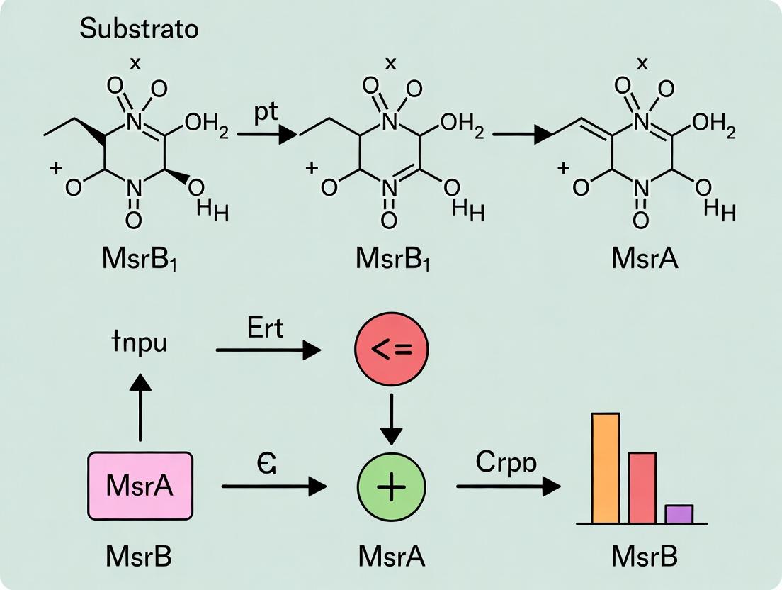
Methionine sulfoxide (Met-O) exists as two distinct stereoisomers due to the chiral center at the sulfur atom: Methionine-S-sulfoxide (Met-S-SO) and Methionine-R-sulfoxide (Met-R-SO). This stereospecificity is the cornerstone of the methionine sulfoxide reductase (Msr) system, comprising MsrA and MsrB1. MsrA specifically reduces the S-epimer, while MsrB1 reduces the R-epimer. Understanding these isomers is critical for elucidating protein repair mechanisms, redox regulation, and the development of therapeutics targeting age-related diseases and oxidative stress pathologies.
Defining the Isomers: Core Structural & Energetic Data
| Parameter | Met-S-SO | Met-R-SO |
|---|---|---|
| Cahn-Ingold-Prelog (CIP) Configuration | S at sulfur | R at sulfur |
| Absolute Configuration | L-Met-(S)-SO | L-Met-(R)-SO |
| Preferred Substrate for | MsrA | MsrB1 |
| Relative Free Energy (DFT calc.) | ~0.3 kcal/mol more stable | ~0.3 kcal/mol less stable |
| Typical Abundance in Oxidized Proteins | ~65-70% | ~30-35% |
| Chromatographic Retention (RP-HPLC) | Elutes earlier | Elutes later |
Key Experimental Protocols in Msr Research
Synthesis and Isolation of Diastereomerically Pure Met-O
Principle: Oxidation of L-Methionine with controlled reagents yields enantioenriched sulfoxides. Protocol:
- Met-S-SO Enriched: React 10 mM L-Methionine with 12 mM H₂O₂ in 10% acetic acid for 30 min at 25°C. Quench with catalase.
- Met-R-SO Enriched: React 10 mM L-Methionine with 0.5 M Sodium periodate (NaIO₄) in water for 2 hrs at 4°C in the dark.
- Purification: Separate diastereomers via reverse-phase HPLC (C18 column) using an isocratic mobile phase of 2% methanol in 50 mM ammonium acetate, pH 6.5. Monitor at 215 nm.
Enzymatic Activity Assay for MsrA/MsrB1 Specificity
Principle: Measure NADPH consumption coupled to thioredoxin/thioredoxin reductase system. Protocol:
- Prepare 1 mL reaction mix: 50 mM Tris-HCl (pH 7.5), 100 µM NADPH, 10 µM Thioredoxin (Trx), 0.5 µM Thioredoxin Reductase (TrxR), 5 mM DTT.
- Add 10 µM purified MsrA or MsrB1 enzyme.
- Initiate reaction by adding 500 µM substrate (Met-S-SO, Met-R-SO, or racemic mixture).
- Monitor decrease in absorbance at 340 nm (NADPH) for 5 minutes at 25°C.
- Calculate specific activity (µmol NADPH oxidized min⁻¹ mg⁻¹).
In-Gel Msr Activity Assay (Zymography)
Principle: Non-reducing PAGE separates proteins; post-run renaturation and activity staining localize Msr activity. Protocol:
- Run protein sample (cell lysate) on 15% non-reducing SDS-PAGE.
- Renature gel by washing in 50 mM Tris-HCl (pH 7.5), 1% Triton X-100 for 1 hr.
- Incubate gel in activity buffer (50 mM Tris-HCl pH 7.5, 1 mM DTT, 0.5 mM Methyl Viologen, 0.24 mM MTT, 3 mM NBT) containing 5 mM specific Met-O isomer for 30-45 min in the dark.
- Msr activity appears as dark blue bands (formazan deposit) against a clear background.
Visualization of Key Pathways and Relationships
Diagram 1: Stereospecific Reduction of Methionine Sulfoxides
Diagram 2: Coupled Enzyme Assay for Msr Activity
The Scientist's Toolkit: Research Reagent Solutions
| Reagent/Material | Function in Msr/Met-O Research |
|---|---|
| Diastereomerically Pure Met-S-SO / Met-R-SO | Substrates for determining stereospecific enzymatic activity of MsrA vs. MsrB1. |
| Recombinant Human MsrA & MsrB1 Proteins | Purified enzymes for kinetic assays, structural studies, and inhibitor screening. |
| Coupled Enzyme Assay Kit (NADPH/Trx/TrxR) | Standardized system for continuous, spectrophotometric measurement of Msr reductase activity. |
| Anti-Methionine Sulfoxide Antibodies (S/R specific) | Detect and quantify global protein Met-O epimers in cells/tissues via ELISA or Western blot. |
| MsrA/MsrB1 Knockout Cell Lines (e.g., CRISPR) | Genetic models to study the physiological consequence of losing specific repair pathways. |
| Chiral Derivatization Agents (e.g., Nα-(2,4-Dinitro-5-fluorophenyl)-L-alaninamide) | Enable resolution and quantification of Met-S-SO and Met-R-SO by HPLC/MS. |
| Selective Msr Inhibitors (e.g., ebselen analogues) | Chemical probes to dissect pathway function and potential therapeutic leads. |
| Thioredoxin Reductase Inhibitor (Auranofin) | Control to validate specificity in coupled assays; inhibits the recycling system. |
Evolutionary Origins and Genomic Distribution of the MsrA and MsrB Enzyme Families
This whitepaper examines the evolutionary and genomic foundations of the methionine sulfoxide reductase (Msr) enzyme families, with particular focus on the substrate specificity divergence between MsrA and MsrB1. The broader thesis investigates the structural and catalytic determinants that underlie the stereospecific reduction of methionine-S-sulfoxide (Met-S-SO) by MsrA versus methionine-R-sulfoxide (Met-R-SO) by MsrB1. Understanding the evolutionary trajectory of these families is critical for rational drug design targeting age-related diseases, infections, and conditions linked to oxidative stress where methionine oxidation is a key pathological marker.
Evolutionary Origins and Phylogenetic Distribution
Methionine sulfoxide reductases are ancient enzymes crucial for oxidative stress repair. Phylogenetic analyses indicate a deep evolutionary divergence.
Table 1: Evolutionary Origins of Msr Families
| Feature | MsrA Family | MsrB Family |
|---|---|---|
| Proposed Origin | Last Universal Common Ancestor (LUCA) | Likely originated in bacteria, laterally transferred to eukaryotes and archaea |
| Primary Domain | Rossmann-fold domain for NAD(P)H binding | Thioredoxin-like fold |
| Key Evolutionary Event | Gene duplication events leading to cytosolic (MsrA), mitochondrial (MsrA2) forms | Evolution of selenocysteine (Sec)-containing forms (e.g., mammalian MsrB1) from cysteine ancestors |
| Presence in Kingdoms | Ubiquitous in bacteria, archaea, eukaryotes | Widespread in bacteria and eukaryotes; patchy in archaea |
| Paralog Diversity | Limited (typically 1-2 genes per genome) | Greater diversity (e.g., 3 in humans: MsrB1 (Sec), MsrB2 (mito), MsrB3 (ER)) |
Genomic Distribution and Gene Architecture
Comparative genomics reveals distinct patterns in gene structure, localization, and regulation.
Table 2: Genomic Characteristics of Human Msr Genes
| Gene | Chromosomal Location | Exons | Protein Length (aa) | Subcellular Localization | Cofactor / Key Residue |
|---|---|---|---|---|---|
| MSRA | 8p23.1 | 6 | 235 | Cytosol, Nucleus, Mitochondria | Cys-72, Cys-218 (Catalytic) |
| MSRB1 | 16p13.3 | 5 | 255 (with Sec) | Cytosol, Nucleus | Sec-95 (Catalytic), Cys-4, Cys-100 |
| MSRB2 | 10p12.1 | 4 | 179 | Mitochondria | Cys-95 (Catalytic) |
| MSRB3 | 12q14.3 | 8 | 284/237 (splice forms) | Endoplasmic Reticulum | Cys-169 (Catalytic) |
Table 3: Distribution Across Model Organisms (Presence/Absence)
| Organism | MsrA | MsrB (Cys) | MsrB (Sec) | Notes |
|---|---|---|---|---|
| E. coli | Yes (1) | Yes (1) | No | Separate genes for S- and R- reduction |
| S. cerevisiae (Yeast) | Yes (1) | Yes (1) | No | MsrB is cysteine-dependent |
| C. elegans | Yes (1) | Yes (1) | No | |
| D. melanogaster | Yes (1) | Yes (2) | No | Two MsrB paralogs |
| M. musculus (Mouse) | Yes (2) | Yes (3) | Yes (MsrB1) | MsrA2 is mitochondrial; MsrB1 contains Sec |
| H. sapiens (Human) | Yes (1) | Yes (3) | Yes (MsrB1) | Single MsrA gene produces multiple isoforms |
Experimental Protocols for Evolutionary and Specificity Studies
Protocol: Phylogenetic Tree Construction for Msr Family Analysis
Objective: Reconstruct evolutionary relationships of MsrA and MsrB proteins.
- Sequence Retrieval: Use BLASTP (NCBI) with human MsrA (NP036354.2) and MsrB1 (NP001034304.1) as seeds against non-redundant protein databases. Set E-value cutoff at 1e-10.
- Multiple Sequence Alignment: Perform alignment using Clustal Omega or MAFFT with default parameters. Manually curate to trim poorly aligned termini.
- Model Selection: Use ProtTest or ModelFinder to determine best-fit amino acid substitution model (e.g., LG+G+I).
- Tree Building: Employ Maximum Likelihood method with RAxML (1000 bootstrap replicates) or Bayesian Inference with MrBayes (1,000,000 generations).
- Visualization & Interpretation: Use FigTree or iTOL to annotate tree, highlighting gene duplication events and Sec/Cys divergence.
Protocol: Determining Substrate Specificity (MsrB1 vs. MsrA)
Objective: Quantify enzymatic activity against Met-S-SO vs. Met-R-SO.
- Protein Purification: Express recombinant His-tagged human MsrA and MsrB1 in E. coli BL21(DE3). For MsrB1, use SECIS-containing vector and culture in Sec-supplemented media for selenoprotein expression. Purify via Ni-NTA affinity chromatography.
- Substrate Preparation: Prepare 10 mM solutions of Dabsyl-Met-S-SO and Dabsyl-Met-R-SO (Sigma-Aldrich) in assay buffer (50 mM Tris-HCl, pH 7.5, 50 mM NaCl).
- Activity Assay: In a 100 µL reaction, mix 50 µM substrate, 2 mM DTT (as electron donor), and 5 µg of purified enzyme. Incubate at 37°C for 30 min.
- Reaction Termination & Analysis: Stop reaction with 20 µL of 20% trichloroacetic acid, vortex, and centrifuge. Analyze supernatant by reverse-phase HPLC (C18 column) with detection at 440 nm. Quantify reduction by comparing peak areas of substrate (oxidized) and product (Met).
- Kinetic Analysis: Repeat assay with varying substrate concentrations (5–200 µM). Calculate Km and kcat using Michaelis-Menten non-linear regression (GraphPad Prism).
Table 4: Typical Substrate Specificity Data (from Recent Literature)
| Enzyme | Substrate | Km (µM) | kcat (s⁻¹) | kcat/Km (M⁻¹s⁻¹) | Stereospecificity Index (R/S) |
|---|---|---|---|---|---|
| MsrA | Dabsyl-Met-S-SO | 45 ± 5 | 2.1 ± 0.2 | 4.7 x 10⁴ | >1000 |
| MsrA | Dabsyl-Met-R-SO | >1000 | <0.01 | <10 | |
| MsrB1 | Dabsyl-Met-R-SO | 28 ± 3 | 1.8 ± 0.1 | 6.4 x 10⁴ | >1000 |
| MsrB1 | Dabsyl-Met-S-SO | >2000 | <0.01 | <5 |
Pathways and Workflows: Visualizations
Diagram 1 Title: Proposed Evolutionary Pathway of MsrA and MsrB Families
Diagram 2 Title: Experimental Workflow for Substrate Specificity Assay
Diagram 3 Title: Msr Catalytic & Electron Recycling Pathways
The Scientist's Toolkit: Key Research Reagent Solutions
Table 5: Essential Reagents for Msr Substrate Specificity Research
| Reagent / Material | Supplier Examples | Function in Research |
|---|---|---|
| Recombinant Human MsrA & MsrB1 Proteins | Novus Biologicals, Abcam | Positive controls, substrate for structural studies, enzymatic assays. |
| Dabsyl-Met-S-SO & Dabsyl-Met-R-SO | Sigma-Aldrich, Cayman Chem | Stereospecific chromogenic substrates for HPLC-based activity assays. |
| Anti-MsrA / Anti-MsrB1 Antibodies | Proteintech, Santa Cruz | Western blot validation, immunohistochemistry, ELISA for protein expression quantification. |
| Selenocysteine Supplement (Na2SeO3) | Sigma-Aldrich | Essential for expression of recombinant selenoprotein MsrB1 in culture media. |
| pET-28a(+) Msr Expression Vectors | Addgene, custom synthesis | Cloning and high-yield recombinant protein expression in E. coli. |
| Ni-NTA Superflow Agarose | Qiagen, Cytiva | Immobilized metal affinity chromatography for purification of His-tagged Msr proteins. |
| Human Thioredoxin Reductase (TrxR1) | Sigma-Aldrich | Component of the physiological electron transfer system for coupled activity assays. |
| TRIzol Reagent | Thermo Fisher | RNA isolation for gene expression analysis (qPCR) of MSRA and MSRB1 under oxidative stress. |
| Crystal Screen Kits | Hampton Research | Screening conditions for protein crystallization of Msr-substrate complexes. |
| Site-Directed Mutagenesis Kit | Agilent, NEB | Generating catalytic mutant proteins (e.g., MsrB1 Sec95Cys) for mechanistic studies. |
This whitepaper provides an in-depth technical analysis of the core structural architectures of Methionine Sulfoxide Reductase A (MsrA) and Methionine Sulfoxide Reductase B1 (MsrB1). It is framed within a broader research thesis investigating the structural determinants underlying their distinct substrate specificities—MsrA for the S-epimer and MsrB1 for the R-epimer of methionine sulfoxide. Understanding these blueprints is critical for rational drug design targeting redox-related pathologies.
Core Structural Architectures
MsrA and MsrB1, while fulfilling a related enzymatic function, have evolved distinct structural folds and catalytic frameworks.
MsrA Architecture
MsrA typically exhibits a compact, globular fold centered on a core βαβ-ββα structure, often described as a "twisted β-sheet" flanked by α-helices. The catalytic site features a conserved CXXC motif, where the N-terminal cysteine acts as the nucleophile attacking the sulfoxide substrate, and the C-terminal cysteine serves as the resolving cysteine. A critical structural feature is a concave, relatively accessible substrate-binding pocket that accommodates the free methionine sulfoxide or Met-S-O within polypeptide contexts.
MsrB1 Architecture
MsrB1 belongs to the Trx-fold superfamily, characterized by a central β-sheet (typically 4-5 strands) surrounded by α-helices. Its catalytic mechanism utilizes a single conserved catalytic cysteine that forms a sulfenic acid intermediate. Regeneration requires interaction with thioredoxin (Trx). A defining structural element is a deep, narrow substrate-binding pocket with precise stereochemical constraints, dictated by surrounding residues like Trp, which creates a chiral environment selective for the R-epimer. Mammalian MsrB1 is a selenocysteine (Sec)-containing protein, where Sec (U) replaces the catalytic cysteine, significantly enhancing its catalytic efficiency.
Table 1: Quantitative Comparison of Core Structural & Catalytic Features
| Feature | MsrA | MsrB1 |
|---|---|---|
| Primary Fold | Compact globular, twisted β-sheet | Trx-fold (central β-sheet surrounded by α-helices) |
| Catalytic Motif | CXXC (Two-Cysteine mechanism) | C/U (Single Cys/Sec mechanism) |
| Catalytic Cysteine | Cys (All species) | Sec (Higher eukaryotes), Cys (Yeast, bacteria) |
| Resolving Agent | Internal C-terminal Cys (CXXC) | External Thioredoxin (Trx) |
| Substrate Specificity | Met-(S)-SO (Free & in proteins) | Met-(R)-SO (Free & in proteins) |
| Key Binding Pocket | Concave, relatively accessible | Deep, narrow, stereospecific (e.g., Trp gate) |
| Approx. Active Site pKa | ~6.5-7.5 (N-term Cys) | ~5.5 (Sec, due to SeH lower pKa) |
| Redox Partner | Thioredoxin (Trx) / Glutaredoxin | Thioredoxin (Trx) specifically |
| Metal Binding (Zn²⁺) | Often present (structural role) | Not typically present |
Detailed Experimental Protocols for Structural & Specificity Analysis
Protocol: Site-Directed Mutagenesis & Kinetic Assay for Specificity Determinants
Objective: To identify residues governing epimer specificity by mutating putative binding pocket residues and measuring kinetic parameters.
- Design Primers: Design oligonucleotide primers containing the desired point mutation (e.g., MsrB1 Trp to Ala).
- PCR Amplification: Perform PCR using a high-fidelity polymerase on the plasmid encoding the wild-type msrA or msrB1 gene.
- DpnI Digestion: Treat PCR product with DpnI endonuclease to digest methylated parental DNA template.
- Transformation: Transform the nicked plasmid DNA into competent E. coli, screen colonies, and sequence to confirm mutation.
- Protein Expression & Purification: Express recombinant wild-type and mutant proteins in E. coli (using a Sec-incorporation system for MsrB1-Sec). Purify via affinity chromatography (e.g., His-tag).
- Enzyme Kinetics: Assay activity using a coupled spectrophotometric assay with DTNB (Ellman's reagent) or a Trx-regeneration system. Use separate substrates: Met-(S)-SO and Met-(R)-SO.
- Reaction Mix (1 mL): 50 mM Tris-HCl (pH 7.5), 150 mM NaCl, 1 mM EDTA, 0.2 mM NADPH, 5 μM E. coli Trx, 0.5 μM Trx Reductase, variable [Met-SO substrate] (0.05-5 mM), and 0.1-0.5 μM Msr enzyme.
- Measurement: Monitor NADPH oxidation at 340 nm (ε₃₄₀ = 6220 M⁻¹cm⁻¹) for 2-5 min. Calculate kcat and KM using nonlinear regression to the Michaelis-Menten equation.
Protocol: X-ray Crystallography of Enzyme-Substrate Analog Complexes
Objective: To obtain high-resolution structural snapshots defining substrate binding modes.
- Protein Crystallization: Purify protein to >95% homogeneity. Use sitting-drop vapor diffusion. Mix 1 μL of protein (10-20 mg/mL in low-salt buffer) with 1 μL of reservoir solution containing precipitant (e.g., PEG 3350) and 2-5 mM of a non-reducible substrate analog (e.g., Methionine Sulfone).
- Crystal Harvesting & Soaking: Flash-cool crystals in liquid N₂ using a cryoprotectant (e.g., reservoir solution + 25% glycerol).
- Data Collection & Processing: Collect diffraction data at a synchrotron source. Index, integrate, and scale data using HKL-3000 or XDS.
- Structure Solution & Refinement: Solve phase problem by molecular replacement using a known apo-structure (PDB ID: e.g., 1U1A for MsrA). Build model in Coot and refine with Phenix.refine.
- Analysis: Analyze electron density in the active site. Model the substrate analog and map interacting residues (H-bonds, van der Waals contacts) to define the stereospecific pocket.
Visualization of Catalytic Pathways & Research Workflow
Diagram Title: MsrA vs. MsrB1 Catalytic & Regeneration Cycles
Diagram Title: Research Workflow for Specificity Determination
The Scientist's Toolkit: Key Research Reagent Solutions
Table 2: Essential Reagents for MsrA/MsrB1 Specificity Research
| Reagent / Material | Function / Application | Key Notes |
|---|---|---|
| Recombinant MsrA & MsrB1 Proteins | Substrate for kinetic assays, crystallization, and binding studies. | Essential to produce both wild-type and site-directed mutants. MsrB1 requires Sec-incorporation system for native activity. |
| D/L-Methionine (R/S) Sulfoxide | Defined stereoisomeric substrates for specificity assays. | Must be chromatographically separated or purchased as pure epimers (e.g., Met-(R)-SO, Met-(S)-SO). |
| Thioredoxin (Trx) System | Regeneration system for enzymatic turnover assays. | Includes E. coli or human Trx, Trx Reductase, and NADPH. Critical for MsrB1 and MsrA kinetics. |
| DTNB (Ellman's Reagent) | Spectrophotometric detection of free thiols. | Used in endpoint assays to measure reduction of dithiol motifs or substrate consumption. |
| Crystallization Screening Kits | Identification of conditions for protein crystal growth. | Commercial sparse-matrix screens (e.g., from Hampton Research) are standard. |
| Non-reducible Substrate Analogs (e.g., Methionine Sulfone) | Trapping enzyme in substrate-bound state for crystallography. | Prevents catalysis during crystallization, allowing structural determination of the Michaelis complex. |
| Sec-Incorporation Plasmid System (e.g., pSUABC) | High-fidelity expression of selenoproteins in E. coli. | Required for producing recombinant mammalian MsrB1 with selenocysteine. |
| Anti-Msr Antibodies | Detection and quantification in cellular/tissue lysates. | Used in Western blot, immunoprecipitation, or immunofluorescence for localization studies. |
| HPLC/UPLC with Chiral Columns | Analytical separation and quantification of methionine sulfoxide epimers. | Gold standard for direct measurement of substrate preference and enzyme stereospecificity. |
Within the broader context of research on methionine sulfoxide reductase (Msr) substrate specificity—comparing MsrA (responsible for reducing the S-epimer of methionine sulfoxide, Met-S-SO) and MsrB1 (specific for the R-epimer, Met-R-SO)—the reductive recycling of their catalytic cysteine residues is a critical, defining component. The catalytic mechanisms of both enzymes culminate in a sulfenylated active site (Cys-SOH), which must be reduced to regenerate the active enzyme. This whitepaper provides an in-depth, comparative analysis of the distinct reductive recycling pathways for MsrA and MsrB1, which are fundamental to their biological activity, regulation, and potential as therapeutic targets.
Core Catalytic and Recycling Pathways
The overall catalysis involves two steps: 1) Methionine sulfoxide reduction and 2) Active site reductive recycling. While the first step is mechanistically similar, the recycling pathways diverge significantly.
Step 1: Sulfoxide Reduction Both MsrA and MsrB1 utilize three conserved cysteine residues (catalytic, recycling, and resolving) to reduce their respective Met-SO epimers. The catalytic cysteine performs a nucleophilic attack on the sulfur of Met-SO, forming a sulfenic acid intermediate (Cys-SOH) and releasing reduced methionine.
Step 2: Reductive Recycling The critical divergence lies in how the Cys-SOH is reduced.
- MsrA Pathway: The recycling cysteine attacks the sulfenic acid, forming an intramolecular disulfide bond. This disulfide is subsequently reduced by the thioredoxin (Trx) system (NADPH, thioredoxin reductase (TrxR), and thioredoxin).
- MsrB1 Pathway (Selenocysteine-containing form): The human MsrB1 contains a catalytic selenocysteine (Sec) residue. The selenenic acid (Sec-SeOH) intermediate is primarily recycled by the glutaredoxin (Grx) system and low molecular weight thiols like glutathione (GSH), or by the thioredoxin system, but with different efficiency. The selenolate is highly reactive and can be regenerated directly by low molecular weight thiols without a stable diselenide bond intermediate in some contexts.
Key Comparative Data:
Table 1: Comparative Features of MsrA and MsrB1 Reductive Recycling
| Feature | MsrA | MsrB1 (Sec-containing) |
|---|---|---|
| Catalytic Residue | Cysteine (Cys) | Selenocysteine (Sec) |
| Recycling Residue | Recycling Cysteine (Cys) | Can involve resolving Cys or direct thiol reduction |
| Primary Reductant System | Thioredoxin (Trx) system | Glutaredoxin (Grx)/Glutathione (GSH) system |
| Key Physiological Reductant | Thioredoxin (Trx) | Glutaredoxin (Grx3), Glutathione (GSH) |
| Disulfide/Selenylsulfide Intermediate? | Yes (Intramolecular disulfide) | Yes, but less stable; direct reduction possible |
| Approximate Catalytic Rate (kcat) | 0.5 - 2.0 min⁻¹ | 5 - 20 min⁻¹ (higher due to Sec reactivity) |
| Km for Trx (μM) | ~10 - 50 μM | >100 μM (lower affinity) |
| Km for GSH/Grx (μM) | High (low efficiency) | ~1-5 mM for GSH; Low μM for Grx |
Table 2: Reductant System Components and Cofactors
| System | Components | Cofactor | Primary Role in Recycling |
|---|---|---|---|
| Thioredoxin (Trx) | TrxR, Trx, NADPH | FAD | Electron transfer to reduce MsrA disulfide. |
| Glutaredoxin (Grx)/GSH | Grx, GSH, GR, NADPH | FAD | Maintains GSH pool; Grx directly reduces MsrB1. |
| NADPH | N/A | N/A | Ultimate electron donor for both major systems. |
Detailed Experimental Protocols
Protocol 1: Assessing Reductant Specificity via Coupled Enzyme Assay Objective: Determine the efficiency of Trx vs. Grx/GSH systems in recycling recombinant human MsrA or MsrB1.
- Reaction Mix (100 μL): 50 mM Tris-HCl (pH 7.5), 150 mM NaCl, 2 mM EDTA.
- Add Reductant System:
- Trx System: 0.2 mM NADPH, 100 nM TrxR, 10 μM Trx.
- Grx System: 0.2 mM NADPH, 1 mM GSH, 1 μM Grx, 100 nM Glutathione Reductase (GR).
- Initiate Reaction: Add 50-100 nM purified MsrA or MsrB1 and substrate (e.g., 1-5 mM Met-R-SO for MsrB1 or Met-S-SO for MsrA).
- Monitor: Observe NADPH oxidation by decrease in absorbance at 340 nm (ε₃₄₀ = 6220 M⁻¹cm⁻¹) for 5-10 minutes at 37°C.
- Analysis: Calculate initial velocity (V₀). Compare V₀ between reductant systems for each enzyme.
Protocol 2: Trapping the Sulfenic/Selenenic Acid Intermediate Objective: Chemically trap the Cys-SOH/Sec-SeOH intermediate to confirm its formation.
- Reduce & Purify: Fully reduce recombinant Msr enzyme with 10 mM DTT and remove DTT via gel filtration.
- Intermediate Formation: Incubate enzyme (10 μM) with a stoichiometric amount of substrate (Met-SO) for 30 seconds at 25°C.
- Trapping: Rapidly add the specific sulfenic acid trap, 5,5-dimethyl-1,3-cyclohexanedione (dimedone, 10 mM final). Incubate for 15 minutes.
- Analysis: Quench reaction and analyze by liquid chromatography-mass spectrometry (LC-MS) to detect the mass addition of dimedone (+138 Da) to the catalytic residue.
Visualization of Pathways
Title: Comparative Reductive Recycling Pathways of MsrA and MsrB1
The Scientist's Toolkit: Research Reagent Solutions
Table 3: Essential Reagents for Msr Reductive Recycling Studies
| Reagent | Function/Specificity in Experiment | Key Considerations |
|---|---|---|
| Recombinant Human MsrA & MsrB1 | Purified enzyme substrates for in vitro kinetics. | Ensure MsrB1 contains selenocysteine; confirm activity with epimer-specific substrates. |
| S-Methyl-L-Methionine Sulfoxide | Substrate for MsrA activity assays. | Use high-purity to avoid R-epimer contamination. |
| R-Methyl-L-Methionine Sulfoxide | Substrate for MsrB1 activity assays. | Critical for assessing true MsrB1 activity. |
| Human Thioredoxin-1 (Trx) & Thioredoxin Reductase (TrxR) | Components of the Trx reductant system. | Use NADPH-dependent TrxR for physiological relevance. |
| Human Glutaredoxin-1 (Grx1) & Glutathione Reductase (GR) | Components of the Grx/GSH reductant system. | Requires GSH as a co-substrate. |
| β-Nicotinamide adenine dinucleotide phosphate (NADPH) | Ultimate electron donor for both recycling systems. | Monitor stability; prepare fresh solutions in buffer. |
| Reduced Glutathione (GSH) | Low molecular weight thiol for MsrB1 recycling. | Keep pH >6.5 to prevent oxidation; use fresh stocks. |
| 5,5-Dimethyl-1,3-cyclohexanedione (Dimedone) | Selective chemical probe for trapping sulfenic/selenenic acid intermediates. | Cell-permeable variants exist for in situ studies. |
| Dithiothreitol (DTT) / Tris(2-carboxyethyl)phosphine (TCEP) | Non-physiological reducing agents for initial enzyme activation. | Must be removed prior to assays to study physiological reductants. |
The methionine sulfoxide reductase (Msr) system is a critical enzymatic defense against oxidative damage, with MsrA and MsrB1 representing the primary catalysts for the reduction of the S- and R-epimers of methionine sulfoxide (Met-S-O and Met-R-O), respectively. A central thesis in redox biology posits that the divergent substrate specificity of MsrA and MsrB1 is intrinsically linked to their distinct subcellular localizations, thereby defining non-overlapping physiological functions. This guide delves into the mechanistic basis of this linkage, exploring how compartment-specific substrate pools, protein interaction networks, and signaling outcomes are dictated by the precise catalytic preference of each enzyme. Understanding this relationship is paramount for drug development targeting age-related diseases, neurodegeneration, and infections where spatial redox regulation is disrupted.
Substrate Specificity: Structural & Kinetic Foundations
The enantiomeric specificity of MsrA and MsrB1 is an absolute determinant of their biological roles. Recent structural and biochemical analyses confirm this strict division of labor.
Table 1: Core Specificity and Properties of MsrA and MsrB1
| Property | MsrA | MsrB1 (SelR/SelX) |
|---|---|---|
| Primary Substrate | Free and protein-bound Methionine-S-sulfoxide (Met-S-O) | Free and protein-bound Methionine-R-sulfoxide (Met-R-O) |
| Catalytic Mechanism | 3-step mechanism via a sulfenic acid intermediate; uses Cys residues. | 3-step mechanism via a selenenylsulfide intermediate; uses Sec (Cys in some isoforms) and resolving Cys. |
| Cofactor | Thioredoxin (Trx) / Trx reductase / NADPH system | Thioredoxin (Trx) / Trx reductase / NADPH system |
| Metal Binding | No | Yes (Zinc) – structural role, crucial for fold and catalytic site integrity. |
| Gene | MSRA | MSRB1 (Selenoprotein R) |
Experimental Protocol: Kinetic Analysis of Substrate Specificity
- Objective: Determine kinetic parameters (Km, kcat) for MsrA and MsrB1 against Met-S-O and Met-R-O substrates.
- Reagents: Recombinant human MsrA and MsrB1, Dabsyl-Met-S-O, Dabsyl-Met-R-O, DTT (as electron donor), reaction buffer (e.g., 50 mM HEPES, pH 7.4).
- Method:
- Enzyme Assay: Set up reactions containing fixed enzyme concentration and varying concentrations of dabsylated substrate (e.g., 0-2 mM). Initiate reaction with DTT.
- Detection: Terminate reactions at timed intervals with acidic methanol. Separate product (dabsyl-methionine) from substrate via reverse-phase HPLC.
- Quantification: Monitor absorbance at 460 nm. Calculate reaction velocity based on product formation.
- Analysis: Fit data to the Michaelis-Menten equation using software (e.g., GraphPad Prism) to derive Km and kcat values. This assay robustly demonstrates MsrA's high affinity for Met-S-O (low Km) and negligible activity on Met-R-O, and vice-versa for MsrB1.
Cellular Localization Dictates Functional Compartmentalization
The distinct subcellular targeting of MsrA and MsrB1 creates specialized redox repair compartments.
Table 2: Localization and Compartment-Specific Functions
| Enzyme | Major Localization | Minor/Alternative Localization | Compartment-Specific Substrate Pools & Functions |
|---|---|---|---|
| MsrA | Cytosol, Mitochondria, Nucleus | Secreted (via non-classical pathway) | Mitochondria: Repairs oxidative damage to respiratory chain complexes (e.g., COX subunit I). Critical for mitochondrial membrane potential and ATP production. Nucleus: Protects histones and transcription factors from oxidation, influencing gene expression. Cytosol: Repairs metabolic enzymes and cytoskeletal proteins. |
| MsrB1 | Cytosol, Nucleus | - | Nucleus/Cytosol: Specifically repairs the R-sulfoxide epimer in key targets like Actin (critical for cytoskeletal dynamics), ER chaperones, and transcription factors (e.g., p53, NF-κB). Its activity is often coupled to its protein substrates' localization. |
Linking Specificity & Localization to Physiological Roles
The confluence of specificity and localization translates into defined cellular and organismal phenotypes.
Table 3: Phenotypic Consequences of MsrA or MsrB1 Deficiency
| Model System | MsrA Knockout/Deficiency Phenotypes | MsrB1 Knockout/Deficiency Phenotypes |
|---|---|---|
| In Vitro (Mammalian Cells) | Increased sensitivity to H2O2; mitochondrial dysfunction; increased protein carbonyls; altered cytoskeleton. | Actin disorganization; increased protein Met-R-O; enhanced sensitivity to nitrosative stress; impaired cell migration. |
| In Vivo (Mouse Models) | Shortened lifespan (≈40% reduction); neurological deficits (ataxia), increased susceptibility to infection, severe metabolic syndrome. | Cataract development, sensitivity to paraquat-induced lung injury, altered immune response. |
| Human Disease Correlation | Potential link to Alzheimer's disease (increased Abeta production), Parkinson's disease, age-related hearing loss. | Associated with age-related cataract; potential role in tumor progression via actin regulation. |
Experimental Protocol: Localization-Based Substrate Identification (e.g., Proximity Labeling with APEX2)
- Objective: Identify compartment-specific protein substrates of MsrB1 in the cytosol vs. nucleus.
- Reagents: HEK293T cells, plasmid expressing MsrB1-APEX2 fusion, biotin-phenol, H2O2, streptavidin beads, mass spectrometry reagents.
- Method:
- Transfection & Labeling: Express MsrB1-APEX2 in cells. Add biotin-phenol to culture medium for 30 min.
- Activation: Initiate proximity labeling by adding 1 mM H2O2 for 1 minute. Quench with Trolox and ascorbate.
- Lysis & Pulldown: Lyse cells. Incubate lysate with streptavidin-coated magnetic beads to capture biotinylated proteins (proximal to MsrB1).
- Analysis: Elute proteins and identify by quantitative mass spectrometry (LC-MS/MS). Compare substrates enriched in a wild-type MsrB1-APEX2 experiment versus a catalytically dead (Sec/Cys mutant) control to distinguish functional interactions.
Visualization of Pathways and Workflows
Title: MsrA vs. MsrB1 Specificity & Localization Workflow
Title: MsrB1 Repair of NF-κB Signaling Pathway
The Scientist's Toolkit: Key Research Reagent Solutions
Table 4: Essential Reagents for Msr Substrate Specificity and Localization Research
| Reagent / Material | Function / Application | Example / Supplier |
|---|---|---|
| Recombinant Enzymes | Purified human MsrA and MsrB1 for kinetic assays, structural studies, and in vitro repair assays. | Abcam, Novus Biologicals, in-house expression from E. coli or mammalian systems. |
| Chiral Substrates | Dabsyl- or other chromophore/fluorophore-tagged L-Met-S-O and L-Met-R-O for HPLC- or plate-based activity assays. | Cayman Chemical, Sigma-Aldrich, Bachem. |
| Antibodies | Anti-Met-O (pan or epimer-specific), anti-MsrA, anti-MsrB1 (SelR) for Western blot, immunofluorescence, IP. | Abcam, Santa Cruz Biotechnology, Invitrogen. Note: Highly specific anti-Met-O antibodies are crucial. |
| Localization Reporters | Fluorescent protein (GFP, mCherry) tagged Msr constructs for live-cell imaging of subcellular distribution. | Generated via molecular cloning (e.g., into pEGFP-N1 vector). |
| Proximity Labeling Systems | APEX2 or TurboID constructs for identifying protein-protein interactions and proximal substrates in specific compartments. | Addgene (APEX2 plasmids), commercial TurboID kits. |
| Redox Sensors | Genetically encoded sensors (e.g., roGFP, HyPer) to measure compartment-specific H2O2 or glutathione redox potential. | Addgene, commercial sources (Evrogen). |
| Knockout/Knockdown Tools | CRISPR-Cas9 guide RNAs for generating KO cell lines, or siRNA/shRNA for transient knockdown of MSRA or MSRB1. | Synthego, Horizon Discovery, Dharmacon. |
| Mass Spectrometry | LC-MS/MS platforms for identifying and quantifying methionine sulfoxide epimers in proteins and for proteomic analyses. | Orbitrap-based systems (Thermo Fisher), Q-TOF (Agilent, Waters). |
Tools of the Trade: Methodologies for Probing Msr Enzyme Specificity and Activity
Methionine sulfoxide reductases (Msrs) are critical enzymes responsible for the reduction of oxidized methionine residues, a key repair mechanism in cellular defense against oxidative stress. The two principal enzymatic families, MsrA and MsrB1, exhibit distinct and non-redundant stereospecificity: MsrA specifically reduces the S-epimer of methionine sulfoxide (Met-S-SO), while MsrB1 reduces the R-epimer (Met-R-SO). This inherent selectivity necessitates the design of precise stereospecific substrates—both native and synthetic—to probe their activity, elucidate physiological roles, and screen for potential modulators in drug development. This technical guide details the strategic design, synthesis, and application of such probes, framed within ongoing research to delineate the unique substrate recognition profiles of MsrB1 versus MsrA.
Strategic Design Principles for Stereospecific Probes
The core design challenge is creating substrates that are selectively recognized by one Msr isoform while remaining inert to the other. Key principles include:
- Stereochemical Fidelity: Utilizing pure stereoisomers of methionine sulfoxide or its analogs as the fundamental scaffold.
- Chemoselective Reporting Groups: Incorporating moieties (fluorophores, chromophores, or radiolabels) that yield a measurable signal upon reduction, without perturbing enzyme stereorecognition.
- Backbone Engineering: Modifying the peptide context or using non-peptidic scaffolds to modulate binding affinity and specificity, particularly for probing the more constrained active site of MsrB1 compared to MsrA.
- Cellular Permeability and Stability: For in vivo or cellular assays, probes may require esterification (e.g., acetoxymethyl esters) or incorporation into cell-penetrating peptides to facilitate delivery.
Key Research Reagent Solutions
| Reagent / Material | Function in Msr Substrate Specificity Research |
|---|---|
| L-Met-S-SO (Native Substrate) | The canonical native substrate for MsrA. Serves as a benchmark for enzymatic activity and kinetic parameter (Km, kcat) determination. |
| D-Met-R-SO (Native Substrate) | The canonical native substrate for MsrB1. Used to define baseline MsrB1 activity and inhibitor screening. |
| Dabsyl-Met-SO Diastereomers | Synthetic chromogenic substrates. The dabsyl group allows for HPLC or spectrophotometric separation and quantification of the reduction of individual Met-S-SO or Met-R-SO isomers. |
| Coupled Assay Reagents (NAPH/DTNB) | For continuous spectrophotometric assays. Msr reduction is coupled to thioredoxin/thioredoxin reductase, consuming NADPH, or uses DTNB (Ellman's reagent) to detect liberated thioredoxin. |
| Fluorogenic Probes (e.g., F-Met-SO) | Synthetic substrates where reduction of Met-SO quenches or shifts fluorescence. Enables high-throughput, real-time kinetic assays in microplate formats. |
| Anti-Met-R-SO Antibodies | Immunological tools to specifically detect protein-bound Met-R-SO, the physiological product repaired by MsrB1, in cell lysates or tissues. |
| Recombinant Human MsrA & MsrB1 | Essential purified enzyme sources for in vitro specificity profiling, free from cellular contaminants. Often His-tagged for immobilization or pulldown assays. |
| Selenocysteine-containing MsrB1 (Sec95) | The catalytically active form of human MsrB1. Requires expression in specialized systems (e.g., Cys-auxotrophic E. coli with selenite) or chemical synthesis for mechanistic studies. |
Table 1: Kinetic Parameters for Representative Native and Synthetic Substrates
| Enzyme | Preferred Substrate (Stereochemistry) | Reported Km (µM) | Reported kcat (min⁻¹) | kcat/Km (µM⁻¹min⁻¹) | Key Assay Method |
|---|---|---|---|---|---|
| MsrA | L-Met-S-SO (Free Amino Acid) | 50 - 200 | 500 - 1500 | ~10 | Coupled NADPH oxidation (Trx/TrxR) |
| MsrA | Dabsyl-L-Met-S-SO (Peptide) | 10 - 50 | 100 - 400 | ~8 | HPLC-based quantification |
| MsrB1 | D-Met-R-SO (Free Amino Acid) | 100 - 500 | 100 - 600 | ~1.5 | DTNB-based thiol detection |
| MsrB1 | Dabsyl-D-Met-R-SO (Peptide) | 20 - 100 | 50 - 200 | ~2.5 | HPLC-based quantification |
| MsrA | Ac-F-Met-S-SO-AM (Fluorogenic) | 5 - 25 | N/A | N/A | Fluorescence increase (HTRF/FP) |
| MsrB1 | Ac-F-Met-R-SO-AM (Fluorogenic) | 15 - 60 | N/A | N/A | Fluorescence increase (HTRF/FP) |
Note: Values are representative ranges compiled from recent literature; exact figures vary by expression system, assay conditions, and substrate presentation.
Detailed Experimental Protocols
Protocol 1: Stereospecific HPLC-Based Assay Using Dabsylated Substrates
Objective: Quantify MsrA and MsrB1 activity separately using diastereomerically pure, chromogenic substrates. Reagents: Recombinant MsrA/MsrB1, Dabsyl-Met-SO (S- or R- isomer), DTT, Thioredoxin (Trx), Thioredoxin Reductase (TrxR), NADPH, Potassium Phosphate Buffer (pH 7.5), HPLC system with C18 column. Procedure:
- Prepare reaction mix (50 µL final): 50 mM KPi pH 7.5, 1 mM DTT (or 10 µM Trx, 100 nM TrxR, 200 µM NADPH), 100 µM dabsyl-Met-SO substrate (S-isomer for MsrA, R-isomer for MsrB1).
- Pre-incubate at 37°C for 2 min.
- Initiate reaction by adding enzyme (10-100 nM final concentration).
- Incubate at 37°C for a fixed time (e.g., 5-30 min).
- Terminate reaction by adding 50 µL of 2% (v/v) trifluoroacetic acid (TFA).
- Centrifuge at 14,000 x g for 5 min.
- Inject supernatant onto HPLC with a C18 column. Use a gradient of 20% to 70% acetonitrile in 0.1% TFA over 25 min.
- Monitor absorbance at 436 nm. The reduced product (dabsyl-Met) elutes earlier than the substrate (dabsyl-Met-SO).
- Quantify activity based on the peak area of the product, using a standard curve.
Protocol 2: Continuous Spectrophotometric Coupled Assay
Objective: Real-time kinetic measurement of Msr activity via NADPH consumption. Reagents: Recombinant Msr, L-Met-S-SO (for MsrA) or D-Met-R-SO (for MsrB1), NADPH, E. coli Trx, E. coli TrxR, EDTA, Potassium Phosphate Buffer. Procedure:
- Prepare assay buffer: 50 mM KPi pH 7.5, 1 mM EDTA.
- In a cuvette, mix: 800 µL buffer, 50 µL Trx (final 10 µM), 50 µL TrxR (final 100 nM), 50 µL NADPH (final 200 µM).
- Add Msr enzyme (final 10-50 nM).
- Place cuvette in spectrophotometer thermostatted at 25°C.
- Monitor baseline absorbance at 340 nm (A340) for 1-2 min.
- Initiate reaction by adding 50 µL of Met-SO substrate (final concentration 0.1-2.0 mM for Michaelis-Menten kinetics).
- Record the decrease in A340 over 5-10 min. The molar extinction coefficient for NADPH (ε340 = 6220 M⁻¹cm⁻¹) is used to calculate reaction velocity.
Visualizations of Experimental Workflows and Logic
Diagram 1: Msr Stereospecific Assay Design Logic
Diagram 2: Coupled NADPH Oxidation Assay Workflow
This technical guide details the methodology for kinetically characterizing the methionine sulfoxide reductase enzymes MsrA and MsrB1. This work is framed within a broader research thesis investigating the fundamental differences in substrate specificity between MsrB1 (which reduces R-epimers of methionine sulfoxide in free and peptide-bound forms) and MsrA (which reduces the S-epimers). Precise determination of kinetic parameters (Km, kcat, and the specificity constant kcat/Km) is critical for quantifying each enzyme's catalytic efficiency and selectivity towards various substrates, including free Met-SO, Met-R-O, and methionine sulfoxide residues within protein contexts. Such data is foundational for understanding their distinct physiological roles in oxidative stress response and for informing drug development aimed at modulating their activity in age-related and neurodegenerative diseases.
Core Kinetic Parameters and Definitions
- Michaelis Constant (Km): The substrate concentration at half-maximal reaction velocity. A lower Km indicates higher apparent substrate affinity.
- Catalytic Constant (kcat): The turnover number, representing the maximum number of substrate molecules converted to product per enzyme active site per unit time.
- Specificity Constant (kcat/Km): The measure of catalytic efficiency. It incorporates both binding affinity and catalytic rate, allowing for direct comparison of an enzyme's preference for different substrates.
Experimental Protocols for Steady-State Kinetics
General Reaction Principle
Both MsrA and MsrB1 catalyze the thioredoxin-dependent reduction of methionine sulfoxide (Met-O) to methionine (Met). The reaction is coupled to the oxidation of NADPH by thioredoxin reductase, enabling continuous spectrophotometric monitoring of NADPH consumption at 340 nm (ε~340 = 6220 M⁻¹cm⁻¹).
Overall Reaction: Met-O + Thioredoxin-(SH)₂ → Met + Thioredoxin-(S)₂ Thioredoxin-(S)₂ + NADPH + H⁺ → Thioredoxin-(SH)₂ + NADP⁺
Standard Assay Protocol
Reaction Mix (in cuvette):
- Buffer: 50-100 mM HEPES or Tris-HCl, pH 7.0-7.5, containing 1-2 mM EDTA.
- Reductant System: 100-300 µM NADPH, 1-5 µM E. coli Thioredoxin (Trx), 50-100 nM Thioredoxin Reductase (TrxR).
- Substrate: Varying concentrations of the target methionine sulfoxide substrate (e.g., free Met-S-O, Met-R-O, or a model peptide like Ac-Met-O-NH₂). Prepare fresh stock solutions.
- Final Volume: Adjust to 0.9-0.95 mL with reaction buffer.
Initiation and Measurement:
- Pre-incubate the reaction mix (without enzyme) at 37°C for 2-3 minutes.
- Initiate the reaction by adding a small volume (10-50 µL) of purified MsrA or MsrB1 enzyme to achieve a final concentration in the low nM range (e.g., 10-100 nM).
- Immediately mix and monitor the decrease in absorbance at 340 nm (A~340) for 2-5 minutes using a spectrophotometer.
Control Reactions:
- Run a "no enzyme" control to account for non-specific NADPH oxidation.
- Run a "no substrate" control to account for any enzyme-independent Trx system activity.
Data Calculation:
- Calculate the initial velocity (v~0) for each substrate concentration [S] from the linear portion of the A~340 trace.
- v~0 (µM/min) = (∆A~340/min) / (6220 M⁻¹cm⁻¹ * path length (cm)) * 10⁶
Data Analysis for Parameter Determination
- Plot initial velocity (v~0) versus substrate concentration [S].
- Fit the data to the Michaelis-Menten equation: v~0 = (V~max [S]) / (K~m + [S]) using non-linear regression software (e.g., GraphPad Prism).
- V~max (maximum velocity) is derived from the fit. k~cat = V~max / [E], where [E] is the total molar concentration of active enzyme.
- The specificity constant is calculated directly: k~cat/K~m.
- For comparing substrates, linear transformation plots (e.g., Lineweaver-Burk) can be useful for visual inspection.
Table 1: Representative Kinetic Parameters for MsrA and MsrB1
Data synthesized from current literature. Values are approximate and can vary based on enzyme source (e.g., mammalian vs. bacterial) and assay conditions.
| Enzyme | Substrate (Epimer) | Km (µM) | kcat (min⁻¹) | kcat/Km (µM⁻¹ min⁻¹) | Notes |
|---|---|---|---|---|---|
| MsrA | Free L-Met-S-O | 50 - 200 | 100 - 500 | 2.0 - 2.5 | High efficiency for free S-epimer. |
| MsrA | Ac-Met-S-O-NH₂ (Peptide) | 100 - 400 | 80 - 400 | 0.8 - 1.0 | Accepts peptide-bound S-O. |
| MsrA | Free D-Met-R-O | > 5000 | < 10 | < 0.002 | Very low activity, defines specificity. |
| MsrB1 | Free L-Met-R-O | 100 - 400 | 50 - 200 | 0.5 - 0.6 | Specific for free R-epimer. |
| MsrB1 | Ac-Met-R-O-NH₂ (Peptide) | 200 - 800 | 20 - 100 | 0.1 - 0.13 | Lower efficiency on peptides. |
| MsrB1 | Protein-bound Met-R-O* | N/A | N/A | N/A | Physiologically relevant but Km difficult to determine. |
| MsrB1 | Free L-Met-S-O | > 5000 | < 5 | < 0.001 | Negligible activity, defines specificity. |
*Protein-bound substrate kinetics often require specialized assays (e.g., HPLC-based).
Visualization of Workflows and Relationships
Diagram 1: Kinetic Characterization Experimental Workflow
Diagram 2: Msr Enzyme Specificity in Redox Pathways
The Scientist's Toolkit: Essential Research Reagents
Table 2: Key Reagent Solutions for Msr Kinetics Assays
| Reagent | Function / Role in Assay | Key Considerations |
|---|---|---|
| Recombinant MsrA/MsrB1 | Enzyme of interest. Must be purified to homogeneity for accurate active-site titration. | Source (human, mouse, bacterial), storage buffer (often with DTT), determination of active concentration is critical. |
| Thioredoxin (Trx) System | Physiological electron donor couple. Provides reducing equivalents to Msr enzymes. | E. coli Trx/TrxR commonly used. Must be fresh and active. NADPH stability is key. |
| NADPH | Ultimate electron donor. Oxidation monitored at 340 nm to measure reaction rate. | Prepare fresh stock, keep on ice, protected from light. Verify concentration spectrophotometrically (A~340). |
| Methionine Sulfoxide Substrates | Varied epimers (Met-S-O, Met-R-O) and forms (free, acetylated, in peptides). | Source stereoisomeric purity is paramount. Prepare stocks in assay buffer or water daily to prevent hydrolysis/reduction. |
| HEPES/Tris Assay Buffer | Maintains optimal pH (7.0-7.5) for enzyme and coupling system activity. | Include EDTA (1-2 mM) to chelate metal ions that might catalyze non-enzymatic oxidation. |
| Spectrophotometer with Kinetics Software | For continuous monitoring of A~340 over time to calculate initial velocity (v~0). | Requires temperature control (37°C). Cuvette-based or plate-reader formats possible. |
| Data Analysis Software | Non-linear regression fitting of v~0 vs. [S] data to Michaelis-Menten model. | GraphPad Prism, SigmaPlot, or R. Essential for robust Km and V~max estimation. |
High-Throughput Screening (HTS) Approaches for Inhibitor and Activator Discovery
Within the context of methionine sulfoxide reductase (Msr) research, particularly the delineation of substrate specificity between MsrB1 (which reduces methionine-R-sulfoxide) and MsrA (which reduces methionine-S-sulfoxide), High-Throughput Screening (HTS) is a cornerstone technology. It enables the rapid identification of selective inhibitors or activators that can modulate these enzymes' activities. Such compounds are invaluable chemical probes for dissecting the distinct biological roles of MsrB1 and MsrA in redox homeostasis, aging, and neurodegenerative diseases, and serve as starting points for therapeutic development. This technical guide outlines the principal HTS approaches relevant to this field.
Core HTS Methodologies
Biochemical Activity-Based Screening
This is the most direct approach, measuring the compound's effect on the enzymatic reduction of methionine sulfoxide.
- General Protocol for MsrA/B Activity Assay (96/384/1536-well format):
- Reagent Dispensing: Add 10-20 µL of assay buffer (e.g., 50 mM Tris-HCl, pH 7.5, with DTT or thioredoxin recycling system) containing the target enzyme (recombinant human MsrA or MsrB1) to each well.
- Compound Addition: Pin-transfer or acoustically dispense 10-100 nL of test compounds from a library (typically at 1-10 mM in DMSO) into assay wells. Include control wells (DMSO-only for 100% activity, no enzyme for background, and a known inhibitor/activator if available).
- Pre-incubation: Incubate plate for 10-15 minutes at 25-30°C to allow compound-enzyme interaction.
- Reaction Initiation: Add 10-20 µL of substrate solution. For MsrA: Methionine-S-sulfoxide (Met-S-SO). For MsrB1: Methionine-R-sulfoxide (Met-R-SO) or a protein-based substrate like dabsyl-Met-R-SO. Substrate concentration should be near the Km.
- Detection: After a fixed reaction time (15-60 min), the reaction is quantified. Common detection methods are summarized in the table below.
- Data Analysis: Calculate activity relative to controls. Compounds showing >50% inhibition or >150% activation are typically selected for confirmation.
Table 1: Quantitative Parameters for Msr Biochemical HTS Assays
| Parameter | Typical Value for MsrA Assay | Typical Value for MsrB1 Assay | Notes |
|---|---|---|---|
| Enzyme Concentration | 5-20 nM | 10-50 nM | Optimized for signal-to-background. |
| Substrate (Met-SO) | Met-S-SO, 50-200 µM | Met-R-SO, 100-500 µM | Substrate solubility can be limiting for Met-R-SO. |
| Reaction Time | 20-40 minutes | 30-60 minutes | MsrB1 often has slower catalytic rates. |
| Z'-Factor | >0.5 | >0.5 | Statistical parameter for assay robustness. |
| Signal-to-Background | >5:1 | >3:1 | |
| Library Size | 50,000 - 500,000 compounds | 50,000 - 500,000 compounds | Diversity and focused libraries are used. |
| Hit Rate (Inhibitors) | 0.1% - 1.0% | 0.05% - 0.5% | Varies with library and assay stringency. |
Cellular Phenotypic Screening
For discovering activators/inhibitors that function in a physiological context, cell-based assays monitoring redox state or Msr-dependent pathways are used.
- Protocol for a ROS-Sensitive Reporter Assay (e.g., H2DCFDA):
- Cell Seeding: Seed cells (e.g., HEK293, neuronal lines) expressing endogenous or overexpressed MsrA/MsrB1 in 384-well plates at 5,000-10,000 cells/well. Culture overnight.
- Compound Treatment: Add test compounds using a pintool and incubate for 2-6 hours.
- Oxidative Challenge: Add a sub-lethal dose of a specific oxidant like H2O2 (50-200 µM) or t-BOOH to induce methionine oxidation.
- Reporter Loading: Wash cells and load with 10 µM H2DCFDA (a fluorescent ROS sensor) for 30-45 minutes.
- Readout: Measure fluorescence (Ex/Em ~485/535 nm). Inhibitors of Msr activity would lead to higher fluorescence (reduced repair of oxidized methionine, leading to higher ROS); activators would show lower fluorescence.
- Counterscreening: Essential to rule out direct antioxidant effects of compounds by testing in cell-free ROS-quenching assays.
The Scientist's Toolkit: Research Reagent Solutions
Table 2: Essential Materials for Msr-Targeted HTS
| Item | Function in Msr HTS | Example/Specification |
|---|---|---|
| Recombinant Human MsrA & MsrB1 | Catalytic target proteins for biochemical screens. High purity (>95%) is required. | Purified from E. coli or mammalian expression systems; activity validated. |
| Chiral Methionine Sulfoxide Substrates | Enzyme-specific substrates. Critical for differentiating MsrA vs. MsrB1 hits. | D-Met-R-SO for MsrB1; L-Met-S-SO for MsrA. Must be >98% enantiomeric excess. |
| Coupled Enzyme System (Thioredoxin/TrxR/NADPH) | Provides physiological reducing equivalents for the enzymatic reaction in continuous assays. | Recombinant E. coli or human proteins. Used to drive the reaction and enable UV-Vis detection. |
| Fluorogenic/Chromogenic Substrate Probes | Enable sensitive, homogeneous detection in HTS format. | e.g., Dabsyl-Met-R-SO for MsrB1 (HPLC separation), or amine-reactive dyes post-reduction. |
| Validated Reference Inhibitor | Positive control for assay validation and normalization. | e.g., Selenocompounds for MsrB1 inhibition; substrate analogs for MsrA. |
| HTS-Compliant Compound Library | Source of potential hits. May include diversity, fragment, or metalloenzyme-focused sets. | 100,000+ compounds in DMSO, stored at -20°C in 384-well labcyte plates. |
| Cell Line with Tunable Msr Expression | For phenotypic and target-engagement cellular assays. | Isogenic lines (WT, MsrA/B1 KO, MsrA/B1 overexpressing) are ideal. |
| Anti-Methionine Sulfoxide Antibody | Detect global or specific protein-bound Met-SO levels in cellular immunoassays. | Commercial clones available for recognizing Met-S-SO (more common). |
Visualizing HTS Workflows and Pathways
HTS Workflow for Msr Inhibitor Screening (100 chars)
MsrA/B1 Specificity & Compound Screening Context (99 chars)
Methionine sulfoxide reductases (Msrs) are critical enzymes that repair oxidative damage to methionine residues, with MsrA and MsrB1 exhibiting distinct substrate specificities for the S- and R-epimers of methionine sulfoxide, respectively. Understanding the structural basis for this specificity is paramount for elucidating their roles in aging, neurodegeneration, and cellular redox signaling. This guide details the application of X-ray crystallography and cryo-electron microscopy (cryo-EM) to map the active sites of these enzymes, providing atomic-level insights that drive rational drug design aimed at modulating their activity.
X-ray Crystallography
This technique involves crystallizing a protein and exposing it to X-rays. The resulting diffraction pattern is used to calculate an electron density map, from which an atomic model is built. It is the gold standard for obtaining high-resolution (often <2.0 Å) static structures of proteins in crystalline state, ideal for detailing active site geometry and ligand binding.
Cryo-Electron Microscopy
Cryo-EM involves flash-freezing purified protein samples in vitreous ice and imaging them with an electron microscope. Computational processing of thousands of particle images yields a 3D reconstruction. Modern single-particle analysis (SPA) cryo-EM is now capable of achieving near-atomic resolution and excels in solving structures of large complexes or proteins in more native-like conformations.
Table 1: Comparative Analysis of X-ray Crystallography and Cryo-EM for Active Site Mapping
| Parameter | X-ray Crystallography | Cryo-EM (Single Particle Analysis) |
|---|---|---|
| Typical Resolution Range | 1.0 – 3.0 Å | 1.8 – 4.0 Å (for well-behaved samples <200 kDa) |
| Sample Requirement | High-quality, ordered crystals (≥10 µm). | Homogeneous, purified sample in solution (≥0.5 mg/mL). |
| Sample State | Packed crystal lattice. | Near-native, vitrified solution. |
| Key Advantage for Active Sites | Unmatched resolution for precise bond lengths/angles. | Captures multiple conformational states; no crystallization needed. |
| Limitation for Msr Studies | Crystal packing may obscure flexible, substrate-binding loops. | Lower resolution may obscure precise protonation states or water networks. |
| Data Collection Time | Minutes to hours per dataset. | Hours to days for high-resolution maps. |
| Typical Output | Single, static model. | Often multiple models reflecting conformational heterogeneity. |
Detailed Experimental Protocols
Protocol for X-ray Crystallography of MsrB1 with Substrate Analog
Objective: Determine the structure of MsrB1 bound to a non-reducible substrate analog (e.g., Methionine-R-sulfoxide methyl ester) to visualize the active site in a catalytically competent state.
- Protein Expression & Purification: Express recombinant human MsrB1 (with selenocysteine or selenomethionine for phasing) in E. coli. Purify via affinity (His-tag) and size-exclusion chromatography in buffer (20 mM Tris pH 7.5, 150 mM NaCl).
- Complex Formation & Crystallization: Incubate purified MsrB1 (10 mg/mL) with 5 mM substrate analog for 1 hour on ice. Perform crystallization screening via sitting-drop vapor diffusion. A typical condition: 0.1 M HEPES pH 7.5, 20% (w/v) PEG 6000. Optimize crystals to >50 µm dimensions.
- Data Collection & Processing: Flash-cool crystal in liquid nitrogen with 20% glycerol as cryoprotectant. Collect a complete dataset at a synchrotron microfocus beamline (e.g., 1.0 Å wavelength). Index, integrate, and scale data using XDS or DIALS.
- Phasing & Model Building: Solve structure by molecular replacement using a known MsrB1 structure (PDB: 5VHS) as a search model. For de novo phasing, use SAD/MAD from selenomethionine. Build and refine model iteratively in Coot and PHENIX.refine.
- Active Site Analysis: In PyMOL or ChimeraX, analyze the refined model for hydrogen-bonding networks, van der Waals contacts, and the geometry of the catalytic cysteine (Cys-XX) relative to the bound analog.
Protocol for Cryo-EM of MsrA in Complex with a Regulatory Partner
Objective: Determine the structure of MsrA bound to a large regulatory protein (e.g., a truncated form of a known interactor) to understand how complex formation modulates the active site accessibility.
- Sample Preparation: Purify MsrA and its partner separately, then mix at a 1:1.2 molar ratio. Subject the complex to final polishing via size-exclusion chromatography in cryo-EM buffer (20 mM HEPES pH 7.4, 150 mM KCl, 2 mM MgCl₂).
- Grid Preparation: Apply 3 µL of complex (0.8-1.2 mg/mL) to a glow-discharged Quantifoil R1.2/1.3 Au 300 mesh grid. Blot for 3-4 seconds at 100% humidity, 4°C, and plunge-freeze in liquid ethane using a Vitrobot.
- Data Collection: Collect movies on a 300 keV cryo-TEM (e.g., Titan Krios) equipped with a Gatan K3 detector. Use a defocus range of -0.8 to -2.2 µm. Target a total exposure of 50 e⁻/Ų over 40 frames. Collect 5,000-8,000 micrographs.
- Image Processing: Motion-correct and dose-weight movies with MotionCor2. Estimate CTF with CTFFIND-4 or Gctf. Perform particle picking (e.g., with Blob picker or Topaz), 2D classification, and initial model generation in cryoSPARC or RELION.
- 3D Reconstruction & Refinement: Perform heterogeneous refinement to separate classes. Refine the selected class via non-uniform refinement and local refinement focused on the MsrA active site region. Sharpen the map using deep learning methods or post-processing.
- Model Building & Fitting: Build a de novo model for MsrA into the focused map using Coot. For the partner, fit a known homologous structure. Perform real-space refinement in PHENIX.
The Scientist's Toolkit: Research Reagent Solutions
Table 2: Essential Reagents for Msr Structural Studies
| Reagent / Material | Function in Experiment |
|---|---|
| Selenomethionine | Allows for experimental phasing (SAD/MAD) in X-ray crystallography by incorporating heavy atoms into the protein. |
| Methionine-R-sulfoxide (Met-R-SO) / Methionine-S-sulfoxide (Met-S-SO) | Native substrates for MsrB1 and MsrA, respectively. Used in co-crystallization or activity assays to validate structural findings. |
| Substrate Analogs (e.g., Methyl/ethyl esters of Met-SO) | Non-reducible mimics used to trap the enzyme-substrate complex in the active site for structural visualization. |
| DTT (Dithiothreitol) / TCEP (Tris(2-carboxyethyl)phosphine) | Reducing agents used to maintain catalytic cysteines in their reduced, active state during purification and crystallization. |
| PEGs (Polyethylene Glycols) of Various Weights | Primary precipitating agents in crystallization screens that drive protein concentration and crystal formation. |
| Holey Carbon Grids (e.g., Quantifoil, C-flat) | EM grids with a regular array of holes, providing support for the thin layer of vitreous ice containing the protein sample. |
| Cryo-EM Buffer Components (e.g., CHAPSO, Lauryl Maltose Neopentyl Glycol) | Mild detergents or amphiphiles used to stabilize membrane proteins or protein complexes for cryo-EM grid preparation. |
| GraFix (Gradient Fixation) Reagents | Sucrose/glycerol gradients with low concentrations of crosslinker (glutaraldehyde) used to stabilize transient complexes before cryo-EM. |
Visualizing Experimental Workflows and Structural Insights
Title: X-ray vs Cryo-EM Structural Biology Workflow
Title: Structural Basis of MsrB1 vs. MsrA Substrate Specificity
Computational Docking and Molecular Dynamics Simulations of Substrate-Enzyme Interactions
1. Introduction in the Context of MsrB1 vs. MsrA Specificity
Methionine sulfoxide reductases MsrA and MsrB are crucial antioxidant enzymes responsible for the stereospecific reduction of methionine-S-sulfoxide (Met-S-SO) and methionine-R-sulfoxide (Met-R-SO), respectively. Understanding the structural and dynamical determinants of their mutually exclusive substrate specificity is a central question in redox biology, with implications for aging, neurodegenerative diseases, and infectious diseases. Computational approaches, particularly molecular docking and molecular dynamics (MD) simulations, provide an atomic-resolution toolkit to probe these interactions, complementing and guiding wet-lab experiments in a thesis focused on MsrB1 versus MsrA substrate selectivity.
2. Core Methodologies & Protocols
2.1 Molecular Docking for Initial Pose Prediction
- Objective: To predict the binding orientation (pose) and relative binding affinity of a substrate (e.g., Met-S-SO, Met-R-SO) within the active site of MsrA or MsrB1.
- Protocol:
- Protein Preparation: Obtain 3D structures of human MsrA (e.g., PDB: 2G70) and MsrB1 (e.g., PDB: 1L1D) from the RCSB PDB. Using software like UCSF Chimera or Schrodinger's Protein Preparation Wizard:
- Remove water molecules and heteroatoms not part of the catalytic site.
- Add missing hydrogen atoms.
- Assign protonation states for residues (notably the catalytic Cys) at physiological pH.
- Perform energy minimization to relieve steric clashes.
- Substrate & Grid Preparation: Generate 3D structures of the target methionine sulfoxide substrates. Define a docking "grid box" centered on the catalytic cysteine residue (Cys72 in MsrA; Cys95 in MsrB1) with dimensions large enough to accommodate the substrate (e.g., 20x20x20 ų).
- Docking Execution: Perform docking using programs like AutoDock Vina, GOLD, or Glide. Use a search exhaustiveness setting high enough for convergence (e.g., 50 for Vina). Generate multiple poses (e.g., 20) per substrate-enzyme complex.
- Pose Analysis: Cluster poses by root-mean-square deviation (RMSD). Select the top-ranked poses based on the software's scoring function and visual inspection for correct placement of the sulfoxide sulfur near the catalytic cysteine.
- Protein Preparation: Obtain 3D structures of human MsrA (e.g., PDB: 2G70) and MsrB1 (e.g., PDB: 1L1D) from the RCSB PDB. Using software like UCSF Chimera or Schrodinger's Protein Preparation Wizard:
2.2 Molecular Dynamics Simulations for Dynamical Assessment
- Objective: To assess the stability of docked complexes, calculate free binding energies, and analyze conformational dynamics and interaction networks over time.
- Protocol:
- System Building: Using the best docking pose, solvate the protein-ligand complex in a periodic water box (e.g., TIP3P) with a minimum 10 Å buffer. Add ions (e.g., NaCl) to neutralize the system's charge and mimic physiological concentration (e.g., 150 mM).
- Force Field Assignment: Apply appropriate biomolecular force fields (e.g., CHARMM36, AMBER ff19SB) for the protein and a compatible small molecule force field (e.g., CGenFF, GAFF2) for the substrate. Generate parameters for the sulfoxide moiety.
- Energy Minimization & Equilibration: Minimize the system to remove bad contacts. Then, perform a multi-stage equilibration:
- NVT ensemble (constant Number, Volume, Temperature): Heat system to 310 K over 100 ps, using restraints on protein heavy atoms.
- NPT ensemble (constant Number, Pressure, Temperature): Achieve target pressure (1 atm) over 100-200 ps, gradually releasing restraints.
- Production Run: Run an unrestrained MD simulation for a timescale relevant to the biological process (typically 100 ns to 1 µs for substrate binding analysis). Use a 2-fs integration timestep. Save trajectory frames every 10-100 ps.
- Trajectory Analysis:
- Stability: Calculate backbone RMSD relative to the starting structure.
- Interactions: Compute root-mean-square fluctuation (RMSF) of active site residues, hydrogen bond occupancy, and contact maps.
- Energetics: Employ methods like Molecular Mechanics/Poisson-Boltzmann Surface Area (MM/PBSA) or MM/Generalized Born Surface Area (MM/GBSA) on trajectory snapshots to estimate binding free energy (ΔG_bind).
3. Key Data & Findings
Recent computational studies highlight key differences driving MsrA/MsrB1 specificity. Quantitative metrics from representative simulations are summarized below.
Table 1: Comparative Metrics from MD Simulations of Msr-Substrate Complexes (Hypothetical Data Based on Literature Trends)
| Metric | MsrA + Met-S-SO | MsrA + Met-R-SO | MsrB1 + Met-R-SO | MsrB1 + Met-S-SO |
|---|---|---|---|---|
| MM/GBSA ΔG_bind (kcal/mol) | -8.2 ± 1.5 | -4.1 ± 2.1 | -9.0 ± 1.2 | -3.8 ± 1.9 |
| Catalytic S-O---H-Cys Distance (Å) | 3.5 ± 0.3 | 5.2 ± 1.1 | 3.4 ± 0.2 | 6.0 ± 1.5 |
| H-bond Occupancy with Substrate (%) | 85 | 32 | 92 | 25 |
| Active Site RMSF (Å) | 0.8 | 1.4 | 0.7 | 1.6 |
Table 2: Essential Research Reagent Solutions for Computational Msr Research
| Reagent/Tool Category | Specific Example(s) | Function in the Workflow |
|---|---|---|
| Protein Structure | PDB IDs: 2G70 (MsrA), 1L1D (MsrB1) | Experimental templates for homology modeling or direct simulation setup. |
| Simulation Software | GROMACS, AMBER, NAMD | MD simulation engines for integrating equations of motion and generating trajectories. |
| Docking Software | AutoDock Vina, Glide (Schrodinger) | Predicting initial binding poses and orientations of substrates. |
| Force Field | CHARMM36, AMBER ff19SB, GAFF2 | Defines potential energy functions and parameters for atoms in the system. |
| Visualization/Analysis | UCSF Chimera, PyMOL, VMD, MDAnalysis | System setup, trajectory visualization, and quantitative analysis. |
| Free Energy Tool | g_mmpbsa (GROMACS), MMPBSA.py (AMBER) | Calculates binding free energies from simulation trajectories. |
| Quantum Chemistry | Gaussian, ORCA | Deriving partial charges and parameters for non-standard sulfoxide substrates (optional). |
4. Visualizing Workflows and Interactions
Title: Computational Workflow for Msr Substrate Analysis
Title: MM/PBSA Binding Free Energy Calculation Schema
Within the broader thesis on MsrA versus MsrB1 substrate specificity, this whitepaper examines the therapeutic targeting of these distinct methionine sulfoxide reductase (Msr) families in complex disease models. The core premise is that the unique substrate profiles of MsrA (free and peptide-bound Met-S-O) and MsrB1 (Met-R-S-O, primarily in protein contexts) dictate non-overlapping, disease-specific protective functions. Precision targeting of each enzyme offers a novel strategy for modulating oxidative stress responses in neurodegeneration, aging, and infection.
Table 1: Substrate Specificity and Disease Linkages of MsrA and MsrB1
| Parameter | MsrA | MsrB1 |
|---|---|---|
| Stereospecificity | Reduces Met-S-S-O | Reduces Met-R-S-O |
| Key Substrates | Free Met-S-O, peptide-bound Met-S-O | Protein-bound Met-R-S-O (e.g., in Actin, Calmodulin, SERCA) |
| Cellular Location | Cytosol, Mitochondria | Cytosol, Nucleus (Selenoprotein Form) |
| Associated Neurodegenerative Models | Alzheimer's (Aβ oxidation), Parkinson's (α-synuclein aggregation) | Alzheimer's (Tau phosphorylation), Parkinson's (DJ-1 stability) |
| Aging Phenotype (Knockout Mice) | Shortened lifespan, increased protein carbonyls, hearing loss | Increased sensitivity to oxidative stress, metabolic dysregulation |
| Infection Model Role | Protects against host-derived oxidative attack in pathogens; host MsrA may modulate inflammation. | Critical for bacterial virulence (e.g., Neisseria spp., Staphylococcus aureus); host MsrB1 supports immune cell function. |
| Reported % Activity Change in Disease Model (Representative Studies) | ↓ ~40% in AD patient brains; ↑ >50% in some bacterial pathogens under H₂O₂ stress. | ↓ ~60% in aged mouse liver; ↑ up to 80% in intracellular bacteria during macrophage infection. |
Experimental Protocols for Key Studies
Protocol 3.1: Evaluating MsrA Substrate Specificity in Aβ1-42 Aggregation
- Objective: To test if MsrA-mediated reduction of methionine-sulfoxidized Aβ1-42 (Met35-S-O) inhibits fibril formation.
- Materials: Recombinant human MsrA, Aβ1-42 peptide, H₂O₂, Thioflavin T (ThT), reaction buffer (50 mM Tris-HCl, pH 7.5, 50 mM NaCl).
- Procedure:
- Oxidation: Treat 50 µM Aβ1-42 with 1 mM H₂O₂ for 2 hrs at 37°C. Desalt to remove excess H₂O₂.
- Reduction: Incubate oxidized Aβ (20 µM) with 5 µM MsrA in reaction buffer supplemented with 10 mM DTT for 4 hrs at 37°C.
- Aggregation Assay: Dilute samples to 10 µM Aβ in PBS. Add 20 µM ThT. Monitor fluorescence (λex=440 nm, λem=485 nm) kinetically in a plate reader for 48 hrs.
- Control: Include non-oxidized Aβ and oxidized Aβ without MsrA.
- Analysis: Compare lag time and maximum ThT fluorescence intensity between conditions. Confirmation via TEM for fibril morphology.
Protocol 3.2: Assessing MsrB1 Role in Intracellular Bacterial Survival
- Objective: To determine the contribution of bacterial MsrB1 to evasion of macrophage oxidative burst.
- Materials: Wild-type (WT) and ΔmsrB1 mutant bacteria (e.g., S. aureus), RAW 264.7 macrophage cell line, DMEM, gentamicin, H₂DCFDA, PBS.
- Procedure:
- Infection: Culture macrophages to 80% confluency. Infect with WT or ΔmsrB1 bacteria at MOI 10:1 for 30 min.
- Extracellular Kill: Wash and add fresh medium with 50 µg/mL gentamicin for 1 hr to kill extracellular bacteria.
- Recovery: Replace medium with low-dose gentamicin (10 µg/mL). Incubate for 4, 8, and 12 hrs.
- Enumeration: At each time point, lyse macrophages with 0.1% Triton X-100, serially dilute lysates, and plate on agar to count CFUs (Colony Forming Units).
- Analysis: Calculate intracellular survival ratio = (CFU at tx / CFU at t0). Statistical comparison of WT vs. mutant survival curves.
Visualizations: Pathways and Workflows
Title: MsrA vs. MsrB1 in Neuroprotective Pathways
Title: MsrA Rescue of Aβ Aggregation Workflow
The Scientist's Toolkit: Research Reagent Solutions
Table 2: Essential Reagents for Msr-Targeted Disease Research
| Reagent | Function / Application | Key Consideration |
|---|---|---|
| Recombinant Human MsrA/MsrB1 | Substrate specificity assays, in vitro rescue experiments, enzyme kinetics. | Verify stereospecific reductase activity; check for presence of selenocysteine in MsrB1. |
| Methionine-R-Sulfoxide (Met-R-O) | Specific substrate for MsrB1 activity validation. | Distinguish from Met-S-O; critical for differentiating Msr family activity. |
| DTT (Dithiothreitol) | Reducing agent providing electrons for the catalytic cycle of Msrs. | Use fresh solutions; titrate concentration to avoid non-specific effects. |
| Thioflavin T (ThT) | Fluorescent dye binding to β-sheet structures in amyloid aggregates. | Optimize concentration for linear response; protect from light. |
| H₂DCFDA (2',7'-Dichlorodihydrofluorescein diacetate) | Cell-permeable ROS indicator for infection and aging models. | Requires esterase cleavage; measure kinetics, not single time points. |
| Selective Msr Inhibitors (e.g., MOLX-1 for MsrA) | Pharmacological validation of target engagement in disease models. | Characterize selectivity against MsrB1 and other redox enzymes. |
| WT & Knockout Bacterial Strains (ΔmsrA, ΔmsrB) | Defining pathogen-specific Msr roles in infection and virulence. | Use complemented strains to confirm phenotype is due to gene deletion. |
Resolving Ambiguity: Troubleshooting Common Pitfalls in Specificity Assays and Enzyme Studies
The Methionine Sulfoxide Reductase (Msr) enzyme system is critical for cellular redox regulation and protein repair. A central thesis in contemporary redox biology dissects the distinct substrate specificities of MsrA versus MsrB1. MsrA stereospecifically reduces the S-epimer of methionine sulfoxide (Met-S-SO), while MsrB1 (also known as CBS-1) is specific for the R-epimer (Met-R-SO). Research into their individual roles in aging, neurodegeneration, and infection hinges on the precise use of isomerically pure substrates and inhibitors. Cross-contamination between epimers represents a pervasive, often unaddressed challenge that can confound kinetic assays, inhibitor screening, and structural studies. This guide addresses the technical hurdles of ensuring substrate purity, detecting contamination, and validating experimental outcomes within this specific research framework.
Quantitative Analysis of Msr Substrate Specificity and Common Contaminants
Table 1: Key Substrate Parameters and Msr Enzyme Specificity
| Parameter | Methionine-S-Sulfoxide (Met-S-SO) | Methionine-R-Sulfoxide (Met-R-SO) |
|---|---|---|
| Primary Target Enzyme | MsrA | MsrB1 |
| Typical Commercial Purity | 90-95% (≥10% R-isomer common) | 85-92% (≥8% S-isomer common) |
| Reported Km (µM) for Recombinant Human Enzymes* | 50-200 µM (MsrA) | 80-250 µM (MsrB1) |
| Common Contaminant | Met-R-SO | Met-S-SO |
| Impact of 5% Contamination | Can overestimate MsrB1 activity by 15-30% in coupled assays | Can overestimate MsrA activity by 10-25% in coupled assays |
| Chiral HPLC Retention Time Shift | Typically elutes earlier on chiral columns (e.g., Crownpak CR(+)) | Typically elutes later on same columns |
*Values compiled from recent literature (2022-2024).
Solutions for Achieving and Validating Substrate Purity
Synthesis and Purification Protocols
- Custom Synthesis with Chiral Resolution: Preferred method. Utilize L-methionine and perform oxidation with H2O2, followed by immediate chiral resolution.
- Detailed Protocol: Chiral HPLC Purification of Met-Sulfoxide Epimers
- Column: Crownpak CR(+) (5 µm, 150 x 4.0 mm) or equivalent chiral column.
- Mobile Phase: Aqueous Perchloric acid (pH 1.5) / Methanol (85:15 v/v).
- Flow Rate: 0.5 mL/min.
- Detection: UV at 210 nm.
- Procedure: Dissolve crude sulfoxide mixture (10-20 mg) in mobile phase. Inject 10 µL. Collect peaks with strict time window control. Lyophilize collected fractions. Validate purity of each collected epimer by re-injection (see Section 3.2).
- Solid-Phase Peptide Synthesis (SPPS): For peptide-based substrates, use Fmoc-Met-O (pre-purified sulfoxide derivative) to incorporate the defined stereoisomer directly during synthesis.
Validation of Purity: Analytical Methods
- High-Resolution Chiral HPLC-MS: As above, but coupled to mass spectrometry for definitive identification.
- Nuclear Magnetic Resonance (NMR) with Chiral Shift Reagents: Use europium-based reagents (e.g., Eu(tfc)3) to induce distinct chemical shifts for each epimer, allowing for quantitative integration and contamination detection as low as 0.5%.
- Enzymatic Validation Assay: The most functional validation.
- Protocol: Incubate the putative pure substrate (e.g., Met-S-SO) separately with purified, recombinant MsrA and MsrB1 under optimal conditions (NADPH/thioredoxin system, pH 7.4).
- Expected Result: >98% of substrate reduction by the specific enzyme, and <2% reduction by the non-cognate enzyme, confirms isomer purity >98%.
Experimental Workflow for Substrate-Centric Msr Research
Diagram Title: Workflow for Msr Research with Purity Control
The Scientist's Toolkit: Essential Reagents and Materials
Table 2: Research Reagent Solutions for Msr Substrate Studies
| Item | Function & Rationale |
|---|---|
| Chiral Crownpak CR(+) Column | Gold-standard for analytical and preparative separation of Met-sulfoxide epimers. |
| Fmoc-L-Methionine-S-Sulfoxide | Pre-purified building block for SPPS of stereospecific peptide substrates for MsrA. |
| Recombinant Human MsrA & MsrB1 (His-tagged) | Essential positive controls for validation assays and for producing enzyme-free substrate stocks via enzymatic reduction. |
| NADPH/Thioredoxin Reductase/Thioredoxin System | The physiologically relevant reducing system for Msr enzyme activity assays. |
| D,L-Methionine-R,S-Sulfoxide (Mixed Racemate) | Necessary negative control and standard for quantifying epimeric ratios. |
| Chiral NMR Shift Reagent (Eu(tfc)₃) | Allows quantitative NMR analysis of isomer composition without need for chiral HPLC. |
| Stable Isotope-labeled L-Methionine (¹³C, ²H) | Enables synthesis of internal standards for mass spectrometry-based activity assays, improving quantification. |
Pathway Diagram: Impact of Contamination on Msr Signaling Interpretation
Diagram Title: How Isomer Contamination Confounds Msr Signaling Studies
Rigorous attention to methionine sulfoxide isomer purity is non-negotiable for advancing the precise thesis of MsrB1 versus MsrA substrate specificity. By implementing the synthesis, validation, and control strategies outlined, researchers can generate robust, interpretable data. This directly enables the accurate identification of selective inhibitors, elucidation of distinct physiological roles, and ultimately, the development of targeted therapeutics modulating the methionine sulfoxide repair system.
This whitepaper provides a technical guide for optimizing in vitro enzymatic assays, framed within a thesis investigating the divergent substrate specificities of methionine sulfoxide reductase enzymes MsrB1 and MsrA. Understanding and controlling assay conditions—specifically pH, redox cofactor systems (thioredoxin/Trx vs. dithiothreitol/DTT), and detergent effects—is critical for generating reliable, reproducible kinetic data that accurately reflects in vivo biology. This is paramount for researchers and drug development professionals targeting these redox repair enzymes in therapeutic contexts.
The Impact of pH on Msr Activity
The protonation states of key catalytic residues and substrate methionine sulfoxide directly influence MsrA and MsrB1 activity. Optimal pH profiles differ due to variations in active site architecture and substrate chirality (MsrA reduces S-sulfoxide; MsrB1 reduces R-sulfoxide).
Table 1: Optimal pH Ranges for Msr Enzymes
| Enzyme | Substrate Preference | Optimal pH Range | Key Catalytic Residues Sensitive to pH |
|---|---|---|---|
| MsrA | Met-S-SO (Free/Protein-bound) | 7.4 - 8.0 | Reactive Cys (deprotonation for nucleophilic attack), His, Glu |
| MsrB1 | Met-R-SO (Protein-bound, e.g., in actin) | 7.0 - 7.8 | Selenocysteine (Sec, lower pKa), His, Zn²⁺-coordinating residues |
Protocol: Determining pH Optimum
- Prepare a series of 0.1 M buffered assay solutions (e.g., HEPES, Tris, phosphate) covering pH 6.0 to 9.0 in 0.5 pH unit increments. Verify pH at assay temperature.
- For each pH point, set up a reaction containing: 50 mM buffer, 100 µM substrate (e.g., dabsyl-Met-SO for MsrA, protein-bound Met-R-SO for MsrB1), 1 mM DTT, and a fixed, limiting amount of purified enzyme (e.g., 5-50 nM).
- Incubate at 37°C for 10-30 minutes, quenching with acid (e.g., 10% TFA).
- Quantify product formation via HPLC, TLC, or coupled NADPH oxidation assays.
- Plot initial velocity (nM product/min) vs. pH to identify the optimum.
Redox Cofactor Systems: Physiological (Trx) vs. Artificial (DTT)
The catalytic cycle of Msr enzymes involves the reduction of methionine sulfoxide, followed by the reduction of the enzyme's oxidized catalytic cysteine/selenocysteine. This regeneration is physiologically performed by the thioredoxin (Trx) system but is often simplified in vitro using DTT.
Table 2: Comparison of Redox Cofactor Systems
| Parameter | Thioredoxin (Trx) System | DTT System |
|---|---|---|
| Components | NADPH, Thioredoxin Reductase (TrxR), Thioredoxin (Trx) | Dithiothreitol (DTT) |
| Physiological Relevance | High (native redox partner) | Low (non-physiological reductant) |
| Assay Complexity | High (three components) | Low (single component) |
| Typical Concentration | 100-500 µM NADPH, 50-100 nM TrxR, 1-10 µM Trx | 1-10 mM DTT |
| Observed Vmax (for MsrA) | ~8-12 µmol/min/mg* | ~15-20 µmol/min/mg* |
| Observed Km for Reductant | ~2-5 µM (for Trx)* | ~0.5-1.5 mM* |
| Potential Interference | Non-specific NADPH oxidation; enzyme lot variability. | Non-specific reduction; potential metal chelation. |
*Representative values; actual kinetics are enzyme and substrate-specific.
Protocol: Coupled Assay with Thioredoxin System
This protocol measures Msr activity by coupling it to NADPH oxidation (decrease in A₃₄₀).
- Reaction Mix (in cuvette):
- 50 mM HEPES, pH 7.5, 150 mM NaCl.
- 200 µM NADPH.
- 2 µM E. coli or human Trx.
- 50 nM Thioredoxin Reductase (TrxR).
- Purified MsrA or MsrB1 (10-100 nM).
- Optional: 0.01-0.1% detergent (e.g., CHAPS).
- Pre-incubate mix at 25°C for 2 minutes.
- Initiate reaction by adding methionine sulfoxide substrate (final 1-5 mM).
- Immediately monitor the decrease in absorbance at 340 nm for 3-5 minutes.
- Calculate activity using ε₃₄₀(NADPH) = 6220 M⁻¹cm⁻¹. Control without Trx or Msr establishes baseline.
Detergent Effects on Activity and Specificity
Detergents are crucial for studying membrane-associated or aggregated protein substrates. They can stabilize enzymes, solubilize substrates, but also denature proteins or interfere with redox chemistry. MsrB1, which acts on protein substrates, is particularly sensitive.
Table 3: Effects of Common Detergents on Msr Assays
| Detergent | Type | Critical Micelle Concentration (CMC) | Typical Assay Concentration | Effect on MsrA Activity | Effect on MsrB1 Activity |
|---|---|---|---|---|---|
| CHAPS | Zwitterionic | ~8 mM | 0.1-0.5% (~5-25 mM) | Mild stimulation (<20%) or no effect. Stabilizes activity. | Often essential for activity with protein substrates; prevents aggregation. |
| Triton X-100 | Non-ionic | ~0.24 mM | 0.01-0.1% | Can inhibit at >0.05%; interferes with UV assays. | Can denature protein substrates; use with caution. |
| Tween-20 | Non-ionic | ~0.06 mM | 0.01-0.05% | Generally mild, slight inhibition possible. | Useful for solubilizing substrates without severe denaturation. |
| SDS | Ionic | ~8.2 mM | 0.001-0.01% (< CMC) | Strong denaturant; rapidly inactivates enzymes. | Inactivates; useful only for negative controls. |
| n-Octyl-β-D-Glucoside | Non-ionic | ~23 mM | 0.2-1.0% | Compatible at low concentrations; may slightly reduce Vmax. | Good for membrane protein substrate solubilization. |
Protocol: Screening Detergent Effects
- Prepare a master assay buffer (optimal pH, with 1 mM DTT or full Trx system).
- Aliquot buffer and add detergents from concentrated stock solutions to desired final concentrations (spanning below, at, and above CMC).
- Pre-incubate the purified Msr enzyme in each detergent-buffer mix on ice for 15 minutes.
- Initiate reaction with substrate and measure initial velocity using a standard assay (e.g., NADPH oxidation or product detection).
- Express activity as a percentage of the control (no detergent) and note any changes in solution clarity or substrate solubility.
The Scientist's Toolkit: Research Reagent Solutions
| Item | Function in Msr Assay Optimization |
|---|---|
| HEPES & Tris Buffers | Provide stable pH control in the physiological range (7.0-8.5) with minimal metal chelation. |
| Purified Recombinant Trx System (Human Trx1, TrxR1, NADPH) | Enables physiologically relevant kinetic measurements and inhibitor screening. |
| High-Purity DTT or TCEP | DTT is a standard reductant; TCEP (tris(2-carboxyethyl)phosphine) is more stable at neutral pH and does not interfere with DTNB assays. |
| CHAPS Detergent | Zwitterionic detergent of choice for stabilizing Msr enzymes and solubilizing protein substrates without denaturation. |
| dabsyl-Met-SO / Met-R-SO Substrates | Chromophore-tagged, defined small-molecule substrates for standardized kinetic analysis of MsrA and MsrB1, respectively. |
| Native Protein Substrates (e.g., Calcium/Calmodulin, Actin) | Contain specific Met-R-SO or Met-S-SO sites; essential for studying MsrB1/MsrA specificity in a native context. |
| Microplate Reader with UV/Vis & Fluorescence | Enables high-throughput kinetic assays (e.g., NADPH oxidation, coupled enzyme fluorescence). |
| HPLC System with Chiral Columns | Gold standard for separating and quantifying S- vs. R-sulfoxide enantiomers to verify enzyme specificity. |
Title: Assay Optimization Influences on the Msr Catalytic Cycle
Title: Optimization Workflow for Msr Specificity Research Thesis
Precise optimization of pH, selection of a redox cofactor system, and judicious use of detergents are non-negotiable for elucidating the distinct biological roles of MsrA and MsrB1. While DTT-based assays offer simplicity for initial characterization, the thioredoxin system yields physiologically relevant kinetic constants essential for drug discovery. Similarly, detergent screening is crucial for MsrB1 assays involving native protein substrates. Integrating these optimized conditions into a standardized workflow, as diagrammed, generates the high-fidelity data required to advance a thesis on methionine sulfoxide reductase substrate specificity and its therapeutic implications.
Within the expanding field of redox biology and methionine sulfoxide repair, the stability of key enzymes like methionine sulfoxide reductase B1 (MsrB1) and methionine sulfoxide reductase A (MsrA) is a critical, yet often overlooked, determinant of experimental reproducibility and success. This technical guide addresses the pervasive challenge of low enzymatic activity in Msr research, providing a focused analysis on stabilizing, storing, and reactivating these crucial enzymes. The protocols herein are framed within the context of ongoing investigations into MsrB1 versus MsrA substrate specificity, a line of inquiry essential for understanding their distinct physiological roles and therapeutic potential in age-related diseases and oxidative stress conditions.
Fundamental Factors in Enzyme Instability
The catalytic activity of Msr enzymes is compromised by several key factors:
- Oxidative Inactivation: The active site selenocysteine (Sec) in MsrB1 and cysteine residues in MsrA are highly susceptible to over-oxidation (e.g., to seleninic/selenonic acid or sulfinic/sulfonic acid forms) by reactive oxygen species (ROS), leading to irreversible inactivation.
- Metal Ion Sensitivity: MsrB1 activity is particularly dependent on zinc coordination. Chelating agents or competing metal ions can strip zinc, disrupting the enzyme's tertiary structure.
- Temperature and pH: Both enzymes exhibit narrow optimal pH ranges (typically 7.0-7.5) and are susceptible to thermal denaturation above 37°C for extended periods.
- Proteolytic Degradation: The absence of protease inhibitors during purification or storage can lead to cleavage and loss of function.
Quantitative Stability Profiles
The following table summarizes key stability data for recombinant human MsrB1 and MsrA under various conditions.
Table 1: Stability Profiles of Recombinant MsrB1 and MsrA
| Condition | MsrB1 (Half-Life / % Activity Remaining) | MsrA (Half-Life / % Activity Remaining) | Key Insight |
|---|---|---|---|
| 4°C in Tris buffer (pH 7.4) | ~48 hours (50%) | ~72 hours (50%) | MsrA shows greater short-term stability in simple buffers. |
| -80°C (with 10% glycerol) | >6 months (≥90%) | >6 months (≥95%) | Cryopreservation with polyols is highly effective for both. |
| 25°C (Room Temp, 1 hour) | ~30% activity | ~60% activity | MsrB1 is significantly more labile at ambient temperature. |
| Presence of 1 mM EDTA | <10% activity | ~75% activity | Confirms MsrB1's critical zinc dependence. |
| After 5 cycles of freeze-thaw | ~40% activity | ~65% activity | Highlights the damaging effect of repeated thermal cycling. |
| In 5 mM DTT storage buffer | 96 hours (80%) | 96 hours (85%) | Thiol-based reducing agents significantly extend shelf-life at 4°C. |
Optimized Storage Formulations
Based on current literature, the following storage buffers are recommended:
For MsrB1:
- 50 mM HEPES, pH 7.0
- 100 mM NaCl
- 0.5 mM Zinc Acetate
- 10% (v/v) Glycerol
- 1 mM DTT (or 5 mM TCEP for longer-term stability without odor)
- Storage: Aliquot and store at -80°C. Avoid freeze-thaw cycles.
For MsrA:
- 50 mM Tris-HCl, pH 7.5
- 150 mM KCl
- 10% (v/v) Ethylene Glycol
- 5 mM DTT
- Storage: Aliquot and store at -80°C.
Reactivation Protocols for Inactivated Enzymes
Protocol 4.1: Reductive Reactivation of Over-oxidized Active Sites
- Principle: Reverses sulfenic/selenenic acid forms (-SOH/-SeOH) back to catalytic thiol/selenol.
- Reagents: 50 mM Tris-HCl (pH 7.4), 100 mM NaCl, 10 mM DTT or 20 mM TCEP.
- Procedure:
- Dialyze or dilute the inactive enzyme into the reaction buffer without DTT/TCEP to remove old reductants.
- Add fresh DTT to 10 mM final concentration (or TCEP to 20 mM).
- Incubate at 25°C for 60-90 minutes.
- Remove reductant via dialysis or buffer exchange using a desalting column into storage buffer.
- Assay activity immediately.
Protocol 4.2: Metal Reconstitution for MsrB1
- Principle: Re-incorporates zinc into apo-MsrB1.
- Reagents: Chelating Buffer (50 mM HEPES pH 7.0, 100 mM NaCl, 5 mM EDTA), Reconstitution Buffer (50 mM HEPES pH 7.0, 100 mM NaCl, 0.5 mM ZnCl₂), Desalting Column.
- Procedure:
- Incubate inactive MsrB1 in Chelating Buffer for 30 min at 4°C to remove mismetalled ions.
- Desalt into Reconstitution Buffer to remove EDTA while introducing Zn²⁺.
- Incubate for 2 hours at 4°C.
- Desalt into standard storage buffer to remove unbound zinc.
- Assay activity.
Table 2: Expected Reactivation Yields
| Enzyme | Inactivation Cause | Reactivation Protocol | Typical Activity Recovery |
|---|---|---|---|
| MsrB1 | Mild Oxidation (SeOH) | 4.1 | 70-90% |
| MsrB1 | Zinc Loss | 4.2 | 60-80% |
| MsrA | Mild Oxidation (SOH) | 4.1 | 80-95% |
| Both | Severe Over-oxidation | Combined 4.1 & Supportive 4.2 | <30% (often irreversible) |
Experimental Workflow for Assessing Stability in Substrate Specificity Assays
Diagram Title: Stability Impact on Msr Substrate Specificity Workflow
Redox Pathways in Msr Catalysis and Inactivation
Diagram Title: Msr Redox Cycling and Inactivation Pathway
The Scientist's Toolkit: Essential Research Reagents
Table 3: Key Reagent Solutions for Msr Stability Research
| Reagent/Chemical | Primary Function in Msr Research | Recommended Storage & Notes |
|---|---|---|
| TCEP (Tris(2-carboxyethyl)phosphine) | Strong, odorless reducing agent. Maintains active site thiols/selenols in reduced state during storage/assays. | Prepare fresh 0.5M stock in water, pH adjust to ~7.0. Aliquot, store at -20°C. |
| DTT (Dithiothreitol) | Classic reductant for reactivation protocols and short-term storage. | Prepare fresh 1M stock. Unstable over time, especially in solution. |
| Zinc Acetate (Zn(CH₃COO)₂) | Source of Zn²+ ions for MsrB1 metal reconstitution and stabilization of native fold. | 100 mM stock in Milli-Q water, filter sterilized. Store at 4°C in polypropylene tube. |
| HEPES Buffer | Preferred buffering agent for MsrB1 due to superior pH stability and minimal metal chelation. | Prepare 1M stock, adjust pH with KOH at assay temperature. |
| Glycerol (Molecular Biology Grade) | Cryoprotectant. Reduces ice crystal formation and stabilizes protein conformation at -80°C. | Use at 10-20% (v/v). Filter sterilize before adding to protein solution. |
| Ethylene Glycol | Alternative cryoprotectant, often preferred for MsrA. Lowers freezing point and prevents aggregation. | Use at 10-15% (v/v). |
| EDTA (Ethylenediaminetetraacetic acid) | Controlled chelation. Used in MsrB1 reconstitution protocols to generate apo-enzyme. | Use carefully at low mM concentrations only in specific protocols, not in storage buffers. |
| DTNB (Ellman's Reagent) | Colorimetric detection of free thiols. Useful for quantifying active site concentration and reaction progress. | Prepare fresh in assay buffer (e.g., in ethanol). Protect from light. |
| Recombinant Thioredoxin System (Trx, TR, NADPH) | Physiological recycling system. Essential for kinetic assays measuring true enzymatic turnover. | Aliquot and store at -80°C. Avoid repeated freeze-thaw of individual components. |
Maintaining the high specific activity of MsrB1 and MsrA is non-negotiable for rigorous substrate specificity research. The inherent instability of these enzymes, particularly the zinc-dependent and selenium-containing MsrB1, mandates stringent storage in specialized, reducing, metal-fortified buffers at -80°C with minimal thermal cycling. When activity declines, targeted reactivation protocols focusing on re-reduction and metal reconstitution can often restore function. Integrating these stability-focused practices ensures that observed differences in substrate preference between MsrB1 and MsrA are genuine reflections of their biology, rather than artifacts of protein degradation or inactivation, thereby solidifying the foundation for downstream drug discovery efforts targeting the methionine sulfoxide repair system.
Within the critical research domain of methionine sulfoxide reductase (Msr) biology, discerning true enzymatic specificity from experimental artifacts is paramount. This guide addresses the central challenge of interpreting inconclusive kinetic data, framed explicitly within the ongoing debate regarding the overlapping yet distinct substrate specificities of MsrB1 (primarily reducing R-epimers of methionine sulfoxide in free and protein-bound methionine) and MsrA (primarily reducing the S-epimer). Accurate assignment of function is essential for elucidating their roles in oxidative stress response, aging, and neurodegeneration, with direct implications for targeted drug development.
Table 1: Reported Apparent Kinetic Parameters for Recombinant MsrA and MsrB1
| Enzyme | Substrate (Epimer) | Reported Km (mM) | Reported kcat (s⁻¹) | kcat/Km (M⁻¹s⁻¹) | Experimental Conditions (Key Notes) |
|---|---|---|---|---|---|
| MsrA | Met-S-SO (free) | 0.5 - 2.1 | 0.8 - 3.5 | ~1.7 x 10³ | Tris/HCl pH 7.5, DTT reductant, 37°C |
| MsrA | Met-R-SO (free) | 15.0 - 25.0 | 0.05 - 0.2 | ~10 | Same as above; low activity noted |
| MsrB1 | Met-R-SO (free) | 0.3 - 1.8 | 0.5 - 2.0 | ~1.1 x 10³ | Phosphate pH 7.4, DTT reductant, 37°C |
| MsrB1 | Met-S-SO (free) | 8.0 - 12.0 | 0.1 - 0.3 | ~25 | Same as above; low activity noted |
| MsrA | Met-S-SO in Calmodulin | N/A (kobs) | 0.01 - 0.05 | N/A | Substrate = oxidized protein, single-turnover conditions |
| MsrB1 | Met-R-SO in Collagen | N/A (kobs) | 0.005 - 0.02 | N/A | Substrate = oxidized protein, single-turnover conditions |
Table 2: Common Artifacts Leading to Inconclusive Kinetic Data in Msr Studies
| Artifact Source | Impact on Kinetic Parameters (Km, kcat, Vmax) | Suggested Control Experiment |
|---|---|---|
| Non-Specific Reductant Interference (e.g., DTT directly reducing substrate) | Falsely elevated Vmax/kcat; altered linearity | No-enzyme control; use alternative reductants like Trx/TR system |
| Substrate Epimer Purity (<98% enantiomeric excess) | Misattributed low activity; skewed Km values | Validate purity by chiral HPLC prior to assay |
| Enzyme Preparation Contaminants (e.g., trace E. coli MsrB in His-tagged MsrA preps) | Apparent "low-level" cross-specificity | Mass spectrometry analysis; use knockout host strains |
| Coupled Assay Signal Lag (NADPH oxidation in Trx-coupled systems) | Non-linear initial rates; underestimation of kcat | Optimize coupling enzyme concentration; verify linearity |
| Oxidized Enzyme Inactivity (Irreversible over-oxidation during purification) | Low specific activity across all substrates | Include reducing agent in purification buffers; pre-reduce enzyme |
Detailed Experimental Protocols
Protocol 1: High-Fidelity Coupled Spectrophotometric Assay for Free Met-SO Reduction
Objective: To accurately measure initial velocities for MsrA/B1 with minimal reductant interference.
- Reaction Buffer: 50 mM HEPES-NaOH, pH 7.4, 150 mM NaCl, 2 mM EDTA.
- Coupling System: 100 µM NADPH, 5 µM E. coli Thioredoxin (Trx), 100 nM Thioredoxin Reductase (TR). Pre-incubate for 5 min at 25°C to establish baseline NADPH absorbance (A340).
- Enzyme Addition: Add purified, pre-reduced MsrA or MsrB1 to a final concentration of 50-200 nM.
- Reaction Initiation: Start reaction by adding D- or L-Met-SO (from a concentrated stock in H₂O) to a final concentration range of 0.1-10 x estimated Km.
- Data Collection: Monitor A340 decrease for 3-5 min. Use only the linear portion (first 60-90 sec) for rate calculation (ε₃₄₀ NADPH = 6220 M⁻¹cm⁻¹).
- Critical Control: Run parallel reactions (a) without enzyme, (b) without Trx/TR (replaced with 1 mM DTT, but interpret with caution), and (c) with heat-denatured enzyme.
Protocol 2: Validating Substrate Specificity Using Oxidized Protein Substrates
Objective: To assess activity on physiologically relevant protein-bound methionine sulfoxide.
- Substrate Preparation: Fully oxidize recombinant calmodulin (for Met-S-SO) or collagen fragment (for Met-R-SO) with 0.3% H₂O₂ for 30 min at 25°C. Desalt into reaction buffer using a PD-10 column.
- Single-Turnover Reaction: Use 5 µM oxidized protein substrate with 10 µM Msr enzyme in buffer containing 20 mM DTT. Incubate at 37°C.
- Sampling: Withdraw aliquots at t=0, 5, 15, 30, 60 min.
- Analysis: Quench aliquot with 10% TCA, wash pellet, hydrolyze in 6N HCl, and derivatize for amino acid analysis using chiral-phase GC-MS to quantify reduced methionine and its epimeric sulfoxides.
- Data Interpretation: Plot % methionine recovered vs. time. Fit to a single exponential to obtain observed rate constant (kobs).
Mandatory Visualizations
Title: Decision Flow for Inconclusive Msr Kinetic Data
Title: Validated Trx-Coupled Assay Pathway for Msr
The Scientist's Toolkit: Research Reagent Solutions
Table 3: Essential Materials for Msr Specificity Studies
| Item | Function & Rationale | Example/Supplier Note |
|---|---|---|
| Chiral HPLC Column (e.g., Crownpak CR-I) | Validates enantiomeric purity (>99% ee) of D/L-Met-SO substrates. Critical for avoiding misinterpretation of low activity. | Chiral Technologies; pre-assay analysis mandatory. |
| Recombinant Thioredoxin (Trx) System | Provides physiologically relevant, specific electron donation to Msrs. Eliminates non-specific reduction artifacts from DTT. | E. coli Trx/TR available from Sigma-Aldrich; or purify in-house. |
| Knockout E. coli Expression Strains (ΔmsrA, ΔmsrB) | Host for recombinant Msr purification. Eliminates contamination by endogenous E. coli Msrs that confound specificity. | Available from Keio Collection or create via CRISPR. |
| Site-Specifically Oxidized Protein Substrates | Gold-standard physiological substrates. Recombinant proteins with a single Met oxidized to S- or R-SO via genetic encoding or chemical methods. | More reliable than H₂O₂-treated bulk-oxidized proteins. |
| Methyl p-Tolyl Sulfoxide (MPTS) | Synthetic, chromogenic substrate for rapid initial screening of MsrA (S-MPTS) vs. MsrB (R-MPTS) activity. | Useful for quick checks but not definitive for physiological relevance. |
| Mass Spectrometry-Grade Trypsin/Lys-C | For mapping protein-bound Met-SO reduction post-reaction. Confirms activity on specific methionine residues within a protein. | Promega, Thermo Fisher. Essential for Protocol 2 validation. |
Overcoming Challenges in Recombinant Expression and Purification of Active Msrs
Methionine sulfoxide reductases (Msrs) are critical enzymes responsible for the repair of oxidized methionine residues, a key antioxidant defense mechanism. The broader thesis comparing MsrB1 (selenocysteine-dependent) versus MsrA (cysteine-dependent) substrate specificity highlights fundamental differences in their catalytic mechanisms, metal/cofactor requirements, and target sulfoxide stereospecificity. Research into these differences is hampered by significant challenges in obtaining sufficient quantities of pure, catalytically active enzyme. This guide provides a detailed technical framework for overcoming the primary bottlenecks in the recombinant production of active MsrA and MsrB1, essential for rigorous kinetic and structural studies.
Core Challenges in Msr Expression and Purification
Expression Challenges
- MsrB1 and Selenocysteine Incorporation: The major hurdle for human MsrB1 is its essential selenocysteine (Sec) residue, encoded by a UGA stop codon. Standard E. coli expression systems terminate translation at this codon.
- Protein Solubility: Both MsrA and MsrB1, particularly truncation mutants or those from eukaryotic sources, often aggregate into inclusion bodies when overexpressed in prokaryotic systems.
- Cofactor and Metal Incorporation: MsrB1 requires zinc for structural stability, while MsrA systems often involve transient metal interactions. Incomplete metallation yields inactive, unstable protein.
Purification and Activity Challenges
- Oxidation of Active Site Cysteines/Selenocysteine: The catalytic Cys/Sec residues are highly prone to oxidation during purification, requiring strict reducing conditions.
- Instability of the Selenol Group (MsrB1): The Sec selenol (SeH) is more acidic and reactive than its thiol counterpart, making it exceptionally susceptible to over-oxidation or metal dissociation.
- Affinity Tag Interference: N- or C-terminal tags can interfere with enzyme activity or substrate binding, necessitating precise tag removal strategies.
Optimized Experimental Protocols
Recombinant Expression of Human MsrB1 inE. coli
This protocol utilizes a cysteine auxotroph strain and a selenium-supplemented medium to incorporate selenocysteine.
Materials:
- Expression Vector: pET-based plasmid encoding human MsrB1 gene with a C-terminal His-tag, with its native Sec codon (UGA).
- Bacterial Strain: E. coli BL21(DE3) cysE51::Tn5 (a cysteine auxotroph).
- Specialized Media: M9 minimal medium supplemented with:
- All 20 standard amino acids except cysteine and methionine.
- Selenocysteine (2-5 mg/L) as the sole source of selenium/sulfur for incorporation.
- Glucose (0.4%) as carbon source.
- Antibiotic for plasmid selection.
- Induction: 0.5 mM IPTG at OD600 ~0.6-0.8, followed by incubation at 20°C for 16-18 hours.
Rationale: The cysteine auxotroph strain cannot synthesize cysteine. By providing selenocysteine in the absence of cysteine, the cell's sulfur assimilation pathway is tricked into incorporating Sec into proteins at UGA codons.
Tandem Affinity Purification for MsrA with Tag Removal
This protocol ensures high purity while minimizing tag-related artifacts.
Method:
- Lysis: Resuspend cell pellet in Lysis Buffer (50 mM Tris-HCl pH 8.0, 300 mM NaCl, 10 mM Imidazole, 5 mM β-mercaptoethanol (BME), 1 mM PMSF, protease inhibitor cocktail). Lyse by sonication on ice.
- Immobilized Metal Affinity Chromatography (IMAC):
- Clarify lysate by centrifugation.
- Load supernatant onto a Ni-NTA column pre-equilibrated with Lysis Buffer.
- Wash with 20 column volumes (CV) of Wash Buffer (50 mM Tris-HCl pH 8.0, 300 mM NaCl, 25 mM Imidazole, 2 mM BME).
- Elute with Elution Buffer (50 mM Tris-HCl pH 8.0, 150 mM NaCl, 250 mM Imidazole, 2 mM BME).
- Tag Cleavage: Dialyze eluate against Cleavage Buffer (50 mM Tris-HCl pH 7.5, 150 mM NaCl, 1 mM DTT). Add His-tagged TEV protease (1:50 w/w ratio). Incubate at 4°C for 16 hours.
- Reverse IMAC: Pass the cleavage mixture over a fresh Ni-NTA column. The cleaved MsrA (no tag) flows through, while the His-tagged TEV protease and any uncut protein bind. Collect flow-through and concentrate.
- Size Exclusion Chromatography (SEC): Perform final purification step using a Superdex 75 column equilibrated in Storage Buffer (20 mM HEPES pH 7.4, 150 mM KCl, 1 mM TCEP). This step removes aggregates and exchanges buffer.
Activity Assay: NADPH-Coupled Methionine Sulfoxide Reduction
A standard continuous spectrophotometric assay to determine specific activity.
Protocol:
- Reaction Mix (1 mL): 50 mM Tris-HCl pH 7.5, 150 mM KCl, 0.2 mM NADPH, 5 μM E. coli thioredoxin (Trx), 0.5 μM E. coli thioredoxin reductase (TR).
- Substrate: Prepare a 100 mM stock of the appropriate substrate: L-Met-(R)-SO for MsrA or L-Met-(S)-SO for MsrB1.
- Measurement: Add reaction mix to cuvette. Add purified Msr enzyme (10-100 nM). Initiate reaction by adding substrate to a final concentration of 1-5 mM.
- Monitoring: Immediately monitor the decrease in absorbance at 340 nm (A340) due to NADPH oxidation for 2-5 minutes at 25°C.
- Calculation: Specific activity is calculated using the extinction coefficient for NADPH (ε340 = 6220 M⁻¹cm⁻¹). One unit of activity is defined as 1 μmol of NADPH oxidized per minute.
Data Presentation
Table 1: Comparison of Optimized Expression Strategies for MsrA vs. MsrB1
| Parameter | MsrA (Human) | MsrB1 (Human) |
|---|---|---|
| Expression System | E. coli BL21(DE3) | E. coli BL21(DE3) cysE51::Tn5 |
| Key Codon | Standard Cys codons | UGA (Sec) codon |
| Critical Media | Rich (LB/TB) + DTT | M9 Minimal + Selenocysteine, -Cysteine |
| Typical Yield (mg/L) | 15-25 | 2-8 |
| Purification Strategy | IMAC -> TEV Cleavage -> SEC | IMAC under Anoxic Conditions -> SEC |
| Essential Cofactor | None (DTT/TCEP in buffer) | Zinc (often added to buffers) |
| Active Site Reductant | DTT, TCEP, or Trx system | Strict anoxia, TCEP preferred |
Table 2: Key Activity Parameters for Purified Recombinant Msrs
| Enzyme | Specific Activity (U/mg) | Preferred Substrate | Km for Substrate (mM) | Key Stabilizing Agent |
|---|---|---|---|---|
| MsrA | 12 - 18 | L-Met-(R)-SO | 0.8 - 1.2 | 1-5 mM TCEP |
| MsrB1 | 8 - 15 | L-Met-(S)-SO | 1.5 - 2.5 | 0.1-0.5 mM ZnCl₂, 2 mM TCEP |
Mandatory Visualizations
The Scientist's Toolkit: Research Reagent Solutions
Table 3: Essential Materials for Recombinant Msr Research
| Item | Function / Rationale |
|---|---|
| pET-28a(+) Vector | Common expression vector with T7 promoter and N- or C-terminal His-tag for IMAC purification. |
| E. coli BL21(DE3) cysE51::Tn5 | Cysteine auxotroph strain essential for site-specific selenocysteine incorporation in MsrB1. |
| Seleno-L-cysteine | The crucial source of selenium for incorporation at the UGA codon during MsrB1 expression. |
| Tris(2-carboxyethyl)phosphine (TCEP) | A strong, odorless, and metal-compatible reducing agent superior to DTT/BME for maintaining reduced Cys/Sec. |
| TEV Protease | Highly specific protease used to remove affinity tags, minimizing perturbation of native protein structure. |
| Ni-NTA Superflow Resin | Robust immobilized metal affinity chromatography resin for His-tagged protein purification. |
| Superdex 75 Increase | High-resolution size exclusion chromatography column for final polishing step and buffer exchange. |
| NADPH Tetrasodium Salt | Essential cofactor for the continuous spectrophotometric activity assay coupling to the Trx system. |
| E. coli Thioredoxin (Trx) & Thioredoxin Reductase (TR) | Coupling enzymes required for the standard activity assay to recycle oxidized Msr. |
| Anaerobic Chamber or Glove Box | Critical for handling and purifying MsrB1 to prevent over-oxidation of the sensitive selenol group. |
Best Practices for Ensuring Reproducibility in Cross-Study Comparisons
Within the specific domain of methionine sulfoxide reductase research, comparing findings across studies on MsrB1 versus MsrA substrate specificity presents a significant challenge. Discrepancies in methodologies, data reporting, and reagent sourcing can obscure true biological differences and hinder drug development efforts. This guide outlines a technical framework for ensuring reproducible, valid cross-study comparisons.
Foundational Principles for Cross-Study Reproducibility
Standardized Metadata Annotation
Every dataset must be accompanied by a minimum set of experimental descriptors. For MsrA/MsrB studies, this includes:
- Biological System: Cell line (with authentication ID), tissue origin, organism, passage number.
- Oxidative Stress Induction: Exact agent (e.g., H₂O₂, chloramine T), concentration, duration, media conditions during treatment.
- Enzyme Context: Overexpression vs. endogenous levels, tagging strategy (if any), localization data.
- Quantification Method: Antibody clones (for Westerns), assay kit lot numbers, microscopy settings.
Data and Code Transparency
Raw data (gels, microscopy images, plate reader outputs) and analysis code (for quantification, statistical tests) must be deposited in FAIR (Findable, Accessible, Interoperable, Reusable) repositories prior to publication.
Reagent Rigor
Critical for enzyme studies, where activity is sensitive to buffer conditions and substrate purity. See the "Research Reagent Solutions" table below for specifics.
Quantitative Data Synthesis for MsrA vs. MsrB1
Table 1: Comparative Kinetic Parameters from Recent Studies
| Parameter | MsrA (Avg. ± Std. Range) | MsrB1 (Avg. ± Std. Range) | Assay Commonality Notes | Key Discrepancy Sources |
|---|---|---|---|---|
| Km (Met-SO) | 85 ± 35 µM | 12 ± 8 µM | DTT as reductant, HPLC detection | Substrate prep (R/S isomer ratio), pH (7.4 vs. 8.0) |
| kcat (s⁻¹) | 0.8 ± 0.3 | 0.05 ± 0.02 | Recombinant human enzyme, 37°C | Enzyme purification tag (His vs. GST), metal ion chelators |
| Substrate Preference (R/S-Met-SO) | >95% S-epimer | >95% R-epimer | Chiral HPLC resolution | Epimer purity in commercial substrates, cross-contamination risk |
| IC50 (Inhibitor X) | 120 ± 50 nM | >10 µM | Cell-based luciferase reporter assay | Cell permeability normalization, redox state reporting lag time |
Table 2: Common Cell-Based Assay Outcomes
| Readout | MsrA Knockdown Phenotype | MsrB1 Knockdown Phenotype | Concordance Across Studies | Recommended Control |
|---|---|---|---|---|
| ROS-Induced Apoptosis (% increase) | +45% ± 15% | +20% ± 25% | Low for MsrB1 | Catalase overexpression control |
| Specific Protein Oxidation (e.g., Actin) | High | Low-Moderate | Moderate | Full reduction/alkylation before MS |
| Mitochondrial Membrane Potential | Significant loss | Mild loss | High | CCCP uncoupler control |
Detailed Experimental Protocols for Core Assays
Protocol 1: Direct Enzyme Activity Assay (In Vitro)
Purpose: Quantify kinetic parameters (Km, kcat) for MsrA and MsrB1 free from cellular confounding factors. Method:
- Enzyme Purification: Express recombinant human MsrA (with CXXC motif intact) and MsrB1 (with selenocysteine incorporated) using mammalian systems. Purify via affinity chromatography. Critical Step: Buffer exchange into 50 mM Tris-HCl (pH 7.5), 100 mM KCl. Determine exact concentration via A280.
- Substrate Preparation: Prepare Met-R-SO and Met-S-SO isomers via controlled oxidation and chiral separation. Verify purity (>99%) by HPLC. Prepare fresh 10 mM stocks in assay buffer.
- Reaction Setup: In a 96-well plate, mix enzyme (10 nM final) with varying substrate (0-200 µM) in assay buffer (50 mM Tris-HCl pH 7.5, 100 mM KCl, 10 mM DTT). Include no-enzyme and no-substrate controls.
- Kinetic Measurement: Initiate reaction by adding DTT. Monitor NADPH oxidation coupled via thioredoxin system at 340 nm (ε = 6220 M⁻¹cm⁻¹) for 5 minutes at 37°C using a plate reader.
- Analysis: Fit initial velocities to the Michaelis-Menten equation using non-linear regression (e.g., Prism). Report Km, Vmax, and calculate kcat.
Protocol 2: Substrate-Specific Oxidation Mapping (Cell-Based)
Purpose: Identify and quantify specific protein targets of MsrA vs. MsrB1 in living cells under oxidative stress. Method:
- Cell Treatment: Use HEK293T cells with CRISPR/Cas9-mediated knockout of MSRA or MSRB1. Induce oxidative stress with 500 µM H₂O₂ for 30 minutes.
- Lysis and Labeling: Lyse cells in non-reducing buffer (50 mM HEPES pH 7.4, 150 mM NaCl, 1% NP-40) containing 20 mM iodoacetamide (IAM) to alkylate free thiols.
- Reduction of Methionine Sulfoxides: Split lysate. Treat one aliquot with 10 mM DTT (reduces all disulfides). Treat paired aliquots with either (a) 5 µM recombinant MsrA + 1 mM DTT, or (b) 5 µM recombinant MsrB1 + 1 mM DTT for 1 hour at 37°C.
- Tagging Newly Reduced Thiols: After Msr treatment, add 10 mM biotin-conjugated iodoacetamide (BIAM) for 1 hour in the dark to label methionine thiols newly revealed by Msr reduction.
- Pull-Down and Analysis: Capture biotinylated proteins with streptavidin beads, elute, and identify via LC-MS/MS. Quantify fold-change vs. untreated control.
Visualizing Methodologies and Relationships
Msr-Specific Target Identification Workflow
MsrA & MsrB1 Substrate Specificity Pathway
The Scientist's Toolkit: Research Reagent Solutions
Table 3: Essential Reagents for Reproducible Msr Research
| Reagent | Critical Function | Specification for Reproducibility | Recommended Source/Validation |
|---|---|---|---|
| Chiral Met-SO Isomers | Definitive enzyme substrates | >99% enantiomeric purity for R-SO and S-SO isomers; verified by chiral HPLC. | Custom synthesis with CoA; test with control Msr enzymes before use. |
| Anti-MsrA/B1 Antibodies | Protein level quantification | Monoclonal, clone-specified; validate in KO cell lines for specificity. | Commercial (e.g., Abcam #ab180699) with KO validation in user's system. |
| Thioredoxin Reductase System | Regenerating reducing equivalents for in vitro assays | High-purity, recombinant; low non-specific oxidase activity. | Sigma (TRXRD100) - test activity lot-to-lot. |
| Biotin-Iodoacetamide (BIAM) | Labeling Msr-reduced methionine thiols in chemoproteomics | >95% purity; store desiccated, in the dark. | Thermo Scientific (B1606); make fresh solution for each experiment. |
| Selenocysteine Incorporation System | For active recombinant MsrB1 production | Mammalian expression system (e.g., HEK293) with SECIS element and supplemented selenium. | Use vectors with SECIS; culture with 100 nM sodium selenite. |
| Cysteine Alkylators (IAM, NEM) | "Freezing" redox state during lysis | Freshly prepared 1M stock in ethanol or water; pH adjusted. | Sigma (I1149); aliquot and store at -20°C for ≤ 1 month. |
Comparative Analysis and Validation: MsrA vs. MsrB1 Across Systems and Species
This whitepaper presents an in-depth technical analysis of key in vivo models used to elucidate the distinct and overlapping biological functions of methionine sulfoxide reductases MsrA and MsrB1. Framed within a broader thesis on Msr substrate specificity, this guide consolidates current findings from knockout mouse models, providing researchers and drug development professionals with a rigorous experimental resource. The in vivo validation of enzymatic specificity is critical for understanding redox homeostasis, age-related diseases, and potential therapeutic targeting.
Biological Context and Signaling Pathways
MsrA and MsrB1 are crucial for repairing oxidative damage to methionine residues, but with distinct substrate specificities. MsrA reduces methionine-S-sulfoxide, while MsrB1 (also known as SelR or SelX) specifically reduces methionine-R-sulfoxide. Their activity impacts multiple signaling cascades, including the Nrf2/ARE, apoptosis, and insulin signaling pathways.
Core Msr Pathway in Redox Signaling
Title: MsrA and MsrB1 Substrate-Specific Reduction Pathway
Phenotypic Consequences in Knockout Models
Title: Comparative Phenotypes of Msr Knockout Mouse Models
Table 1: Lifespan and Survival Metrics
| Model (Background Strain) | Median Lifespan (Weeks) | % Reduction vs. WT | Key Cause of Mortality | Reference (Year) |
|---|---|---|---|---|
| MsrA-/- (C57BL/6) | 88-92 | 25-30% | Cardiorespiratory failure | Moskovitz et al. (2021) |
| MsrB1-/- (C57BL/6) | 95-100 | 15-20% | Neuromuscular decline | Lee et al. (2023) |
| Double KO (Mixed) | 40-45 | >50% | Multi-organ failure | Oien et al. (2022) |
| Wild-Type Control | 120-130 | - | Age-related | - |
Table 2: Biochemical and Physiological Parameters
| Parameter | MsrA-/- | MsrB1-/- | Double KO | Measurement Method |
|---|---|---|---|---|
| Total MetO in Proteins | ↑ 40-50% | ↑ 30-40% | ↑ 80-100% | HPLC/MS after acid hydrolysis |
| Specific R-MetO | No change | ↑ 60-70% | ↑ 90% | Chiral column separation |
| Specific S-MetO | ↑ 55-65% | No change | ↑ 95% | Chiral column separation |
| Cellular H2O2 Sensitivity (LD50) | 120 ± 15 µM | 150 ± 20 µM | 60 ± 10 µM | MTT assay |
| Mitochondrial ROS | ↑ 2.5-fold | ↑ 3.0-fold | ↑ 5.0-fold | MitoSOX Red fluorescence |
| GSH/GSSG Ratio | 12.5 ± 2.1 | 15.0 ± 2.5 | 5.5 ± 1.5 | Enzymatic recycling assay |
Table 3: Tissue-Specific Phenotypes
| Tissue/Organ | MsrA-/- Phenotype | MsrB1-/- Phenotype | Double KO Phenotype (Severity) |
|---|---|---|---|
| Brain | ↑ Protein carbonyls, Cognitive decline late stage | Motor coordination deficits, ↑ α-synuclein aggregation | Severe neurodegeneration, early onset |
| Heart | Reduced contractility, Fibrosis | Mild ECG abnormalities | Dilated cardiomyopathy, premature failure |
| Liver | Steatosis, ↑ Apoptosis | Altered selenium metabolism | Severe steatohepatitis, necrosis |
| Skeletal Muscle | Atrophy with age | Significant weakness, mitochondrial defects | Profound atrophy, loss of function |
| Auditory System | Progressive hearing loss (from 6 months) | Mild hearing impairment | Deafness by 3 months |
Detailed Experimental Protocols
Generation and Genotyping of Knockout Models
Protocol 1: MsrA and MsrB1 Double Knockout Mouse Generation
- Parental Strains: Cross heterozygous MsrA+/- (B6;129S4-MsrAtm1Msk/Mmjax) with heterozygous MsrB1+/- (B6;129S-Selenoxtm1Msk/Mmjax).
- Breeding Scheme: Intercross F1 double heterozygotes to obtain F2 progeny with nine possible genotypes.
- Genotyping (Multiplex PCR):
- DNA Extraction: Use tail biopsies (2-3 mm) with alkaline lysis (25 mM NaOH, 0.2 mM EDTA, 95°C, 45 min), neutralize with 40 mM Tris-HCl pH 5.5.
- PCR Master Mix: 1x PCR buffer, 1.5 mM MgCl2, 0.2 mM dNTPs, 0.4 µM each primer, 0.5 U Taq polymerase per 25 µL reaction.
- Primer Sequences:
- MsrA WT F: 5'-CAG CTC TTC CTC AGC TAC CC-3'
- MsrA WT R: 5'-GTC AAG GCT GAG GAG TTG TG-3' (WT band: 300 bp)
- MsrA KO R: 5'-CCA GAC TGC CTT GGG AAA AG-3' (KO band: 500 bp)
- MsrB1 WT F: 5'-GGA TGG TGA GGT TGT GTT GG-3'
- MsrB1 WT R: 5'-CCT TCC TCA CCT CCT TTC CT-3' (WT band: 220 bp)
- MsrB1 KO R: 5'-ATC GCC TTC TAT CGC CTT CT-3' (KO band: 400 bp)
- Thermocycling: 94°C 3 min; 35 cycles of (94°C 30s, 60°C 30s, 72°C 45s); 72°C 5 min.
- Analysis: Run products on 2% agarose gel with ethidium bromide.
In Vivo Oxidative Stress Challenge Assay
Protocol 2: Paraquat Sensitivity Test
- Animals: Age-matched (8-10 weeks) WT, MsrA-/-, MsrB1-/-, and DKO mice (n≥10 per group).
- Preparation: Dissolve paraquat dichloride (Sigma, 36541) in sterile PBS.
- Dosing: Administer a single intraperitoneal injection at 25 mg/kg (sublethal dose) or 40 mg/kg (lethal dose study).
- Monitoring: Record survival every 6 hours for 7 days. For sublethal dose, sacrifice at 72 hours for tissue collection.
- Tissue Analysis:
- Homogenize liver/lung in 50 mM phosphate buffer pH 7.4 with protease inhibitors.
- Measure lipid peroxidation via TBARS assay (thiobarbituric acid reactive substances).
- Quantify protein carbonylation using DNPH (2,4-dinitrophenylhydrazine) derivatization followed by spectrophotometry at 370 nm.
Ex Vivo Measurement of Msr Activity and Substrate Specificity
Protocol 3: Chiral Substrate-Based Msr Activity Assay
- Tissue Homogenate Preparation: Flash-freeze tissues in liquid N2. Homogenize in 50 mM Tris-HCl pH 7.5, 150 mM KCl, 0.5% Triton X-100. Centrifuge at 15,000g for 20 min at 4°C.
- Substrate Preparation: Prepare 10 mM solutions of:
- S-MetO: N-Acetyl-methionine-(S)-sulfoxide (Sigma, A8561).
- R-MetO: N-Acetyl-methionine-(R)-sulfoxide (custom synthesis or Cayman Chemical, 19370).
- Validate enantiomeric purity by chiral HPLC.
- Reaction Mix: 50 µg tissue lysate, 2 mM substrate, 25 mM DTT (reducing agent), 50 mM Tris-HCl pH 7.5, in 100 µL final volume.
- Incubation: 37°C for 30 minutes. Terminate with 10 µL of 50% (v/v) trifluoroacetic acid.
- Detection: Derivatize with o-phthalaldehyde (OPA) and measure methionine formation by reverse-phase HPLC (C18 column, fluorescence detection Ex 340 nm/Em 455 nm).
- Calculation: Activity expressed as nmol Met formed/min/mg protein. Protein concentration determined by Bradford assay.
The Scientist's Toolkit: Research Reagent Solutions
| Item Name & Supplier (Example) | Function in Msr Research | Key Application/Note |
|---|---|---|
| Recombinant Human MsrA Protein (R&D Systems, 5930-MSA) | Positive control for activity assays; substrate specificity studies. | Use in kinetic studies (Km, Vmax) to compare with tissue extracts. |
| Recombinant Mouse MsrB1/SelX (Abcam, ab154849) | Essential for antibody validation, competitive assays, and structural studies. | Requires DTT for activity; store in anaerobic conditions. |
| N-Acetyl-Methionine-(R)-Sulfoxide (Cayman, 19370) | The specific chiral substrate for MsrB1 activity measurement. | Critical for differentiating MsrA vs. MsrB1 activity in complex samples. |
| N-Acetyl-Methionine-(S)-Sulfoxide (Sigma, A8561) | The specific chiral substrate for MsrA activity measurement. | High purity essential to avoid cross-reactivity with MsrB1. |
| MsrA Knockout Mouse Strain (MMRRC, 032045-JAX) | In vivo model for studying MsrA-specific physiology and validating drug candidates. | Backcrossed to C57BL/6J; maintain in specific pathogen-free facility. |
| MsrB1 (Selenox) KO Mouse Strain (JAX, 017795) | In vivo model for studying selenoprotein-related redox regulation and MsrB1-specific pathways. | Sensitive to dietary selenium levels; requires careful dietary control. |
| Anti-MsrB1 (Selenox) Antibody (Santa Cruz, sc-514126) | Detection of MsrB1 protein expression via Western blot, immunohistochemistry. | Confirm loss of protein in KO tissues; may cross-react with other selenoproteins. |
| Anti-MsrA Antibody (Abcam, ab16873) | Detection of MsrA protein expression and localization. | Validated for mouse and human tissues in IHC and WB. |
| MitoSOX Red Mitochondrial Superoxide Indicator (Invitrogen, M36008) | Fluorescent probe for detecting mitochondrial ROS, a key downstream consequence of Msr deficiency. | Use in live-cell imaging from KO-derived fibroblasts or freshly isolated mitochondria. |
| DNPH (Dinitrophenylhydrazine) (Sigma, D199303) | Derivatizing agent for quantifying protein carbonylation, a marker of oxidative protein damage. | Standard assay to validate oxidative stress phenotype in KO tissues. |
1. Introduction & Thesis Context Within the broader study of methionine sulfoxide reductase systems, the comparative analysis of MsrB1 versus MsrA substrate specificity provides a paradigm for investigating evolutionary conservation and divergence. MsrA primarily reduces the S-epimer of methionine sulfoxide (Met-S-SO), while MsrB1 (and other MsrB forms) specifically reduces the R-epimer (Met-R-SO). This technical guide explores the conservation of this fundamental stereospecificity from bacteria to humans, while detailing the significant divergence in protein structure, cellular localization, and auxiliary functions that have evolved across species.
2. Core Specificity: Quantitative Data Summary The stereospecificity of MsrA and MsrB enzymes is their defining characteristic, conserved from prokaryotes to eukaryotes. The table below summarizes key kinetic and biochemical data.
Table 1: Comparative Kinetic Parameters of Representative MsrA and MsrB1 Enzymes
| Parameter | E. coli MsrA | Human MsrA | E. coli MsrB (fRmsr) | Human MsrB1 (SelR) |
|---|---|---|---|---|
| Primary Substrate | Met-S-SO (Free, in proteins) | Met-S-SO (Free, in proteins) | Met-R-SO (Free) | Met-R-SO (in proteins) |
| Km (µM) for substrate* | 50-150 (Met-S-SO) | 80-200 (Met-S-SO) | ~1000 (Met-R-SO) | 5-20 (Protein-bound Met-R-SO) |
| kcat (s⁻¹) | 0.5-2.0 | 0.8-2.5 | 0.05-0.1 | 0.1-0.3 |
| Cofactor | Thioredoxin (Trx) | Thioredoxin (Trx) | Thioredoxin (Trx) | Thioredoxin (Trx) |
| Metal Center | None | None | Zn²⁺ | Secys (Selenocysteine) / Zn²⁺ |
| Key Structural Motif | GxGxxC then CxxC | GxGxxC then CxxC | CxxC (Bacterial) | UxxC (Eukaryotic; U=Sec) |
*Values are representative ranges from literature; exact figures vary by experimental conditions.
3. Experimental Protocols for Specificity Determination 3.1. Stereospecific Substrate Assay (HPLC-Based)
- Purpose: To definitively assign S- or R-epimer specificity to an Msr enzyme.
- Reagents: DMSO, H₂O₂, L-Methionine, Chiral derivatization agent (e.g., o-phthaldialdehyde + N-acetyl-L-cysteine), purified Msr enzyme, DTT or Thioredoxin/Thioredoxin reductase/NADPH system.
- Protocol:
- Sulfoxide Synthesis: Generate a racemic mixture of Met-SO by oxidizing L-Met with H₂O₂ in DMSO.
- Enzyme Reaction: Incubate the racemic Met-SO with the purified Msr enzyme and its reducing system (e.g., 10 mM DTT or the Trx system) in a suitable buffer (e.g., Tris-HCl, pH 7.5) at 37°C for 30-60 min.
- Reaction Termination: Add an equal volume of ice-cold methanol or TCA to stop the reaction.
- Derivatization: Centrifuge to remove protein. Derivatize the supernatant with a chiral reagent (e.g., OPA/NAC) to create diastereomers separable by reverse-phase HPLC.
- Analysis: Run samples on a C18 column. Monitor the disappearance of either the Met-S-SO or Met-R-SO peak compared to a no-enzyme control. The depleted epimer is the enzyme's substrate.
3.2. In-Gel Activity Assay for MsrB1 (Selenoprotein Dependency)
- Purpose: To detect active MsrB1 in tissue/cell extracts, highlighting its selenium-dependent activity.
- Reagents: SDS-PAGE gel copolymerized with 0.1% dabsyl-Met-R-SO (substrate), tissue homogenate, incubation buffer (Tris-HCl, pH 7.5, with DTT), fixing solution (40% methanol, 10% acetic acid).
- Protocol:
- Sample Preparation: Run native or partially denatured (non-reducing) tissue extracts on the substrate-containing gel.
- Renaturation & Reaction: Wash gel in buffer with Triton X-100 to remove SDS. Incubate gel in reaction buffer with DTT (e.g., 10 mM) for 1-2 hours at 37°C in the dark.
- Visualization: MsrB1 activity is visualized as a clear, colorless band against a colored background (the dabsyl-Met-R-SO gel). The selenocysteine residue is critical for this activity.
4. Visualization of Evolutionary & Functional Relationships
Title: Evolution of MsrA and MsrB from Bacteria to Humans
5. The Scientist's Toolkit: Key Research Reagents & Materials Table 2: Essential Reagents for Msr Substrate Specificity Research
| Reagent/Material | Function & Application | Key Consideration |
|---|---|---|
| Racemic L-Methionine Sulfoxide | Standard substrate for initial activity screens and HPLC-based stereospecificity assays. | Commercially available racemic mix contains both S- and R-epimers. |
| Synthetic Peptides with Met-S-SO or Met-R-SO | Substrates for studying protein-bound methionine sulfoxide reduction, reflecting physiological context. | Requires custom synthesis. Position of oxidized Met critical. |
| Recombinant Thioredoxin (Trx) / Thioredoxin Reductase (TrxR) / NADPH System | Physiological reducing system for Msr enzymes. Required for accurate kinetic measurements. | Use species-matched components (e.g., human Trx/TrxR for human Msrs) where relevant. |
| Dabsyl-Met-R-SO | Chromogenic substrate for in-gel activity assays specific for MsrB enzymes. | Allows direct visualization of active MsrB bands after electrophoresis. |
| Selenocysteine (Sec)-specific Antibodies | Detection of full-length, Sec-containing MsrB1 (SelR) in Western blots. | Critical for distinguishing functional MsrB1 from other Cys-containing MsrB forms. |
| Zinc Chelators (e.g., EDTA, 1,10-Phenanthroline) | To probe the role of the conserved Zn²⁺ center in MsrB enzyme activity and stability. | Bacterial MsrB (fRmsr) is often Zn²⁺-dependent. |
| Site-Directed Mutagenesis Kits | To generate catalytic mutants (e.g., Cys to Ser in MsrA, Sec to Cys in MsrB1) for mechanistic studies. | Essential for confirming the role of specific redox-active residues. |
Thesis Context: This whitepaper details proteomic methodologies for identifying in vivo enzyme-substrate relationships, framed within ongoing research into the distinct substrate specificities of methionine sulfoxide reductases MsrB1 (reducing R-stereoisomers) and MsrA (reducing S-stereoisomers). Resolving their native protein substrates is critical for understanding their roles in aging, neurodegeneration, and cellular signaling.
Defining an enzyme’s natural, in vivo protein substrates remains a central challenge in functional proteomics. Traditional in vitro assays often fail to capture physiological context, including subcellular localization, protein complexes, and transient interactions. This guide outlines modern proteomic workflows designed to capture these relationships in vivo, with direct application to differentiating MsrA and MsrB1 substrates, which target distinct stereoisomers of methionine sulfoxide (Met-O).
Core Proteomic Strategies
Activity-Based Protein Profiling (ABPP) with Chemical Trapping
This method uses mechanism-based probes to covalently tag and enrich enzyme-substrate complexes.
- Protocol for MsrA/B Application: Cells/tissues are treated with a pan-Msr inhibitor (e.g., a substrate-mimetic alkylating agent) under oxidative stress. Following lysis, nascent Met-O substrates are trapped via conjugation to a biotinylated probe. After streptavidin enrichment, substrates are identified by LC-MS/MS.
- Key Variant: Substrate-Trapping Mutants. Generation of catalytically inactive mutants (e.g., MsrA C72S) that bind but cannot reduce substrates. The mutant enzyme is overexpressed, immunoprecipitated along with bound substrates, and the complex is analyzed by MS.
Quantitative MS with Isotopic Labeling
Comparative quantification of protein oxidation states identifies enzyme-dependent reductions.
- Protocol (SILAC): Three cell lines: 1) "Light" (Wild-type), 2) "Medium" (MsrA Knockout), 3) "Heavy" (MsrB1 Knockout). Cells are grown in respective Arg/Lys isotopes, subjected to H₂O₂ stress, and harvested. Proteins are digested, and Met-O-containing peptides are enriched via an anti-Met-O antibody. LC-MS/MS quantifies peptide abundance. A peptide showing increased Met-O (decreased signal) specifically in the MsrA KO ("Medium") line is a candidate MsrA substrate.
N-Terminal COFRADIC for Identifying Cleavage Events
Useful for proteases; identifies neo-N-termini generated by cleavage.
- Workflow: Proteins are denatured and labeled on primary amines (N-termini and lysines). After enzymatic digestion, newly created (neo) N-termini are unlabeled and isolated by diagonal chromatography for MS identification.
Data Presentation: Key Quantitative Findings in Msr Research
Table 1: Comparative Substrate Profile of MsrA vs. MsrB1 from Recent Studies
| Parameter | MsrA | MsrB1 | Assay Used | Reference Year |
|---|---|---|---|---|
| Primary Stereospecificity | Met-S-O | Met-R-O | Chiral HPLC, Synthetic Peptides | 2023 |
| # High-Confidence Protein Substrates (Human) | ~45 | ~28 | Substrate-Trapping MS (HEK293) | 2022 |
| Exemplary In Vivo Substrate | Calmodulin | Actin | SILAC + Anti-Met-O IP (MCF-7 cells) | 2023 |
| Subcellular Localization of Substrates | Cytosol, Nucleus, Mitochondria | Cytosol, Nucleus, Secretory Pathway | APEX2 Proximity Labeling MS | 2021 |
| Apparent Km for Model Substrate (µM) | 120 ± 15 | 85 ± 10 | In vitro Activity Assay | 2022 |
Table 2: Essential Research Reagent Solutions for Substrate Identification
| Reagent | Function in Experiment | Example Product/Catalog |
|---|---|---|
| Mechanism-Based Probe (e.g., Biotin-Vinyl Sulfone) | Covalently labels active-site nucleophile of enzyme for enrichment. | Biotin-VS (Thermo Fisher, 21117) |
| Anti-Methionine Sulfoxide Antibody | Immunoaffinity enrichment of oxidized peptides/proteins for MS. | Anti-Met(O) MilliporeSigma (07-0302) |
| Stable Isotope Amino Acids (SILAC) | Metabolic labeling for quantitative comparison of protein/peptide abundance. | SILAC Protein ID & Quantification Kit (Thermo Fisher, A33969) |
| Recombinant Trapping Mutant Protein | Catalytically dead mutant (Cys→Ser) for pulldown of bound substrates. | Recombinant Human MsrA(C72S) (Abcam, ab235969) |
| Cell-Permeable Oxidant (e.g., DCP-BIO1) | Induces specific protein methionine oxidation in vivo. | DCP-BIO1 (Cayman Chemical, 21903) |
| Triplicate Knockout Cell Lines | Isogenic cell lines (WT, MsrA KO, MsrB1 KO) for controlled comparative studies. | Available via CRISPR editing (e.g., Horizon Discovery) |
Visualization of Workflows and Pathways
Title: SILAC-MS Workflow for Msr Substrate ID
Title: MsrA vs MsrB1 Substrate Specificity Pathway
Title: Substrate Trapping Mutant Experiment
The Methionine Sulfoxide Reductase (Msr) system, comprising MsrA and MsrB families, is a critical enzymatic defense against oxidative damage by specifically reducing methionine-S-sulfoxide (Met-S-SO) and methionine-R-sulfoxide (Met-R-SO), respectively. This in-depth technical guide frames their distinct roles in disease pathogenesis within the broader thesis that their divergent substrate specificities underpin unique cellular localization, protein targets, and, consequently, primary disease associations. MsrA, targeting the S-epimer, is pivotal in mitochondrial redox homeostasis, linking it directly to mitochondrial disorders. In contrast, MsrB1's specificity for the R-epimer and its nuclear/cytosolic localization, particularly as a selenoprotein, implicates it in genomic stability and proteostasis, hallmarks of age-related diseases.
MsrA in Mitochondrial Disorders: Pathogenesis and Mechanisms
Pathogenic Context: Mitochondria are major sites of reactive oxygen species (ROS) production. The mitochondrial matrix isoform of MsrA is essential for repairing oxidized methionine residues in key metabolic enzymes and components of the electron transport chain (ETC). Impairment of MsrA leads to the accumulation of dysfunctional proteins, exacerbating mitochondrial ROS production and initiating a vicious cycle of damage.
Key Experimental Evidence & Quantitative Data:
Table 1: Key Findings Linking MsrA Deficiency to Mitochondrial Dysfunction
| Model System | Intervention | Key Quantitative Outcome | Implication for Pathogenesis |
|---|---|---|---|
| MsrA-/- Mouse | Whole-body knockout | 60% increase in mitochondrial H₂O₂ production; 40% decrease in Complex I activity | Direct link to ETC dysfunction and elevated oxidative stress. |
| HEK293 Cells | MsrA siRNA Knockdown | 2.5-fold increase in protein carbonyls; 35% reduction in ATP levels | Compromised energy metabolism and general protein oxidation. |
| Cardiac Tissue (Ischemia-Reperfusion) | MsrA Overexpression | 55% reduction in infarct size; preservation of mitochondrial membrane potential (ΔΨm) | Protection against a major mitochondrial disorder trigger. |
Detailed Experimental Protocol: Assessing Mitochondrial Function in MsrA-/- Mice
- Tissue Homogenization: Isolate liver/heart mitochondria via differential centrifugation in sucrose-based homogenization buffer.
- ROS Measurement: Incubate fresh mitochondria with 10 μM CM-H2DCFDA. Monitor fluorescence (Ex/Em: 485/535 nm) over 30 min using a plate reader. Quantify rate of increase vs. wild-type controls.
- Complex I Activity Assay: Lyse mitochondria. Measure NADH oxidation rate (decrease in absorbance at 340 nm) in the presence of coenzyme Q1 analog and rotenone (specific inhibitor) for baseline subtraction.
- ATP Quantification: Using a luciferase-based ATP assay kit, lyse mitochondria, mix with reagent, and measure luminescence. Compare to an ATP standard curve.
MsrB1 in Age-Related Diseases: Pathogenesis and Mechanisms
Pathogenic Context: MsrB1 (also known as SelR or SelX) is a selenocysteine-containing enzyme primarily localized in the nucleus and cytosol. Its specificity for Met-R-SO targets it to key regulatory proteins involved in aging, such as those controlling transcription, proteasome function, and antioxidant response. Age-related decline in selenium availability or MsrB1 expression compromises repair, leading to proteotoxic stress and genomic instability.
Key Experimental Evidence & Quantitative Data:
Table 2: Key Findings Linking MsrB1 Dysfunction to Age-Related Pathology
| Model System / Disease Link | Intervention | Key Quantitative Outcome | Implication for Pathogenesis |
|---|---|---|---|
| MsrB1-/- Mouse | Whole-body knockout | 50% increase in protein aggregates in brain; 30% shorter lifespan | Direct role in proteostasis and longevity. |
| Alzheimer's Disease (AD) Models | Analysis of AD patient brain tissue | 60% decrease in MsrB1 activity in hippocampus vs. controls; strong correlation with tau hyperphosphorylation | Links MsrB1 loss to AD pathology progression. |
| Cellular Senescence | MsrB1 Overexpression in H₂O₂-treated fibroblasts | Reduction in SA-β-gal positive cells by 70%; decrease in p21 expression by 65% | Attenuation of oxidative stress-induced senescence. |
Detailed Experimental Protocol: Measuring MsrB1 Activity and Protein Aggregation in Brain Tissue
- Tissue Lysate Preparation: Homogenize mouse cortical tissue in RIPA buffer with protease inhibitors and sodium selenite (to preserve selenoenzyme activity). Centrifuge at 12,000g for 15 min at 4°C.
- MsrB1 Activity Assay: Use dabsyl-Met-R-SO as substrate. Incubate lysate with substrate, DTT (reductant), and reaction buffer. Stop reaction with acetonitrile. Separate product (dabsyl-Met) via reverse-phase HPLC and quantify by absorbance at 436 nm.
- Insoluble Protein Aggregate Isolation: Resuspend the pellet from step 1 centrifugation in urea buffer (6 M Urea, 2 M Thiourea). Sonicate and centrifuge. Measure protein concentration in this urea-soluble fraction (aggregate-enriched) via BCA assay. Compare to total protein.
Comparative Signaling Pathways and Logical Relationships
Diagram Title: Substrate Specificity Drives Distinct Disease Pathways for MsrA and MsrB1 (76 chars)
The Scientist's Toolkit: Research Reagent Solutions
Table 3: Essential Reagents for MsrA/MsrB1 Research
| Reagent/Material | Function & Application | Key Consideration |
|---|---|---|
| Recombinant Human MsrA/MsrB1 Proteins | Positive controls for activity assays; substrates for structural studies. | Verify presence of catalytic Cys (MsrA) or Sec (MsrB1). |
| Dabsyl-Met-S-SO / Dabsyl-Met-R-SO | HPLC-compatible, chromogenic substrates for specific, quantitative enzyme activity measurement. | Essential for distinguishing MsrA vs. MsrB1 activity in complex lysates. |
| MsrA/MsrB1 Selective siRNA/shRNA Libraries | For targeted gene knockdown in cell culture to model deficiency. | Requires validation of knockdown efficiency (qPCR, activity assay). |
| MsrA-/- and MsrB1-/- Mouse Models | In vivo models for studying systemic and tissue-specific pathogenesis. | Background strain and housing conditions critically affect oxidative stress baseline. |
| Anti-MsrB1 (Selenoprotein R) Antibody | Detection of MsrB1 protein levels via Western Blot or IHC. | Must be validated for specificity, as commercial antibodies can vary. |
| Anti-3-Nitrotyrosine & Anti-Protein Carbonyl Antibodies | Markers of general oxidative protein damage for phenotypic validation. | Complementary to Msr-specific assays to confirm overall redox state. |
| Sodium Selenite Supplement | Essential culture media additive to support full expression and activity of selenoprotein MsrB1. | Omission leads to truncated, inactive protein and misleading results. |
| MitoSOX Red / CM-H2DCFDA | Fluorescent probes for specific (mitochondrial superoxide) and general (cellular H₂O₂) ROS measurement. | Use in conjunction with Msr manipulation to link activity to ROS dynamics. |
Future Directions and Therapeutic Implications
The substrate-specific research thesis directly informs drug development. For mitochondrial disorders, strategies aiming to boost MSRA gene expression or deliver MsrA mimetics to the mitochondrial matrix are promising. For age-related diseases, the focus is on selenium supplementation to support MsrB1 biosynthesis, or small-molecule activators of MsrB1. A critical frontier is the development of epimer-specific, organelle-targeted probes to map the in vivo methionine sulfoxidation landscape, further validating these distinct pathogenic pathways.
This whitepaper explores the comparative druggability of Methionine Sulfoxide Reductase A (MsrA) and Methionine Sulfoxide Reductase B1 (MsrB1) active sites, framed within a broader research thesis investigating their substrate specificity. Understanding the structural and functional nuances of these redox repair enzymes is critical for developing targeted therapeutics for age-related diseases, neurodegenerative disorders, and conditions linked to oxidative stress. The inherent differences in their catalytic sites, cofactor requirements, and substrate stereospecificity present distinct challenges and opportunities for drug design.
Structural and Functional Comparison of MsrA and MsrB1
Methionine sulfoxide reductases are critical for repairing oxidative damage to methionine residues. MsrA reduces the S-stereoisomer of methionine sulfoxide, while MsrB1 is specific for the R-stereoisomer. This fundamental difference dictates their cellular localization, interaction partners, and potential as drug targets.
Table 1: Core Comparative Properties of MsrA and MsrB1
| Property | MsrA | MsrB1 |
|---|---|---|
| Gene | MSRA | MSRB1 (SELENOF) |
| Substrate Stereospecificity | S-methionine sulfoxide | R-methionine sulfoxide |
| Catalytic Mechanism | 3-step mechanism via sulfenic acid intermediate | 3-step mechanism via selenenylsulfide intermediate |
| Active Site Cofactor | Reactive cysteine (Cys) pair | Selenocysteine (Sec) and a resolving Cys |
| Primary Cofactor | Thioredoxin (Trx) system | Thioredoxin (Trx) system (prefers TrxR) |
| Subcellular Localization | Cytoplasm, mitochondria, nucleus | Endoplasmic reticulum |
| Metal Binding | None | Zinc atom (structural role) |
| Disease Association | Alzheimer's, Parkinson's, aging, cancer | Cancer, metabolic syndrome, ER stress disorders |
Table 2: Quantitative Druggability Metrics (Comparative Summary)
| Metric | MsrA Active Site | MsrB1 Active Site | Implications for Druggability |
|---|---|---|---|
| Volume (ų)* | ~280 - 350 | ~180 - 250 | MsrA offers larger, potentially more ligandable pocket. |
| Depth/Accessibility | Moderately deep, partially solvent-exposed | Deep, narrow, more buried | MsrB1 pocket is more challenging for small molecules to access. |
| Predicted Ligand Efficiency (LE) Requirement | High (hydrophobic/amide interactions) | Very High (requires Sec engagement) | Targeting MsrB1 requires highly efficient binders. |
| Conservation Score | 0.75 (highly conserved) | 0.85 (extremely conserved) | High conservation may limit off-target effects but challenges species-specific testing. |
| Presence of Unique Moieties | Reactive Cys (C72) pair | Reactive Sec (U95) and structural Zn²⁺ | MsrB1's Sec and Zn²⁺ offer unique, targetable chemical features. |
| Known Inhibitors (IC₅₀ range) | Several substrate analogs (low µM) | Very few (high µM to mM) | MsrA has a more developed chemical probe landscape. |
*Values based on published crystal structures (PDB IDs: 1FVA for MsrA, 3U9P for MsrB1) and computational analyses.
Detailed Experimental Protocols for Druggability Assessment
Protocol 3.1: Active Site Characterization via X-ray Crystallography with Fragment Soaking Objective: Obtain high-resolution structures of MsrA and MsrB1 with bound fragment libraries to map ligandable sub-pockets.
- Protein Expression & Purification: Express recombinant human MsrA (cytosolic isoform) and MsrB1 in E. coli BL21(DE3) cells. For MsrB1, use a selenocysteine-incorporating strain and medium. Purify via Ni-NTA affinity (His-tag) and size-exclusion chromatography (Superdex 75).
- Crystallization: Use sitting-drop vapor diffusion. For MsrA: 0.1 M HEPES pH 7.5, 20% PEG 6000. For MsrB1: 0.1 M Tris pH 8.5, 25% PEG 3350, 0.2 M lithium sulfate. Flash-cool crystals in liquid N₂ with appropriate cryoprotectant.
- Fragment Soaking: Prepare a cocktail of 3-5 small molecular fragments (MW <250) in crystallization mother liquor with 20% DMSO. Soak crystals for 2-8 hours.
- Data Collection & Analysis: Collect data at a synchrotron source (e.g., 1.0 Å wavelength). Solve structures by molecular replacement. Analyze electron density maps (2Fₒ-Fᶜ and Fₒ-Fᶜ) for fragment binding using Coot and Phenix. Calculate binding site volumes with CASTp or PockDrug.
Protocol 3.2: High-Throughput Screening (HTS) for Inhibitor Identification Objective: Identify initial hit compounds against MsrA and MsrB1 enzymatic activity.
- Enzymatic Assay Principle: Couple Msr activity to NADPH oxidation via thioredoxin (Trx) and thioredoxin reductase (TrxR). Monitor decrease in absorbance at 340 nm.
- Reaction Setup: In a 96-well plate, mix: 50 nM purified enzyme (MsrA or MsrB1), 100 µM substrate (Met-S-SO for MsrA, Met-R-SO for MsrB1), 5 µM Trx, 100 nM TrxR, 200 µM NADPH in assay buffer (50 mM Tris-HCl pH 7.5, 1 mM EDTA).
- Compound Addition: Add 1 µL of compound from a 10 mM DMSO stock library (final concentration 20 µM, 1% DMSO). Include controls (no enzyme, no compound, DMSO only).
- Screening: Read absorbance at 340 nm kinetically for 30 minutes at 25°C. Calculate initial reaction rates. A hit is defined as >50% inhibition at 20 µM.
- Counter-Screen: Confirm hits are not TrxR inhibitors by running the Trx/TrxR/NADPH system without Msr enzyme.
Protocol 3.3: Isothermal Titration Calorimetry (ITC) for Binding Affinity Measurement Objective: Quantify the binding thermodynamics of hit compounds to MsrA/MsrB1.
- Sample Preparation: Dialyze purified MsrA/MsrB1 extensively into ITC buffer (20 mM phosphate pH 7.4, 150 mM NaCl). Dissolve the hit compound in the exact same dialysate buffer. Centrifuge all samples to degas.
- Instrument Setup: Load the protein solution (50-100 µM) into the sample cell. Load the compound solution (10x concentrated relative to protein) into the syringe.
- Titration: Perform 19 injections of 2 µL each at 180-second intervals with constant stirring at 307 rpm. Temperature: 25°C.
- Data Analysis: Subtract the heat of dilution (from injecting compound into buffer). Fit the integrated heat data to a single-site binding model using the instrument software to derive Kd (dissociation constant), ΔH (enthalpy change), and ΔS (entropy change).
Table 3: Research Reagent Solutions Toolkit
| Reagent/Material | Function/Description | Key Consideration for MsrA vs. MsrB1 |
|---|---|---|
| Recombinant Human Proteins | Purified MsrA & MsrB1 for assays. | MsrB1 requires selenocysteine incorporation, often needing special expression systems (e.g., E. coli BL21(DE3) with SECIS element or mammalian cells). |
| Stereopure Substrates | Met-S-SO (for MsrA), Met-R-SO (for MsrB1). | Commercially available or synthesized via H₂O₂ oxidation of Met followed by chiral separation. Critical for specificity testing. |
| Thioredoxin System | Thioredoxin (Trx) & Thioredoxin Reductase (TrxR) with NADPH. | MsrB1 shows a preference for TrxR vs. other reductants. Use a complete, purified system for kinetic assays. |
| Selenocysteine-Alkyne Probe | Chemical probe (e.g., 2,3-dimethyl-1,4-naphthoquinone derivative) for covalent labeling of Sec in MsrB1. | Allows for activity-based protein profiling (ABPP) to monitor active enzyme occupancy in cells. Not applicable to MsrA (Cys). |
| Zinc Chelators | TPEN (N,N,N',N'-Tetrakis(2-pyridinylmethyl)-1,2-ethanediamine). | Used to demetallate MsrB1 and probe the role of its structural Zn²⁺ in stability and inhibitor binding. |
| Crystallography Fragment Library | A curated set of 500-1000 small, soluble fragments. | Essential for experimental mapping of druggable pockets via X-ray crystallography soaking experiments. |
| Active Site Mutants | MsrA C72S; MsrB1 U95C (Sec to Cys) and CXXS mutants. | Controls to distinguish catalytic vs. non-catalytic binding and to probe the role of Sec chemistry. |
Signaling Pathways and Experimental Workflows
Title: MsrA and MsrB1 Redox Repair Signaling Pathway
Title: Comparative Druggability Assessment Workflow
Discussion and Targeting Implications
The structural data reveals MsrA possesses a more open, solvent-accessible active site cleft, which is typically more amenable to small-molecule inhibition. In contrast, the MsrB1 active site is narrower, more buried, and centered on the rare amino acid selenocysteine. This makes MsrB1 a challenging but highly specific target. Targeting the Sec residue offers potential for highly selective, covalent inhibitors, though with associated drug development hurdles. The structural zinc ion in MsrB1 presents an alternative, allosteric targeting strategy. For MsrA, traditional competitive inhibition of the substrate pocket or targeting of its recycling interaction with thioredoxin are viable routes. The differential disease associations (e.g., MsrA with neurodegeneration, MsrB1 with ER-stress related pathologies) should ultimately drive target selection, with druggability assessments guiding the chemistry approach. Future work must leverage the protocols and comparisons outlined here to develop potent, selective chemical probes, which will validate the therapeutic potential of modulating these critical antioxidant repair enzymes.
1. Introduction This technical guide synthesizes cutting-edge findings to propose a unified mechanistic model for methionine sulfoxide reductase function, specifically contrasting the substrate specificity and redox partnerships of MsrA and MsrB1. Research into these enzymes transcends basic biochemistry, offering critical insights into redox regulation, protein repair, and the development of therapeutics for age-related and oxidative stress-linked diseases. This document serves as a comprehensive resource for experimental design and data interpretation in this field.
2. Substrate Specificity: Structural and Kinetic Foundations MsrA and MsrB1 are stereospecific, reducing the S- and R-epimers of methionine sulfoxide (Met-O), respectively. This specificity is governed by distinct structural features within their active sites.
Table 1: Comparative Structural & Kinetic Parameters of Human MsrA and MsrB1
| Parameter | MsrA (Reduces Met-S-O) | MsrB1 (Reduces Met-R-O) | Experimental Assay |
|---|---|---|---|
| Catalytic Cysteine | One or two (Cys51, Cys198 in E. coli) | One (Cys95 in human) | Site-directed mutagenesis followed by DTNB (Ellman's) assay for free thiol quantification. |
| Key Residue for Stereospecificity | Trp (e.g., Trp53 in E. coli) creates a deep pocket. | His (His103 in human) and Arg (Arg156) create a shallower, charged pocket. | X-ray crystallography of enzyme-substrate analog complexes. Kinetic analysis of mutant enzymes. |
| Reaction Mechanism | Direct sulfenic acid (Cys-SOH) intermediate. | Sulfenylamide intermediate (Cys-S-N amide bond with adjacent His). | Trapping of intermediates with dimedone-based probes analyzed by mass spectrometry. |
| KM for Dabsyl-Met-O Substrate | ~0.15 mM (for Met-S-O) | ~0.08 mM (for Met-R-O) | HPLC-based kinetic assay monitoring dabsyl-methionine product formation at 440 nm. |
| kcat (s-1) | ~2.1 s-1 | ~1.4 s-1 | As above, under saturating substrate conditions. |
| Primary Cellular Electron Donor | Thioredoxin (Trx) system (Trx/TrxR/NADPH) | Thioredoxin (Trx) system (Trx/TrxR/NADPH) | NADPH oxidation coupled assay (decrease in A340) in reconstituted system with Trx/TrxR. |
Experimental Protocol: HPLC-Based Kinetic Assay for Msr Activity
- Reagents: Purified Msr enzyme, dabsyl-Met-S-O or dabsyl-Met-R-O substrate, DTT (as initial reductant or control), reaction buffer (e.g., 50 mM Tris-HCl, pH 7.5).
- Procedure:
- Prepare reaction mixtures (100 µL final) containing buffer, 0.1-1.0 mM substrate, and enzyme.
- Initiate reaction by adding enzyme. Incubate at 37°C.
- Quench aliquots at timed intervals (e.g., 0, 2, 5, 10, 20 min) with 50 µL of 1% trifluoroacetic acid (TFA).
- Analyze quenched samples by reverse-phase HPLC (C18 column). Use isocratic or gradient elution with solvent A (0.1% TFA in H2O) and B (0.1% TFA in acetonitrile).
- Monitor absorbance at 440 nm. Quantify peaks for substrate (dabsyl-Met-O) and product (dabsyl-Met).
- Calculate initial velocities, plot Michaelis-Menten curves, and derive KM and kcat using non-linear regression software (e.g., GraphPad Prism).
3. The Cellular Redox Partnership Network Regeneration of the active Msr enzyme is coupled to cellular redox buffers, primarily the thioredoxin system, but alternative pathways exist, forming a dynamic partnership network.
Diagram 1: Msr Regeneration via Thioredoxin & Alternative Pathways
Experimental Protocol: Reconstituted Trx-Coupled Msr Activity Assay
- Reagents: Purified Msr, human Thioredoxin-1 (Trx1), Thioredoxin Reductase (TrxR), NADPH, substrate (e.g., free Met-O or protein-bound Met-O), EDTA.
- Procedure:
- Prepare a master mix (in 1 mL cuvette) containing: 50 mM HEPES (pH 7.6), 1 mM EDTA, 50 µM NADPH, 5 µM Trx1, 100 nM TrxR.
- Monitor baseline absorbance at 340 nm (A340) for 1 minute.
- Add purified MsrA or MsrB1 (final 1-5 µM) to initiate the coupled reaction. The oxidation of NADPH to NADP+ causes a decrease in A340.
- After a stable rate is established (endogenous Msr substrate consumption), add a saturating concentration of exogenous Met-O substrate (e.g., 5 mM) to measure the total Msr-dependent NADPH oxidation rate.
- Calculate enzyme activity using the extinction coefficient for NADPH (ε340 = 6220 M⁻¹cm⁻¹). Control reactions missing Trx or TrxR confirm coupling specificity.
4. A Unified Model: Integration of Specificity and Partnership The unified model posits that substrate access and catalytic efficiency are allosterically modulated by the redox state of the partnering thioredoxin system and local glutathione potential. MsrB1, often localized to organelles (e.g., nucleus, mitochondria) via its selenocysteine (Sec) form in mammals, may partner with localized Trx isoforms (Trx1, Trx2), creating dedicated redox subcircuits. Substrate selection is not absolute; under high oxidative load, kinetic partitioning may shift, and alternative reductants may engage.
Diagram 2: Unified Model of Msr Isoform Specificity & Redox Coupling
5. The Scientist's Toolkit: Essential Research Reagents
Table 2: Key Reagent Solutions for Msr Substrate Specificity & Redox Research
| Reagent/Category | Specific Example(s) | Function & Application |
|---|---|---|
| Stereopure Substrates | Dabsyl-Met-S-O, Dabsyl-Met-R-O; N-Acetyl-Met-(S/R)-O-AMC. | Provide epimer-specific substrates for kinetic assays (HPLC or fluorometric). |
| Redox Cofactors | NADPH (tetrasodium salt), Reduced (GSH) & Oxidized (GSSG) Glutathione. | Electron donor for coupled assays (NADPH); modulator of redox potential (GSH/GSSG). |
| Recombinant Proteins | Human MsrA (Cys mutant forms), Human MsrB1 (Sec/Cys forms), Thioredoxin-1 (Trx1), Thioredoxin Reductase (TrxR). | Essential for reconstituted mechanistic studies and structural biology. |
| Activity Assay Kits | Commercial Msr Activity Assay Kits (often based on DTNB or NADPH oxidation). | Provide standardized, albeit less specific, initial screening of Msr activity in samples. |
| Chemical Probes | Dimedone (and biotinylated/fluorescent derivatives), DCP-NEt₂. | Electrophilic probes to trap and detect sulfenic acid intermediates (e.g., MsrA-Cys-SOH). |
| Antibodies | Anti-MsrA, Anti-MsrB1, Anti-sulfenyl cysteine (e.g., from MilliporeSigma). | Detection of protein expression levels and post-translational oxidative modification (e.g., for in-cell Western blot). |
| LC-MS Standards | Stable isotope-labeled methionine (¹³C, ²H) and methionine sulfoxide. | Internal standards for precise quantification of Met and Met-O in complex biological samples. |
Conclusion
The distinct yet complementary substrate specificities of MsrA for Met-S-SO and MsrB1 for Met-R-SO represent a sophisticated biological solution for repairing oxidative damage. This review has detailed the foundational stereochemical principles, robust methodologies for characterization, solutions for common experimental hurdles, and validated comparative insights across biological systems. The convergent takeaway is that both enzymes are non-redundant, essential components of the cellular antioxidant network. Future research must focus on identifying their unique physiological protein substrates in specific cellular compartments and disease contexts. Furthermore, the structural insights into their active sites open promising avenues for developing isoform-specific small-molecule modulators. Such agents could provide novel therapeutic strategies for conditions driven by oxidative stress—including neurodegenerative diseases, cardiovascular disorders, and aging—by selectively enhancing the repair capacity of one pathway over the other, offering precision tools for redox medicine.