Cell-Specific Redox Hormesis: Navigating the Double-Edged Sword of Oxidative Stress in Targeted Therapeutics
Redox hormesis, the biphasic dose-response relationship where low-level oxidative stress induces adaptive benefits while high levels cause damage, presents a promising yet complex therapeutic target.
Cell-Specific Redox Hormesis: Navigating the Double-Edged Sword of Oxidative Stress in Targeted Therapeutics
Abstract
Redox hormesis, the biphasic dose-response relationship where low-level oxidative stress induces adaptive benefits while high levels cause damage, presents a promising yet complex therapeutic target. However, its effects are profoundly cell type-specific, governed by unique metabolic profiles, antioxidant capacities, and signaling networks. This article provides researchers, scientists, and drug development professionals with a comprehensive framework. We explore the foundational mechanisms of cell-specific redox signaling, detail methodological approaches for its study and therapeutic application, address key experimental challenges in modeling and optimization, and critically evaluate validation strategies and comparative analyses across tissues. By synthesizing current research, this review aims to guide the rational design of precision therapies that exploit redox hormesis while mitigating off-target toxicity.
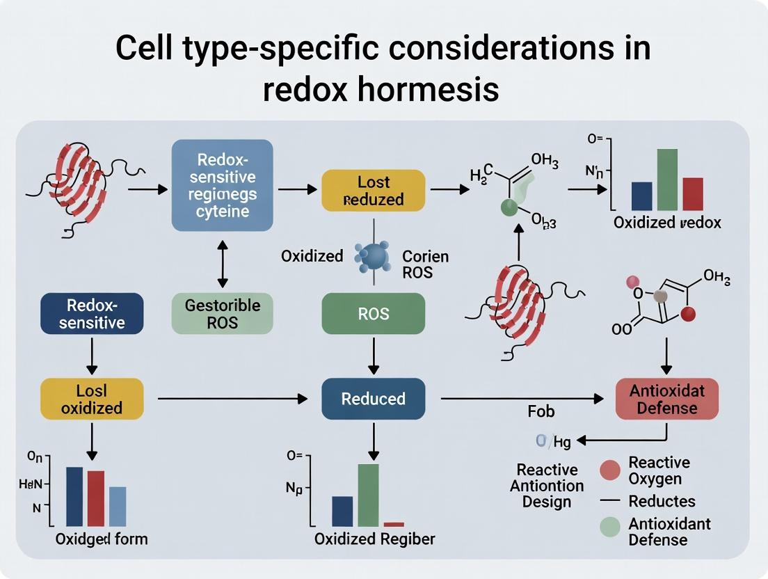
Decoding the Blueprint: Foundational Principles of Cell-Specific Redox Signaling and Hormesis
Technical Support Center
Welcome to the Cell-Specific Redox Hormesis Support Hub. This center addresses common experimental challenges in defining hormetic zones across different cell types. All content is framed within the thesis context: "Cell type-specific considerations in redox hormesis research."
Troubleshooting Guide & FAQs
Q1: In my experiment with Compound X, I observe cytotoxicity at low doses but protective effects at higher doses, which is the inverse of the expected J-shaped curve. What could be the cause?
- A: This is a classic sign of cell type-specific basal redox tone. Your cell line may have inherently high levels of antioxidant enzymes (e.g., high basal Nrf2 activity or catalase expression). A "low" dose for a standard cell line might be sufficient to push these already-stressed cells into apoptosis. Conversely, a higher dose may more effectively activate adaptive transcriptional programs.
- Actionable Protocol:
- Measure Basal State: Quantify baseline ROS (using CellROX Deep Red or H2DCFDA) and key antioxidants (GSH/GSSG ratio, catalase activity) in your cell type vs. a control line.
- Titrate More Broadly: Extend your dose range downward by an order of magnitude.
- Inhibit Antioxidants: Use a low, non-toxic dose of an Nrf2 inhibitor (e.g., ML385) or a catalase inhibitor (e.g., 3-AT) to sensitize cells and re-test the low-dose range.
- A: Neuronal cells typically have lower antioxidant capacity and higher metabolic rates, making them more sensitive to oxidative insult. The hormetic zone will be narrower and shifted leftward (toward lower concentrations).
- Actionable Protocol:
- Pilot Range-Finding: Perform a high-resolution viability assay (e.g., resazurin) across a very low H2O2 range (e.g., 1-100 µM for neurons vs. 10-500 µM for hepatocytes).
- Assay at Multiple Timepoints: Measure viability and ROS at 2h, 6h, 24h, and 48h. The hormetic "window" is temporally constrained.
- Cell-Specific Endpoint: For neurons, include a differentiated state and measure neurite outgrowth or synaptic markers as a functional hormetic endpoint, not just viability.
Q3: My data shows a clear U-shaped dose-response, but the adaptive (hormetic) peak is very shallow and statistically weak. How can I enhance the signal?
- A: A shallow hormetic zone often indicates suboptimal timing between the initial insult and the measurement of the adaptive benefit.
- Actionable Protocol (Preconditioning Paradigm):
- Prime Cells: Treat cells with the putative hormetic dose (Dhorm) for a short period (e.g., 1-2 hours).
- Wash Out: Remove the compound and allow a recovery period (e.g., 6-24 hours) for the adaptive mechanisms to upregulate.
- Challenge: Apply a higher, toxic challenge dose (Dchallenge) of the same or a different stressor.
- Assay: Measure viability/function 24h post-challenge. A significant protective effect compared to non-primed cells confirms and amplifies the hormetic response.
Q4: How do I properly control for cell confluency in long-term hormesis assays?
- A: Confluency dramatically alters redox signaling and metabolic state. A hormetic dose defined at 30% confluency may be toxic at 80% confluency.
- Actionable Protocol:
- Standardize Seeding: Seed cells at a precise density to reach the desired confluency (e.g., 40-50%) at the exact time of compound addition.
- Include a Confluency Control Plate: Use a live-imaging system or parallel plates to monitor confluence daily.
- Normalize to Metabolic Rate: Use a metabolic assay (like Seahorse) to normalize the "effective dose" to the cell's real-time metabolic activity, which is confluency-dependent.
Table 1: Cell Type-Specific Hormetic Zones for Common Redox-Active Agents Data synthesized from recent literature (2022-2024). "Hormetic Range" indicates the dose window where a statistically significant adaptive benefit (110-140% of control) is observed.
| Stressor Agent | Cell Type | Typical Toxic Threshold | Hormetic Range (Adaptive Peak) | Key Adaptive Pathway Activated | Primary Cell-Specific Reason for Variation |
|---|---|---|---|---|---|
| Hydrogen Peroxide (H₂O₂) | Hepatocyte (HepG2) | >250 µM | 10 - 80 µM (~120% viability) | Nrf2/ARE, FOXO | High constitutive detox capacity (CAT, GPx). |
| Cardiomyocyte (H9c2) | >150 µM | 5 - 40 µM (~115% viability) | Nrf2/ARE, AMPK | High mitochondrial density & ROS flux. | |
| Neuron (SH-SY5Y) | >50 µM | 1 - 15 µM (~125% neurite outgrowth) | BDNF/Nrf2 crossover | Low GSH pools, high PUFA membrane content. | |
| Sulforaphane | Colon Cancer (HCT116) | >20 µM | 0.1 - 2.0 µM (~135% clonogenic survival) | Nrf2, HSP70 | Rapid metabolism and Keap1 saturation kinetics. |
| Primary T-cells | >10 µM | 0.05 - 0.5 µM (~140% IL-2 production) | Nrf2, NF-κB | Dynamic redox-sensitive signaling in immune activation. | |
| Metformin | Mammary Epithelial (MCF10A) | >50 mM | 0.1 - 5 mM (~130% stress resistance) | AMPK, mitohormesis | Dose-dependent inhibition of complex I vs. adaptive mitochondrial remodeling. |
Experimental Protocols
Protocol 1: Core Workflow for Defining a Cell-Specific Hormetic Zone Title: Determining the Biphasic Dose-Response Curve
Objective: To precisely define the hormetic and toxic zones of a redox-active compound for a specific cell type.
Materials: See "Research Reagent Solutions" below. Procedure:
- Cell Preparation: Seed cells in 96-well plates at a pre-optimized, sub-confluent density (e.g., 30-40% confluency at treatment time). Include 8 replicate wells per condition.
- Dose-Response Matrix: Prepare a 2-fold serial dilution of the test compound across a broad range (e.g., 8-10 concentrations). Include vehicle and positive (toxic) controls.
- Treatment Paradigm:
- Acute Cytotoxicity: Treat cells for 24-48h, then assay for viability (e.g., Resazurin reduction).
- Preconditioning Assay: Treat cells with doses for 2-4h, wash out, recover for 18h, then challenge with a known toxic dose of the same agent for 24h before viability assay.
- Multi-Parameter Endpoint Analysis: At assay endpoint, use a multiplexed approach:
- Viability: Resazurin incubation (1-4h), measure fluorescence (λex 560/λem 590).
- ROS: Load cells with 10 µM H2DCFDA for 30 min prior to assay termination, measure fluorescence (λex 495/λem 529).
- (Optional) Live-Cell Imaging: Use Incucyte or similar for real-time confluency and health monitoring.
- Data Analysis: Normalize all data to vehicle control (100%). Plot dose-response curves. The hormetic zone is defined where viability/stress resistance is >110% of control (p<0.05). The toxic threshold is the lowest dose where viability falls to <90% of control.
Protocol 2: Validating Nrf2 Pathway Activation in the Hormetic Zone Title: Confirming Adaptive Transcriptional Response
Objective: To confirm that the observed hormetic effect is mediated by the Nrf2 antioxidant response pathway.
Procedure:
- Treatment: Treat cells with vehicle, the optimal hormetic dose (from Protocol 1), and a toxic dose for 6h.
- Nuclear Extraction: Use a commercial nuclear extraction kit. Confirm purity via Western Blot for Lamin B1 (nuclear) and GAPDH (cytosolic).
- Western Blot: Run 20-30 µg of nuclear protein on SDS-PAGE. Probe for Nrf2. Use Lamin B1 as loading control.
- Downstream Target Q-PCR: In parallel, extract total RNA, synthesize cDNA, and perform qPCR for classic Nrf2 targets (e.g., HMOX1, NQO1, GCLC). Normalize to ACTB or GAPDH. Use the 2^(-ΔΔCt) method.
- Functional Assay: Measure the activity of NQO1 (using menadione as a substrate) 24h post-treatment.
Diagrams
Diagram Title: Workflow for Defining Cell-Specific Hormetic Zone
Diagram Title: Nrf2 Pathway Activation in Redox Hormesis
The Scientist's Toolkit: Research Reagent Solutions
| Reagent / Material | Function / Rationale | Example Product / Catalog Number |
|---|---|---|
| CellROX Deep Red Reagent | Fluorogenic probe for measuring total cellular ROS. More stable and specific than H2DCFDA. | Thermo Fisher Scientific, C10422 |
| Nrf2 Inhibitor (ML385) | Specific inhibitor of Nrf2 binding to ARE. Essential for validating pathway necessity in hormesis. | Sigma-Aldrich, SML1833 |
| Nuclear Extraction Kit | For clean separation of nuclear and cytosolic fractions to assess Nrf2 translocation. | NE-PER Kit, Thermo Fisher, 78833 |
| Resazurin Sodium Salt | Cell-permeable redox indicator for long-term, non-destructive viability monitoring. | Sigma-Aldrich, R7017 |
| Recombinant Human BDNF | Critical positive control for neuronal hormesis studies where neurotrophic pathways are engaged. | PeproTech, 450-02 |
| Seahorse XFp Analyzer Cartridge | For real-time metabolic profiling (glycolysis, OXPHOS) to link hormesis to mitohormesis. | Agilent Technologies, 103025-100 |
| C11-BODIPY^(581/591) | Lipid peroxidation sensor. Critical for cell types with high PUFA content (neurons, hepatocytes). | Thermo Fisher Scientific, D3861 |
| 3-Amino-1,2,4-triazole (3-AT) | Catalase inhibitor. Used to modulate intrinsic antioxidant capacity and sensitize cells. | Sigma-Aldrich, A8056 |
Technical Support Center: Troubleshooting & FAQs
Welcome to the Redox Hormesis Technical Support Center. This resource is designed for researchers investigating cell type-specific redox signaling and homeostasis. The following guides address common experimental pitfalls when quantifying and manipulating ROS sources and sinks across different cell models, a core consideration for thesis work on Cell type-specific considerations in redox hormesis research.
Frequently Asked Questions (FAQs)
Q1: My measurement of total cellular ROS (e.g., using DCFDA) shows wildly different baseline levels between my primary hepatocytes and cultured HeLa cells. Is this an artifact? A: This is likely a real biological variation, not an artifact. Different cell types express different complements and activities of ROS sources and sinks. Hepatocytes have high metabolic activity and abundant mitochondria (a major ROS source) but also high levels of antioxidant enzymes like Catalase and GPx. Epithelial cancer lines like HeLa may have altered mitochondrial metabolism and NOX activity. Always:
- Normalize assays to cell count or protein content.
- Use compartment-specific probes (e.g., MitoSOX for mitochondrial superoxide) instead of just global probes.
- Establish a baseline profile for each cell type using the table below as a guide.
Q2: I inhibited NOX with apocynin, but see no change in my ROS readout in my neuronal cell culture. What could be wrong? A: Apocynin requires activation by cellular peroxidases and its efficacy is highly cell type-dependent. Neuronal cells may have low relevant peroxidase activity. Furthermore, the dominant ROS source in your neuronal model may be the mitochondrial electron transport chain (ETC), not NOX.
- Troubleshooting Steps:
- Verify NOX isoform expression in your cell type via qPCR or western blot (e.g., NOX2 is key in phagocytes, NOX4 in fibroblasts).
- Use an alternative, direct NOX inhibitor like GKT136901 (for NOX1/4) or a peptide inhibitor (gp91ds-tat) as a confirmatory tool.
- Combine with mitochondrial inhibition (e.g., rotenone) to assess source contribution.
Q3: When I overexpress SOD2 (MnSOD) in my cardiac fibroblast model to increase antioxidant capacity, I sometimes see increased oxidative damage markers. Why? A: This is a classic example of disrupted redox hormesis and signaling. SOD2 converts superoxide (O₂•⁻) to hydrogen peroxide (H₂O₂). A sudden, localized increase in H₂O₂ without a concomitant increase in H₂O₂-removing sinks (like GPx or Catalase) can create a peroxidative environment. H₂O₂ is also a key signaling molecule; altering its micro-distribution disrupts pathways.
- Solution: Consider co-overexpression or boosting of downstream sinks (e.g., Catalase, Peroxiredoxins) or analyze a more complete antioxidant profile.
Q4: My Thioredoxin Reductase (TrxR) activity assay results are inconsistent between my purified protein and cell lysate experiments. A: Cellular TrxR activity is highly sensitive to sample preparation and the presence of inhibitors (e.g., auranofin). Ensure:
- Lysis buffer contains appropriate protease inhibitors and a protecting agent like EDTA.
- Avoid repeated freeze-thaw cycles of lysates.
- Run a positive control (e.g., auranofin inhibition) to confirm assay specificity in complex lysates.
Table 1: Representative relative expression/activity levels. Values are normalized, hypothetical units (0-10 scale) for illustrative comparison based on common literature findings. Actual quantitative values vary by study and measurement technique.
| Cell Type | Major ROS Source | NOX Activity | Mitochondrial ROS Potential | Major ROS Sink | SOD Activity | GPx/Catalase Activity | Thioredoxin System Activity |
|---|---|---|---|---|---|---|---|
| Macrophage | NOX2 (Phagocytic Burst) | 9 (High) | 4 (Moderate) | GPx, Catalase | 5 | 8 (High) | 6 |
| Hepatocyte | Mitochondria (ETC), CYP450 | 2 (Low) | 8 (High) | Catalase (very high), GPx, SOD | 7 | 9 (Very High) | 8 (High) |
| Neuron | Mitochondria (ETC) | 1 (Very Low) | 7 (High) | GPx, Prx, Trx System | 6 | 5 (Moderate) | 7 (High) |
| Cardiac Myocyte | Mitochondria (ETC), NOX4 | 4 (Moderate) | 9 (Very High) | GPx, Trx System | 8 (High) | 6 (Moderate) | 8 (High) |
| Cancer Cell Line | Mitochondria, NOX1/4 | Variable | Variable (Often High) | Variable (Often Adapted) | Variable | Variable | Variable (Often High) |
Detailed Experimental Protocols
Protocol 1: Cell Type-Specific ROS Source Profiling using Pharmacological Inhibition
Objective: To determine the relative contribution of NOX vs. Mitochondrial ETC to baseline ROS in a new cell type. Reagents: See Scientist's Toolkit below. Procedure:
- Plate cells in a black-walled, clear-bottom 96-well plate at optimal density. Include replicates (n>=6).
- Treatment Groups: Pre-treat cells for 1 hour with:
- Group A: Vehicle control (e.g., DMSO).
- Group B: NOX inhibitor (e.g., 100µM Apocynin or 10µM GKT136901).
- Group C: Mitochondrial ETC Complex I inhibitor (e.g., 5µM Rotenone).
- Group D: Combination of B & C.
- Load cells with 10µM CM-H2DCFDA (for general cytosolic ROS) or 5µM MitoSOX Red (for mitochondrial superoxide) in pre-warmed, serum-free media. Incubate 30-45 min at 37°C.
- Wash 2x with PBS. Add fresh phenol-free medium.
- Measure fluorescence immediately (Ex/Em: 495/529 nm for DCF; 510/580 nm for MitoSOX) using a plate reader. Take kinetic reads every 5-10 min for 60 min.
- Data Analysis: Normalize fluorescence to cell number (via post-assay nuclei stain). Calculate the % reduction in fluorescence for each inhibitor group compared to vehicle control. The source contributing most will show the greatest inhibition of the ROS signal.
Protocol 2: Assessing Antioxidant Sink Capacity via Enzyme Activity Assays
Objective: To measure Catalase and GPx activity in cell lysates from different tissues. Key Consideration: Run a protein assay (e.g., BCA) on all lysates first to normalize activity to total protein. A. Catalase Activity (UV Spectrophotometric Method):
- Prepare lysates in cold PBS (pH 7.0) with 0.1% Triton X-100. Centrifuge (10,000g, 15 min, 4°C), keep supernatant.
- In a cuvette, mix: 50mM Potassium Phosphate buffer (pH 7.0) and 10-20µg of lysate protein. Bring volume to 1.9mL.
- Start reaction by adding 0.1mL of freshly prepared 30mM H₂O₂ solution.
- Immediately measure the decrease in absorbance at 240 nm (A240) every 10 seconds for 2 minutes. H₂O₂ decomposition by catalase is directly measured.
- Calculation: One unit decomposes 1µmol H₂O₂ per min at pH 7.0. Use the extinction coefficient for H₂O₂ (0.0436 M⁻¹cm⁻¹). Activity = (ΔA240/min * Total Vol) / (0.0436 * Protein mg).
B. GPx Activity (Coupled NADPH Oxidation Assay):
- Prepare master mix (per reaction): 50mM Tris-HCl (pH 7.6), 1mM EDTA, 0.2mM NADPH, 1 Unit/mL Glutathione Reductase (GR), 1mM GSH.
- In a 96-well plate, add 180µL master mix + 10-20µg lysate protein. Pre-incubate 5 min at 25°C.
- Initiate reaction by adding 20µL of 0.5mM tert-butyl hydroperoxide (or Cumene hydroperoxide).
- Monitor the decrease in A340 due to NADPH oxidation for 3-5 minutes.
- Calculation: Activity = (ΔA340/min * Total Vol) / (6.22 mM⁻¹cm⁻¹ * Protein mg). 6.22 is the extinction coefficient of NADPH.
Visualizations
Diagram 1: Key ROS Sources & Sinks in a Generic Cell
Diagram 2: Experimental Workflow for Cell-Specific Redox Profiling
The Scientist's Toolkit: Essential Research Reagents
Table 2: Key reagents for studying ROS sources and sinks.
| Reagent Category | Specific Example(s) | Function & Application Notes |
|---|---|---|
| General ROS Probes | CM-H2DCFDA, CellROX Green | Cell-permeable, measure broad-spectrum intracellular ROS. Limitation: Non-specific, photo-sensitive, can be autoxidized. |
| Compartment-Specific Probes | MitoSOX Red (Mitochondrial SO), HyPer (Cytosolic H₂O₂), roGFP (Redox sensor) | Target specific organelles or measure defined species (H₂O₂). Provides spatial resolution. |
| NOX Inhibitors | Apocynin, GKT136901, GKT831, gp91ds-tat peptide | Pharmacologic or peptide-based inhibition to probe NOX contribution. Check isoform specificity and cell permeability. |
| ETC/Mito Inhibitors | Rotenone (Complex I), Antimycin A (Complex III), CCCP (Uncoupler) | Induce mitochondrial ROS or collapse membrane potential to assess mitochondrial source role. |
| Antioxidant Enzyme Assay Kits | Catalase Activity Assay Kit (Colorimetric/UV), GPx Assay Kit (Coupled NADPH) | Standardized, optimized kits for reliable activity measurement from cell/tissue lysates. |
| Thioredoxin System Probes | Auranofin (TrxR Inhibitor), Monobromobimane (for reduced Trx) | Pharmacologic inhibition or fluorescent labeling to assess Trx system status and function. |
| Critical Substrates/Cofactors | NADPH, Glutathione (GSH/GSSG), H₂O₂ solutions | Essential for enzyme activity assays. Use fresh, accurately titrated H₂O₂ stocks. |
Technical Support Center
Troubleshooting Guides & FAQs
Q1: In my hepatocyte experiments, low-dose H₂O₂ fails to activate the expected Nrf2-mediated antioxidant response. What could be wrong? A: This is a common cell type-specific issue. Primary hepatocytes have high basal antioxidant (e.g., GSH) levels and rapid ROS detoxification. The "low dose" may be sub-threshold.
- Troubleshooting Steps:
- Quantify intracellular ROS: Confirm ROS accumulation using a probe like CM-H2DCFDA. Your "low dose" may be instantly scavenged.
- Titrate your inducer: Perform a detailed dose-response (e.g., 1-200 µM H₂O₂) and time-course (5 min to 24h) for Nrf2 target genes (e.g., NQO1, HMOX1) via qPCR.
- Inhibit baseline detoxification: Pre-treat with a sub-toxic dose of L-Buthionine-sulfoximine (BSO, 100 µM, 24h) to deplete GSH. This can sensitize hepatocytes to redox hormesis.
- Check Nrf2 localization: Use immunofluorescence to confirm Nrf2 nuclear translocation post-stimulation.
Q2: I observe simultaneous activation of pro-inflammatory NF-κB and anti-inflammatory Nrf2 in my macrophage models upon oxidant exposure. Is this contradictory? A: No, this is a key feature of the signaling nexus, especially in immune cells. The outcome depends on ROS flux, timing, and parallel pathways.
- Troubleshooting Guide:
- Issue: Non-specific or overwhelming ROS stimulus.
- Solution: Use more physiologically relevant oxidants (e.g., oxidized LDL for macrophages) or precise ROS generators (e.g., glucose oxidase for steady H₂O₂ production).
- Protocol Refinement:
- Monitor Kinetics: NF-κB activation is often transient (peaks at 15-30 min), while Nrf2 response is sustained (peaks at 2-6h). Perform detailed time-course analyses.
- Modulate AMPK: Pharmacologically activate AMPK (e.g., with AICAR) or inhibit it (e.g., Compound C). AMPK can inhibit NF-κB and promote Nrf2, shifting the balance.
- Measure Functional Outputs: Don't just read pathway activation. Assay cytokine secretion (IL-1β, TNF-α for NF-κB) and glutathione levels (for Nrf2) to determine the functional outcome.
Q3: How can I accurately measure the switch from adaptive Nrf2 activation to apoptotic signaling in neuronal cells? A: Neuronal cells are sensitive to oxidative stress. The switch is defined by a loss of homeostasis.
- Critical Checkpoints:
- Mitochondrial ROS & Health: Use MitoSOX Red to measure mtROS and JC-1 dye for mitochondrial membrane potential. A permanent ΔΨm collapse indicates the apoptotic switch.
- Key Marker Table:
Adaptive Phase (Nrf2 Dominant) Apoptotic Phase (Switch) Keap1 cysteine modification Cytochrome c release (cytosolic fraction) Nrf2 nuclear accumulation (IF) Cleaved caspase-3 (western blot) HMOX1 mRNA ↑ (qPCR) PARP cleavage (western blot) Cell viability >85% (MTT assay) Cell viability <70% (MTT assay) - Protocol: Pre-treat with a specific Nrf2 inhibitor (e.g., ML385) prior to ROS stimulus. If apoptosis markers appear significantly earlier, it confirms Nrf2 was holding the adaptive state.
Experimental Protocols
Protocol 1: Assessing Cell-Type-Specific Nrf2/NF-κB Crosstalk Title: Co-Immunoprecipitation and Fractionation for Nrf2/NF-κB p65 Interaction Analysis. Method:
- Cell Treatment & Lysis: Treat cells (e.g., macrophages vs. fibroblasts) with a hormetic ROS dose (e.g., 50 µM tert-butyl hydroperoxide, tBHP, for 2h). Lyse in IP Lysis Buffer containing 25 mM Tris, 150 mM NaCl, 1% NP-40, 1 mM EDTA, 5% glycerol, and fresh protease/phosphatase inhibitors.
- Nuclear/Cytosolic Fractionation: Use a commercial fractionation kit. Validate purity by blotting for Lamin B1 (nuclear) and α-tubulin (cytosolic).
- Co-Immunoprecipitation (Co-IP): Incubate 500 µg of nuclear fraction protein with 2 µg of anti-Nrf2 antibody overnight at 4°C. Add Protein A/G magnetic beads for 2h. Wash beads 3x with lysis buffer.
- Immunoblotting: Elute proteins in 2X Laemmli buffer. Run SDS-PAGE and blot for NF-κB p65 and Nrf2. An interaction suggests direct crosstalk, which may vary by cell type.
Protocol 2: Quantifying AMPK's Role in Redox Fate Decisions Title: Live-Cell Imaging of ROS, AMPK, and Cell Viability. Method:
- Cell Seeding & Staining: Seed cells in a 96-well glass-bottom plate. At ~70% confluency, load with:
- ROS: CellROX Green Reagent (5 µM).
- AMPK Activity: Use a FRET-based AMPK biosensor (e.g., AMPKAR) if available.
- Viability: Hoechst 33342 (nuclei) and propidium iodide (PI, dead cells).
- Stimulation & Imaging: Treat with a gradient of oxidant (e.g., 0, 25, 50, 100 µM H₂O₂) directly on the microscope stage of a live-cell imaging system. Maintain at 37°C/5% CO₂.
- Data Acquisition: Acquire images every 5 minutes for 24 hours using appropriate filter sets.
- Analysis: Quantify CellROX and AMPKAR fluorescence (cytosolic region) over time. Correlate the timing and amplitude of peaks with subsequent PI incorporation.
Diagrams
Short Title: Cell-Type-Specific ROS Signaling Nexus
Short Title: Experimental Workflow for Redox Hormesis
The Scientist's Toolkit: Key Research Reagent Solutions
| Reagent/Category | Function in Redox Hormesis Research | Example Product/Assay |
|---|---|---|
| ROS Inducers (Precise) | Generate specific types and fluxes of ROS for controlled stimulation. | tert-Butyl hydroperoxide (tBHP), Glucose Oxidase (steady H₂O₂), Menadione (superoxide generator). |
| ROS Scavengers & Inhibitors | Confirm ROS-mediated effects by quenching or blocking production. | N-acetylcysteine (NAC, general), MitoTEMPO (mitochondrial), Apocynin (NOX inhibitor). |
| Pathway-Specific Activators/Inhibitors | Pharmacologically manipulate key nodes to establish causality. | Nrf2: sulforaphane (activator), ML385 (inhibitor). NF-κB: PMA (activator), BAY 11-7082 (inhibitor). AMPK: AICAR (activator), Compound C (inhibitor). |
| Intracellular ROS Probes | Quantify and visualize ROS levels in live or fixed cells. | CM-H2DCFDA (general cytosolic ROS), MitoSOX Red (mitochondrial superoxide), CellROX kits. |
| Antibodies for Key Targets | Detect activation, translocation, and expression of pathway components. | Phospho-specific: p-AMPK (Thr172), p-IκBα (Ser32). Total protein: Nrf2, NF-κB p65, Keap1. Localization: Lamin B1 (nuclear), COX IV (mitochondrial). |
| Live-Cell Viability/Apoptosis Assays | Continuously monitor cell fate decisions post-ROS exposure. | Real-time assays using Incucyte with Annexin V (apoptosis) or Caspase-3/7 dyes. |
| GSH/GSSG Detection Kit | Measure the central redox couple critical for Nrf2 signaling and hormesis. | Colorimetric or fluorometric GSH/GSSG Ratio Assay Kits. |
Technical Support Center: Troubleshooting & FAQs for Redox-Metabolism Assays
This support center addresses common experimental challenges in studying metabolic identity (Warburg vs. OXPHOS) and its interplay with nutrient sensing and redox homeostasis. Content is framed within cell type-specific considerations for redox hormesis research.
FAQs & Troubleshooting Guides
Q1: In my Seahorse XF Glycolysis Stress Test, I observe a very low glycolytic capacity and reserve in my cancer cell line, contrary to the expected Warburg effect. What could be the cause? A: This can be cell type-specific. High OXPHOS dependency can mask glycolytic flux.
- Checklist:
- Cell Density & Seeding: Over-confluent cells may exhibit contact inhibition and reduced glycolysis. Optimize cell number (see Table 1).
- Culture Medium Pre-conditioning: Cells cultured in high glucose may downregulate glycolysis. Incubate in assay medium (e.g., XF Base Medium + 2 mM Glutamine + 1 mM Pyruvate + 10 mM Glucose) for 1 hour prior to assay.
- Mitochondrial Stress Test First: Run a Mito Stress Test to confirm OXPHOS activity. Some cancers (e.g., oxidative carcinomas) primarily use OXPHOS.
- Inhibitor Potency: Verify the concentration and freshness of oligomycin. Use 1.0 µM as a starting point.
Q2: When measuring intracellular ROS (e.g., with DCFDA or CellROX) in response to nutrient shifts, I get inconsistent results between different cell types. How should I standardize this? A: ROS readouts are highly sensitive to metabolic context and basal redox state.
- Troubleshooting Steps:
- Quench Ambient ROS: Include a positive control (e.g., 100-200 µM H₂O₂) and a negative control (N-acetylcysteine, NAC) in every experiment to define your signal range.
- Nutrient Synchronization: Prior to assay, starve cells in a low-nutrient buffer (e.g., PBS or low-glucose medium) for 45-60 minutes, then stimulate with specific nutrients (glucose, glutamine). This synchronizes nutrient sensing pathways.
- Cell-Type Specific Baselines: Always measure basal ROS for each cell type. Cells with high OXPHOS (e.g., cardiomyocytes) may have a higher baseline than Warburg-phenotype cells under standard culture.
- Kinetic vs. Endpoint: Perform kinetic reads for 60-90 minutes post-stimulation, as ROS bursts can be transient.
Q3: My AMPK/ mTOR nutrient-sensing western blots show poor activation/ inhibition upon glucose deprivation, especially in my immortalized cell line. What protocols improve detection? A: Signaling responses can be blunted in immortalized lines. Enhance sensitivity.
- Detailed Protocol:
- Starvation Rigor: Use a true "starvation" medium: Glucose-free DMEM, no serum, no glutamine (to prevent anaplerosis). Pre-incubate for 15-30 min.
- Positive Control: Include 1 µM AICAR (AMPK activator) and/or 100 nM Rapamycin (mTOR inhibitor) in parallel wells.
- Lysis: Use ice-cold RIPA buffer with fresh protease and phosphatase inhibitors. Scrape cells on ice immediately.
- Phospho-Specific Antibodies: For p-AMPKα (Thr172) and p-S6K/S6RP (Thr389/Ser235/236), block membrane with 5% BSA in TBST, not milk, to reduce background.
Q4: How do I accurately dissect the contribution of mitochondrial vs. cytosolic ROS in my nutrient-sensing experiments? A: Use a combination of genetic and pharmacologic tools with live-cell imaging.
- Experimental Workflow:
- Inhibit & Detect: Treat cells with 5 µM Rotenone/Antimycin A (complex I/III inhibitor, increases mtROS) or 1 µM Oligomycin (complex V inhibitor, can decrease mtROS). Measure with MitoSOX Red (for mitochondrial superoxide) and DCFDA (broad ROS) concurrently.
- Genetic Probes: Express roGFP2-Orp1 (for cytosolic H₂O₂) or mito-roGFP2-Grx1 (for mitochondrial matrix H₂O₂) and measure by ratiometric fluorescence.
- Scavenger Confirmation: Co-treat with mitochondria-targeted antioxidant MitoTEMPO (e.g., 100 µM) to confirm mtROS origin.
Data Presentation Tables
Table 1: Optimized Cell Seeding Density for Key Metabolic Assays (96-well plate)
| Cell Type / Phenotype | Seahorse XF Assay (cells/well) | Intracellular ROS Assay (cells/well) | Metabolic Labeling (e.g., ¹³C-Glucose) |
|---|---|---|---|
| Primary Fibroblasts (OXPHOS) | 15,000 - 20,000 | 10,000 - 15,000 | 1.0e6 - 2.0e6 / 6cm dish |
| Hepatocellular Carcinoma (Warburg) | 10,000 - 15,000 | 8,000 - 12,000 | 0.8e6 - 1.5e6 / 6cm dish |
| Immortalized Neurons | 30,000 - 40,000 | 20,000 - 25,000 | 2.0e6 - 3.0e6 / 6cm dish |
| Activated T-Cells | 150,000 - 200,000 | 100,000 - 150,000 | 5.0e6 - 10.0e6 / 6cm dish |
Table 2: Key Metabolic Parameters from Glycolysis Stress Test in Different Cell Types (Representative Data)
| Parameter | OXPHOS-Dependent Cell (e.g., Primary Hepatocyte) | Warburg-Phenotype Cell (e.g., Glioblastoma) | Unit |
|---|---|---|---|
| Basal Glycolysis | 20-40 | 80-150 | mpH/min |
| Glycolytic Capacity | 30-60 | 120-250 | mpH/min |
| Glycolytic Reserve | 10-25 | 40-100 | mpH/min |
| Basal ECAR/OCR Ratio | 0.5 - 1.2 | 2.5 - 5.0 | Ratio |
Experimental Protocols
Protocol: Measuring Redox Response to Acute Glucose Withdrawal Objective: To assess the immediate ROS hormesis triggered by glucose sensing in different cell types.
- Seed cells in black-walled, clear-bottom 96-well plates at densities from Table 1.
- Culture for 24h in standard growth medium.
- Load ROS Sensor: Replace medium with phenol-red-free RPMI containing 10 µM DCFDA or 5 µM CellROX Green. Incubate 30 min at 37°C.
- Wash & Stimulate: Wash 2x with pre-warmed PBS. Immediately add:
- Control: Complete medium (25 mM Glucose).
- Test: Glucose-free medium + 10% dialyzed FBS.
- Positive Control: Complete medium + 200 µM H₂O₂.
- Read Fluorescence: Using a plate reader (Ex/Em: 485/535 nm), take kinetic readings every 5 minutes for 90 minutes. Maintain 37°C.
- Normalize: Normalize fluorescence to cell number (e.g., post-read Crystal Violet stain).
Protocol: ¹³C-Glucose Tracing for Glycolytic vs. TCA Flux Objective: To quantify metabolic identity via isotopic labeling.
- Prepare Tracer Medium: Use DMEM without glucose, glutamine, or sodium pyruvate. Supplement with 10% dialyzed FBS, 4 mM glutamine, and 25 mM [U-¹³C]-Glucose.
- Label Cells: At ~80% confluency, wash cells (in 6cm dish) with PBS and add 2 mL tracer medium. Incubate for 1, 4, or 24 hours (time-course).
- Quench & Extract: Aspirate medium, wash rapidly with 0.9% ice-cold saline. Add 1 mL 80% ice-cold methanol/water. Scrape. Transfer to tube, vortex, incubate at -80°C for 15 min. Centrifuge at 16,000 g, 15 min, 4°C.
- Analyze: Dry supernatant under N₂ gas. Derivatize (e.g., Methoxyamination and silylation). Analyze by GC-MS. Quantify M+2 (glycolytic lactate), M+3 (pyruvate dehydrogenase entry), and M+4/5/6 (TCA cycle intermediates) isotopologues.
Diagrams
Title: AMPK-mTOR Nutrient Sensing Pathway & Redox Implications
Title: Metabolic Identity Determines Redox Hormesis Threshold
The Scientist's Toolkit: Research Reagent Solutions
| Item / Reagent | Function / Application in Metabolic-Redox Studies |
|---|---|
| Seahorse XF Glycolysis Stress Test Kit (Agilent) | Measures extracellular acidification rate (ECAR) to quantify glycolytic flux: basal glycolysis, capacity, and reserve. |
| MitoSOX Red Mitochondrial Superoxide Indicator (Thermo Fisher) | Live-cell, fluorogenic probe selectively targeted to mitochondria, oxidized by superoxide. |
| [U-¹³C]-Glucose (Cambridge Isotope Laboratories) | Stable isotope tracer for GC-MS or LC-MS metabolic flux analysis (MFA) to map glycolytic and TCA pathway contributions. |
| Compound C / Dorsomorphin (AMPK Inhibitor) | Pharmacological inhibitor of AMPK. Used to dissect AMPK's role in nutrient sensing-induced redox shifts. |
| 2-NBDG (2-(N-(7-Nitrobenz-2-oxa-1,3-diazol-4-yl)Amino)-2-Deoxyglucose) | Fluorescent glucose analog for real-time visualization and semi-quantification of glucose uptake in live cells. |
| CellMembrane Peroxy Yellow 1 (PEPY1) (Sigma) | A rationetric, peroxynitrite-selective fluorescent probe for detecting specific RNS in live cells. |
| Anti-Phospho-AMPKα (Thr172) Antibody (Cell Signaling) | Key antibody for detecting activated AMPK via western blot, a readout of low energy/nutrient stress. |
| MitoTEMPO (Sigma) | Mitochondria-targeted superoxide dismutase mimetic and antioxidant. Used to selectively scavenge mtROS and test its functional role. |
| Rotenone & Antimycin A (Sigma) | Mitochondrial ETC Complex I and III inhibitors. Used in combination to induce maximal mtROS production for positive controls. |
| Dialyzed Fetal Bovine Serum (FBS) | Serum with low-molecular-weight metabolites (like glucose) removed. Essential for controlled nutrient manipulation experiments. |
Technical Support Center: Troubleshooting & FAQs for Redox Hormesis Experiments
This support center addresses common experimental challenges when studying redox hormesis across canonical cell types. The guidance is framed within the thesis: Understanding cell type-specific redox handling capacities and signaling networks is critical for elucidating the biphasic dose-response of hormesis and its therapeutic potential.
Frequently Asked Questions (FAQs)
Q1: Why do we observe cell death in primary neuron cultures at ROS levels that induce a protective hormetic response in hepatocytes? A: Neurons have a high metabolic rate and lower baseline levels of certain antioxidant enzymes (e.g., catalase) compared to hepatocytes, making them more vulnerable to oxidative stress. The redox hormesis threshold is significantly lower. Solution: Titrate your pro-oxidant (e.g., H₂O₂) concentration over a much lower range (e.g., 1-50 µM) for neurons versus hepatocytes (e.g., 50-500 µM). Always confirm cell-type specific viability curves.
Q2: Our measurements of Nrf2 activation in cardiomyocytes are inconsistent. What could be the issue? A: Cardiomyocytes have a unique redox landscape due to continuous mitochondrial flux and may regulate Nrf2 kinetics differently. Common pitfalls include incorrect timepoints for peak nuclear translocation (may be later than in other cells) and interference from media components like phenol red. Solution: Use a serum-free, phenol-red free media during stimulation. Perform a time-course experiment (0, 2, 4, 8, 12, 24h) to identify the optimal window for Nrf2 measurement in this cell type.
Q3: When treating cancer cell lines with a pro-oxidant drug, we see an increase in proliferation instead of oxidative stress-induced death. Is this normal? A: Yes, this is a canonical challenge in cancer redox biology. Many cancer cells have a constitutively elevated basal ROS state and adapted antioxidant systems (e.g., upregulated Nrf2, xCT cystine transporter). A low-dose pro-oxidant challenge can further stimulate pro-growth signaling (e.g., via AP-1). Solution: Characterize the basal ROS and antioxidant capacity of your specific cancer line. The therapeutic window for pro-oxidant therapies lies above this adapted threshold, which may be higher than for non-transformed cells.
Q4: Our hepatocyte data shows unexpected toxicity at very low doses of a suspected hormetic agent. What should we check? A: First, rule out cell culture contamination. Second, consider the compound's metabolism. Hepatocytes possess robust Phase I CYP enzymes that may convert the compound into a more toxic metabolite at any dose. Solution: Run an LC-MS analysis to check for metabolite formation. Consider using a CYP inhibitor (e.g., 1-ABT) co-treatment to see if the low-dose toxicity is ablated.
Troubleshooting Guides
Issue: No biphasic response observed; only a monotonic decrease in cell viability.
- Check 1: Dose range. Your concentration steps may be too large or not centered on the correct threshold. Perform a high-resolution dose-response.
- Check 2: Assay sensitivity. The assay (e.g., MTT) may not detect subtle increases in metabolic activity. Supplement with a direct proliferation assay (e.g., BrdU) or a measure of mitochondrial fitness (Seahorse assay).
- Check 3: Timepoint. The adaptive response may be transient. Measure outcomes at multiple timepoints post-treatment (e.g., 24h, 48h, 72h).
Issue: High variability in ROS measurements (e.g., with CM-H2DCFDA) between cell types.
- Check 1: Dye loading. Different cell types have varying esterase activity and efflux pump (e.g., MDR) expression. Optimize loading concentration and time. Use a control with an esterase inhibitor.
- Check 2: Quenching. The high antioxidant capacity of some hepatocytes or cardiomyocytes can rapidly quench the ROS signal. Try a more sensitive dye (e.g., CellROX) or a real-time kinetic assay.
- Check 3: Compartmentalization. Dyes may localize differently. Validate with mitochondrial- or cytosolic-specific ROS probes.
Table 1: Canonical Cell Type Redox Parameters & Hormetic Thresholds
| Parameter | Neurons (Primary) | Cardiomyocytes (H9c2) | Hepatocytes (HepG2) | Cancer Cells (HeLa) |
|---|---|---|---|---|
| Basal ROS (Relative Units) | Low-Moderate | Moderate (High mitochondrial) | Low | High |
| Key Antioxidant | GSH, SOD1 | GSH, Trx2, SOD2 | Catalase, GSH, SOD1 | GSH, GPx4, xCT |
| Primary Redox Sensor | Nrf2, p38 MAPK | NF-κB, Nrf2 | Nrf2, ARE | Nrf2, AP-1, HIF-1α |
| Typical H₂O₂ Hormesis Range | 1 – 25 µM | 10 – 100 µM | 50 – 400 µM | 50 – 200 µM* |
| Typical H₂O₂ Toxicity Threshold | ~50 µM | ~250 µM | ~500 µM | >500 µM* |
| Key Hormetic Pathway Output | BDNF expression, Enhanced resilience | Mitochondrial biogenesis (PGC-1α) | Detoxification enzyme synthesis | Proliferation, Drug resistance |
*Highly variable and cell line dependent.
Experimental Protocols
Protocol 1: Determining Cell-Type Specific Redox Hormesis Window Objective: To establish the biphasic dose-response curve for a pro-oxidant agent.
- Seed cells in 96-well plates at optimal density (Neurons: 50k/well; Hepatocytes: 20k/well).
- After adherence, treat with a serial dilution of your pro-oxidant (e.g., H₂O₂) in at least 10 concentrations across a broad range (see Table 1 for guidance).
- Incubate for 24h in standard culture conditions.
- Replace media with fresh, complete media (to remove agent) and incubate for an additional 48h.
- Assay viability using two distinct methods (e.g., MTT for metabolism and Calcein-AM for esterase activity/live cells). Normalize to untreated control.
- Analyze data: Plot % viability vs. log[concentration]. A hormetic response shows a significant increase (110-140%) at low doses before the toxic decline.
Protocol 2: Measuring Nrf2 Nuclear Translocation via Immunofluorescence Objective: To visualize the activation of the key antioxidant response pathway.
- Seed cells on glass coverslips in 24-well plates.
- Treat with a low, hormetic dose of your agent (determined from Protocol 1) for 0, 1, 2, 4, and 8 hours.
- Fix with 4% PFA for 15 min, permeabilize with 0.1% Triton X-100 for 10 min.
- Block with 5% BSA for 1 hour.
- Incubate with primary antibody against Nrf2 (1:250) overnight at 4°C.
- Incubate with fluorescent secondary antibody (e.g., Alexa Fluor 488) and nuclear stain (DAPI) for 1 hour at RT.
- Image using a fluorescence microscope. Quantify the ratio of nuclear to cytoplasmic fluorescence intensity using image analysis software (e.g., ImageJ).
Diagrams
Diagram 1: Comparative Redox Signaling Pathways in Canonical Cell Types
Diagram 2: Workflow for Defining Cell-Type Specific Redox Hormesis
The Scientist's Toolkit: Research Reagent Solutions
Table 2: Essential Reagents for Redox Hormesis Studies
| Reagent | Function & Application | Cell Type-Specific Note |
|---|---|---|
| CM-H2DCFDA | General cytosolic ROS probe. Becomes fluorescent upon oxidation. | Neurons: Optimize loading; low signal. Cancer Cells: May efflux rapidly. |
| MitoSOX Red | Selective detection of mitochondrial superoxide. | Critical for Cardiomyocytes and other high mitochondrial-activity cells. |
| CellROX Green/Orange | More robust, less-quenched general ROS probes for challenging cells. | Recommended for hepatocytes with high antioxidant flux. |
| BSO (Buthionine sulfoximine) | Inhibitor of glutathione synthesis. Depletes GSH to sensitize cells to ROS. | Useful to probe dependency on GSH in cancer cells and hepatocytes. |
| ML385 | Selective inhibitor of Nrf2. Blocks the antioxidant response pathway. | Control for Nrf2-dependent effects in hepatocytes and neurons. |
| Erastin | Inducer of ferroptosis via system xc⁻ inhibition and ROS. | Tool to study the ferroptosis susceptibility of hepatocytes vs. cancer cells. |
| NAC (N-Acetyl Cysteine) | Antioxidant precursor, boosts cellular GSH. | Used to confirm ROS-mediated effects; can abolish hormesis if added during induction phase. |
| H₂O₂ | Canonical, direct pro-oxidant. Short-lived, easy to titrate. | Gold standard for initial hormesis titration across all cell types (see Table 1 for ranges). |
From Bench to Bedside: Methodologies for Studying and Harnessing Cell-Specific Redox Hormesis
Technical Support Center: Troubleshooting & FAQs
FAQ 1: My roGFP2 ratio indicates a more oxidized state than expected. What could be wrong?
- Answer: This is a common issue. First, confirm proper probe expression and localization via microscopy (check for expected subcellular pattern). Second, ensure your calibration with DTT and H₂O₂ was performed correctly on the same imaging setup. Common culprits include:
- pH Interference: roGFP is pH-sensitive. Use a pH-insensitive mutant (e.g., roGFP2-R12) or concurrently image with a pH probe like pHluorin to rule out artifacts.
- Bleaching: roGFP is prone to photobleaching, which can alter ratios. Minimize light exposure and use low illumination intensities.
- Expression Level: Extremely high expression can saturate cellular redox buffering systems, leading to non-physiological readings. Titrate your transfection conditions.
- Cell Health: Apoptotic or stressed cells exhibit highly oxidized states. Check morphology and viability.
FAQ 2: My HyPer signal is unstable or decays rapidly during time-lapse imaging. How can I fix this?
- Answer: HyPer can photobleach and its response can be irreversible under strong illumination.
- Protocol Fix: Use minimal excitation light intensity and reduce acquisition frequency. Consider using a more photostable variant like HyPer7.
- Environmental Control: Ensure the imaging chamber maintains proper temperature and CO₂ levels, as cellular stress affects H₂O₂ metabolism.
- Calibration: Perform a full calibration at the end of each experiment using consecutive additions of DTT (reduced baseline) and then H₂O₂ (oxidized baseline) to define the dynamic range for that specific experiment.
FAQ 3: My metabolomics data shows high variability between biological replicates in my redox-stressed samples. How do I improve consistency?
- Answer: Redox metabolites are notoriously labile. Standardize your quenching and extraction protocol meticulously.
- Critical Protocol Step: For cell culture, rapidly aspirate media and quench metabolism with cold (< -40°C) methanol, acetonitrile, or a mixture, followed by immediate scraping or sonication on ice. The entire process should take <30 seconds per sample.
- Neutralization: For some extraction buffers, neutralize pH post-extraction to prevent degradation of acid-/base-sensitive metabolites.
- Internal Standards: Use a broad set of stable isotope-labeled internal standards added at the very beginning of extraction to correct for losses during sample processing.
FAQ 4: When performing single-cell roGFP assays in a heterogeneous co-culture, how do I attribute the redox state to the correct cell type?
- Answer: You must implement a cell-type-specific labeling strategy.
- Methodology: Create a dual- or triple-vector system, or a single multi-cistronic vector where the roGFP/HyPer probe is co-expressed with a fluorescent marker (e.g., mCherry, mCerulean) under a cell-type-specific promoter. Alternatively, use a Cre-lox system for lineage-specific expression.
- Analysis: During image analysis, use the marker channel to create a mask, ensuring redox ratio measurements are only taken from positively identified cells of interest.
Experimental Protocols
Protocol 1: Calibration of roGFP2 in Adherent Cells
Purpose: To convert the 405/488 nm excitation ratio into a quantitative estimate of redox potential. Steps:
- Plate cells expressing roGFP2 in the desired compartment (e.g., cytosol, mitochondria) on an imaging dish.
- Image Acquisition: Acquire ratio-metric images (excitation 405 nm and 488 nm, emission 500-540 nm) using a live-cell compatible microscope setup.
- Full Reduction: Replace medium with medium containing 10 mM DTT (dithiothreitol). Incubate for 5-10 min and acquire image set (Rmin).
- Wash: Gently wash cells 3x with PBS or imaging medium.
- Full Oxidation: Replace medium with medium containing 1-5 mM H₂O₂. Incubate for 5-10 min and acquire image set (Rmax).
- Calculation: Calculate the oxidation degree: OxD(roGFP) = (R - Rmin) / (Rmax - R). The redox potential can be derived using the Nernst equation.
Protocol 2: LC-MS Metabolomics Sample Preparation for Redox Metabolites
Purpose: To quench metabolism and extract redox-relevant metabolites (e.g., GSH/GSSG, NADH/NAD⁺) for LC-MS analysis. Steps:
- Quenching: For a 6-well plate, rapidly aspirate media and add 1 mL of pre-chilled (-80°C) 40:40:20 Methanol:Acetonitrile:Water containing internal standards.
- Harvest: Immediately scrape cells on dry ice or in a -20°C cold block. Transfer extract to a pre-cooled microcentrifuge tube.
- Extraction: Vortex vigorously for 30 seconds. Incubate at -20°C for 1 hour.
- Pellet Debris: Centrifuge at 16,000 x g for 15 minutes at 4°C.
- Collect Supernatant: Transfer clarified supernatant to a new vial.
- Dry and Reconstitute: Dry under a gentle stream of nitrogen or in a vacuum concentrator. Reconstitute in an appropriate volume of LC-MS injection solvent (e.g., 97:3 Water:Acetonitrile) for analysis.
Data Presentation
Table 1: Key Properties of Genetically Encoded Redox Probes
| Probe | Redox-Sensitive Element | Target | Dynamic Range (Rmax/Rmin) | pH Sensitivity | Primary Application |
|---|---|---|---|---|---|
| roGFP2 | Disulfide bond (cpYFP) | Glutathione redox potential (EGSH) | ~5-7 | High | Static or slow-changing redox potential |
| roGFP2-Orp1 | Disulfide bond (via Orp1) | H₂O₂ (via Orp1) | ~3-4 | Moderate | Specific detection of H₂O₂ dynamics |
| HyPer (3/7) | Disulfide bond (cpYFP+ OxyR) | H₂O₂ | ~4-10 (HyPer7) | Low | Real-time, specific H₂O₂ imaging |
| Grx1-roGFP2 | Disulfide bond (via Grx1) | EGSH (Grx1-coupled) | ~5-7 | High | Thermodynamically defined EGSH |
Table 2: Common Troubleshooting for Single-Cell Redox Assays
| Symptom | Possible Cause | Suggested Solution |
|---|---|---|
| Low Signal-to-Noise Ratio | Low probe expression; High background autofluorescence | Optimize transfection; Use red-shifted probes; Apply spectral unmixing. |
| Ratio Drift Over Time | Photobleaching; Changes in focus or environmental conditions | Use antifade reagents; Implement perfect focus system; Use an environmental chamber. |
| Inconsistent Calibration | Incomplete reduction/oxidation; Cell death during calibration | Test calibration reagent concentrations/timing; Include viability dye. |
| Poor Cell-Type Resolution | Promoter leakiness; Marker bleed-through | Use tighter cell-type-specific promoters; Optimize filter sets for spectral separation. |
The Scientist's Toolkit: Research Reagent Solutions
| Item | Function |
|---|---|
| roGFP2 or HyPer7 Plasmid | Genetically encoded sensor for ratiometric imaging of redox state or H₂O₂. |
| Cell-Type-Specific Promoter Plasmid (e.g., GFAP for astrocytes, CD11b for microglia) | Drives sensor expression in specific cell lineages within heterogeneous samples. |
| Dithiothreitol (DTT) | Strong reducing agent used for in situ calibration of roGFP probes (defines Rmin). |
| Hydrogen Peroxide (H₂O₂) | Oxidizing agent used for in situ calibration of roGFP/HyPer (defines Rmax). |
| Cold Methanol/Acetonitrile | Quenches cellular metabolism instantly to preserve in vivo metabolite levels. |
| Stable Isotope-Labeled Internal Standards (e.g., ¹³C-GSH, D₈-NAD⁺) | Normalizes for sample preparation variability in metabolomics. |
| Matrigel or other ECM | Provides physiological 3D context for studying redox signaling in relevant tissue models. |
Visualizations
Diagram 1: roGFP2 Calibration & Quantification Workflow
Diagram 2: Cell-Type-Specific Redox Analysis in a Co-Culture
Diagram 3: Redox Hormesis Signaling Pathway Simplified
Troubleshooting Guide & FAQs
Q1: My iPSC-derived neurons show high variability in ROS response to a pro-hormetic stimulus compared to primary neuronal cultures. What could be the cause? A: This is a common issue stemming from differentiation efficiency and maturity. Variability often arises from residual pluripotent cells or inconsistent expression of mature neuronal markers (e.g., MAP2, Synapsin). First, quantify differentiation efficiency via flow cytometry for a pan-neuronal marker like βIII-Tubulin (TUJ1). If efficiency is below 85%, optimize differentiation protocol. Second, assess functional maturity by measuring spontaneous calcium oscillations; immature cultures show blunted and inconsistent responses. Use a standardized 21-day differentiation protocol with dual-SMAD inhibition followed by neuronal maturation factors (BDNF, GDNF, cAMP). Always include a positive control, like a known Nrf2 activator (e.g., sulforaphane), to benchmark the redox response window.
Q2: When treating intestinal organoids with a potential hormetic compound, how do I distinguish a protective adaptive response from overt cytotoxicity? A: This requires a multi-parametric endpoint analysis. Relying on a single viability assay (e.g., ATP content) is insufficient. Implement a tiered approach:
- High-throughput viability screen: Use CellTiter-Glo 3D to establish a baseline LD10-LD50 curve.
- Adaptive response markers: At sub-cytotoxic doses (typically LD10-LD20), measure preconditioning effects. Challenge organoids with a subsequent high-dose stressor (e.g., 500 µM H₂O₂) 24 hours post-treatment and assess cell death (Propidium Iodide uptake via confocal imaging).
- Redox-specific molecular endpoints: Quantify Nrf2 nuclear translocation (immunofluorescence) or upregulation of downstream genes (HO-1, NQO1 via qPCR) at the sub-cytotoxic dose. A true hormetic response shows a "low-dose induction, high-dose inhibition" pattern in these markers.
Q3: My in vivo redox hormesis data from mouse liver contradicts my findings in primary hepatocytes. How should I reconcile this? A: Discrepancies are expected due to systemic factors absent in vitro. Follow this diagnostic checklist:
- Check for compensatory pathways: In vivo, hormetic stress in hepatocytes can trigger inter-organ signaling (e.g., via FGF21 from liver affecting adipose or brain). Run a cytokine/endocrine panel on serum from treated mice.
- Verify compound pharmacokinetics: The effective dose reaching hepatocytes in vivo may differ from your in vitro concentration. Measure compound levels in portal blood or liver tissue via LC-MS if possible.
- Assess niche interactions: Primary hepatocytes lose non-parenchymal cell interactions. Co-culture with Kupffer cells or LSECs, or analyze liver sections for immune cell infiltration (CD68+ staining) which can modulate the redox landscape.
Q4: Primary cells from aged donors show no adaptive glutathione upregulation to mild oxidative stress, unlike cells from young donors. Is this a model failure? A: No, this likely accurately reflects age-related redox inflexibility, a key consideration for hormesis research. Confirm the finding with these steps:
- Validate the donor age and health status metadata.
- Measure baseline redox buffers: Quantify total and reduced glutathione (GSH) and the GSH/GSSG ratio in young vs. aged cells before treatment. Aged cells often have a lower baseline ratio.
- Test the Nrf2-Keap1 sensing mechanism: Assess nuclear accumulation of Nrf2 post-stimulus. An impaired response may indicate age-related Keap1 dysregulation or altered kinase (e.g., p62, PKC) activity. This "failure" in the model is a critical biological insight.
Data Presentation Tables
Table 1: Key Characteristics and Considerations for Redox Hormesis Studies
| Model System | Physiological Relevance | Throughput | Cost (Relative) | Genetic Manipulability | Key Redox Hormesis Consideration |
|---|---|---|---|---|---|
| Primary Cells | High (donor-specific) | Low | High | Very Low | Donor age, health, and isolation stress significantly alter baseline ROS and adaptive capacity. |
| iPSC-Derived Lineages | Medium-High (disease-specific) | Medium | Medium-High | High (via base editing) | Differentiation batch variability; often exhibit fetal-like redox metabolism. |
| Organoids | High (3D cytoarchitecture) | Medium-Low | High | Medium (via lentivirus) | Hypoxic cores can create heterogeneous redox microenvironments; measure gradients. |
| In Vivo Models | Highest (systemic context) | Very Low | Very High | Variable (transgenics) | Inter-organ signaling dominates; requires non-invasive redox probes (e.g., roGFP). |
Table 2: Quantitative Redox Endpoint Comparison Across Models (Example: Response to 5 µM Sulforaphane)
| Endpoint | Primary Hepatocytes | iPSC-Cardiomyocytes | Cerebral Organoids | Mouse Liver (in vivo) |
|---|---|---|---|---|
| NQO1 mRNA Induction (Fold-change) | 4.2 ± 0.8 | 3.1 ± 1.2* | 2.5 ± 0.9 (edge) / 1.5 (core)* | 3.8 ± 0.6 |
| GSH/GSSG Ratio (24h post-tx) | +35% ± 5% | +22% ± 10%* | N/A (heterogeneous) | +40% ± 8% |
| Optimal Preconditioning Window | 6-12h | 12-24h | 24-48h | 12-18h |
| LD₁₀ for Cytotoxicity (µM) | ~15 µM | ~25 µM | ~50 µM (whole organoid) | ~10 mg/kg |
*Indicates higher variability (Coefficient of Variation >25%).
Experimental Protocols
Protocol 1: Measuring Compartment-Specific ROS in Cerebral Organoids using a Genetically Encoded Sensor. Objective: To quantify cytosolic vs. mitochondrial H₂O₂ dynamics in response to a hormetic stressor within 3D cerebral organoids. Materials: Cerebral organoids (~day 60), lentivirus for cyto-roGFP2-Orp1 or mito-roGFP2-Orp1, polybrene (8 µg/mL), confocal microscope with environmental chamber, imaging medium (Neurobasal + B27). Steps:
- Sensor Transduction: At organoid day 30, microinject 1 µL of high-titer lentivirus (>1x10^8 IU/mL) into the organoid core. Alternatively, incubate with virus + polybrene for 24h during early formation (day 10-15).
- Recovery & Expression: Culture for 3-4 weeks for stable sensor expression. Confirm via fluorescence microscopy.
- Live Imaging: Place organoid in imaging chamber at 37°C, 5% CO₂. Acquire ratiometric images (excitation 405/488 nm, emission 510 nm) at baseline.
- Treatment & Kinetics: Perfuse with 100 µM tert-butyl hydroperoxide (tBHP) for 5 min to establish maximum oxidation. Wash and perfuse with 5 µM of the test hormetic compound. Image every 30 seconds for 60 minutes.
- Data Analysis: Calculate the 405/488 nm fluorescence ratio for cytosolic and mitochondrial compartments separately. Normalize ratios to baseline (0%) and tBHP maximum (100%).
Protocol 2: Assessing Redox-Flexibility in Primary Cells from Aged Donors. Objective: To evaluate the adaptive glutathione response to mild H₂O₂ preconditioning in primary human dermal fibroblasts from young and aged donors. Materials: Primary HDFs (Young: ≤25 yrs, Aged: ≥70 yrs), Seahorse XFp Analyzer, GSH/GSSG-Glo Assay Kit, CellRox Green dye, 96-well plates. Steps:
- Preconditioning: Seed cells at 10,000/well. At 80% confluency, treat with a low, sub-cytotoxic dose of H₂O₂ (empirically determined, typically 10-50 µM) for 1 hour in serum-free medium.
- Challenge & Metabolic Profiling (Seahorse): 24h post-preconditioning, replace medium with Seahorse XF DMEM, pH 7.4. Perform a Mito Stress Test. A successful adaptive response in young cells will show enhanced spare respiratory capacity (SRC).
- GSH/GSSG Quantification: In parallel plates, lyse cells 24h post-preconditioning. Use the GSH/GSSG-Glo Assay per manufacturer's instructions. Calculate the GSH/GSSG ratio. Aged cells will show a diminished increase in this ratio compared to young.
- ROS Burst Measurement: At time of preconditioning, include wells stained with 5 µM CellRox Green. Image immediately after the 1-hour H₂O₂ pulse. Quantify fluorescence intensity. This confirms the initial stimulus magnitude was equivalent across donor groups.
Diagrams
Diagram 1: Core Nrf2-Keap1 Signaling in Redox Hormesis
Diagram 2: Experimental Workflow for Cross-Model Hormesis Validation
The Scientist's Toolkit: Research Reagent Solutions
| Item | Function in Redox Hormesis Research |
|---|---|
| roGFP2-Orp1 (Genetically Encoded Sensor) | Allows ratiometric, compartment-specific (cytosol, mitochondria) live-cell measurement of H₂O₂ dynamics, critical for defining sub-lethal hormetic doses. |
| GSH/GSSG-Glo Assay (Promega) | Luminescence-based assay for specific quantification of reduced (GSH) and oxidized (GSSG) glutathione ratios, a primary endpoint of redox adaptive capacity. |
| CellTiter-Glo 3D Cell Viability Assay | Optimized for 3D models like organoids; measures ATP content to accurately establish cytotoxicity curves (LD10, LD50) in spheroids. |
| Sulforaphane (Cayman Chemical) | Well-characterized Nrf2 activator used as a positive control compound to benchmark the hormetic response pathway across different model systems. |
| MitoSOX Red (Thermo Fisher) | Fluorogenic dye selectively targeted to mitochondria; detects superoxide (O₂•⁻) formation, a key upstream ROS in mitochondrial hormesis. |
| Nrf2 siRNA (Santa Cruz Biotechnology) | Tool for knock-down experiments to confirm the essential role of the Nrf2 pathway in observed adaptive responses to mild stress. |
| Seahorse XF Analyzer (Agilent) | Measures real-time cellular metabolic parameters (OCR, ECAR); a functional readout for enhanced mitochondrial spare capacity post-hormetic conditioning. |
| Hypoxia Chamber (Billups-Rothenberg) | For creating physiologically relevant low-oxygen environments (e.g., 1-5% O₂) to study redox stress and signaling in stem cell niches or organoid cores. |
Technical Support Center
Troubleshooting Guide: NOX Isoform Targeting
Issue 1: Low Specificity of NOX2 Inhibition in Mixed Macrophage Populations
- Problem: Off-target effects on phagocytic vs. non-phagocytic cells.
- Solution: Implement fluorescence-activated cell sorting (FACS) using lineage-specific surface markers (e.g., CD11b, F4/80) prior to treatment. Use isoform-selective inhibitors like GSK2795039 (NOX2) at a validated, low-nanomolar range and confirm via gp91phox immunoblot.
- Prevention: Pre-characterize cell population heterogeneity using single-cell RNA-seq for CYBB (NOX2) expression before designing experiments.
Issue 2: Unintended Compensatory Upregulation of Non-Targeted NOX Isoforms
- Problem: Knocking down NOX4 in endothelial cells leads to increased NOX1/2 activity, confounding results.
- Solution: Design a multiplex monitoring protocol. Simultaneously measure mRNA levels of all relevant NOX isoforms (NOX1, NOX2, NOX4) and DUOX1/2 via qPCR at 24h, 48h, and 72h post-intervention.
- Prevention: Use combination strategies of siRNA with low-dose pharmacological inhibitors to block compensatory activity.
Issue 3: Inconsistent ROS Burst from ETC Complex I Inhibition
- Problem: Variability in superoxide generation using rotenone across batches of primary neurons.
- Solution: Standardize mitochondrial health assessment prior to experiment. Use a viability/health table. Only proceed with cells where ≥85% meet thresholds.
- Prevention: Pre-treat cells with low-dose oligomycin (1-10 nM) to gently hyperpolarize mitochondria, creating a more uniform baseline for pro-oxidant challenge.
Frequently Asked Questions (FAQs)
Q1: What is the most reliable method to verify cell-type-specific delivery of a NOX4-targeting agent in vivo? A1: Use a conjugate approach. Link your agent (e.g., NOX4 siRNA) to a cell-penetrating peptide (CPP) with a known tropism (e.g., targeting lung endothelium). Perform confocal microscopy on tissue sections using antibodies against the CPP and a cell-specific marker (e.g., CD31). Quantify co-localization using image analysis software (e.g., ImageJ Coloc2).
Q2: How do I distinguish between ROS originating from NOX vs. ETC in a live-cell assay? A2: Employ a sequential inhibition and temporally-resolved detection protocol. First, treat cells with a NOX inhibitor (e.g., VAS2870, 10 µM) and measure ROS (e.g., with H2DCFDA). Wash, then treat with an ETC Complex I inhibitor (e.g., piericidin A, 50 nM) and measure again. Use a mitochondria-targeted ROS sensor (MitoSOX Red) for specificity. Normalize to cell count.
Q3: When targeting ETC complexes, what are key controls for confirming on-target, pro-oxidant effects versus general cytotoxicity? A3: Essential controls include:
- Rescue with Mitochondria-targeted Antioxidant: Pre-treatment with MitoTEMPO (e.g., 100 µM) should attenuate the ROS signal and cell death.
- Genetic Control: Use cells with CRISPR-mediated knockout of the target complex subunit (e.g., NDUFS1 for Complex I) versus wild-type. The pro-oxidant effect should be absent in KO cells.
- Metabolic Control: Measure extracellular acidification rate (ECAR) via Seahorse assay. Specific ETC inhibition should decrease oxygen consumption rate (OCR) without an immediate spike in ECAR, which would indicate a shift to glycolysis due to general stress.
Q4: What are the critical parameters for designing a "precision" pro-oxidant dosing protocol for redox hormesis studies? A4: You must establish a biphasic dose-response curve for each cell type. Key parameters are in the table below.
Table 1: Established Biphasic Dose Ranges for Pro-Oxidants in Common Cell Models
| Cell Type | Target | Pro-Oxidant Agent | Hormetic (Low) Dose Range (ROS Increase: 30-80%) | Toxic (High) Dose Range (ROS Increase: >200%) | Key Readout for Benefit |
|---|---|---|---|---|---|
| Cardiomyocyte (HL-1) | ETC Complex III | Antimycin A | 1 - 10 nM | > 100 nM | Increased Nrf2 nuclear translocation |
| Hepatic Stellate Cell | NOX1/4 | GKT137831 | 50 - 200 nM | > 5 µM | Reduced α-SMA expression |
| Neuron (Primary) | ETC Complex I | MPP+ | 1 - 5 µM | > 50 µM | Increased BDNF secretion |
| Tumor-Associated Macrophage | NOX2 | Phorbol Myristate Acetate (PMA) | 0.1 - 1 ng/mL | > 10 ng/mL | Shift to M1-like phenotype (iNOS+) |
Table 2: Common Reagents for Distinguishing ROS Species in Precision Studies
| Reagent Name | Target ROS | Specificity Level | Key Interference/Note |
|---|---|---|---|
| MitoSOX Red | Mitochondrial Superoxide | High | Can be oxidized by cytosolic enzymes if mitochondria membrane is compromised. |
| Amplex Red (with HRP) | Hydrogen Peroxide (H2O2) | Medium-High | Measures extracellular H2O2; sensitive to ambient light. |
| Dihydroethidium (DHE) | Superoxide | Medium | Oxidation products bind DNA; specificity requires HPLC validation. |
| HyPer7 (genetically encoded) | Cytosolic H2O2 | Very High | Requires transfection; ratiometric measurement. |
Experimental Protocols
Protocol 1: Validating Cell-Type-Specific NOX4 Knockdown and Functional Output
- Transfection: Seed target cells (e.g., hepatic stellate cells, LX-2) at 60% confluence. Transfect with 20 nM NOX4-specific siRNA or scrambled control using a lipid-based transfection reagent optimized for low-serum conditions.
- Efficiency Check: At 48h post-transfection, harvest cells for:
- qPCR: Isolate RNA, reverse transcribe, and run qPCR for NOX4 and housekeeping gene (e.g., GAPDH). Calculate fold-change using the 2^(-ΔΔCt) method. Target >70% knockdown.
- Western Blot: Lyse cells in RIPA buffer, run 30 µg protein on SDS-PAGE, transfer, and probe with anti-NOX4 and anti-β-actin antibodies.
- Functional ROS Assay: At 72h post-transfection, load cells with 5 µM H2DCFDA in serum-free media for 30 min. Wash and treat with a relevant stimulus (e.g., TGF-β1, 5 ng/mL). Measure fluorescence (Ex/Em: 485/535 nm) kinetically for 60 minutes.
- Phenotypic Readout: Parallel wells at 96h post-transfection are fixed and immunostained for α-smooth muscle actin (α-SMA) to quantify fibrotic activation.
Protocol 2: Measuring ETC Complex-Specific ROS Burst with Temporal Resolution
- Cell Preparation: Plate cells in a black-walled, clear-bottom 96-well plate. Pre-incubate with 5 µM MitoSOX Red in complete media for 30 min at 37°C.
- Baseline Measurement: Wash cells twice with warm PBS. Add fresh, phenol-red-free media. Place plate in a pre-warmed (37°C) microplate reader with a CO2 control module. Record baseline MitoSOX fluorescence (Ex/Em: 510/580 nm) for 10 minutes (read every minute).
- Inhibitor Injection: Using the instrument's injector, add the ETC inhibitor (e.g., Rotenone at a final concentration of 50 nM for Complex I, or Antimycin A at 100 nM for Complex III) to the test wells. Include vehicle control injection.
- Kinetic Recording: Immediately continue fluorescence readings every minute for 60-90 minutes.
- Data Analysis: Normalize fluorescence to the average baseline reading for each well. Plot normalized fluorescence over time. The initial slope (first 15-20 min) represents the specific ETC-derived ROS burst.
The Scientist's Toolkit: Research Reagent Solutions
Table 3: Essential Reagents for Precision Pro-Oxidant Research
| Item | Function/Application | Example Product/Catalog # (Illustrative) |
|---|---|---|
| Isoform-Selective NOX Inhibitors | To dissect contributions of specific NADPH oxidase isoforms. | GKT137831 (NOX1/4); GSK2795039 (NOX2); VAS2870 (pan-NOX). |
| ETC Complex Inhibitors | To induce pro-oxidant bursts from specific mitochondrial sites. | Rotenone (Complex I); Thenoyltrifluoroacetone (TTFA, Complex II); Antimycin A (Complex III). |
| Cell-Lineage Specific Antibodies | For FACS sorting or immuno-validation of target cell population. | Anti-CD31 (Endothelial); Anti-GFAP (Astrocytes); Anti-F4/80 (Macrophages). |
| Genetically-Encoded ROS Sensors | For compartment-specific (e.g., cytosol, matrix), ratiometric ROS measurement. | HyPer7 (H2O2, cytosol); roGFP2-Orp1 (H2O2, matrix); cpYFP (Superoxide, matrix). |
| Mitochondria-Targeted Antioxidant | To confirm mitochondrial origin of ROS signals. | MitoTEMPO (mito-SOD mimetic + catalase). |
| Seahorse XFp / XFe96 Analyzer | To profile mitochondrial function (OCR) and glycolytic rate (ECAR) in real-time. | Agilent Technologies - Enables calculation of basal respiration, ATP production, proton leak, and maximal respiration. |
Visualizations
Diagram 1: Precision Pro-Oxidant Experimental Decision Workflow
Diagram 2: Redox Hormesis vs. Toxicity Signaling Pathways
Troubleshooting Guides and FAQs
Q1: Why do I observe minimal NRF2 activation/ARE-luciferase response in my primary neuronal culture when using a known NRF2 activator like sulforaphane (SFN), while it works robustly in my hepatocyte line? A: This is a classic example of cell-type specificity. Neuronal cells often have a higher basal antioxidant capacity and different KEAP1/NRF2 regulatory kinetics. Check the following:
- Dose: Neurons may require higher concentrations. Perform a detailed dose-response (e.g., 0.5 µM to 20 µM SFN) over 24 hours.
- Timing: NRF2 nuclear translocation and downstream gene expression peaks later in neurons (often 12-24h post-treatment) compared to dividing cell lines (6-12h).
- Viability: Confirm treatment is not causing cytotoxicity (e.g., LDH assay), which can confound results.
- Protocol: Pre-treat with a low dose of a proteasome inhibitor (e.g., MG-132, 1 µM for 1h) to stabilize NRF2 and confirm pathway integrity.
Q2: My qPCR data for NQO1 and HMOX1 show high variability after tert-Butylhydroquinone (tBHQ) treatment across different cell types. What could be the cause? A: Variability often stems from suboptimal timing of RNA harvest relative to the peak of gene induction, which is cell-type dependent.
- Troubleshooting Step: Conduct a detailed time-course experiment. Harvest RNA at multiple time points (e.g., 3, 6, 9, 12, 18, 24h) after treatment with a standard dose (e.g., 50 µM tBHQ).
- Solution: Use the data from this time-course to establish the optimal harvest window for each cell type and gene target. See Table 1 for typical ranges.
Q3: I am not seeing the expected protective effect against oxidative stress (e.g., H₂O₂ challenge) after pre-treatment with an NRF2 activator. What should I check? A: This relates directly to the hormetic principle of timing and dose.
- Cause 1: Insufficient Priming. The pre-treatment duration may be too short to upregulate the full antioxidant enzyme portfolio. Extend the pre-treatment time to allow for protein synthesis (e.g., >16h).
- Cause 2: Exhaustion or Feedback Inhibition. The pre-treatment dose may be too high, causing excessive pathway activation leading to feedback inhibition (e.g., via KEAP1-independent degradation or ATF3-mediated repression) or off-target effects. Titrate the pre-treatment dose downward.
- Critical Control: Always include a group treated only with the NRF2 activator (no H₂O₂) to assess its standalone impact on your viability/assay readout.
Q4: How do I confirm that observed effects are specifically mediated by NRF2 and not off-target pathways? A: NRF2 knockdown/knockout is essential.
- Core Protocol: Use siRNA or CRISPR-Cas9 to generate NRF2-deficient cells in your model system. Repeat the key experiments (activator treatment, followed by gene expression or oxidative challenge).
- Interpretation: If the protective or gene-inductive effects are abolished in NRF2-deficient cells but remain in scramble/control cells, the effects are NRF2-dependent. Always confirm knockdown/knockout efficacy via western blot.
Data Tables
Table 1: Cell-Type Specific Kinetic Profiles of Common NRF2 Target Genes
| Cell Type | Activator (Dose) | Peak NQO1 mRNA Induction (Time Post-Treatment) | Peak HMOX1 mRNA Induction (Time Post-Treatment) | Key Consideration |
|---|---|---|---|---|
| Primary Hepatocytes | Sulforaphane (10 µM) | 9-12 hours | 6-9 hours | High basal NRF2 activity; sensitive to cytotoxicity. |
| Primary Neurons | Dimethyl Fumarate (30 µM) | 18-24 hours | 12-18 hours | Slow response; require careful osmolarity control. |
| Pulmonary Epithelial (BEAS-2B) | tBHQ (50 µM) | 6-9 hours | 3-6 hours | Rapid, robust induction; HMOX1 can be highly variable. |
| Macrophages (RAW 264.7) | CDDO-Im (100 nM) | 12-18 hours | 9-12 hours | Inflammatory context (LPS) can significantly modulate response. |
Table 2: Troubleshooting Guide: Dose-Response Outcomes & Interpretations
| Observed Outcome | Possible Interpretation | Recommended Action |
|---|---|---|
| Bell-shaped efficacy curve (Low & high doses ineffective, mid-dose protective) | Classic hormetic response. High dose may cause off-target toxicity or pathway feedback. | Focus on narrow mid-dose range. Test high dose for cytotoxicity. |
| No response at any dose | Cell type may be inherently insensitive; KEAP1 mutation; activator not bioavailable. | Validate activator in a positive control cell line. Try alternative activators. Check cellular uptake. |
| Linear increase in effect with dose, then plateau | Saturation of the NRF2-KEAP1 interaction or downstream transcriptional machinery. | The plateau dose is sufficient for maximal pathway activation in that system. |
| High basal gene expression, minimal further induction | Cell type exists in a persistently "primed" redox state (e.g., some cancer lines). | Measure protein activity (e.g., NQO1 enzymatic assay) instead of mRNA. |
Experimental Protocols
Protocol 1: Determining Cell-Type Specific NRF2 Activation Kinetics Objective: To establish the optimal timing for assessing NRF2 pathway activation in a new cell type. Materials: Cells of interest, NRF2 activator (e.g., SFN), lysis buffer, qPCR reagents, antibodies for NRF2 western blot. Method:
- Seed cells in appropriate multi-well plates.
- At ~80% confluency, treat with a standardized mid-range dose of activator (e.g., 5 µM SFN).
- Harvest protein and RNA at time points: 0, 1, 3, 6, 9, 12, 18, 24 hours post-treatment.
- For Protein: Perform nuclear fractionation or whole-cell lysis followed by Western blot for NRF2. Lamin B1 or Histone H3 (nuclear), GAPDH (cytoplasmic) as controls.
- For RNA: Extract RNA, synthesize cDNA, perform qPCR for canonical targets (NQO1, HMOX1, GCLC).
- Plot expression levels vs. time to identify peak activation windows.
Protocol 2: Redox Hormesis Challenge Assay Objective: To test the preconditioning (hormetic) effect of NRF2 activators against a subsequent oxidative insult. Materials: Cells, NRF2 activator, oxidative stressor (e.g., H₂O₂, menadione), cell viability assay kit (e.g., MTT, Resazurin). Method:
- Seed cells for viability assay.
- Pre-treatment Phase: Treat cells with a range of NRF2 activator doses (e.g., 0.1, 1, 10 µM SFN) for 16-24 hours.
- Wash: Gently wash cells 2x with PBS to remove the activator.
- Challenge Phase: Treat cells with a standardized, sub-lethal to lethal dose of oxidative stressor (e.g., 200-600 µM H₂O₂ for 2-6 hours, determined by prior titration).
- Viability Assessment: Remove challenge medium, add viability assay reagent, and incubate per manufacturer's instructions. Measure absorbance/fluorescence.
- Analysis: Compare viability of pre-treated + challenged cells to challenged-only and untreated controls. The hormetic effect is seen as a significant increase in viability at specific pre-treatment doses.
Diagrams
Title: NRF2-KEAP1 Signaling Pathway
Title: Experimental Workflow for Dose-Time Optimization
The Scientist's Toolkit: Research Reagent Solutions
| Reagent / Material | Primary Function in NRF2/Redox Hormesis Research |
|---|---|
| Sulforaphane (SFN) | A well-characterized, potent electrophilic activator derived from broccoli sprouts. Induces NRF2 by modifying KEAP1 cysteine residues. The gold standard for many studies. |
| Dimethyl Fumarate (DMF) | A clinically used (MS treatment) NRF2 activator. More stable than some electrophiles but requires careful dose control due to potential off-target effects. |
| tBHQ (tert-Butylhydroquinone) | A synthetic phenolic antioxidant and potent NRF2 inducer. Often used in mechanistic studies due to its defined action. |
| CDDO-Im (Bardoxolone methyl analog) | A potent synthetic triterpenoid activator. Used for high-potency induction, often in nanomolar ranges. Useful for challenging cell types. |
| NRF2 siRNA / CRISPR-Cas9 Kit | Essential for validating the specificity of observed effects to the NRF2 pathway. Knockdown/knockout controls are mandatory. |
| ARE-Luciferase Reporter Plasmid | Allows for real-time, dynamic monitoring of NRF2/ARE transcriptional activity upon treatment in live cells. |
| Nuclear Extraction Kit | Critical for assessing NRF2 nuclear translocation, a key step in pathway activation. |
| NQO1 Enzymatic Activity Assay Kit | Functional readout of NRF2 pathway activity, often more reliable than mRNA levels, especially in cells with high basal expression. |
| H₂O₂ / Menadione | Common in vitro oxidants used to apply a controlled oxidative challenge in hormesis/preconditioning experiments. |
| CellROX / DCFH-DA Probes | Fluorescent dyes used to measure general cellular ROS levels before and after treatments. |
Technical Support Center
Welcome to the Redox Hormesis Technical Support Center. The following FAQs and troubleshooting guides address common issues in cell type-specific redox research for the stated therapeutic applications. All content is framed within the thesis context: Cell type-specific considerations in redox hormesis research.
FAQs & Troubleshooting Guides
Q1: When priming mesenchymal stem cells (MSCs) with low-dose H₂O₂, we observe excessive differentiation or cell death instead of enhanced proliferation and paracrine function. What are the likely causes and solutions?
- A: This is a classic issue of exceeding the hormetic threshold, which is highly cell type- and passage-dependent.
- Cause 1: Inaccurate Baseline Redox State. Early-passage MSCs from young donors have a more robust endogenous antioxidant capacity (higher Nrf2 activity, glutathione levels) than late-passage or donor-derived MSCs.
- Troubleshooting: Quantify baseline reactive oxygen species (ROS) using CellROX Green and glutathione using mBCI. Establish a dose-response curve for your specific cell source (see Protocol 1).
- Cause 2: Serum Batch Variability. Serum composition greatly affects antioxidant buffering capacity.
- Troubleshooting: Use chemically defined, serum-free media during the priming protocol. If serum is necessary, pre-test and standardize a large serum batch.
- Solution: Implement Protocol 1 (below) to define the precise hormetic window.
Q2: In experiments using pro-oxidants to sensitize cancer cells to chemotherapy, our control cancer cell line shows high viability despite treatment. Is the redox sensitization approach failing?
- A: Not necessarily. Cancer cell lines have vastly different basal redox states and adaptive responses.
- Cause 1: Constitutively Active Nrf2. Many cancer lines (e.g., A549 lung, HepG2 liver) have high basal Nrf2 activity, making them resistant to exogenous pro-oxidants.
- Troubleshooting: Inhibit Nrf2 (with ML385 or siRNA) prior to pro-oxidant treatment. Measure KEAP1 mutation status for your cell line.
- Cause 2: Altered Metabolic Phenotype. Glycolytic (Warburg) cells vs. oxidative phosphorylation-dependent cells respond differently to mitochondrial vs. NADPH oxidase-targeting pro-oxidants.
- Troubleshooting: Profile metabolic phenotype (Seahorse assay) and match the pro-oxidant mechanism to the vulnerability (e.g., use menadione for glycolytic cells, piericidin A for OXPHOS-dependent cells).
- Solution: Refer to Table 1 for cell line-specific considerations and Protocol 2.
Q3: When testing neuroprotective agents that activate mild mitochondrial ROS, our neuronal cultures (e.g., primary cortical neurons) become highly fragile and variable. How can we improve reproducibility?
- A: Neurons are exquisitely sensitive to redox shifts, and primary cultures have inherent variability.
- Cause 1: Glial Contamination. Astrocytes, often present at 10-20%, can buffer ROS and confound results.
- Troubleshooting: Use cytosolic (e.g., DCFDA) and mitochondrial (e.g., MitoSOX Red) ROS probes simultaneously. Enrich neuronal purity via immunopanning or use defined co-culture systems.
- Cause 2: Timing of Intervention. The therapeutic window for "pre-conditioning" vs. "post-conditioning" is narrow.
- Troubleshooting: For priming/protection, apply the mild oxidative stimulus 24 hours prior to the insult (e.g., rotenone, glutamate). Apply it during or after the insult, and it will likely exacerbate damage.
- Solution: Follow the neuron-optimized Protocol 3 meticulously.
Experimental Protocols
Protocol 1: Defining the Hormetic Window for MSC Priming
Objective: To determine the optimal low-dose H₂O₂ concentration that enhances MSC function without causing toxicity.
- Seed passage 4-6 MSCs in 96-well plates at 5,000 cells/well. Use serum-free medium for the last 12 hours before treatment.
- Prepare a dilution series of H₂O₂ in PBS (0, 25, 50, 75, 100, 150, 200 µM).
- Treat cells for 1 hour at 37°C.
- Replace medium with fresh, complete growth medium.
- Assay at 24h & 48h:
- Viability: Use MTT or Calcein-AM assay.
- Proliferation: Use EdU incorporation assay.
- Functional Readout: Collect conditioned medium and analyze VEGF or IL-6 via ELISA.
- Analysis: The hormetic window is the concentration range where viability is ≥100% of control and functional secretion is maximally elevated.
Protocol 2: Redox Sensitization of Cancer Cells to Cisplatin
Objective: To pre-sensitize Nrf2-hyperactive cancer cells to a standard chemotherapeutic.
- Seed A549 cells in 96-well plates.
- Pre-treat with either DMSO (control) or 5 µM ML385 (Nrf2 inhibitor) for 6 hours.
- Co-treat with a sub-toxic dose of menadione (2 µM) or vehicle for 1 hour.
- Wash and treat with a titration of cisplatin (0, 2, 5, 10, 20 µM) for 48 hours.
- Assay viability using a resazurin-based assay.
- Analysis: Calculate combination index (CI) using Chou-Talalay method. A CI < 1 indicates synergistic sensitization.
Protocol 3: Mitochondrial ROS Pre-conditioning in Primary Neurons
Objective: To protect primary cortical neurons from excitotoxicity via mild mitochondrial uncoupling.
- Culture primary rat cortical neurons (DIV 7-10) in neurobasal/B27 medium.
- Pre-condition by treating with 50 nM of the uncoupler FCCP or vehicle (DMSO) for 30 minutes.
- Wash thoroughly with fresh medium and return to incubator for 24 hours.
- Induce Insult: Treat with 50 µM glutamate for 30 minutes.
- Wash and return to conditioned neurobasal medium.
- Assay at 24h post-insult:
- Cell Death: Measure LDH release in medium.
- Viability: Perform live/dead staining (Calcein-AM/EthD-1).
- Mitochondrial Health: Use TMRE staining (ΔΨm) and MitoSOX.
Data Presentation
Table 1: Cell Type-Specific Redox Parameters and Hormetic Thresholds
| Cell Type | Example Line | Key Endogenous Antioxidant | Typical Hormetic Trigger (Pro-oxidant) | Approximate Threshold (Dose/Time) | Sensitive Readout |
|---|---|---|---|---|---|
| Mesenchymal Stem Cell | Human Bone Marrow MSC | Glutathione (GSH) | Low-dose H₂O₂ | 50-100 µM, 1 hr | VEGF Secretion (ELISA) |
| Carcinoma | A549 (Lung) | Nrf2 (Constitutively High) | Menadione + Nrf2 Inhibitor | 2 µM + 5 µM ML385, 1 hr | Cisplatin IC50 Shift |
| Carcinoma | MCF-7 (Breast) | Thioredoxin | Auranofin | 1 µM, 4 hr | Doxorubicin Synergy |
| Primary Neuron | Rat Cortical | SOD2, Mitochondrial GSH | FCCP (Uncoupler) | 50 nM, 30 min | Glutamate-Induced LDH Release |
| Cardiomyocyte | H9c2 | Heme Oxygenase-1 | Doxorubicin (Low-Dose) | 100 nM, 12 hr | Ischemia-Reperfusion Viability |
* *Thresholds are highly dependent on culture conditions, passage, and assay. Must be empirically determined in each lab.
Signaling Pathway & Workflow Diagrams
The Scientist's Toolkit: Research Reagent Solutions
| Reagent/Material | Function in Redox Hormesis Research | Example Use Case |
|---|---|---|
| CellROX Deep Red / Green | Fluorescent probes for general cellular ROS detection. | Quantifying baseline oxidative stress after pro-oxidant treatment in cancer cells. |
| MitoSOX Red | Mitochondria-specific superoxide indicator. | Measuring mtROS during neuronal pre-conditioning with FCCP. |
| Monochlorobimane (mBCI) | Cell-permeable dye that conjugates with glutathione (GSH). | Assessing antioxidant capacity of MSCs before priming experiments. |
| Nrf2 Inhibitor (ML385) | Small molecule that binds to Nrf2 and prevents its binding to DNA. | Sensitizing Nrf2-high cancer cell lines (A549) to redox therapies. |
| Auranofin | Thioredoxin reductase (TrxR) inhibitor, induces oxidative stress. | Targeting the thioredoxin system in breast cancer cell lines (MCF-7). |
| FCCP (Carbonyl cyanide-p-trifluoromethoxyphenylhydrazone) | Mitochondrial uncoupler, increases respiration and mild mtROS. | Inducing mitohormetic response in primary neurons or cardiomyocytes. |
| Menadione (Vitamin K3) | Redox-cycling quinone generating superoxide, primarily cytosolic. | Imposing a pro-oxidant challenge in glycolytic cancer cells. |
| MitoTEMPO | Mitochondria-targeted superoxide scavenger. | Control reagent to confirm the role of mtROS in observed protective effects. |
| Seahorse XF Analyzer Reagents | Measures mitochondrial respiration and glycolytic function in live cells. | Profiling metabolic phenotype to match cancer cells with appropriate redox stressor. |
| Human VEGF / IL-6 ELISA Kits | Quantifies paracrine factor secretion. | Functional readout for successful priming of MSCs. |
Navigating Experimental Pitfalls: Optimization Strategies for Reliable Redox Hormesis Research
Technical Support Center
Troubleshooting Guide
Issue 1: My ROS-sensitive fluorescence probe (e.g., DCFH-DA) shows no signal (floor effect) in my treated cells.
- Possible Cause: The chosen probe's detection threshold is too high for the basal or induced ROS level in your specific cell type. Alternatively, the cellular antioxidant capacity is scavenging the ROS before the probe can react.
- Solution: Validate assay sensitivity by including a positive control (e.g., treat cells with a known ROS inducer like menadione or H2O2). Switch to a more sensitive probe (e.g., CellROX Deep Red) or a genetically encoded sensor (e.g., HyPer) with a lower detection limit. Consider inhibiting major antioxidant pathways (e.g., with BSO to deplete glutathione) briefly prior to measurement, but only if consistent with your hormesis experimental goals.
Issue 2: My ROS probe signal is saturated (ceiling effect) immediately upon stimulation, preventing observation of kinetic changes.
- Possible Cause: The probe concentration is too high, or the ROS burst exceeds the linear range of the probe's detection capacity.
- Solution: Titrate the probe concentration to find the linear response range. Use a probe with a higher dynamic range (e.g., H2DCFDA vs. DCFH-DA). Dilute the cell lysate or reduce the excitation light intensity/measurement time to avoid photodetector saturation. Employ ratiometric probes (e.g., roGFP) which are less prone to artifacts from probe concentration or cell thickness.
Issue 3: High background fluorescence interferes with low-level ROS measurements.
- Possible Cause: Autofluorescence of the cell type (e.g., hepatocytes, chloroplasts), media components (e.g., phenol red), or non-specific oxidation of the probe.
- Solution: Use probes with longer excitation/emission wavelengths (>500 nm) to reduce interference. Switch to phenol-red free media. Include a non-fluorescent control (probe-loaded, unstimulated cells) and an autofluorescence control (unloaded cells) for background subtraction. Pre-incubate probes under inert atmosphere to minimize auto-oxidation.
Issue 4: Inconsistent ROS signals between different cell types under identical pro-hormetic treatments.
- Possible Cause: Cell type-specific differences in probe uptake, esterase activity (for dye activation), subcellular localization, antioxidant buffer capacity, and baseline mitochondrial activity.
- Solution: Normalize ROS signals to cell number or protein content. Confirm probe localization and activation efficiency using microscopy or flow cytometry controls. Perform a ROS assay battery targeting different ROS species (e.g., superoxide with MitoSOX, hydrogen peroxide with PF6-AM) to account for species-specific generation and scavenging.
Frequently Asked Questions (FAQs)
Q1: What are the most critical controls for a reliable ROS hormesis experiment? A: Essential controls include: 1) A vehicle control (solvent only), 2) A positive control (known ROS inducer), 3) A negative/antioxidant control (e.g., N-acetylcysteine + treatment), 4) An autofluorescence control (no dye), and 5) A probe-only control (dye in buffer) to monitor auto-oxidation.
Q2: How do I choose between chemical probes and genetically encoded sensors for my cell type? A: Chemical dyes (e.g., DCF, MitoSOX) are easier to implement but suffer from artifacts like leakage and non-specific oxidation. They are suitable for initial screening. Genetically encoded sensors (e.g., HyPer, roGFP) provide subcellularly targeted, ratiometric, and more quantitative readouts but require transfection/transduction, which may not be efficient in all primary cell types.
Q3: How can I verify that a measured signal represents a true hormetic response rather than an artifact? A: A true hormetic ROS response should be biphasic. Establish a full dose-response curve (at least 6-8 concentrations) and a time-course. The low-dose stimulatory effect must be reproducible and statistically significant, and it should be abolished by co-treatment with a specific antioxidant or scavenger, linking the signal to the functional outcome (e.g., increased cell proliferation or stress resistance).
Q4: Why is it important to measure ROS in specific subcellular compartments in hormesis research? A: Redox signaling is highly compartmentalized. A pro-hormetic mitochondrial ROS (mtROS) signal may be distinct from a cytotoxic ER or peroxisomal ROS signal. Cell types with different metabolic profiles (e.g., neurons vs. macrophages) generate ROS in different compartments, requiring targeted probes (e.g., MitoSOX for mitochondria, HyPer-ER for endoplasmic reticulum) for accurate interpretation.
| Probe Name | Target ROS Species | Excitation/Emission (nm) | Key Advantages | Key Limitations & Floor/Ceiling Risks | Best for Cell Types |
|---|---|---|---|---|---|
| DCFH-DA / H2DCFDA | General ROS (H2O2, ONOO-) | ~492-495/517-527 | Widely used, inexpensive. | Prone to auto-oxidation (high background/floor), photobleaching, non-specificity. Ceiling effects common. | Robust, rapidly proliferating lines (e.g., HeLa, HEK293). |
| MitoSOX Red | Mitochondrial Superoxide | 510/580 | Mitochondrially targeted. | Can be oxidized by other oxidants; signal may saturate (ceiling) with high mtROS. | Cells with active mitochondrial metabolism (cardiomyocytes, neurons). |
| CellROX Reagents | General oxidative stress | Multiple wavelengths | Reduced photo-bleaching, varied fluorophores. | Can still saturate; requires careful titration for linear range. | Multiplexing in various cell types, including primaries. |
| HyPer (genetic) | H2O2 | 420/500 & 500/516 (Ratiometric) | Ratiometric, quantitative, subcellularly targetable. | Requires genetic modification; pH-sensitive. | Studies requiring precise, compartmentalized H2O2 measurement in transfectable cells. |
| roGFP (genetic) | Glutathione redox potential | 400/510 & 475/510 (Ratiometric) | Ratiometric, reversible, measures redox potential. | Requires genetic modification; response time can be slow. | Long-term redox homeostasis studies in stable cell lines. |
Experimental Protocol: Validating Dynamic Range for a ROS Hormesis Experiment
Title: Protocol for Establishing the Linear Range of a ROS-Sensitive Probe in a New Cell Type.
Objective: To determine the appropriate treatment conditions and probe concentration that avoid floor and ceiling effects when measuring a hormetic ROS response.
Materials:
- Cell culture of interest
- Test compound for hormesis induction
- ROS probe (e.g., H2DCFDA)
- Positive control oxidant (e.g., 100-500 µM tert-Butyl hydroperoxide, tBHP)
- Fluorescent plate reader or flow cytometer
- Phenol-red free assay buffer
Procedure:
- Cell Seeding: Seed cells in a 96-well black-walled plate at a density that will be ~80% confluent at the time of assay.
- Probe Loading Titration: After adherence, load cells with at least 4 different concentrations of the ROS probe (e.g., 1, 2.5, 5, 10 µM H2DCFDA) in serum-free, phenol-red free media/buffer. Incubate per manufacturer's protocol (typically 30-45 min at 37°C).
- Wash: Gently wash cells 2x with warm assay buffer to remove extracellular dye.
- Stimulus Titration: Add a range of concentrations of your hormetic agent (e.g., 8 concentrations spanning 3 log scales) and the positive control (tBHP) to the loaded cells. Include vehicle controls.
- Kinetic Measurement: Immediately place the plate in a pre-warmed (37°C) plate reader. Measure fluorescence (Ex/Em ~492/527 nm for DCF) every 5-10 minutes for 1-2 hours.
- Data Analysis: Plot fluorescence over time. Identify the probe concentration and time point where:
- The vehicle control signal is clearly above instrument background (avoids floor).
- The signal from the highest dose of your hormetic agent or positive control is within the linear range of the detector and does not plateau (avoids ceiling).
- The low-dose stimulatory effect is discernible from the vehicle control.
Visualizations
Diagram 1: Workflow for ROS Assay Dynamic Range Optimization
Diagram 2: Cell-Type Specific Factors Influencing ROS Signal
The Scientist's Toolkit: Research Reagent Solutions
| Reagent / Material | Function in ROS Hormesis Studies | Key Consideration |
|---|---|---|
| H2DCFDA | Cell-permeable, general oxidative stress indicator. Becomes fluorescent upon ROS oxidation. | Susceptible to artifacts; requires careful control for floor/ceiling effects. |
| MitoSOX Red | Mitochondria-targeted fluorogenic dye for selective superoxide detection. | Specificity for superoxide is not absolute; use with mitochondrial inhibitors for validation. |
| HyPer7 cDNA | Genetically encoded, rationetric, and highly sensitive fluorescent sensor for H2O2. | Enables subcellular targeting and accurate quantification, but requires transfection. |
| N-Acetylcysteine (NAC) | Membrane-permeable antioxidant precursor (increases glutathione). | Used as a negative control to confirm ROS-dependent effects and quench excessive signals. |
| tert-Butyl Hydroperoxide (tBHP) | Stable organic peroxide used as a positive control to induce measurable ROS. | Establishes the upper detection limit (ceiling) of the assay in your cell type. |
| BSO (Buthionine Sulfoximine) | Inhibitor of glutathione synthesis. | Used to lower antioxidant capacity, helping to reveal low-level (hormetic) ROS signals. |
| Rotenone/Antimycin A | Mitochondrial electron transport chain inhibitors. | Used to stimulate mitochondrial ROS generation as a compartment-specific control. |
| Phenol Red-Free Media | Cell culture media without the pH-sensitive dye phenol red. | Eliminates background fluorescence, crucial for sensitive measurements. |
Troubleshooting Guides & FAQs
FAQ 1: Baseline ROS Measurement Inconsistencies
Q: Why do I measure drastically different baseline ROS levels (e.g., using H2DCFDA or MitoSOX) in the same cell line across different labs or even in my own experiments over time? A: Baseline reactive oxygen species (ROS) are exquisitely sensitive to culture condition "drift." The primary culprits are variations in dissolved oxygen (O2), glucose concentration, and serum batch. High O2 tension (atmospheric 20% vs. physiological 1-5%) potently elevates basal ROS. Fluctuating glucose levels alter flux through metabolic pathways like the pentose phosphate pathway, changing NADPH availability and the reduced redox state. Serum components (antioxidants, hormones, growth factors) vary by batch and profoundly affect endogenous antioxidant defenses. Always standardize and report these parameters.
FAQ 2: Confounding Hormesis Response Data
Q: My pro-oxidant treatment induces hormesis (a beneficial adaptive response) in one experiment but shows pure toxicity in a follow-up. What culture condition variables should I audit? A: This classic inconsistency often traces back to the cells' starting redox setpoint, conditioned by their culture context. A cell with a higher baseline ROS due to high O2 or low serum may be pushed over the threshold from adaptive to toxic by the same pro-oxidant dose. Systematically check:
- Passage Number & Confluence: Higher passages or over-confluence can induce senescence-associated ROS.
- Serum Starvation: If used for synchronization, it can deplete endogenous antioxidants.
- Glucose Depletion: Media acidification (color change) indicates metabolic exhaustion and redox stress.
- Antibiotic Use: E.g., penicillin-streptomycin can subtly affect mitochondrial function.
FAQ 3: Reproducing Physiological Redox Signaling In Vitro
Q: How can I better model the in vivo redox environment for my cell type-specific hormesis studies? A: The standard "one-size-fits-all" culture (DMEM, 20% O2, 10% FBS) is often hyperoxic and hyper-glycemic. Adopt a condition-mimicking approach:
- For hepatocytes: Use high physiological glucose (5-10 mM) and consider incorporating relevant hormones (insulin, glucagon).
- For neuronal cells: Culture at lower glucose (2.5-5 mM) and physiological O2 (3-5%).
- For cancer cells: Specify if modeling normoxic or hypoxic tumor regions. Use galactose instead of glucose to force oxidative phosphorylation and alter redox baselines. Protocol: Establishing a Physiological O2 Baseline:
- Place cells in a humidified, temperature-controlled hypoxia workstation or chamber.
- Flush with a pre-mixed gas of 5% CO2, balance N2, and a defined O2 level (e.g., 5% for most tissues, 1-2% for stem cell niches).
- Equilibrate cells for a minimum of 24-48 hours before experimentation, with media pre-equilibrated in the same atmosphere.
- Perform all experimental manipulations (treatment, staining) within the chamber or using pre-equilibrated solutions to avoid re-oxygenation artifacts.
Table 1: Effect of Oxygen Tension on Common Redox Probes in Epithelial Cells
| O2 Tension (%) | H2DCFDA Fluorescence (A.U.) | GSH/GSSG Ratio | Mitochondrial Membrane Potential (JC-1 agg/monomer) |
|---|---|---|---|
| 1 (Physiological) | 100 ± 15 | 12.5 ± 2.1 | 8.2 ± 0.9 |
| 5 (Common in vitro phys.) | 180 ± 22 | 9.8 ± 1.5 | 7.5 ± 0.7 |
| 20 (Atmospheric) | 450 ± 45 | 4.2 ± 0.8 | 5.1 ± 0.6 |
Table 2: Influence of Serum and Glucose on Antioxidant Capacity
| Culture Condition | Catalase Activity (U/mg protein) | SOD2 Expression (Fold Change) | NADPH/NADP+ Ratio |
|---|---|---|---|
| High Glucose (25 mM), 10% FBS | 1.0 (ref) | 1.0 (ref) | 1.0 (ref) |
| Low Glucose (5 mM), 10% FBS | 1.4 ± 0.2 | 1.8 ± 0.3 | 1.5 ± 0.2 |
| High Glucose (25 mM), 0.5% FBS | 0.6 ± 0.1 | 0.5 ± 0.1 | 0.4 ± 0.1 |
| Physiological Gluc (5 mM), 2% FBS | 1.2 ± 0.2 | 1.5 ± 0.2 | 1.1 ± 0.1 |
Experimental Protocol: Systematic Redox Baseline Profiling
Title: Protocol for Auditing Culture-Condition-Dependent Redox Baselines
Purpose: To standardize the measurement of key redox parameters under defined culture conditions to ensure reproducibility in hormesis experiments.
Materials: See "The Scientist's Toolkit" below.
Procedure:
- Cell Preparation: Seed cells at a standardized density (e.g., 30-40% confluence) in parallel plates/wells.
- Condition Modulation: After attachment, switch cells to the test media formulations (varying O2, glucose, serum) for 48 hours. Include a "lab standard" condition as control.
- Parallel Assay Harvest (at 48h):
- Plate 1 (ROS): Load with 5 µM H2DCFDA (general ROS) or 5 µM MitoSOX Red (mito-ROS) in PBS for 30 min at 37°C. Wash, trypsinize, and analyze via flow cytometry (FITC channel for H2DCFDA, PE for MitoSOX).
- Plate 2 (GSH/GSSG): Lyse cells in cold assay buffer. Use a commercial GSH/GSSG ratio detection kit (fluorometric). Process according to manufacturer instructions.
- Plate 3 (Western Blot): Harvest in RIPA buffer. Run 20-30 µg protein, probe for antioxidant enzymes (SOD2, Catalase, Nrf2, HO-1) and a loading control (β-actin).
- Plate 4 (Metabolites): Perform a methanol/water extraction for LC-MS analysis of NADPH, NADP+, lactate, and TCA cycle intermediates.
- Data Integration: Correlate condition variables with the multi-parametric redox readouts to define your cell line's specific "redox map."
Pathway & Workflow Visualizations
Diagram Title: How Culture Conditions Influence Redox Hormesis Thresholds
Diagram Title: Redox Baseline Auditing Experimental Workflow
The Scientist's Toolkit: Essential Research Reagents
Table 3: Key Reagents for Redox Baseline Characterization
| Reagent / Material | Function & Rationale |
|---|---|
| Tri-gas Incubator (O2/CO2/N2 Control) | Precisely controls dissolved O2 to mimic physiological (1-5%) or pathological hypoxia (<1%). Critical for setting authentic redox baselines. |
| Galactose-based Media | Replaces glucose to force ATP production via oxidative phosphorylation, reducing glycolytic flux and altering mitochondrial ROS generation. |
| Charcoal/Dextran-Stripped FBS | Removes endogenous hormones and lipophilic factors. Reduces batch variability and allows defined hormone supplementation. |
| Genetically Encoded Biosensors (e.g., Grx1-roGFP2, HyPer) | Provide ratiometric, compartment-specific (cytosol, mitochondria) real-time measurement of H2O2 or GSH/GSSG, minimizing probe artifacts. |
| LC-MS/MS System | Gold standard for absolute quantification of redox-sensitive metabolites (NADPH/NADP+, GSH/GSSG, TCA intermediates). |
| Seahorse XF Analyzer | Measures real-time oxygen consumption rate (OCR) and extracellular acidification rate (ECAR). Links bioenergetic phenotype to redox state. |
| Nrf2/HO-1 Reporter Cell Line | Enables high-throughput screening of how culture conditions prime the antioxidant response element (ARE) pathway. |
| Cysteine/Cystine-Free Base Media | Allows precise control of extracellular thiol/disulfide pools, which directly couple to intracellular GSH synthesis and redox potential. |
Troubleshooting Guides & FAQs
FAQ: Conceptual & Experimental Design
Q1: What is the fundamental definition of a "hormetic window" in redox biology? A: The hormetic window is the specific, low-dose range of a redox-active agent (e.g., H₂O₂, pharmacological agents) that induces an adaptive, protective response, leading to improved cellular function and stress resistance. Doses below this window have no effect, while doses above it cause oxidative damage and cytotoxicity. This window is unique for each cell type due to differences in baseline antioxidant capacity, metabolic rate, and receptor expression.
Q2: Why does the same pro-oxidant concentration cause hormesis in one cell type but toxicity in another? A: Primary reasons include:
- Basal Redox State: Cell types have varying levels of endogenous antioxidants (e.g., glutathione, catalase).
- Metabolic Activity: Highly metabolic cells (e.g., cardiomyocytes, neurons) may generate more intrinsic ROS, altering the net dose.
- Expression of Sensing & Signaling Machinery: Variation in the expression of Nrf2, KEAP1, SIRTuins, and NF-κB pathways determines the threshold and magnitude of the adaptive response.
- Proliferation Status: Non-dividing primary cells often have a narrower hormetic window compared to immortalized cell lines.
Q3: What are the most common pitfalls in determining the hormetic window? A:
- Insufficient Time-Course Data: Measuring only a single endpoint misses the kinetic nature of the response (e.g., early ROS spike, followed by adaptive gene expression).
- Over-reliance on Viability Assays: Using only cell viability (MTT, Resazurin) may miss subtle adaptive changes. Functional assays (mitochondrial function, phagocytosis) are crucial.
- Ignoring Cell Density: Confluence dramatically affects redox signaling and nutrient availability.
- Using a Single ROS Probe: Different probes (DCFH-DA for general ROS, MitoSOX for mitochondrial superoxide) report on distinct species.
Troubleshooting Guide: Common Experimental Issues
Issue: Inconsistent or unreproducible hormetic effects across experimental repeats.
- Possible Cause & Solution: Variation in serum batch or cell passage number. Solution: Use the same validated serum batch for a full study and limit experiments to a narrow, defined range of cell passages (e.g., passages 5-15 for primary cells).
Issue: No adaptive response is observed at any low dose; only linear toxicity.
- Possible Cause & Solution: The chosen "low" doses are still too high for the sensitive cell type. Solution: Perform a ultra-fine gradient dose-response (e.g., 0.1 µM to 5 µM increments) of the pro-oxidant. Pre-condition cells by transiently knocking down (siRNA) a key antioxidant (e.g., GPx1) to sensitize them and potentially reveal a window.
Issue: The beneficial effect appears, but the mechanism is unclear.
- Possible Cause & Solution: Lack of temporal resolution in pathway analysis. Solution: Implement a detailed time-course experiment post-treatment (e.g., 15 min, 30 min, 2h, 6h, 24h) to sequentially capture KEAP1-Nrf2 dissociation, Nrf2 nuclear translocation (imaging/western blot), and downstream gene expression (qPCR for HO-1, NQO1).
Data Presentation: Quantitative Hormetic Windows Across Cell Types
Table 1: Exemplary Hormetic Windows for Hydrogen Peroxide (H₂O₂) in Various Mammalian Cell Types.
| Cell Type | Hormetic Window (H₂O₂ Concentration) | Optimal Exposure Time | Key Adaptive Outcome Measured | Primary Sensor/Pathway |
|---|---|---|---|---|
| Primary Human Fibroblasts | 10 - 25 µM | 1 - 2 hours | Increased cell proliferation, elevated glutathione levels | Nrf2/ARE |
| Cardiomyocytes (Rodent) | 0.5 - 5 µM | 30 - 60 minutes | Improved mitochondrial membrane potential, resistance to ischemia-reperfusion | Nrf2, SIRT1/PGC-1α |
| HepG2 (Liver Carcinoma) | 50 - 100 µM | 2 - 4 hours | Upregulation of detoxification enzymes (NQO1), increased chemoresistance | Nrf2/ARE |
| Primary Neurons | 1 - 10 µM | 15 - 30 minutes | Enhanced synaptic plasticity markers, protection against Aβ toxicity | BDNF/TrkB, Nrf2 |
| RAW 264.7 (Macrophages) | 25 - 75 µM | 30 - 90 minutes | Enhanced phagocytic activity, modulated cytokine release | NF-κB, MAPK |
Experimental Protocols
Protocol 1: Establishing a Baseline Dose-Response Curve
Objective: To define the toxic threshold and identify potential hormetic low-dose ranges for a novel redox-active compound (Compound X) in a new cell type. Method:
- Plate cells in 96-well plates at optimal density (e.g., 70% confluence).
- Treatment: After 24h, treat with a logarithmic series of Compound X concentrations (e.g., 0.01, 0.1, 1, 10, 50, 100, 500 µM) for 2 hours in serum-free media.
- Recovery: Replace with fresh complete media and incubate for 22 hours.
- Viability Assay: Quantify cell viability using a resazurin reduction assay. Measure fluorescence (Ex560/Em590).
- Analysis: Normalize data to vehicle control (0 µM). The hormetic zone is typically the 1-2 concentrations just below the point where viability falls below 100% of control.
Protocol 2: Time-Course Analysis of Nrf2 Pathway Activation
Objective: To confirm the molecular mechanism of hormesis within the identified low-dose window. Method:
- Plate cells in 6-well plates. At ~80% confluence, treat with the optimal hormetic dose of Compound X (e.g., 25 µM) from Protocol 1.
- Lysate Collection: Harvest cells (using RIPA buffer + protease/phosphatase inhibitors) at time points: 0 (control), 15 min, 30 min, 1h, 2h, 4h, 8h post-treatment.
- Western Blotting:
- Separate proteins via SDS-PAGE and transfer to PVDF membrane.
- Probe for:
- Cytosolic Fraction: KEAP1, Phospho-Nrf2 (Ser40).
- Nuclear Fraction (using a kit): Nrf2.
- Total Lysate: Nrf2 target protein (HO-1), loading controls (β-actin, Lamin B1).
- Interpretation: Successful hormetic signaling shows transient KEAP1 degradation/physical dissociation, increased p-Nrf2 and nuclear Nrf2 within 30 min-2h, followed by sustained HO-1 protein upregulation at 4-8h.
Diagrams
The Scientist's Toolkit: Research Reagent Solutions
Table 2: Essential Reagents for Redox Hormesis Research
| Reagent / Kit | Primary Function in Hormesis Studies | Example Application |
|---|---|---|
| CellROX / DCFH-DA Probes | Fluorogenic detection of general intracellular ROS. | Quantifying the initial ROS burst following pro-oxidant treatment. |
| MitoSOX Red | Selective detection of mitochondrial superoxide. | Determining if the hormetic trigger is specifically mitochondrial. |
| GSH/GSSG Ratio Assay Kit | Quantifies the reduced/oxidized glutathione ratio, a key redox buffer. | Measuring the adaptive improvement in cellular antioxidant capacity after low-dose stress. |
| Nrf2 Transcription Factor Assay Kit (ELISA-based) | Measures Nrf2 DNA-binding activity in nuclear extracts. | Objectively confirming Nrf2 pathway activation at the functional level. |
| Cellular Antioxidant Activity (CAA) Assay | Measures antioxidant capacity in living cells using DCFH-DA and ABAP. | Evaluating the functional increase in cellular antioxidant activity post-hormetic conditioning. |
| Seahorse XF Analyzer Reagents | Real-time measurement of mitochondrial respiration and glycolysis (OCR, ECAR). | Assessing the hormetic effect on metabolic fitness and bioenergetics. |
| siRNA against KEAP1 or Nrf2 | Gene knockdown to manipulate the core hormetic signaling pathway. | Validating the necessity of the KEAP1-Nrf2 axis for the observed adaptive effect. |
Technical Support Center
Troubleshooting Guides & FAQs
Q1: Our pro-drug, activated by a tissue-specific reductase, shows systemic toxicity in mouse models. What could be wrong?
- A: This indicates potential off-target activation. Troubleshooting steps:
- Quantify Enzyme Distribution: Measure the activating reductase's activity in non-target organs (e.g., liver, kidney) versus the target tissue using enzymatic assays. See Protocol 1.
- Analyze Pro-drug Stability: Check for non-enzymatic hydrolysis or oxidation of the pro-drug in plasma. Incubate the pro-drug in mouse plasma in vitro and analyze degradation products via HPLC.
- Check Metabolite Profile: Compare metabolites from target vs. non-target tissue homogenates using LC-MS. Off-target toxicity often arises from unexpected metabolites.
- A: This indicates potential off-target activation. Troubleshooting steps:
Q2: Our nanoparticle delivery system for catalase-mimetic compounds shows poor accumulation in the target inflammatory tissue. How can we improve specificity?
- A: Poor accumulation often relates to nanoparticle surface properties.
- Characterize Protein Corona: Isolate nanoparticles from blood plasma ex vivo and analyze the adsorbed protein corona via proteomics. A dense, non-specific corona masks targeting ligands.
- Verify Ligand Functionality: Confirm that your targeting ligand (e.g., antibody, peptide) remains accessible and functional post-nanoparticle conjugation using a binding assay (e.g., ELISA, surface plasmon resonance).
- Optimize Physicochemical Parameters: Systemically vary and test nanoparticle size (aim for 50-150 nm for EPR effect) and surface charge (slightly negative to neutral reduces non-specific uptake). See Table 1 for design parameters.
- A: Poor accumulation often relates to nanoparticle surface properties.
Q3: We are using a genetic tool (Cre-Lox) to overexpress a redox modulator in a specific cell type, but we see leaky expression in off-target cells. How do we resolve this?
- A: Leaky expression compromises cell-type specificity.
- Validate Driver Specificity: Use a fluorescent reporter (e.g., tdTomato) under your chosen Cre driver line. Perform rigorous immunohistochemistry co-staining with cell-type-specific markers across all major tissues to confirm true specificity.
- Employ AND-Gate Logic: Implement a dual-recombinase system (e.g., Cre-FlpO). Require two promoters, each specific to your target cell, to be active for the transgene to express, drastically increasing specificity.
- Check for Transient Cre Activity: Use a tamoxifen-inducible Cre-ER⁺² system with precise dosing and a strict washout period to limit activity to a defined window.
- A: Leaky expression compromises cell-type specificity.
Experimental Protocols
Protocol 1: Quantitative Assessment of Tissue-Specific Reductase Activity for Pro-drug Activation.
- Objective: To measure the specific activity of a pro-drug-activating enzyme across different tissues.
- Materials: Fresh or snap-frozen tissues, homogenization buffer, pro-drug substrate, NADPH cofactor, detection reagent (e.g., for colorimetric/fluorimetric readout), microplate reader.
- Method:
- Homogenize 100 mg of each tissue in 1 mL of ice-cold buffer. Centrifuge at 10,000×g for 15 min at 4°C.
- Collect supernatant. Determine total protein concentration via BCA assay.
- In a 96-well plate, mix 50 µg of tissue lysate, 200 µM pro-drug, and 1 mM NADPH in assay buffer (total vol. 200 µL).
- Immediately measure absorbance/fluorescence (at appropriate λ) kinetically for 30 minutes.
- Calculate enzyme activity as nmol of product formed per minute per mg of total protein (nmol/min/mg). See Table 2 for example data.
Protocol 2: Conjugation and Validation of Targeting Ligands on Redox Nanoparticles.
- Objective: To functionalize nanoparticles with a targeting ligand and verify bioactivity.
- Materials: PEGylated nanoparticles (e.g., PLGA-PEG-COOH), targeting peptide/antibody, EDC/NHS coupling reagents, centrifugation filters, target receptor protein/positive control cells.
- Method:
- Activate nanoparticle surface carboxyl groups with EDC/NHS for 15 min. Purify via centrifugation.
- Incubate with targeting ligand (e.g., 50 µg per mg nanoparticles) in coupling buffer for 2 hours.
- Quench reaction, wash nanoparticles 3x with buffer to remove unreacted ligand.
- Validation: Use a modified ELISA. Coat a plate with the target receptor. Add serial dilutions of your conjugated nanoparticles. Detect binding using an antibody against your nanoparticle core material (e.g., anti-PLGA) followed by an HRP-conjugated secondary. Compare signal to non-targeted nanoparticles.
Data Presentation
Table 1: Impact of Nanoparticle Properties on Target Tissue Accumulation
| Property | Range Tested | Optimal for Inflammatory Targeting | Effect on Specificity |
|---|---|---|---|
| Size | 30-250 nm | 80-120 nm | Larger (>150 nm) cleared by liver/spleen; smaller (<50 nm) kidney filtered. |
| Surface Charge (Zeta Potential) | -40 mV to +20 mV | -10 mV to 0 mV | Highly negative or positive charges increase non-specific protein adsorption. |
| PEG Density | 1-20% PEGylation | 5-10% PEGylation | Reduces opsonization; >10% can hinder active targeting ligand binding. |
| Ligand Density | 10-100 ligands/particle | 30-50 ligands/particle | Too low: insufficient binding. Too high: can accelerate clearance. |
Table 2: Example Tissue-Specific Reductase Activity (nmol/min/mg protein)
| Tissue | Wild-Type Mouse | Disease Model Mouse | Fold Change (Disease vs. WT) |
|---|---|---|---|
| Target Intestinal Epithelium | 15.2 ± 2.1 | 45.6 ± 5.8 | 3.0 |
| Liver | 120.5 ± 15.3 | 118.7 ± 12.4 | 1.0 |
| Kidney | 22.4 ± 3.3 | 25.1 ± 3.9 | 1.1 |
| Plasma | 0.5 ± 0.1 | 0.5 ± 0.1 | 1.0 |
Mandatory Visualizations
Title: Mechanism of Off-Target Pro-drug Activation
Title: Targeted Nanoparticle ROS Scavenging
The Scientist's Toolkit: Research Reagent Solutions
| Item | Function in Confining Redox Interventions |
|---|---|
| Tissue-Specific Promoter-Driven Cre Mice | Enables genetic manipulation (e.g., overexpression/knockout of redox genes) exclusively in defined cell populations. |
| ROS-Activatable Pro-drugs | Chemically caged redox modulators designed to release the active compound only upon reaction with a specific ROS (e.g., H₂O₂) overexpressed in the target diseased tissue. |
| PEGylated Nanoparticles with Click Chemistry Handles | Provides a versatile, long-circulating delivery platform. Click chemistry handles (e.g., DBCO, azide) allow for precise, modular conjugation of targeting ligands. |
| Cell-Type-Specific Surface Receptor Antibodies | Used both for validating animal models and as targeting moieties for ligand-directed delivery systems (e.g., antibody-drug conjugates, immunonanoparticles). |
| Activity-Based Protein Profiling (ABPP) Probes for Redox Enzymes | Chemical tools to globally map the functional activity of enzyme families (e.g., peroxiredoxins, glutathione reductases) across tissues, identifying true target/off-target sites. |
| Inducible Genetic Systems (Cre-ER⁺², Tet-On/Off) | Allows temporal control over the expression of redox modulators, confining the intervention to a specific time window and reducing adaptive off-target effects. |
Technical Support Center
Core Thesis Context: This support center provides troubleshooting guidance for experiments within the broader thesis on Cell type-specific considerations in redox hormesis research. Reproducibility challenges are particularly acute in this field due to cell-type-specific redox buffering capacities, differential expression of antioxidant enzymes, and variable hormetic response thresholds.
FAQs & Troubleshooting Guides
Q1: My cell viability assay shows an inconsistent hormetic response (low-dose stimulation, high-dose inhibition) to the same pro-oxidant between experiments using the same primary hepatocyte line. What are the key variables to control? A: Inconsistent hormetic curves often stem from subtle variations in redox context. Follow this checklist:
- Passage Number & Confluency: Standardize cell seeding density and use cells within a narrow passage range (e.g., P4-P8). Senescent cells have altered redox baselines.
- Serum Batch Variation: Use a single, large batch of fetal bovine serum (FBS) for a full study series. Different batches have varying antioxidant (e.g., albumin) content.
- Assay Timing: Measure viability at exact, pre-defined time points (e.g., 24h post-treatment). Hormetic windows are temporally sensitive.
- Positive Control: Include a standard pro-oxidant (e.g., a precise dilution of H₂O₂) in every plate to normalize inter-assay variability.
Q2: When measuring ROS with fluorescent probes (e.g., DCFH-DA, MitoSOX), my background signal is high and variable across cell types (neurons vs. fibroblasts). How can I improve signal specificity? A: High background is common. Implement this protocol:
- Probe Loading Optimization: Titrate probe concentration (typically 5-20 µM) and loading time (30-45 min) for each new cell type. Overloading causes artifactual oxidation.
- Scavenger Controls: Include wells pre-treated with a membrane-permeable antioxidant (e.g., 5 mM N-acetylcysteine, NAC) for 1 hour before probe loading and pro-oxidant treatment. The signal in these wells is your non-specific background; subtract it.
- Instrument Settings: Use identical gain, exposure time, and laser power across all experiments. Always perform a "no-probe" control to account for autofluorescence.
- Quench Extracellular Probe: After loading, wash cells 3x with warm, dye-free buffer to minimize extracellular probe contribution.
Q3: My Western blot results for antioxidant proteins (e.g., Nrf2, HO-1, SOD2) are not reproducible, especially when comparing different cell models. A: This is a critical cell type-specific issue.
- Sample Preparation: Use fresh lysis buffer with fresh protease and phosphatase inhibitors. For nuclear extracts (Nrf2), standardize the sub-fractionation protocol rigorously.
- Loading Normalization: Do not rely solely on total protein assays. Use a housekeeping protein validated for each cell type (e.g., β-actin for fibroblasts, NeuN for neurons may not be suitable). Confirm the housekeeper is unchanged by your treatments.
- Antibody Validation: Use antibodies with cited validation in your specific cell type. Always include a positive control lysate (e.g., from cells treated with a known Nrf2 activator like sulforaphane).
Q4: How should I report the concentration of a pro-oxidant like menadione to ensure reproducibility, given its activity depends on cellular metabolism? A: Reporting just "µM" is insufficient. You must contextualize the dose. Adopt this reporting standard:
- State: The exact formulation (e.g., Menadione sodium bisulfite, Sigma-Aldrich catalog #M5750).
- Report: Solvent (e.g., DMSO), final solvent concentration in media (e.g., 0.1% v/v).
- Quantify Effect: Alongside the molar concentration, report the resultant increase in intracellular ROS (as a % over control) or the percentage of cell death induced at the high dose for your specific cell type. This functional description is crucial for cross-study comparison.
Table 1: Common Pro-Oxidants in Redox Hormesis: Critical Parameters for Reproducibility
| Pro-Oxidant | Common Working Range | Key Mechanism | Cell Type-Specific Consideration | Stability & Handling |
|---|---|---|---|---|
| Hydrogen Peroxide (H₂O₂) | 1-200 µM | Direct oxidant, diffusible. | Cells with high catalase activity (e.g., hepatocytes) rapidly degrade it; use shorter exposures. | Unstable in solution. Aliquot, store at -20°C, use fresh dilution in cold buffer. |
| Menadione | 5-50 µM | Redox cycles via NQO1, generating O₂•⁻. | Activity directly depends on expression of NQO1 and other reductases, which varies widely. | Light-sensitive. Prepare stock in DMSO, protect from light. |
| tert-Butyl Hydroperoxide (tBHP) | 50-500 µM | Organic peroxide, mimics lipid peroxide. | Resistance correlates with glutathione peroxidase (GPx) activity. | More stable than H₂O₂. Store at 4°C for short term. |
| Paraquat | 100 µM - 1 mM | Accepts electrons from Complex I, generates O₂•⁻. | Uptake is transporter-dependent; epithelial cells are often more sensitive. | Stable in solution. Store at RT. |
Table 2: Troubleshooting Common Assay Variability
| Assay | Primary Source of Variability | Standardization Protocol | Recommended Control for Normalization |
|---|---|---|---|
| MTT/WST-1 Viability | Serum concentration, incubation time. | Use serum-free media during assay incubation. Fix incubation time (±2 min). | Include a "0% viability" control (1% SDS) and "100% viability" control (untreated) on every plate. |
| DCFH-DA (Total ROS) | Probe autoxidation, esterase activity. | Load probe in serum-free, phenol-red free media. Use a plate reader with temperature control. | NAC-scavenged control (subtract this value from all readings). |
| MitoSOX (Mitochondrial O₂•⁻) | Non-specific nuclear staining, photo-oxidation. | Use low probe concentration (e.g., 2.5 µM), incubate for 30 min max, image immediately. | Co-treatment with mitochondrial uncoupler (e.g., FCCP) to reduce signal. |
| GSH/GSSG Ratio | Rapid autoxidation of GSH during processing. | Use ice-cold lysis buffer with alkylating agent (e.g., NEM) to freeze thiol status. | Process all samples within 30 seconds of lysis. Use a standard curve for each assay run. |
Experimental Protocols
Protocol 1: Standardized Cell Type-Specific Viability & Hormesis Curve Generation Objective: To reliably generate a dose-response curve for a pro-oxidant, capturing the hormetic zone.
- Cell Seeding: Seed cells in 96-well plates at a density predetermined to reach 70% confluency at the time of treatment (e.g., 10,000 cells/well for primary fibroblasts, 50,000 for hepatocytes). Use 8 replicates per dose.
- Treatment Preparation: Prepare a 2X serial dilution series of the pro-oxidant in full growth media. Include a vehicle control (e.g., 0.1% DMSO).
- Treatment: At 24h post-seeding, remove old media and add 100 µL of the 2X treatment media directly. This avoids disruption from a media change.
- Incubation: Treat for exactly 24 hours in standard culture conditions.
- Viability Assay: Perform MTT assay. Add 10 µL of 5 mg/mL MTT solution per well. Incubate for 4 hours. Remove media, add 100 µL DMSO, shake for 10 min.
- Analysis: Measure absorbance at 570 nm with a reference at 650 nm. Normalize the mean of each dose to the mean of the vehicle control (100% viability).
Protocol 2: Specific Intracellular ROS Measurement with DCFH-DA Objective: To quantify generalized intracellular ROS levels with minimized background.
- Cell Preparation: Seed cells in a black-walled, clear-bottom 96-well plate.
- Probe Loading: At assay time, wash cells 1x with warm PBS. Load with 10 µM DCFH-DA in phenol-red free, serum-free media. Incubate for 45 min at 37°C.
- Washing & Treatment: Wash cells 3x with warm PBS. Add pre-warmed treatment media (with pro-oxidant or vehicle) prepared in phenol-red free, serum-containing media.
- Measurement: Immediately place plate in a pre-warmed (37°C) plate reader. Measure fluorescence (Ex/Em: 485/535 nm) every 5 minutes for 60-90 minutes. Kinetic reading is more informative than a single endpoint.
- Data Processing: Subtract the fluorescence of the NAC-scavenged control wells. Report results as the Area Under the Curve (AUC) for the 60-minute measurement period or as the slope of the initial linear increase.
Diagrams
Title: Workflow and Variability in Redox Experiments
Title: Nrf2 Pathway Activation in Redox Hormesis
The Scientist's Toolkit: Research Reagent Solutions
Table 3: Essential Reagents for Standardized Redox Hormesis Research
| Item / Reagent | Function & Rationale | Key Consideration for Reproducibility |
|---|---|---|
| Cell Culture: Defined FBS Batch | Provides consistent growth factors and antioxidants. Using a single, large batch eliminates a major source of inter-experiment variability. | Aliquot and store at -20°C. Test new batches with a standard pro-oxidant challenge before full adoption. |
| Pro-Oxidant: Menadione Sodium Bisulfite | A well-characterized redox-cycling agent that generates superoxide, useful for probing the hormetic window. | Its effect is highly dependent on cellular reductase levels (cell-type specific). Always report catalog and lot number. |
| ROS Probe: CM-H2DCFDA (Cell-permeant) | More stable esterified form of DCFH-DA, with a chloromethyl group for better cellular retention. Reduces leakage artifact. | Susceptible to photobleaching. Aliquot stock, store at -20°C desiccated, and protect from light during use. |
| Antioxidant Control: N-Acetylcysteine (NAC) | A broad-spectrum, cell-permeant ROS scavenger and glutathione precursor. Serves as a critical negative control for ROS assays. | Prepare fresh in buffer or media, adjust pH to 7.4 before use. Its efficacy can vary with cell type due to uptake. |
| Lysis Buffer: RIPA with Fresh Inhibitors | For consistent protein extraction for Western blotting of antioxidant proteins (Nrf2, HO-1, SOD). | Must include fresh protease and phosphatase inhibitors for every use. For Nrf2, a nuclear extraction kit is preferred. |
| Viability Assay: Resazurin (AlamarBlue) | A fluorometric/colorimetric indicator that is non-toxic, allowing kinetic measurements on the same cells. | More sensitive than MTT. Signal can be affected by cellular metabolic phenotype; standardize incubation time precisely. |
| Housekeeping Antibody: Validated for Cell Type | For normalizing protein or mRNA data. β-Actin/Tubulin are not invariant in all cell types or treatments. | Validate under your experimental conditions. For neuronal cells, consider β-III Tubulin; for fibroblasts, GAPDH may be suitable. |
| qPCR Assays: Pre-validated Primer Sets | For measuring mRNA levels of antioxidant response genes (HMOX1, NQO1, GCLC). | Use primer sets with published validation (e.g., from PrimerBank). Include no-RT and no-template controls. Efficiency must be 90-110%. |
Comparative Analysis and Validation: Benchmarking Redox Hormesis Effects Across Cell Lineages
Troubleshooting Guide & FAQs
Q1: My MitoSOX staining for mitochondrial ROS shows high background fluorescence, masking specific signal. What could be the cause and solution?
A: High background is often due to probe overloading or incomplete washing. Mitochondrial superoxide is short-lived. Ensure you are using the recommended concentration (typically 2-5 µM) and incubating at 37°C for 10-30 minutes, not longer. Include a control with a mitochondrial superoxide quencher like MitoTEMPO (100-200 µM). Crucially, after incubation, wash cells 3 times with warm, serum-free buffer or media. Analyze immediately by flow cytometry or microscopy. For adherent cells, consider gentle trypsinization and resuspension in buffer for flow cytometry to reduce background from extracellular probe.
Q2: When measuring LC3-II via immunoblot to monitor autophagy, I see multiple bands or inconsistent changes. How do I interpret this?
A: LC3-II (lipidated form) migrates at ~14-16 kDa, while LC3-I is at ~16-18 kDa. Multiple bands can indicate degradation, non-specific antibody binding, or improper sample preparation. Key steps:
- Include Controls: Always run parallel samples treated with an autophagy inducer (e.g., 50 nM Rapamycin for 4-6h) and an inhibitor (e.g., 100 nM Bafilomycin A1 for the final 2-4h of treatment). Bafilomycin A1 blocks autophagosome-lysosome fusion, causing LC3-II to accumulate, confirming flux.
- Sample Prep: Lyse cells directly in hot SDS sample buffer or RIPA buffer with protease inhibitors. Sonicate briefly to break DNA viscosity.
- Normalization: Normalize LC3-II bands to a loading control (e.g., GAPDH, Actin) AND a protein whose levels are not affected by autophagy (e.g., HSP90). Compare LC3-II levels in the presence vs. absence of Bafilomycin A1 to measure autophagic flux, not just abundance.
Q3: My SA-β-Gal assay for senescence shows staining in non-senescent control cells. How can I validate the result?
A: SA-β-Gal activity at pH 6.0 is a biomarker but not definitive. False positives can arise from confluent culture, serum starvation, or extended trypsinization. Validation is mandatory:
- Proliferation Marker: Co-stain for Ki-67 (negative in senescent cells).
- Senescence-Associated Secretory Phenotype (SASP): Measure secretion of IL-6 or IL-8 by ELISA. Use conditioned media from equal cell numbers.
- Positive Control: Include a positive control by treating cells with 10 Gy of X-ray radiation or 1 µM Doxorubicin for 48h, then culture for 5-7 days.
- Cell-Type Specificity: Note that some cell types (e.g., certain primary epithelial cells) have intrinsically higher lysosomal β-Gal activity.
Q4: I am treating different cell lines with the same pro-oxidant hormetic agent, but I observe cell-type-specific effects on mitochondrial membrane potential (ΔΨm). How should I adjust my protocol?
A: ΔΨm, measured by dyes like JC-1 or TMRM, is highly cell-type-dependent. Basal ΔΨm varies.
- JC-1 Protocol Adjustment: For cells with low ΔΨm (e.g., some lymphocytes), the J-aggregate (red) may form poorly. Use TMRM (20-200 nM, 15-30 min incubation) instead and measure by flow cytometry with careful compensation for background. Always include a ΔΨm depolarizer control (e.g., 50 µM CCCP) to set the low baseline.
- Normalization: Express data as a ratio of aggregate/monomer (JC-1) or as the difference in mean fluorescence intensity (TMRM) relative to the CCCP-treated control. Do not compare absolute values across cell types.
Q5: When trying to measure redox hormesis, my low-dose "hormetic" treatment sometimes shows high variability in endpoint assays. What are key experimental parameters to stabilize?
A: Redox hormesis zones are narrow. Standardize:
- Cell Density: Use a consistent, mid-log phase density (e.g., 30-50% confluence at treatment start). Hormetic effects are highly density-dependent.
- Serum Batch: Use the same batch of serum for an entire study. Serum components can scavenge ROS.
- Treatment Duration & Recovery: A short pulse (e.g., 1-2h) followed by a recovery period (24-48h) is often more reflective of hormetic adaptation than continuous exposure. Define this timeline precisely.
- Antioxidant Media: For precise control, consider using media without phenol red and with defined, low levels of antioxidants during the treatment pulse.
Research Reagent Solutions Toolkit
| Reagent | Function in Research | Key Considerations |
|---|---|---|
| MitoSOX Red | Fluorescent probe for selective detection of mitochondrial superoxide. | Cell-permeable, oxidized by superoxide in mitochondria. Use with a mitochondrial superoxide quencher (e.g., MitoTEMPO) for specificity. |
| JC-1 Dye | Cationic dye for measuring mitochondrial membrane potential (ΔΨm). | Forms red fluorescent J-aggregates at high ΔΨm, green monomers at low ΔΨm. Sensitive to temperature and incubation time. |
| Bafilomycin A1 | V-ATPase inhibitor that blocks autophagosome-lysosome fusion. | Used to measure autophagic flux. Critical control for LC3 immunoblot or GFP-LC3 assays. Toxic with prolonged exposure. |
| Rapamycin | mTOR inhibitor and canonical autophagy inducer. | Serves as a positive control for autophagy induction. Use at low nM concentrations (20-100 nM). |
| SA-β-Gal Staining Kit | Histochemical detection of β-galactosidase activity at pH 6.0. | Biomarker for senescence. Requires careful pH control and inclusion of proper positive/negative cell controls. |
| TMRM | Cell-permeable, potentiometric dye for ΔΨm measurement. | More reliable than JC-1 for cells with low ΔΨm. Used in quenching mode for imaging or non-quenching for flow cytometry. |
| MitoTEMPO | Mitochondria-targeted superoxide dismutase mimetic and antioxidant. | Used to scavenge mitochondrial superoxide specifically. Key for validating MitoSOX signal and probing mechanism. |
| N-Acetylcysteine (NAC) | General antioxidant and glutathione precursor. | Used to determine if effects of a pro-oxidant are redox-dependent. A "reversibility" control for hormesis studies. |
| CellROX Reagents | Fluorogenic probes for measuring general cellular ROS (Oxidative Stress). | CellROX Green (nuclear), Orange (cytoplasmic), Deep Red. Measure after live-cell incubation, fix if needed. |
Table 1: Common Endpoint Assays for Redox Hormesis Beyond Viability
| Endpoint | Common Assay | Key Readout | Typical Timeline Post-Treatment | Cell-Type Specific Consideration |
|---|---|---|---|---|
| Viability | ATP-based Luminescence (e.g., CellTiter-Glo) | Luminescence (RLU) | 24-72h | ATP levels vary by metabolic profile (e.g., glycolytic vs. oxidative). |
| Mitochondrial ROS | MitoSOX + Flow Cytometry | Median Fluorescence Intensity (MFI) | Immediate (30 min - 2h post-pulse) | Basal mitochondrial content and antioxidant capacity differ widely. |
| Mitochondrial Function | Seahorse XF Analyzer | OCR (Oxygen Consumption Rate) | 24h (adaptation period) | Cell seeding density and substrate (glucose/galactose) are critical. |
| Autophagic Flux | LC3-II Immunoblot +/- Bafilomycin A1 | LC3-II/GAPDH ratio | 4-24h | Basal autophagy rates vary; some cells (e.g., neurons) have high flux. |
| Cellular Senescence | SA-β-Gal + Ki-67 co-staining | % SA-β-Gal+/Ki-67- cells | 5-7 days post-insult | Primary cells senesce easier; some immortalized lines are resistant. |
| SASP | IL-6/IL-8 ELISA | pg/mL/µg total protein | 24-48h (conditioned media) | Secretome profile is highly cell-type specific (e.g., fibroblasts vs. endothelial). |
Table 2: Example Redox Hormesis Protocol Parameters Across Cell Types
| Cell Type | Example Pro-Oxidant (H₂O₂) | Hormetic Pulse Dose | Cytotoxic Dose (IC₅₀) | Key Adaptation Endpoint |
|---|---|---|---|---|
| Primary Human Fibroblasts | 25-100 µM | 50 µM, 1h | ~200 µM, 1h | Increased autophagy & Nrf2 activity at 24h. |
| HepG2 (Liver Carcinoma) | 100-500 µM | 250 µM, 1h | ~750 µM, 1h | Enhanced mitochondrial respiration (OCR) at 24h. |
| SH-SY5Y (Neuronal) | 10-50 µM | 25 µM, 1h | ~100 µM, 1h | Upregulated SOD2, protection against Aβ toxicity. |
| Primary Cardiomyocytes | 5-25 µM | 10 µM, 30 min | ~50 µM, 30 min | Improved mitochondrial coupling & reduced IR-induced death. |
Detailed Experimental Protocols
Protocol 1: Measuring Autophagic Flux via LC3-II Immunoblot Objective: To distinguish true autophagy induction from impaired degradation. Reagents: LC3B antibody, GAPDH antibody, Bafilomycin A1 (Cat. # B1793), Rapamycin (Cat. # R8781), RIPA Lysis Buffer, SDS-PAGE reagents. Procedure:
- Seed cells in 6-well plates. After treatment, set up 4 conditions per cell line: a) Control, b) Bafilomycin A1 (100 nM, 4h), c) Test Compound, d) Test Compound + Bafilomycin A1.
- Treat cells according to your hormesis timeline (e.g., 1h pulse with pro-oxidant, then replace media).
- 4 hours before harvesting, add Bafilomycin A1 to conditions (b) and (d).
- Harvest cells by scraping in cold PBS, pellet, and lyse in 100 µL RIPA buffer + protease inhibitors on ice for 15 min. Centrifuge at 14,000g for 15 min at 4°C.
- Determine protein concentration, prepare samples with Laemmli buffer, boil for 5 min.
- Load 20-30 µg protein per lane on a 15% SDS-PAGE gel. Transfer to PVDF membrane.
- Block, then incubate with anti-LC3B (1:1000) and anti-GAPDH (1:5000) overnight at 4°C.
- Develop with HRP-conjugated secondaries and ECL. Quantify band intensity.
- Calculation: Autophagic Flux = (LC3-II in Test+Baf) - (LC3-II in Test alone). Compare to control flux.
Protocol 2: Multiparameter Flow Cytometry for ΔΨm and Mitochondrial ROS Objective: Concurrently assess mitochondrial health and oxidative stress in single cells. Reagents: TMRM (Cat. # T668), MitoSOX Red (Cat. # M36008), Zombie NIR Viability Dye (Cat. # 423105), Flow Cytometry Staining Buffer. Procedure:
- Prepare single-cell suspensions. Include a control for ΔΨm depolarization (50 µM CCCP, 30 min).
- Viability Staining: Resuspend cells in PBS with Zombie NIR dye (1:1000), incubate 15 min at RT in dark. Wash with buffer.
- ΔΨm Staining: Resuspend cells in pre-warmed media with 50 nM TMRM. Incubate 30 min at 37°C in dark. Wash with warm buffer. Keep samples warm.
- Mitochondrial ROS Staining: Resuspend cells in pre-warmed media with 2 µM MitoSOX Red. Incubate 15 min at 37°C in dark.
- Wash cells twice with warm buffer and resuspend in ice-cold staining buffer. Analyze immediately on a flow cytometer.
- Gating: Exclude debris, then select single, live (Zombie NIR-negative) cells. Plot TMRM vs. MitoSOX. Compare median fluorescence intensities of treated vs. control populations.
Diagrams
Diagram 1: Redox Hormesis Experimental Workflow
Diagram 2: Key Signaling Pathways in Redox Hormesis & Endpoints
Technical Support Center: Troubleshooting Redox Hormesis Experiments
Frequently Asked Questions (FAQs)
Q1: In my skeletal muscle cell culture (C2C12), I observe cell death, not improved resilience, upon treatment with low-dose H₂O₂. What could be the cause? A: This is a common issue where the "low dose" is not calibrated for the specific cell type. Muscle satellite cells and myotubes have distinct basal redox states and antioxidant capacity (e.g., high mitochondrial content). A dose that induces hormesis in fibroblasts may be toxic in muscle cells.
- Troubleshooting Steps:
- Measure Basal ROS: Use a probe like CellROX Deep Red to quantify baseline ROS in your specific differentiated myotubes versus proliferating myoblasts.
- Dose Titration: Perform a full-range dose-response (e.g., 1-200 µM H₂O₂) and assess viability (MTT, Calcein-AM) and a hormetic marker (e.g., p-AMPK, Nrf2 nuclear translocation) at multiple time points (2, 6, 24h post-treatment).
- Quench Promptly: For acute stimulation, ensure H₂O₂ is thoroughly removed after the intended exposure period (e.g., 30-60 min) by washing 2x with fresh media to prevent chronic oxidative stress.
Q2: When isolating primary hepatocytes for redox stress experiments, viability plummets after the peroxide challenge compared to immortalized HepG2 cells. How can I improve primary cell robustness? A: Primary hepatocytes are exquisitely sensitive to redox perturbations due to their primary role in xenobiotic metabolism. The isolation process itself induces "preconditioning" redox stress.
- Troubleshooting Steps:
- Pre-conditioning: Allow a 36-48 hour recovery period post-seeding before any experiment. Use a dedicated hepatocyte maintenance medium.
- Antioxidant Media: Supplement recovery media with a physiological antioxidant cocktail (e.g., 100 µM ascorbic acid, 50 µM α-tocopherol, 1x Insulin-Transferrin-Selenium) only during recovery. Remove before hormesis experiments.
- Metabolic Priming: Use media containing 5-10 mM glucose and 1-2 mM pyruvate to support NADPH regeneration via the pentose phosphate pathway, crucial for glutathione recycling.
Q3: My measurements of glutathione (GSH/GSSG) ratio in brain tissue homogenates show extreme variability and rapid oxidation post-mortem. How can I get reliable data? A: Brain regions (e.g., hippocampus vs. striatum) have vastly different redox circuitry. The rapid post-mortem oxidation is a key technical hurdle.
- Troubleshooting Steps:
- Rapid Fixation: Euthanize animals using focused microwave irradiation (for in vivo snap-fixing) or immediately freeze the brain in liquid nitrogen within 60-90 seconds of decapitation.
- Region-Specific Homogenization: Microdissect regions on a cold stage. Homogenize in an acidic buffer (with 5% metaphosphoric acid or N-ethylmaleimide) to instantly stabilize thiols.
- Avoid Freeze-Thaw: Aliquot homogenates and assay immediately. Use commercial kits (e.g., GSH/GSSG-Glo) designed for tissue lysates.
Q4: When stimulating immune cells (e.g., PBMCs or THP-1) with LPS to study Nrf2-mediated anti-inflammatory hormesis, the pro-inflammatory response overwhelms any redox adaptation. How can I uncouple these pathways? A: The timing and order of stimuli are critical. LPS is a potent inducer of NOX2-derived ROS, which can create a conflicting signal.
- Troubleshooting Steps:
- Prime First: Pre-treat cells with a sub-threshold Nrf2 inducer (e.g., 50 nM sulforaphane or 1 µM tert-butylhydroquinone) for 4-6 hours. Then wash and apply LPS.
- Use a Specific Redox Agent: Instead of H₂O₂, use a NOX2-specific priming agent like low-dose PMA (1-5 nM) to mimic physiological immune-derived ROS signaling.
- Measure Specific Outputs: Don't just rely on TNF-α. Measure IL-10 production or the expression of heme oxygenase-1 (HO-1), which integrates the anti-inflammatory and redox hormetic response.
Table 1: Comparative Basal Redox Parameters Across Tissues/Cell Types
| Tissue/Cell Type | Typical GSH/GSSG Ratio | Major ROS Source(s) | Key Antioxidant System(s) | Reference Hormetic H₂O₂ Dose (Acute, 1h) |
|---|---|---|---|---|
| Skeletal Muscle (C2C12 myotube) | 80-120:1 | Mitochondria (Complex I, III), NOX4 | Glutathione, Thioredoxin, SOD2 | 10-50 µM |
| Neuron (Primary cortical) | 30-60:1 | Mitochondria, NMDA-R activity, NOX2 | Glutathione (neuronal supply reliant), Catalase (low) | 5-20 µM |
| Hepatocyte (Primary mouse) | 40-80:1 | Cytochrome P450 (CYP2E1), Peroxisomes | Glutathione (high capacity), Catalase, GSTs | 25-100 µM |
| Macrophage (M1-polarized) | 10-30:1 | NOX2 (phagocytic burst), iNOS | PhGPx, HO-1 (inducible), SOD2 | 50-200 µM |
Table 2: Common Assay Pitfalls and Validated Alternatives
| Assay Goal | Common Problematic Assay | Issue | Recommended Validated Protocol |
|---|---|---|---|
| Total ROS Detection | DCF-DA in muscle/brain cells | Artifact from heme peroxidases, pH changes, autoxidation | Use MitoSOX Red for mitochondrial O₂⁻, HPF for •OH/ONOO⁻, CellROX Deep Red for general stress. |
| GSH/GSSG Ratio | Colorimetric kits in liver/immune cells | Poor sensitivity, GSSG overestimation due to auto-oxidation | LC-MS/MS for absolute quantification or GSH/GSSG-Glo bioluminescent assay. |
| Nrf2 Activation | Whole cell lysate Western Blot | Misses critical nuclear translocation | Immunofluorescence for nuclear accumulation or ELISA-based Nrf2 DNA-binding assay. |
Detailed Experimental Protocols
Protocol 1: Tissue-Specific Redox Hormesis Dose-Response Profiling Objective: To determine the hormetic window (low-dose protective, high-dose toxic) for a pro-oxidant in a specific cell type. Materials: See "Scientist's Toolkit" below. Steps:
- Cell Preparation: Plate cells in tissue-appropriate media. Differentiate/polarize if needed (e.g., C2C12 myotubes, M1 macrophages).
- Pro-oxidant Dilution: Prepare a 10x stock of H₂O₂ (or other agent) in PBS. Create a 10-point dilution series in plain media (no serum), covering a broad range (e.g., 1 µM to 1 mM).
- Acute Stimulation: Aspirate culture media. Apply pro-oxidant media for exactly 60 minutes at 37°C.
- Recovery & Wash: Aspirate pro-oxidant media. Wash cells gently 2x with warm PBS. Add fresh complete media.
- Endpoint Analysis (24h post-recovery):
- Viability/Cytotoxicity: Perform Calcein-AM (live, green)/Ethidium homodimer-1 (dead, red) dual staining. Image and quantify.
- Hormetic Marker: Lyse cells and perform Western Blot for p-AMPK/AMPK ratio or Nrf2 target (e.g., NQO1).
- Data Analysis: Plot viability and marker induction vs. log[dose]. The hormetic window is the dose range where viability is ≥110% of control AND hormetic markers are significantly upregulated.
Protocol 2: Ex Vivo Analysis of Glutathione States in Brain Tissue Objective: To accurately measure the reduced/oxidized glutathione ratio in discrete brain regions. Materials: Micropunches, cold stage, 0.1 M phosphate-EDTA buffer (pH 7.5), 10% metaphosphoric acid (MPA), Micro BCA kit, GSH/GSSG detection kit (fluorometric or bioluminescent). Steps:
- Rapid Tissue Harvest: Decapitate animal and immediately extract brain (<60 sec). Flash-freeze in isopentane on dry ice or liquid N₂.
- Microdissection: Cut 300 µm coronal sections on a cryostat at -20°C. Using a micropunch tool on a cold stage, isolate regions of interest (e.g., hippocampus).
- Acid Homogenization: Transfer tissue to a tube with 100 µL ice-cold 10% MPA. Homogenize with a sonic dismembrator on ice. Incubate on ice for 10 min.
- Neutralization: Centrifuge at 13,000 x g for 10 min (4°C). Transfer supernatant to a new tube. For total GSH assay, use 10 µL of supernatant + 90 µL 0.1 M phosphate-EDTA buffer. For GSSG-specific assay, first derivatize GSH in the supernatant with 2-vinylpyridine.
- Assay: Follow kit instructions precisely. Normalize GSH levels to total protein content from a parallel homogenate prepared in RIPA buffer.
Visualizations
Title: Muscle Cell Redox Hormesis Signaling Pathway
Title: General Workflow for Tissue-Specific Redox Hormesis Studies
The Scientist's Toolkit: Research Reagent Solutions
| Item | Function & Tissue-Specific Note |
|---|---|
| CellROX Deep Red Reagent | Fluorogenic probe for general oxidative stress. Best for: Muscle, liver cells. Avoid for: Immune cells with high phagocytic activity (high background). |
| MitoSOX Red | Selective detection of mitochondrial superoxide. Critical for: Neuronal and muscle cell studies. Use with flow cytometry or high-content imaging. |
| GSH/GSSG-Glo Assay | Luminescent-based, high-throughput assay for GSH/GSSG ratio in cell lysates. Superior for: Liver and immune cell screens. Requires separate wells for total GSH and GSSG. |
| Sulforaphane | Natural isothiocyanate, potent Nrf2 inducer. Use for: Priming experiments in neurons and hepatocytes. Note: EC₅₀ varies greatly by cell type (50 nM in neurons to 2 µM in hepatocytes). |
| AMPK (Phospho-Thr172) Antibody | Key marker for metabolic hormesis. Essential for: Muscle and liver studies. Confirm with AICAR (AMPK agonist) as a positive control. |
| Hemin | HO-1 inducer via Nrf2-independent pathways. Use as: A positive control for HO-1 expression in macrophage hormesis models. |
| Acidified Lysis Buffer (with NEM or MPA) | Preserves thiol redox state during tissue/cell lysis. Non-negotiable for: Accurate GSH/GSSG measurements in any tissue, especially brain. |
Troubleshooting Guide & FAQs: Technical Support Center
This technical support center provides guidance for researchers working within the thesis framework: "Cell type-specific considerations in redox hormesis research." The FAQs address common experimental challenges in exploiting the differential redox stress landscape between malignant and normal cells.
FAQs & Troubleshooting
Q1: In our cell culture models, we observe high variability in the baseline ROS levels between different batches of the same cancer cell line. What are the primary sources of this variability and how can we control for it? A: Batch-to-batch variability is common. Key sources and solutions:
- Serum Lot Variability: Fetal bovine serum (FBS) has variable antioxidant/redox-active component concentrations.
- Solution: Use a single, large lot of charcoal/dextran-stripped FBS for an entire study series. Pre-test and qualify the lot.
- Passage Number & Confluence: Metabolic and redox states shift with high passages and at different densities.
- Solution: Use cells within a narrow, low passage window (e.g., P5-P15). Harvest at a consistent, sub-confluent density (e.g., 70-80%).
- Measurement Timing & Stress: Ambient light, temperature fluctuations, and trypsinization can artificially alter ROS.
- Solution: Perform assays immediately after harvesting under dim light. Use gentle, non-enzymatic dissociation buffers when possible. Include a standardized positive control (e.g., a bolus of H₂O₂) in every assay.
Q2: When testing pro-oxidant agents, we struggle to achieve a therapeutic window where cancer cells are killed but normal primary cells (e.g., fibroblasts) survive. What parameters should we optimize? A: This is the core challenge. Focus on these experimental parameters:
- Dose-Response Granularity: Use a tight, low-dose concentration range (e.g., 0.1 - 10 µM) with many data points to identify the hormetic zone for normal cells and the cytotoxic threshold for cancer cells.
- Co-treatment with Metabolic Inhibitors: Cancer cells often rely on glucose metabolism (glycolysis) and glutaminolysis to maintain their reducing power (NADPH). Co-treatment with low-dose 2-deoxy-D-glucose (2-DG, 1-5 mM) or glutaminase inhibitor CB-839 can sensitize cancer cells specifically.
- Microenvironment Mimicry: Normal cells in standard culture may not reflect in vivo robustness. Use physiological O₂ (5% O₂ for most tissues, 1-2% for tumor core) and consider 3D co-culture models.
Q3: Our results from MTT/WST-1 viability assays after redox stress often contradict results from clonogenic assays. Which is more reliable? A: The clonogenic assay is the gold standard for measuring long-term reproductive cell death, especially for therapies causing oxidative stress.
- Issue: MTT/WST-1 assays measure metabolic activity via NAD(P)H-dependent oxidoreductases. A pro-oxidant insult may transiently inhibit these enzymes, giving a false positive for death, or may induce compensatory metabolic changes.
- Protocol - Clonogenic Survival Assay:
- Seed a low, known number of cells (e.g., 200-1000) into 6-well plates.
- After 24h, treat with your redox-modulating agent for the desired duration.
- Remove drug, replace with fresh medium, and incubate for 7-14 days, allowing colonies (>50 cells) to form.
- Fix with 70% ethanol, stain with 0.5% crystal violet.
- Count colonies. Calculate surviving fraction: (Colonies counted / Cells seeded) / (Plating efficiency of untreated control).
- Recommendation: Use MTT for initial high-throughput screening, but always confirm key findings with a clonogenic assay.
Q4: We want to measure the glutathione (GSH)/GSSG ratio as a key redox buffer metric, but samples degrade rapidly. What is the optimal protocol? A: Rapid quenching is critical due to rapid GSH auto-oxidation.
- Detailed Protocol for Cell Pellet Collection:
- Pre-chill: Chill PBS and required reagents on ice.
- Rapid Washing: Aspirate media, wash cells once quickly with ice-cold PBS.
- Instant Quenching: Add 0.5-1 mL of ice-cold 5% (w/v) meta-phosphoric acid (MPA) or a commercial thiol-quenching buffer directly to the dish/well.
- Immediate Scrape: Use a cold cell scraper to lyse cells in the quenching buffer instantly.
- Processing: Transfer lysate to a pre-cooled microtube. Perform two freeze-thaw cycles (liquid N₂/37°C water bath). Centrifuge at 13,000 x g for 10 min at 4°C.
- Assay: Use the clear supernatant immediately in a commercial enzymatic recycling assay (DTNB-based) or HPLC. For GSSG, pre-derivatize GSH with 2-vinylpyridine.
Table 1: Representative Differential Responses of Cancer vs. Normal Cells to Pro-oxidant Agents
| Agent / Stressor | Cancer Cell Line (IC₅₀ / Effective Dose) | Normal Cell Line (IC₅₀ / Tolerated Dose) | Therapeutic Index (Normal IC₅₀ / Cancer IC₅₀) | Key Redox Mechanism |
|---|---|---|---|---|
| Piperlongumine | A549 (Lung Ca.): 8 µM | MRC-5 (Lung Fibroblast): 25 µM | ~3.1 | GSTP1 inhibition, ROS elevation |
| Auranofin | HeLa (Cervical Ca.): 1.5 µM | HFF (Fore skin Fibroblast): 5 µM | ~3.3 | Thioredoxin Reductase (TrxR) inhibition |
| Elesclomol + Cu | HL-60 (Leukemia): 50 nM | PBMCs (Primary): >500 nM | >10 | Cu²⁺ ionophore, induces mitochondrial ROS |
| Menadione | MCF-7 (Breast Ca.): 15 µM | MCF-10A (Breast Epithelial): 45 µM | ~3.0 | NQO1 bioactivation, redox cycling |
Table 2: Key Baseline Redox Parameters in Model Systems
| Cell Type | Approx. GSH (nmol/mg protein) | Approx. GSH/GSSG Ratio | Basal ROS (Relative Fluorescence Units) | Key Vulnerable Pathway |
|---|---|---|---|---|
| Primary Dermal Fibroblast | 25-35 | 60-100 | Low (100-150) | Adaptable Nrf2-Keap1 signaling |
| HT-29 (Colon Cancer) | 40-60 | 20-40 | High-Moderate (300-500) | High basal Nrf2, reliant on glycolysis |
| Primary Hepatocyte | 35-45 | 80-120 | Low (100-200) | Robust xenobiotic metabolism |
| HepG2 (Liver Cancer) | 60-80 | 10-30 | High (400-600) | Constitutive PI3K/Akt, impaired p53 |
Pathway & Workflow Diagrams
Diagram Title: Mechanism of Differential Redox Stress Leading to Selective Killing
Diagram Title: Iterative Experimental Workflow for Redox Therapeutic Development
The Scientist's Toolkit: Research Reagent Solutions
Table 3: Essential Reagents for Differential Redox Stress Experiments
| Reagent | Function & Rationale | Example Product/Catalog # (for reference) |
|---|---|---|
| CellROX Green/Orange/Deep Red | Fluorogenic probes for measuring general oxidative stress in live cells. Different wavelengths allow multiplexing or organelle-specific targeting. | Thermo Fisher Scientific, C10444 |
| MitoSOX Red | Specifically targets the mitochondria and is oxidized by superoxide. Critical for assessing mitochondrial ROS, a key player in redox stress therapies. | Thermo Fisher Scientific, M36008 |
| GSH/GSSG-Glo Assay | Luminescence-based assay for measuring total glutathione and the GSH/GSSG ratio from cell lysates. Homogeneous, no TCA precipitation needed. | Promega, V6611 |
| Menadione (Vitamin K3) | A classic redox-cycling agent used as a positive control to generate superoxide and test a cell's redox buffering capacity. | Sigma-Aldrich, M5625 |
| 2-Deoxy-D-Glucose (2-DG) | A glycolytic inhibitor. Used in combination studies to deplete cancer cells of NADPH, sensitizing them to pro-oxidant agents. | Cayman Chemical, 14325 |
| Auranofin | FDA-approved thioredoxin reductase (TrxR) inhibitor. A benchmark tool compound for inducing oxidative stress selectively in cancer cells. | Tocris Bioscience, 2161 |
| N-Acetylcysteine (NAC) | A precursor for glutathione synthesis and direct antioxidant. Used as a negative control/rescue agent to confirm ROS-mediated effects. | Sigma-Aldrich, A9165 |
| ML385 | A specific inhibitor of NRF2. Used to block the adaptive antioxidant response and probe its role in protecting normal vs. cancer cells. | MedChemExpress, HY-100523 |
Technical Support Center: Troubleshooting & FAQs
Frequently Asked Questions
Q1: Our in vitro cell model shows a clear biphasic (hormetic) response to a pro-oxidant compound, but we see only toxicity in the corresponding mouse model. What are the primary causes? A: This common discrepancy often stems from: 1) Pharmacokinetic/ADME Differences: The compound may not reach the target tissue at the hormetic concentration in vivo due to metabolism, plasma protein binding, or clearance. 2) Microenvironmental Disconnect: The in vitro model lacks the complex cell-cell interactions, extracellular matrix, and systemic signaling (e.g., from immune cells) present in vivo that modulate redox networks. 3) Cell Type-Specific Thresholds: The redox buffering capacity and antioxidant response elements (ARE) activation thresholds may differ significantly between your cultured cells and the primary cells in the animal. Perform plasma/tissue pharmacokinetics and measure tissue-specific oxidative stress markers (e.g., 4-HNE, protein carbonyls) at multiple time points.
Q2: How do we validate that a fluorescent redox probe (e.g., DCFH-DA, MitoSOX) reading in vitro is predictive of a meaningful in vivo redox shift? A: Fluorescent probes are prone to artifacts. Validation requires a multi-assay approach:
- Correlate with Orthogonal Assays: In vitro, correlate probe signal with direct measurements like GSH/GSSG ratio (via HPLC), thioredoxin activity, or specific oxidation of roGFP biosensors.
- Ex Vivo Validation: Post-sacrifice, immediately homogenize tissues and run the same orthogonal assays. Use the table below to compare signatures.
Q3: When establishing a hormetic dose in vitro for subsequent in vivo testing, which cell type should we prioritize: transformed cell lines or primary cells? A: Primary cells are superior for predicting in vivo outcomes. Transformed cell lines often have altered redox metabolism (e.g., increased basal ROS, mutated Nrf2 pathways). If using a cell line, ensure its redox-relevant signaling pathways (Keap1-Nrf2-ARE, PI3K/Akt, NF-κB) are intact and correlate its response with primary cell data from the literature or pilot experiments.
Q4: Our in vivo outcomes show high variability in redox biomarker readouts between animals. How can we reduce this noise? A: Key controls are:
- Strict Circadian Timing: Collect tissues at the same time of day due to circadian regulation of redox enzymes.
- Fasting State: Standardize feeding/fasting as diet dramatically affects redox state.
- Sex and Age: Use age- and sex-matched cohorts. Redox capacity declines with age and differs by sex.
- Environmental Enrichment: Minimize variable stress by standardizing housing conditions.
Troubleshooting Guides
Issue: Lack of Correlation Between In Vitro ROS Assay and In Vivo Efficacy
- Check 1: Verify Target Engagement In Vivo. Use tissue mass spectrometry or a tagged compound to confirm the drug reaches the target organ.
- Check 2: Assess Compensatory Mechanisms. In vivo systems may upregulate antioxidant enzymes (e.g., SOD, catalase) between doses, dampening the effect. Measure enzyme activities.
- Actionable Protocol: Perform a time-course experiment measuring both the initial oxidative stress marker (e.g., lipid peroxidation at 6h) and the subsequent adaptive response (e.g., Nrf2 nuclear translocation and target gene expression at 24h) in the target tissue.
Issue: Inconsistent Results with Luminescence-based GSH/GSSG Assays from Tissue Homogenates
- Check 1: Sample Preparation Artifacts. Rapid oxidation ex vivo skews ratios. Immediately freeze-clamp tissues in liquid N₂ and homogenize in assay buffer containing preservatives (e.g., N-ethylmaleimide).
- Check 2: Hemoglobin Interference. High blood content can quench signal. Perfuse organs with saline prior to collection if possible, or use a column-based assay that removes interferents.
- Actionable Protocol: Follow this validated tissue processing protocol:
- Pre-chill all tools.
- Euthanize animal, rapidly expose target organ.
- Perfuse in situ with ice-cold PBS via heart or relevant artery.
- Excise tissue slice (<100 mg), freeze-clamp immediately in liquid N₂.
- Homogenize on dry ice using a pre-chilled mortar and pestle.
Data Presentation: Key Correlative Metrics
Table 1: Quantitative Correlation of In Vitro Redox Signatures with In Vivo Outcomes
| In Vitro Metric (Cell-Based Assay) | Corresponding In Vivo Metric (Tissue-Based) | Strong Correlation Indicator | Typical Assay Method | Expected Lag/Adaptation Time in Vivo |
|---|---|---|---|---|
| EC₅₀ for Nrf2 Nuclear Translocation | ARE-Driven Gene Expression (e.g., Hmox1, Nqo1) Fold-Change | R² > 0.85 | Imaging / qPCR (in vitro); RNA-seq / qPCR (in vivo) | 12-48 hours |
| IC₅₀ for Cell Viability (MTT/XTT) | Maximum Tolerated Dose (MTD) or Organ Toxicity Score | R² ~ 0.6-0.75 | Colorimetric assay (in vitro); Histopathology (in vivo) | 24-72 hours |
| GSH/GSSG Ratio Shift (at hormetic dose) | Tissue GSH/GSSG Ratio & Cysteine Oxidation Proteomics | R² > 0.8 | Luminescent/Colorimetric kit (in vitro); HPLC/MS (in vivo) | 2-8 hours |
| Mitochondrial Superoxide (MitoSOX) Increase | Tissue 8-OHdG & Mitochondrial Protein Carbonylation | R² ~ 0.7-0.8 | Flow Cytometry (in vitro); ELISA / Immunoblot (in vivo) | 1-6 hours |
| p-AMPK/p-Akt Activation Window | Tissue p-AMPK/p-Akt & Metabolic Markers (e.g., Plasma Lactate) | R² > 0.75 | Immunoblot (in vitro); Multiplex IHC / MSD assay (in vivo) | 15 min - 4 hours |
Experimental Protocols
Protocol 1: Generating a Correlative In Vitro Redox Signature Title: Multi-Parametric In Vitro Redox Profiling for Hormesis Prediction. Objective: To generate a composite, quantitative redox signature from cultured cells that can be used to predict the in vivo hormetic window. Materials: See "Research Reagent Solutions" below. Procedure:
- Dose-Response Matrix: Plate primary hepatocytes or relevant cell type in 96-well plates and treat with 8 concentrations of test compound (from nM to high µM) across 6 time points (15 min, 1h, 4h, 12h, 24h, 48h). Include vehicle and positive controls (e.g., sulforaphane for Nrf2, menadione for superoxide).
- Parallel Assay Workflow: For each time point/concentration, run parallel plates/wells for:
- Viability: MTT assay.
- Total ROS: DCFH-DA assay (with careful controls for auto-oxidation).
- GSH/GSSG: GSH/GSSG-Glo Assay.
- Pathway Activation: Fix cells for immunocytochemistry of Nrf2, p-AMPK, γH2AX.
- Data Integration: Calculate Z-scores for each parameter relative to vehicle control. Generate a heatmap and derive the "optimal hormetic zone" (OHZ) as the concentration/time window where viability is ≥110%, ROS is moderately elevated (140-160%), and Nrf2 is nuclear.
Protocol 2: Validating the Signature In Vivo Title: Translational Validation of Redox Hormesis in a Rodent Model. Objective: To test if the in vitro-derived OHZ predicts a beneficial adaptive response in the target tissue. Materials: C57BL/6 mice, test compound, equipment for tissue collection/homogenization, ELISA/Western blot kits for biomarkers listed in Table 1. Procedure:
- Dosing: Administer three doses to separate animal groups (n=8): Dose A (mapped from OHZ), Dose B (10x higher, predicted toxic), and Vehicle.
- Temporal Sampling: Euthanize animals at two time points: Early (e.g., 4h, for initial stress markers) and Late (e.g., 48h, for adaptive response).
- Tissue Analysis: Collect target organ (e.g., liver). Snap-freeze for:
- Early Time Point: Lipid peroxidation (MDA/4-HNE ELISA), protein carbonyls, phosphorylation status of stress kinases.
- Late Time Point: GSH/GSSG ratio (via HPLC), antioxidant enzyme activities (Catalase, SOD), Nrf2 target gene expression (qPCR).
- Correlation Analysis: Statistically compare the magnitude and direction of change for each biomarker with the predictions from the in vitro signature model.
The Scientist's Toolkit: Research Reagent Solutions
| Item | Function in Redox Hormesis Research | Example Product/Catalog # (Generic) |
|---|---|---|
| CellROX / DCFH-DA Probes | Fluorogenic sensors for general cellular ROS. Critical for defining the hormetic ROS peak. | Thermo Fisher Scientific, C10444 (CellROX Green) |
| MitoSOX Red | Mitochondria-targeted fluorogenic probe for superoxide. Differentiates mitochondrial vs. cytosolic ROS. | Thermo Fisher Scientific, M36008 |
| GSH/GSSG-Glo Assay | Luminescent-based assay for quantifying the reduced/oxidized glutathione ratio, a central redox couple. | Promega, V6611 |
| roGFP Biosensors | Genetically encoded, rationetric sensors for specific redox potentials (e.g., Grx1-roGFP for GSH/GSSG). | Available via Addgene; transfection/transduction required. |
| Nrf2 (D1Z9C) XP Rabbit mAb | Specific antibody for detecting total and nuclear Nrf2, the master regulator of the antioxidant response. | Cell Signaling Technology, 12721S |
| Phospho-AMPKα (Thr172) Antibody | Detects activation of AMPK, a key energy sensor activated by mild oxidative stress. | Cell Signaling Technology, 2535S |
| OxiSelect Protein Carbonyl ELISA Kit | Quantifies protein carbonylation, a marker of severe oxidative damage. | Cell Biolabs, STA-310 |
| 8-OHdG ELISA Kit | Measures 8-hydroxy-2'-deoxyguanosine, a marker of oxidative DNA damage. | Abcam, ab201734 |
| Mass Spectrometry-Grade Antioxidants (e.g., butylated hydroxytoluene) | Added to tissue homogenates to prevent ex vivo oxidation during sample prep for '-omics' studies. | Sigma-Aldrich, B1378 |
Visualizations
Diagram Title: Predictive Validation Workflow from In Vitro to In Vivo
Diagram Title: Keap1-Nrf2-ARE Adaptive Response Pathway
Diagram Title: Troubleshooting Failed Correlation
Technical Support Center
Troubleshooting Guides & FAQs
FAQ 1: My proteomic data shows no significant changes despite clear transcriptomic shifts. What could be the cause?
- Answer: This is a common issue known as the transcript-protein discordance. Potential causes and solutions include:
- Post-translational Modifications (PTMs): Redox stressors often induce PTMs (e.g., oxidation, S-glutathionylation) that alter protein function without changing abundance. Solution: Integrate PTM-enrichment protocols (e.g., biotin-switch assays) into your proteomic workflow.
- Temporal Lag: Protein turnover rates differ. Solution: Conduct a longitudinal time-course study. Collect samples for proteomics at later time points than for transcriptomics.
- Translation Inhibition: Severe oxidative stress can globally inhibit translation. Solution: Perform polysome profiling or Ribo-seq to assess translational efficiency alongside RNA-seq.
- Technical Issues: Solution: Verify protein extraction efficiency (check for insoluble aggregates), use internal standard spike-ins (e.g., SILAC, TMT), and ensure sufficient proteomic depth (≥10,000 protein groups).
FAQ 2: How can I distinguish an adaptive hormetic signal from a toxic one in my multi-omics data?
- Answer: This requires multi-parametric analysis. Key differentiators are summarized in Table 1.
- Primary Workflow: 1) Apply a low-dose (hormetic) and high-dose (toxic) of your redox stressor (e.g., H₂O₂). 2) Perform transcriptomics (RNA-seq) and proteomics (LC-MS/MS) at matched time points. 3) Integrate data using pathway over-representation analysis (ORA) or gene set enrichment analysis (GSEA), focusing on the pathways in Table 1.
FAQ 3: My cell-type-specific response is masked by heterogeneity in bulk sequencing. What are my options?
- Answer: To address this within the thesis context of cell type-specific considerations, consider:
- Wet-Lab Solution: Use fluorescence-activated cell sorting (FACS) to isolate specific cell populations from a co-culture or tissue sample prior to omics analysis. Label cells with specific antibodies or fluorescent reporters (e.g., GFP under a cell-type-specific promoter).
- Dry-Lab Solution: Employ computational deconvolution tools (e.g., CIBERSORTx, MuSiC) on your bulk RNA-seq data. This requires a validated reference signature matrix of your cell types of interest.
- Advanced Solution: Implement single-cell or single-nucleus RNA sequencing (scRNA-seq/snRNA-seq). This is the gold standard for uncovering cell-type-specific transcriptional responses to redox stress.
FAQ 4: What is the best statistical approach for integrating my transcriptomic and proteomic datasets?
- Answer: A tiered approach is recommended:
- Individual Analysis: Analyze each dataset separately (DESeq2 for RNA-seq, Limma for proteomics). Identify significantly altered genes/proteins (p-adj < 0.05, |log2FC| > 0.5).
- Correlation Analysis: Perform pairwise correlation (Spearman) between log2FC values of proteins and their corresponding mRNAs. Expected: Moderate positive correlation (~0.4-0.6). Lower correlation may indicate strong post-transcriptional regulation.
- Pathway-Centric Integration: Use tools like OmicsIntegrator2 or PaintOmics3 to map both gene and protein hits onto KEGG/Reactome pathways. Pathways with significant hits in both layers are high-confidence targets.
- Multi-Omic Clustering: Use MOFA2 (Multi-Omics Factor Analysis) to identify latent factors driving variation across both data modalities in an unsupervised manner.
Experimental Protocols
Protocol 1: Time-Course Sampling for Adaptive vs. Toxic Redox Stress
- Cell Treatment: Plate cells (specify type, e.g., primary hepatocytes vs. cardiomyocytes). Apply:
- Low Dose (Adaptive): 25-100 µM H₂O₂ for 1 hour. Replace with fresh media. Harvest samples at T=0 (control), 2h, 6h, 24h post-treatment.
- High Dose (Toxic): 500-1000 µM H₂O₂ for 1 hour. Replace with fresh media. Harvest samples at the same intervals.
- RNA Extraction (for Transcriptomics): Use TRIzol reagent with DNase I treatment. Assess integrity via Bioanalyzer (RIN > 8.5). Proceed with poly-A selection and stranded library prep for RNA-seq.
- Protein Extraction (for Proteomics): Lyse cells in RIPA buffer supplemented with 10 mM N-ethylmaleimide (to alkylate free thiols and preserve redox PTMs) and protease/phosphatase inhibitors. Centrifuge at 16,000×g for 15 min at 4°C to remove debris. Quantify via BCA assay.
Protocol 2: Enrichment for S-glutathionylated Proteins (Redox Proteomics)
- Cell Lysis & Blocking: Lyse control and treated cells in HEN buffer (250 mM HEPES, 1 mM EDTA, 0.1 mM Neocuproine, pH 7.7) with 100 mM methyl methanethiosulfonate (MMTS) to block free thiols. Incubate 20 min at 50°C.
- Precipitation & Reduction: Precipitate proteins with acetone. Wash pellet. Resuspend in HEN buffer with 1% SDS.
- Biotinylation: Add 20 mM ascorbate and 4 mM N-[6-(Biotinamido)hexyl]-3'-(2'-pyridyldithio)propionamide (biotin-HPDP) to reduce and label glutathionylated disulfides. Incubate 1 hour at RT.
- Pull-down: Precipitate proteins again, resuspend in neutralization buffer. Incubate with streptavidin-agarose beads overnight at 4°C.
- Wash & Elution: Wash beads stringently. Elute bound proteins with Laemmli buffer containing 100 mM DTT. Analyze by western blot or LC-MS/MS.
Data Presentation
Table 1: Comparative Signatures of Adaptive vs. Toxic Redox Stress
| Feature | Adaptive (Hormetic) Redox Stress | Toxic Redox Stress |
|---|---|---|
| Nrf2/ARE Pathway | Sustained, coordinated upregulation of NQO1, HMOX1, GCLC. | Transient or failed activation; late-stage downregulation. |
| Inflammatory Markers | Mild, transient increase in IL6, TNFα. | Robust, sustained upregulation of inflammasome genes (e.g., NLRP3). |
| Antioxidant Enzymes | Increased activity & abundance of SOD, Catalase, GPx. | Inactivation via over-oxidation (e.g., Cys sulfonation of peroxiredoxins). |
| Metabolic Shift | Upregulation of PPP genes (e.g., G6PD); increased NADPH. | Mitochondrial dysfunction markers (e.g., BNIP3); ATP depletion. |
| Proteostasis | Increased chaperones (HSP70, HSP27) & proteasome subunits. | Marked increase in ER stress markers (ATF4, CHOP); ubiquitin accumulation. |
| Apoptosis/Cellular Fate | Upregulation of pro-survival Bcl-2 proteins. | Cleavage of caspases-3/-7; release of cytochrome c. |
Diagrams
Diagram 1: Nrf2-Keap1 Signaling Pathway
Diagram 2: Multi-Omics Experimental Workflow
The Scientist's Toolkit: Research Reagent Solutions
| Item | Function & Relevance |
|---|---|
| Menadione (Vitamin K3) | A redox-cycling quinone that generates superoxide, used to induce controlled mitochondrial oxidative stress. |
| TBHP (tert-Butyl hydroperoxide) | A stable organic peroxide; a membrane-permeable source of ROS to study generalized oxidative stress. |
| ML385 | Specific small-molecule inhibitor of Nrf2. Essential for validating the role of the Nrf2 pathway in observed adaptive responses. |
| BSO (Buthionine sulfoximine) | Inhibitor of glutathione synthesis. Used to deplete cellular GSH and sensitize cells to redox stressors. |
| Biotin-HPDP | Thiol-reactive biotinylation reagent for labeling and pulling down S-glutathionylated or S-nitrosylated proteins (see Protocol 2). |
| TMTpro 16plex | Tandem Mass Tag reagents for multiplexed quantitative proteomics, allowing simultaneous analysis of up to 16 conditions (e.g., a full time-course). |
| CellROX / DCFH-DA | Fluorescent probes for general cellular ROS detection by flow cytometry or microscopy. Note: DCFH-DA has significant limitations (artifacts, non-specificity). |
| MitoSOX Red | Mitochondria-targeted superoxide indicator. Critical for assessing the source of ROS generation. |
| siRNA Pool (Keap1) | For genetically validating the Keap1-Nrf2 axis in a cell-type-specific manner via knockdown. |
| Recombinant Human TGF-β1 | Used to pre-condition certain cell types (e.g., fibroblasts) to study how the cellular background (e.g., fibrotic state) alters the redox stress response. |
Conclusion
The pursuit of redox hormesis as a therapeutic paradigm underscores a critical shift from global antioxidant supplementation to precision redox medicine. Success hinges on a deep understanding of cell type-specific architectures, where the same ROS molecule can signal survival in one context and death in another. This requires moving beyond one-size-fits-all approaches to embrace sophisticated models, targeted delivery, and rigorous, context-dependent validation. Future directions must focus on mapping the 'redoxome' of human cell types, developing clinical biomarkers for the hormetic zone, and engineering smart pro-oxidants that activate only in target cells. By integrating foundational knowledge with advanced methodologies and robust comparative validation, researchers can unlock the potential of redox hormesis to develop transformative therapies for neurodegeneration, cardiovascular disease, cancer, and aging.