Beyond ROS: Decoding NADPH Oxidase NOX Isoforms as Master Regulators of Cellular Signaling and Disease Pathways
This comprehensive review synthesizes current knowledge on the NADPH oxidase (NOX) family, moving beyond their traditional role as generators of reactive oxygen species (ROS) to highlight their essential functions in...
Beyond ROS: Decoding NADPH Oxidase NOX Isoforms as Master Regulators of Cellular Signaling and Disease Pathways
Abstract
This comprehensive review synthesizes current knowledge on the NADPH oxidase (NOX) family, moving beyond their traditional role as generators of reactive oxygen species (ROS) to highlight their essential functions in physiological signaling. Targeted at researchers and drug development professionals, the article provides a foundational understanding of NOX isoform diversity, expression, and regulation. It details state-of-the-art methodological approaches for studying NOX activity and localization, addresses common challenges in experimental validation and quantification, and offers a comparative analysis of isoform-specific signaling roles in cardiovascular, neurological, and immune systems. The review concludes by evaluating NOX isoforms as promising, yet complex, therapeutic targets for a range of pathologies.
NOX Family Fundamentals: From Gene Structure to Physiological ROS Signaling
NADPH oxidase (NOX) enzymes are transmembrane proteins dedicated to the controlled generation of reactive oxygen species (ROS), primarily superoxide anion (O₂•⁻) or hydrogen peroxide (H₂O₂). Once viewed solely as pathological effectors of oxidative stress, NOX-derived ROS are now recognized as crucial secondary messengers in physiological cellular signaling. This whitepaper, framed within the broader thesis of NOX in redox signaling research, provides an in-depth technical guide to the seven mammalian isoforms—NOX1 through NOX5 and DUOX1/2. We define their unique structural properties, regulatory mechanisms, tissue distribution, and functional roles, providing researchers and drug development professionals with a consolidated, current resource.
Structural & Functional Classification of NOX Isoforms
All NOX family members share a conserved core: a C-terminal dehydrogenase domain containing FAD and NADPH binding sites, and six transmembrane domains housing two non-identical heme groups. They diverge in their regulatory subunits, activators, and primary ROS products.
Table 1: Core Characteristics of NOX/DUOX Isoforms
| Isoform | Core Regulatory Partners/Subunits | Primary ROS Product | Tissue Expression (Key Sites) | Physiological Roles |
|---|---|---|---|---|
| NOX1 | p22phox, NOXO1, NOXA1, Rac1/2 | O₂•⁻/H₂O₂ | Colon, Vascular Smooth Muscle, Endothelium | Host Defense, Blood Pressure Regulation, Cell Proliferation |
| NOX2 | p22phox, p47phox (NOXO2), p67phox (NOXA2), p40phox, Rac1/2 | O₂•⁻ | Phagocytes, Endothelium, Neurons | Microbial Killing, Angiogenesis, CNS Signaling |
| NOX3 | p22phox, NOXO1, NOXA1, Rac1 | O₂•⁻ | Inner Ear (Vestibular System) | Otoconia Biogenesis, Balance |
| NOX4 | p22phox | H₂O₂ (Constitutive) | Kidney, Endothelium, Osteoclasts | Oxygen Sensing, Fibrosis, Bone Resorption |
| NOX5 | Ca²⁺ (EF-hand domains) | O₂•⁻ | Testis, Lymphoid Tissue, Vascularure | Sperm Capacitation, Lymphocyte Signaling, Vascular Dysfunction |
| DUOX1 | DUOXA1 (Maturation Factor), Ca²⁺ | H₂O₂ | Thyroid, Respiratory & GI Epithelia | Thyroid Hormone Synthesis, Mucus Production, Innate Immunity |
| DUOX2 | DUOXA2 (Maturation Factor), Ca²⁺ | H₂O₂ | Thyroid, GI Epithelium | Thyroid Hormone Synthesis, Gut Microbiota Defense |
Table 2: Quantitative Biochemical Properties
| Isoform | Km for NADPH (μM) | Optimal pH | Activation Trigger | Specific Inhibitor (Example) |
|---|---|---|---|---|
| NOX1 | ~30-50 | Neutral 7.0-7.5 | PMA, Angiotensin II | ML171 (NoxA1ds) |
| NOX2 | ~40-60 | Neutral 7.0-7.5 | PMA, fMLP, Opsonized Particles | GSK2795039, gp91ds-tat |
| NOX3 | ~50 | Slightly Acidic | Constitutive (High Basal) | VAS2870 |
| NOX4 | ~100 | Alkaline 8.0-9.0 | Constitutive (Oxygen-Sensing) | GKT137831, GLX351322 |
| NOX5 | ~20-30 | Neutral 7.0-7.5 | Intracellular Ca²⁺ Rise | ML090 (EF-hand binder) |
| DUOX1/2 | ~10-20 | Neutral 7.0-7.5 | Intracellular Ca²⁺ Rise | Diphenyleneiodonium (DPI) |
Key Experimental Protocols in NOX Research
Protocol: Measurement of Cellular Superoxide Production (Cytochrome c Reduction Assay)
Principle: Superoxide reduces ferricytochrome c to ferrocytochrome c, measurable at 550 nm. Specificity is confirmed by adding superoxide dismutase (SOD). Materials: Cell culture, Phosphate-Buffered Saline (PBS), Ferricytochrome c, SOD, microplate reader. Procedure:
- Prepare cells in a 96-well plate.
- Replace medium with PBS containing 80 μM ferricytochrome c +/- 400 U/mL SOD.
- Add agonist (e.g., 100 nM PMA for NOX2) or vehicle.
- Immediately measure absorbance at 550 nm kinetically every 30 seconds for 30-60 minutes.
- Calculate SOD-inhibitable reduction rate using the extinction coefficient Δε550 = 21.1 mM⁻¹cm⁻¹. Analysis: Superoxide production = [(Ratesample - Ratesample+SOD) / 21.1] * dilution factor.
Protocol: Detection of H₂O₂ Production (Amplex Red Assay)
Principle: In the presence of horseradish peroxidase (HRP), H₂O₂ reacts with Amplex Red to generate fluorescent resorufin. Materials: Amplex Red reagent (10-acetyl-3,7-dihydroxyphenoxazine), HRP, Hanks' Balanced Salt Solution (HBSS), fluorescence microplate reader. Procedure:
- Prepare working solution: 50 μM Amplex Red + 0.1 U/mL HRP in HBSS.
- Incubate cells with working solution in the dark.
- Add stimuli (e.g., Calcium ionophore for NOX5/DUOX).
- Measure fluorescence (Ex/Em = 530-560/590 nm) kinetically.
- Generate a standard curve with known H₂O₂ concentrations. Note: This assay detects extracellular H₂O₂. Use inhibitors like catalase for specificity.
Protocol: NOX Complex Immunoprecipitation & Co-Localization
Principle: To study regulatory subunit interactions (e.g., p47phox with NOX2). Materials: Lysis buffer (with 1% non-ionic detergent, protease inhibitors), specific antibodies, Protein A/G beads. Procedure:
- Lyse cells under non-denaturing conditions.
- Pre-clear lysate with beads for 1h.
- Incubate supernatant with anti-NOX or anti-subunit antibody overnight at 4°C.
- Add beads for 2h, then wash extensively.
- Elute proteins and analyze by Western Blot for co-precipitating partners.
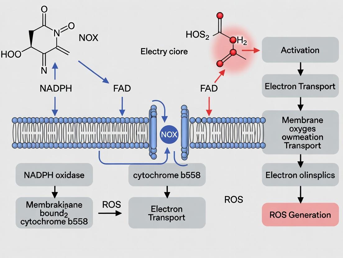
Signaling Pathways and Regulatory Logic
NOX activation integrates into diverse signaling cascades. The diagrams below illustrate two canonical pathways.
The Scientist's Toolkit: Essential Research Reagents
Table 3: Key Research Reagent Solutions for NOX Studies
| Reagent/Category | Example Product/Code | Function & Application |
|---|---|---|
| Isoform-Selective Inhibitors | GKT137831 (NOX1/4), ML171 (NOX1), GSK2795039 (NOX2) | Pharmacological dissection of isoform-specific functions in cells and in vivo. |
| Peptide Inhibitors | gp91ds-tat (NOX2), NoxA1ds (NOX1/3) | Cell-permeable peptides blocking subunit interaction; high specificity. |
| Activation Agonists | Phorbol Myristate Acetate (PMA), Angiotensin II, Formyl Peptide (fMLP) | Activate PKC-dependent (NOX1/2) or GPCR-dependent pathways. |
| ROS Detection Probes | Dihydroethidium (DHE), MitoSOX, Amplex Red, H2DCFDA | Fluorescent/luminescent detection of specific ROS (O₂•⁻, H₂O₂) in compartments. |
| Validated Antibodies | Anti-NOX1-5, Anti-p22phox, Anti-p47phox (from reputable suppliers) | Western Blot, Immunoprecipitation, Immunofluorescence for expression and localization. |
| Knockout/Knockdown Tools | siRNA/shRNA libraries, CRISPR/Cas9 knockout cell lines (commercial) | Genetic validation of isoform-specific phenotypes. |
| Activity Assay Kits | NADPH Consumption Assay Kits, Lucigenin-based CL Kits | Direct in vitro enzymatic activity measurement of immunoprecipitated NOX. |
| Calcium Modulators | Ionomycin, Thapsigargin, BAPTA-AM | To activate (ionomycin) or inhibit (BAPTA) Ca²⁺-sensitive NOX5/DUOX isoforms. |
The NOX enzyme family represents a sophisticated, tightly regulated system for ROS-based signal transduction. Each isoform's unique properties—defined by its regulatory partners, ROS product, tissue distribution, and activation kinetics—tailor it to specific physiological roles. Current research frontiers include elucidating the structural basis for NOX4's constitutive H₂O₂ production, defining the precise roles of DUOX in mucosal immunity, and developing next-generation isoform-selective inhibitors with therapeutic potential for fibrosis, cardiovascular disease, and cancer. This precise understanding is fundamental for advancing the thesis of targeted NOX modulation in human health and disease.
NADPH oxidases (NOXes) are transmembrane enzyme complexes that catalyze the production of reactive oxygen species (ROS), primarily superoxide anion (O₂•⁻), by transferring electrons from cytosolic NADPH to extracellular or phagosomal oxygen. Within the context of physiological signaling research, NOX-derived ROS are recognized not merely as toxic by-products but as crucial second messengers regulating diverse processes including cell proliferation, differentiation, and immune response. The catalytic core of the NOX2 complex, the most extensively studied isoform, is formed by the membrane-bound heterodimer of gp91phox (NOX2) and p22phox. Its activity is tightly controlled by the assembly of cytosolic regulator subunits: p47phox, p67phox, p40phox, and the small GTPase Rac. This whitepaper provides an in-depth structural and mechanistic analysis of this complex assembly, serving as a technical guide for researchers and drug development professionals aiming to modulate NOX function.
Core Subunit Architecture and Quantitative Data
Membrane-Bound Catalytic Subunits
The catalytic center resides in the NOX2/p22phox heterodimer. NOX2 is a heme-containing flavoprotein with key cofactor-binding domains. p22phox serves as a stabilizing partner and docking site for cytosolic regulators.
Table 1: Structural and Functional Properties of Membrane-Bound Catalytic Subunits
| Subunit | Gene | Transmembrane Helices | Key Domains/Motifs | Molecular Weight (kDa) | Critical Residues/Binding Partners |
|---|---|---|---|---|---|
| NOX2 (gp91phox) | CYBB | 6 | FAD-binding, NADPH-binding, 2 heme groups (histidine-ligated) | ~65 | His101, His115, His209, His222 (heme ligation); Cys244 (FAD binding) |
| p22phox | CYBA | 2 | PRD (Proline-Rich Domain) at cytosolic C-terminus | ~22 | Pro156, Gln160 (binds p47phox SH3 domain); essential for NOX2 stability |
Cytosolic Regulators
Activation involves translocation of cytosolic subunits to form the active complex at the membrane.
Table 2: Structural Domains and Functions of Cytosolic Regulatory Subunits
| Subunit | Key Domains | Molecular Weight (kDa) | Primary Function | Critical Regulatory Sites |
|---|---|---|---|---|
| p47phox | PX, two SH3 domains (SH3A, SH3B), AIR (Auto-Inhibitory Region), PRR (Proline-Rich Region) | ~47 | Organizer subunit; senses phosphoinositides & phosphorylation; bridges membrane and other regulators | Ser303, Ser304, Ser328 (PKC phosphorylation sites); SH3B binds p22phox PRD. |
| p67phox | TPR (Tetratricopeptide Repeat), Activation Domain (AD), PBI, two SH3 domains | ~67 | Essential activator; AD binds and likely induces conformational change in NOX2 | Arg86, His338, Asp500 (in AD, critical for electron transfer activation). |
| p40phox | PX, SH3 domain | ~40 | Accessory regulator; stabilizes complex; enhances activity via PX binding to PtdIns(3)P | PX domain binds PtdIns(3)P; SH3 binds p47phox PRR. |
| Rac (Rac1/2) | GTPase domain, Polybasic region, C-terminal tail | ~21 | Molecular switch; binds p67phox and membranes; induces final active conformation | Gly12 (G12V oncogenic), Thr35 (binds p67phox PBI domain). |
Mechanism of Assembly and Activation
In resting state, p47phox is auto-inhibited: its SH3 domains are masked by intramolecular binding to its AIR. p67phox and p40phox are constitutively associated via a PBI-PBX domain interaction. Upon cellular stimulation (e.g., by PMA, fMLP), signaling pathways lead to:
- Phosphorylation of p47phox: Primarily by PKC on multiple serines in its AIR, causing a conformational unmasking of its SH3 domains.
- Membrane Recruitment: The p47phox PX domain binds to phosphoinositides (e.g., PtdIns(3,4)P₂), while its exposed SH3B domain engages the PRD of p22phox. The p40phox PX domain binds specifically to PtdIns(3)P, enriching the complex at phagosomal membranes.
- GTP-Rac Recruitment: GTP-loaded Rac (Rac2 in neutrophils) translocates to the membrane and binds both the p67phox PBI domain and the membrane via its prenylated tail.
- Catalytic Activation: The p67phox Activation Domain engages the dehydrogenase domain of NOX2, facilitated by Rac binding, to induce electron transfer from NADPH.
Diagram 1: NOX2 Complex Activation Pathway (100 chars)
Key Experimental Protocols
In Vitro Reconstitution of NOX Activity (Cell-Free Assay)
This gold-standard assay directly measures the electron transfer capability of the assembled complex using purified components.
Detailed Protocol:
- Membrane Preparation: Isolate neutrophil plasma membranes (or membranes from NOX2/p22phox-expressing cells) via nitrogen cavitation and differential centrifugation. These provide the catalytic core. Store at -80°C.
- Cytosolic Component Preparation: Express and purify recombinant full-length p47phox, p67phox, p40phox, and Rac1 (preloaded with GTPγS, a non-hydrolyzable GTP analog) from E. coli or insect cells. For p47phox, a phospho-mimetic mutant (e.g., S303E/S304E/S328E) is often used to bypass the need for kinase treatment.
- Reaction Setup: In a 96-well plate, combine:
- 20 μg of membrane protein.
- 100 nM each of recombinant p47phox, p67phox, p40phox.
- 500 nM Rac1-GTPγS.
- 100 μM NADPH (electron donor).
- 50 μM cytochrome c (electron acceptor, detects O₂•⁻).
- Assay buffer: 65 mM HEPES, pH 7.0, 0.17 M sucrose, 500 μM MgCl₂, 1 mM EGTA, 100 μM DTPA (chelator).
- Measurement: Initiate reaction by adding NADPH. Continuously monitor the reduction of cytochrome c at 550 nm (ε₅₅₀ = 21.1 mM⁻¹cm⁻¹) using a spectrophotometer for 5-10 minutes. The initial linear rate is calculated as superoxide-dependent SOD-inhibitable activity.
Co-Immunoprecipitation (Co-IP) for Complex Assembly Analysis
Used to validate protein-protein interactions in a cellular context.
Detailed Protocol:
- Cell Stimulation & Lysis: Stimulate NOX-expressing cells (e.g., differentiated PLB-985 or HEK293-NOX2) with PMA (100 ng/mL, 5-10 min). Lyse cells in a mild non-ionic detergent buffer (e.g., 1% Triton X-100, 150 mM NaCl, 50 mM Tris pH 7.4, plus protease/phosphatase inhibitors).
- Immunoprecipitation: Pre-clear lysate with Protein A/G beads. Incubate 500 μg of lysate with 2 μg of antibody against a subunit of interest (e.g., anti-p22phox) or a control IgG overnight at 4°C with gentle rotation. Capture immune complexes with Protein A/G beads for 2 hours.
- Washing & Elution: Wash beads 3-4 times with lysis buffer. Elute bound proteins by boiling in 2X Laemmli SDS-PAGE sample buffer.
- Detection: Analyze eluates by SDS-PAGE and Western blotting, probing for putative binding partners (e.g., probe for p47phox and p67phox in a p22phox IP).
The Scientist's Toolkit: Key Research Reagents
Table 3: Essential Reagents for NOX Complex Research
| Reagent/Solution | Function/Application in NOX Research | Key Details/Considerations |
|---|---|---|
| Diphenyleneiodonium (DPI) | Broad-spectrum, irreversible flavoprotein inhibitor. | Inhibits NOX by binding FAD moiety. Positive control for activity inhibition (IC₅₀ ~10-100 nM). |
| Phorbol 12-Myristate 13-Acetate (PMA) | Potent PKC activator. | Used to stimulate p47phox phosphorylation and NOX2 complex assembly in cells (typical: 100 ng/mL). |
| Superoxide Dismutase (SOD) | Enzyme that catalyzes O₂•⁻ dismutation. | Used in assays (e.g., cytochrome c reduction) to confirm superoxide is the measured product (SOD-inhibitable signal). |
| Cytochrome c (Ferric) | Electron acceptor for superoxide. | Used in cell-free and cellular assays. Reduction monitored at 550 nm. Membrane-impermeable; measures extracellular O₂•⁻. |
| GTPγS & GDPβS | Non-hydrolyzable GTP and GDP analogs. | Used to lock Rac in active (GTPγS) or inactive (GDPβS) state for in vitro reconstitution studies. |
| Anti-NOX2/gp91phox Antibody (e.g., Clone 54.1) | Specific detection of NOX2 subunit. | Critical for Western blot, immunofluorescence, and flow cytometry (e.g., diagnosing CGD). |
| Recombinant Cytosolic Factors (p47, p67, p40, Rac) | Purified proteins for in vitro reconstitution. | Available from commercial suppliers or purified in-house. Phospho-mimetic p47phox mutants bypass kinase requirement. |
| L-012 & Lucigenin | Chemiluminescent probes for ROS detection. | Highly sensitive, used for cellular and in vivo imaging of NOX activity. L-012 is more specific for superoxide. |
Diagram 2: NOX Research Experimental Workflows (96 chars)
Within the broader thesis on NADPH oxidase (NOX) in physiological signaling research, understanding the precise tissue and subcellular localization of each isoform is paramount. NOX enzymes, which catalyze the reduction of molecular oxygen to generate reactive oxygen species (ROS), are not merely sources of oxidative stress. They are critical signaling hubs in health, development, and disease. Their function is intrinsically linked to their specific expression patterns and compartmentalization within cells. This guide provides an in-depth analysis of the operational niches of the seven NOX isoforms (NOX1-5, DUOX1-2) in mammalian systems.
The following tables consolidate data on isoform-specific expression across tissues and subcellular compartments, derived from recent transcriptomic, proteomic, and immunohistochemical studies.
Table 1: Primary Tissue Expression of NOX Isoforms in Health & Development
| Isoform | High-Expression Tissues/Cells (Adult) | Key Roles in Development |
|---|---|---|
| NOX1 | Colon epithelium, vascular smooth muscle, uterus, prostate, microglia | Gut epithelial maturation, postnatal vascular remodeling |
| NOX2 | Phagocytes (neutrophils, macrophages), endothelial cells, cardiomyocytes, neurons, hematopoietic stem cells | Brain development (neuronal migration, progenitor proliferation), innate immune system ontogeny |
| NOX3 | Inner ear (vestibular and cochlear epithelia), fetal kidney, fetal brain | Critical for otoconia formation and balance; role in early renal and neural patterning |
| NOX4 | Ubiquitous; highest in kidney, vasculature (endothelium, SMC), heart, bone, lung fibroblasts | Angiogenesis, stem cell differentiation, bone mineralization, kidney organogenesis |
| NOX5 | Testis, lymphoid tissue, vascular endothelium (species-dependent; absent in rodents) | Sperm capacitation, lymphocyte activation, vascular function (esp. in humans) |
| DUOX1 | Thyroid, respiratory epithelium, salivary glands, prostate | Thyroid hormone synthesis, innate mucosal defense, lung branching morphogenesis |
| DUOX2 | Thyroid, gastrointestinal tract (especially colon), respiratory epithelium | Thyroid hormone synthesis, gut microbial homeostasis, post-injury intestinal repair |
Table 2: Characteristic Subcellular Localization of NOX Isoforms
| Isoform | Primary Subcellular Compartments | Membrane Association & Key Partners |
|---|---|---|
| NOX1 | Plasma membrane (lipid rafts), endosomes, caveolae | Requires p22phox, NOXO1, NOXA1, Rac1. Localization directed by NOXO1. |
| NOX2 | Plasma membrane, phagosomal membrane, secretory vesicles (in resting phagocytes) | Requires p22phox, p47phox (NOXO2), p67phox (NOXA2), p40phox, Rac2. Phox proteins direct targeting. |
| NOX3 | Plasma membrane (apical in inner ear hair cells) | Requires p22phox; can utilize NOXO1/NOXA1 or phagocyte oxidase components. |
| NOX4 | Focal adhesions, endoplasmic reticulum, nucleus, mitochondria, plasma membrane | Constitutively active with p22phox. Localization dictates signaling output (e.g., ER: calcium signaling). |
| NOX5 | Cytoplasm (upon low Ca2+), Plasma membrane (upon activation) | Calcium-dependent, does not require p22phox or cytosolic subunits. Contains EF-hand domains. |
| DUOX1/2 | Apical plasma membrane of polarized epithelia (e.g., thyrocytes, bronchial cells) | Require DUOXA1/2 maturation factors for ER exit and apical localization. Generate extracellular H2O2. |
Detailed Methodologies for Localization Studies
Protocol 3.1: Immunofluorescence Confocal Microscopy for Subcellular NOX Localization
- Objective: To visualize endogenous NOX isoform distribution in fixed cells or tissue sections.
- Key Reagents:
- Validated Isoform-Specific Primary Antibodies: Critical due to homology between isoforms. Must be verified using KO tissue controls.
- Organelle-Specific Markers: e.g., Anti-Calnexin (ER), Anti-TOM20 (Mitochondria), Anti-E-Cadherin (Plasma membrane), Phalloidin (Actin).
- Cell Permeabilization Buffer: 0.1-0.3% Triton X-100 in PBS. Concentration optimized for membrane-bound vs. cytosolic epitope access.
- High-Resolution Mounting Medium with DAPI: For nuclear counterstaining and photostability.
- Procedure:
- Culture cells on glass coverslips or prepare 5-10 µm frozen tissue sections.
- Fix with 4% paraformaldehyde (PFA) for 15 min at RT. Avoid methanol for membrane protein preservation.
- Permeabilize and block with 5% normal serum in PBS-Triton for 1 hour.
- Incubate with primary antibody cocktail (NOX + organelle marker) overnight at 4°C.
- Wash and incubate with species/isotype-specific secondary antibodies conjugated to distinct fluorophores (e.g., Alexa Fluor 488, 568) for 1 hour.
- Mount and image using a confocal laser scanning microscope. Acquire z-stacks for 3D localization.
- Analysis: Perform colocalization analysis (e.g., Pearson's correlation coefficient, Manders' overlap coefficient) using software like ImageJ/Fiji or Imaris.
Protocol 3.2: Subcellular Fractionation and Western Blot Analysis
- Objective: To biochemically isolate and quantify NOX isoforms in specific cellular compartments.
- Key Reagents:
- Differential Centrifugation Buffers: Homogenization buffer (0.25 M sucrose, 10 mM HEPES, pH 7.4 with protease inhibitors) and sucrose density gradients.
- Protease and Phosphatase Inhibitor Cocktails: Essential to prevent degradation and maintain modification states.
- Membrane Protein Extraction Kits: For separating integral membrane proteins (like NOXes) from cytosolic fractions.
- Compartment-Specific Antibodies (for blotting): e.g., Na+/K+ ATPase (plasma membrane), Calreticulin (ER), Cytochrome C (mitochondria), LAMP1 (lysosomes).
- Procedure:
- Homogenize cells/tissue in ice-cold isotonic buffer using a Dounce homogenizer or needle.
- Perform sequential centrifugation: 800 x g (nuclei/debris), 10,000 x g (heavy mitochondria), 100,000 x g supernatant (cytosol) and pellet (light membranes/microsomes).
- For higher resolution, load the 10,000 x g supernatant onto a discontinuous sucrose gradient (e.g., 1.0 M, 1.5 M, 2.0 M) and ultracentrifuge at 100,000 x g overnight.
- Collect fractions and precipitate proteins. Perform SDS-PAGE and Western blotting.
- Probe blots sequentially for NOX isoforms and compartment markers to assign localization.
Protocol 3.3: In Situ Hybridization for Developmental Expression Mapping
- Objective: To map spatial and temporal mRNA expression of NOX isoforms during embryogenesis.
- Key Reagents:
- RNAscope or Similar HCR Probes: Isoform-specific, double-Z oligonucleotide probe sets provide high specificity and sensitivity.
- RNase-free reagents and equipment: To prevent RNA degradation.
- Developmental tissue series: Paraffin-embedded or OCT-embedded embryos at multiple stages.
- Procedure: Follow manufacturer's protocol for multiplex fluorescent in situ hybridization. Allows co-detection of multiple NOX mRNAs and key developmental markers in the same section.
Signaling Pathways and Experimental Workflows: Visualizations
Diagram Title: NOX1 Signaling in Epithelial Proliferation (76 chars)
Diagram Title: Experimental Workflow for NOX4 Localization (68 chars)
The Scientist's Toolkit: Key Research Reagent Solutions
Table 3: Essential Reagents for NOX Localization and Activity Studies
| Reagent Category | Specific Example & Function | Application Notes |
|---|---|---|
| Validated Antibodies | Anti-NOX4 (C-terminal, extracellular): For immunofluorescence and Western blot without permeabilization (membrane-bound). | Always confirm specificity with KO controls. Distinguish between total vs. surface pools. |
| Chemical Inhibitors | GLX351322 (NOX4-specific): Small molecule inhibitor used to probe NOX4-specific function in localization contexts. | Use alongside genetic knockdown (siRNA) to confirm on-target effects. |
| Genetic Tools | siRNA/shRNA Libraries (isoform-specific): Knockdown to validate antibody specificity and study localization consequences. | Off-target effects are a concern; use pooled siRNAs or CRISPRi for better validation. |
| CRISPR-Cas9 KO Cell Lines: Generate definitive negative controls and study developmental roles in engineered stem cells. | Essential for establishing antibody specificity and functional assays. | |
| Activity Probes | H2O2-sensitive fluorescent probes (HyPer, roGFP): Targeted to organelles (ER, mitochondria) to measure localized ROS. | Can be expressed as fusion proteins. Allows real-time, compartment-specific ROS detection upon NOX activation. |
| Localization Reporters | NOX isoform-GFP Fusion Constructs: For live-cell imaging of trafficking. Must be C-terminally tagged to avoid interference with complex assembly. | Overexpression can mislocalize; use endogenous tagging via CRISPR/Cas9 knock-in for optimal results. |
| Subcellular Markers | CellLight BAC-GFP Organelle Tags (Thermo Fisher): Reliable fluorescent labeling of specific organelles in live cells. | Cructive for definitive colocalization studies in dynamic processes. |
The historical view of reactive oxygen species (ROS) as solely damaging agents has been conclusively overturned. A central thesis in contemporary physiological signaling research posits that NADPH oxidases (NOX enzymes) are dedicated, regulated sources of ROS that function as deliberate second messenger generators. This paradigm shift places NOX-derived ROS—primarily hydrogen peroxide (H₂O₂)—alongside canonical second messengers like cAMP and Ca²⁺. Controlled, spatiotemporally restricted ROS production modulates redox-sensitive signaling nodes, regulating processes from cell proliferation and differentiation to immune response and cell death. This whitepaper details the mechanisms, experimental evidence, and methodologies underpinning this fundamental concept.
NOX Isoforms: Specialized ROS-Generating Enzymes
NOX enzymes are multi-subunit complexes that catalyze the reduction of molecular oxygen using NADPH as an electron donor. Different isoforms enable localized, quantifiable ROS production.
Table 1: NOX Isoforms and Their Signaling Contexts
| Isoform | Primary Tissue/Cell Expression | Physiological Signaling Roles | Key Regulatory Subunits |
|---|---|---|---|
| NOX1 | Colon, vascular smooth muscle | Angiogenesis, blood pressure regulation, host defense | NOXO1, NOXA1, Rac1 |
| NOX2 | Phagocytes, endothelium, neurons | Microbial killing, post-injury inflammation, memory formation | p47phox, p67phox, p40phox, Rac2 |
| NOX3 | Inner ear | Otoconia biogenesis (balance) | p47phox, NOXO1? |
| NOX4 | Kidney, endothelium, osteoclasts | Oxygen sensing, fibrosis, osteoclastogenesis | Poldip2 (constitutive activity) |
| NOX5 | Spleen, testis, vascular tissue | Sperm capacitation, lymphocyte activation | Ca²⁺-binding EF hands |
| DUOX1/2 | Thyroid, lung, epithelia | Thyroid hormone synthesis, mucosal host defense | DUOXA1/2 maturation factors |
Core Signaling Mechanisms: How ROS Acts as a Second Messenger
H₂O₂, due to its relative stability and membrane permeability, is the primary ROS second messenger. It regulates signaling via two principal mechanisms:
1. Reversible Oxidation of Redox-Sensitive Cysteine Residues: H₂O₂ oxidizes specific cysteine thiols (-SH) in target proteins to sulfenic acid (-SOH), altering protein conformation, activity, localization, and interactions. 2. Inhibition of Phosphatases: A cardinal example is the oxidation and inhibition of Protein Tyrosine Phosphatases (PTPs) and the tumor suppressor phosphatase PTEN. This inhibition potentiates kinase-driven signaling cascades (e.g., MAPK, PI3K/AKT).
Diagram 1: NOX-Dependent ROS Signaling Node
Title: Core NOX-ROS-PTP Signaling Axis
Quantitative Data: Measuring ROS and Its Effects
Table 2: Quantifiable Metrics in NOX/ROS Signaling Research
| Parameter | Typical Measurement Method | Example Quantitative Range (Cell-Based Assay) | Significance |
|---|---|---|---|
| ROS Production (Rate) | Amplex Red (H₂O₂), Lucigenin (O₂⁻), DCFDA (Cellular ROS) | 10-100 pmol H₂O₂/min/µg protein (NOX4) | Direct readout of NOX activity. |
| Protein Oxidation | Biotin-Switch Assay (Sulfenation), dimedone-based probes | 2-5 fold increase in sulfenation upon stimulation | Maps direct redox targets. |
| PTP Inhibition | In vitro phosphatase activity assay with DTT rescue | >80% activity loss upon H₂O₂ (10-100 µM) | Demonstrates functional consequence. |
| Downstream Phosphorylation | Western blot (p-ERK, p-AKT), phospho-tyrosine arrays | 3-10 fold increase p-ERK/p-AKT, blocked by NOX inhibition | Measures amplified signaling output. |
| Transcriptional Output | qPCR of Nrf2/ARE or NF-κB targets (e.g., HO-1, IL-8) | 5-50 fold mRNA induction | Quantifies long-term genetic changes. |
Key Experimental Protocols
Protocol 1: Validating NOX-Dependent ROS in a Signaling Pathway
Objective: To establish that a specific cellular stimulus triggers ROS production via a NOX enzyme, and that this ROS is required for downstream signaling.
Materials: See "The Scientist's Toolkit" below.
Method:
- Cell Stimulation: Plate cells in 96-well black plates or culture dishes. Serum-starve (e.g., 4-6 hrs).
- Inhibitor Pre-treatment (30-60 min prior):
- NOX Inhibition: Diphenyleneiodonium (DPI, 10 µM), VAS2870 (10 µM), or GKT136901 (1 µM).
- ROS Scavenging: N-acetylcysteine (NAC, 5 mM), Polyethylene glycol-catalase (PEG-Cat, 250 U/mL).
- Include vehicle controls (e.g., DMSO).
- Stimulation & ROS Detection: Add stimulus (e.g., PDGF, 20 ng/mL; TNF-α, 10 ng/mL). Simultaneously, load with a cell-permeable ROS-sensitive fluorescent probe (e.g., CM-H2DCFDA, 5 µM). Incubate for desired time (typically 15-60 min).
- Quantification:
- Microplate Reader: Measure fluorescence (Ex/Em ~492/517 nm for DCF) kinetically or at endpoint.
- Flow Cytometry: Harvest cells, analyze median fluorescence intensity (MFI) of 10,000 cells.
- Microscopy: Image live cells; quantify mean fluorescence per cell.
- Downstream Analysis: In parallel dishes (without probe), lyse cells post-stimulation for Western blot analysis of phospho-proteins (e.g., p-ERK1/2).
Interpretation: A NOX/ROS inhibitor should attenuate both the stimulus-induced fluorescence increase and the downstream phosphorylation.
Protocol 2: Detecting Protein Sulfenation (Reversible Oxidation)
Objective: To identify specific proteins that undergo cysteine sulfenation in response to NOX-derived ROS.
Method (Biotin-Switch Based, e.g., using DYn-2):
- Cell Treatment & Probe Labeling: Stimulate cells in the presence/absence of NOX inhibitors. During stimulation, add the sulfenic acid-specific probe DYn-2 (50 µM) to the medium.
- Cell Lysis: Lyse cells in non-reducing, non-denaturing lysis buffer (avoid DTT/β-mercaptoethanol).
- Click Chemistry: Perform copper-catalyzed azide-alkyne cycloaddition (CuAAC) between the DYn-2 alkyne and a biotin-azide tag (e.g., 100 µM). Add CuSO₄ (1 mM), THPTA ligand (100 µM), and sodium ascorbate (1 mM). Incubate 1 hr at RT.
- Streptavidin Pulldown: Incubate lysate with streptavidin-agarose beads overnight at 4°C. Wash stringently.
- Elution & Analysis: Elute proteins with Laemmli buffer containing DTT (to reduce disulfides). Analyze by Western blot for proteins of interest (to identify specific targets) or by mass spectrometry for global profiling.
Advanced Pathway Visualization
Diagram 2: NOX4 in TGF-β-Induced Profibrotic Signaling
Title: NOX4 Amplifies TGF-β Fibrotic Signaling
The Scientist's Toolkit: Key Research Reagents
Table 3: Essential Reagents for NOX/ROS Signaling Research
| Reagent/Category | Example Specific Items | Function & Application | Key Considerations |
|---|---|---|---|
| NOX Inhibitors | Diphenyleneiodonium (DPI), VAS2870, GKT136901/831, apocynin | Pharmacological inhibition of NOX enzyme activity to establish causality. | DPI is non-specific (flavoproteins); apocynin requires peroxidation for activity. Isoform-specific inhibitors preferred. |
| ROS Scavengers | N-acetylcysteine (NAC), PEG-SOD, PEG-Catalase, Tempol | Chemical quenching of ROS to confirm role in signaling. | Distinguish between O₂⁻ (SOD) and H₂O₂ (Catalase). NAC boosts glutathione. |
| ROS Detection Probes | CM-H2DCFDA (general ROS), Amplex Red (H₂O₂), MitoSOX (mito. O₂⁻), HyPer (genetically encoded H₂O₂) | Quantitative and spatial detection of ROS production. | DCFDA is non-specific and can auto-oxidize. Use specific probes and ratiometric sensors (HyPer) for accuracy. |
| Sulfenation Probes | DYn-2, DCP-Bio1, SOH-4 | Chemoselective labeling of sulfenated cysteines for detection or pulldown. | Require click chemistry for detection. Use in non-reducing conditions. |
| Genetic Tools | siRNA/shRNA (NOX isoforms), CRISPR-Cas9 KO cells, Dominant-negative Rac1 (N17) | Genetic validation of specific NOX isoform involvement. | Controls for isoform specificity and compensatory mechanisms. |
| Activity Assays | NADPH consumption assay, lucigenin/cytochrome c reduction (O₂⁻), hydrogen peroxide electrode | Direct in vitro or cell-free measurement of NOX complex activity. | Often require membrane fractions from overexpressing systems. |
| Antibodies | Anti-NOX isoforms, anti-phospho-tyrosine, anti-sulfenic acid (e.g., SOH antibody), anti-phospho-kinases (p-ERK, p-AKT) | Detection of protein expression, localization, and redox/phospho-states. | Validate antibodies for specific applications (WB, IF). SOH antibodies may have limited targets. |
Within the framework of NADPH oxidase (NOX) research, understanding upstream activators is paramount for elucidating physiological and pathophysiological signaling. NOX enzymes, particularly NOX1-5 and Duox1-2, are critical sources of regulated reactive oxygen species (ROS) that function as second messengers. This guide details the core upstream activators—Growth Factors, Cytokines, GPCRs, and Mechanical Forces—that converge on NOX activation, framing their roles within physiological signaling pathways and therapeutic targeting.
Growth Factor-Mediated NOX Activation
Growth factors such as Epidermal Growth Factor (EGF), Platelet-Derived Growth Factor (PDGF), and Vascular Endothelial Growth Factor (VEGF) activate NOX isoforms via receptor tyrosine kinases (RTKs). This leads to ROS-dependent amplification of downstream pathways like PI3K/Akt and MAPK/ERK, crucial for cell proliferation, migration, and survival.
Key Pathway: Ligand binding → RTK autophosphorylation → Recruitment of p47phox/p67phox (NOX2) or NOXA1 (NOX1) via PI3K/Rac GTPase → NOX complex assembly → Localized ROS production.
Experimental Protocol: Assessing NOX Activation by EGF in Cell Culture
- Cell Preparation: Seed serum-starved adherent cells (e.g., vascular smooth muscle cells) in 6-well plates.
- Stimulation: Treat cells with EGF (e.g., 100 ng/mL) for time points (0, 2, 5, 15, 30 min).
- ROS Detection: Load cells with 10 µM CM-H2DCFDA (fluorogenic probe) for 30 min pre-stimulation. Measure fluorescence intensity (Ex/Em: 495/529 nm) via plate reader or fluorescence microscopy.
- Inhibition Control: Pre-treat with NOX inhibitor (e.g., 10 µM VAS2870 or 100 µM apocynin) or ROS scavenger (e.g., 500 U/mL PEG-catalase) for 1 hour before stimulation.
- Validation: Perform immunoblotting for phosphorylated EGFR (Tyr1068) and ERK1/2 (Thr202/Tyr204) to correlate ROS burst with pathway activation.
Diagram 1: Growth Factor RTK Signaling to NOX Activation
Cytokine-Induced NOX Signaling
Pro-inflammatory cytokines (TNF-α, IL-1β, IFN-γ) activate NOX2 primarily in immune cells but also in stromal cells. Signaling occurs through cytokine receptor engagement, leading to activation of NF-κB and JAK/STAT pathways, which can upregulate NOX subunit expression and induce complex assembly.
Key Pathway: Cytokine binding → Receptor dimerization → JAK/NF-κB activation → Transcriptional upregulation of NOX subunits/p22phox & increased Rac activity → Enhanced NOX complex formation and ROS burst.
Experimental Protocol: Measuring TNF-α-Induced NOX2 Activity in Macrophages
- Cell Stimulation: Differentiate THP-1 cells into macrophages with PMA (100 nM, 48h). Stimulate with TNF-α (20 ng/mL) for 0-24h.
- Gene Expression: Extract RNA at intervals (0, 2, 6, 24h). Perform qRT-PCR for CYBB (NOX2), NCF1 (p47phox), and NCF2 (p67phox) using SYBR Green.
- ROS Assay: Post-stimulation, incubate cells with 5 µM L-012 (high-sensitivity luminescent probe for superoxide) and add PMA (100 nM) as a positive control. Measure chemiluminescence for 60 min.
- Inhibition: Use JAK inhibitor (e.g., Tofacitinib, 1 µM) or NF-κB inhibitor (e.g., BAY 11-7082, 5 µM) to confirm pathway specificity.
Diagram 2: Cytokine Receptor Signaling Leading to NOX Activation
GPCR-Triggered NOX Activation
GPCRs (e.g., angiotensin II AT1R, chemokine receptors) are potent activators of NOX1, NOX2, and NOX4. Ligand binding initiates Gαq/11 and Gβγ signaling, activating phospholipase Cβ (PLCβ), generating IP3/DAG, and activating Protein Kinase C (PKC) and Rac. This is a primary mechanism in cardiovascular signaling.
Key Pathway: Agonist (e.g., Ang II) → GPCR → Gαq/11 activation → PLCβ → PKC activation → Phosphorylation of p47phox → Translocation to membrane NOX → ROS generation.
Experimental Protocol: Analyzing Angiotensin II (Ang II)-Dependent NOX Activation
- Tissue/Cell Model: Use primary vascular smooth muscle cells (VSMCs) or intact aortic rings.
- Stimulation: Treat with Ang II (100 nM) for 15-60 minutes.
- Membrane Translocation Assay: Fractionate cells into cytosol and membrane fractions via differential centrifugation. Perform Western blot for p47phox, Rac1, and membrane marker (e.g., Na+/K+ ATPase).
- Functional Readout: Measure superoxide production via lucigenin (5 µM) enhanced chemiluminescence in aortic rings. Include pretreatment with AT1R blocker (Losartan, 10 µM) or PKC inhibitor (GF109203X, 5 µM).
- Calcium Dependence: Chelate intracellular Ca2+ with BAPTA-AM (10 µM) to assess Ca2+-dependent NOX activation.
Mechanotransduction and NOX Activation
Mechanical forces (shear stress, cyclic stretch, pressure overload) activate NOX, particularly NOX2 and NOX4, in endothelial cells, cardiomyocytes, and osteocytes. Integrins, focal adhesion kinases (FAK), and stretch-activated ion channels are key sensors.
Key Pathway: Mechanical force → Integrin conformational change/ Ion channel opening → FAK/Src/PI3K activation → Rac1 GTP loading → NOX activation → ROS modulating mechano-adaptive responses.
Experimental Protocol: Shear Stress-Induced NOX Activity in Endothelial Cells
- Flow System: Seed human umbilical vein endothelial cells (HUVECs) on slides compatible with a parallel-plate flow chamber.
- Shear Application: Subject cells to laminar shear stress (e.g., 15 dyn/cm²) for periods from 5 min to 24h. Static cells as control.
- Real-time ROS Measurement: Load cells with CellROX Deep Red (5 µM) 30 min before endpoint. Quantify fluorescence intensity per cell using automated microscopy.
- Mechanistic Dissection: Transfect with siRNA against Ptk2 (FAK) or Rac1. Use specific inhibitors: integrin blocker (RGD peptide, 1 mM) or TRPV4 channel inhibitor (GSK2193874, 100 nM).
- Downstream Analysis: Assess phosphorylation of paxillin (Tyr118) and VEGFR2 (Tyr1175) as markers of mechanotransduction.
Diagram 3: Mechanical Force Transduction to NOX Activation
Table 1: Characteristic Parameters of NOX Activation by Upstream Stimuli
| Activator Class | Prototypical Agonist | Primary NOX Isoform | Onset of ROS Burst | Key Measured Output (Example) | Common Inhibitors/Interventions |
|---|---|---|---|---|---|
| Growth Factors | EGF (100 ng/mL) | NOX1, NOX2 | 2-5 minutes | 2-3 fold increase in DCF fluorescence vs. basal | AG1478 (EGFRi), VAS2870 (NOXi) |
| Cytokines | TNF-α (20 ng/mL) | NOX2 | 30 min (acute), sustained over 24h | 5-fold increase in L-012 chemiluminescence | BAY 11-7082 (NF-κBi), Tofacitinib (JAKi) |
| GPCRs | Angiotensin II (100 nM) | NOX1, NOX2, NOX4 | 5-15 minutes | 50% increase in lucigenin signal in vessels | Losartan (AT1Ri), Gallein (Gβγi) |
| Mechanical Forces | Laminar Shear (15 dyn/cm²) | NOX2, NOX4 | 10-30 minutes | 1.8-fold increase in CellROX intensity | RGD peptide, GSK2193874 (TRPV4i) |
Table 2: Key Research Reagent Solutions for Studying NOX Upstream Activation
| Reagent / Material | Category | Function in Experiment | Example Product/Catalog # |
|---|---|---|---|
| CM-H2DCFDA | Fluorescent ROS probe | Detects general intracellular ROS (H2O2, peroxynitrite) upon oxidation. | Thermo Fisher Scientific, C6827 |
| L-012 | Chemiluminescent probe | Highly sensitive detection of superoxide (O2•−) from cells or tissues. | Wako Pure Chemical, 120-04891 |
| CellROX Deep Red | Far-red fluorescent ROS probe | For live-cell imaging, more resistant to oxidation, measures multiple ROS. | Thermo Fisher Scientific, C10422 |
| Recombinant Human EGF | Growth Factor | Activates RTK pathways to stimulate NOX1/2. | PeproTech, AF-100-15 |
| Recombinant Human TNF-α | Cytokine | Induces inflammatory NOX2 activation and subunit expression. | R&D Systems, 210-TA |
| Angiotensin II | GPCR agonist | Activates AT1R to trigger Gαq/PKC-dependent NOX activation. | Sigma-Aldrich, A9525 |
| VAS2870 | NOX inhibitor | Pan-NOX inhibitor, used to confirm NOX-dependent ROS signals. | Sigma-Aldrich, SML0273 |
| Rac1 Activation Assay Kit | Biochemical assay | Pulldown of active GTP-bound Rac1, critical for NOX assembly. | Cytoskeleton, Inc., BK035 |
| siRNA against p47phox (NCF1) | Genetic tool | Knockdown to confirm specific NOX subunit requirement. | Dharmacon, M-010534-01 |
| Parallel-Plate Flow Chamber System | Mechanobiology tool | Applies defined laminar shear stress to endothelial cell monolayers. | Ibidi, µ-Slide I 0.4 Luer |
Integrated Experimental Workflow
Diagram 4: Core Workflow for Studying NOX Upstream Activators
The precise activation of NOX enzymes by distinct upstream triggers—growth factors, cytokines, GPCRs, and mechanical forces—forms a complex signaling network where ROS act as specific second messengers. Dissecting these pathways with rigorous protocols, quantitative assays, and appropriate controls is essential for advancing the thesis that NOX-derived ROS are central, regulated mediators in physiology. This knowledge is foundational for developing targeted therapies in conditions of dysregulated ROS signaling, such as hypertension, fibrosis, and chronic inflammation.
Tools of the Trade: Cutting-Edge Methods to Measure NOX Activity, Localization, and Function
Within the context of NADPH oxidase (NOX) research, precise detection of reactive oxygen species (ROS) is paramount. NOX enzymes, unlike mitochondrial sources, produce ROS as primary signaling molecules, requiring methods that distinguish specific ROS types (e.g., superoxide [O₂•⁻], hydrogen peroxide [H₂O₂]) with high spatial and temporal resolution. This guide details core direct detection methodologies—chemiluminescence probes, fluorescent dyes, and Electron Spin Resonance (ESR) spectroscopy—as applied to NOX-derived ROS in physiological signaling studies.
Chemiluminescence Probes
Chemiluminescence probes emit light upon oxidation, offering high sensitivity with minimal background. They are ideal for real-time, whole-population ROS measurements in cell suspensions or tissue homogenates.
Key Probes: Lucigenin and L-012
- Lucigenin (bis-N-methylacridinium nitrate): Long used for O₂•⁻ detection. Its reduction to a lucigenyl radical followed by reaction with O₂•⁻ yields light emission (~430 nm). Concerns regarding redox-cycling artifact necessitate careful use at low concentrations (<5 µM).
- L-012 (8-amino-5-chloro-7-phenylpyridopyridazine-1,4(2H,3H)dione): A highly sensitive luminol derivative. In the presence of peroxidase (e.g., released MPO) and ROS (O₂•⁻, H₂O₂, ONOO⁻), it produces intense chemiluminescence. It is widely used for detecting NOX2 activity in phagocytes and vascular systems.
Table 1: Comparison of Chemiluminescence Probes
| Probe | Primary ROS Detected | Emission Peak | Key Advantage | Key Limitation/Consideration | Typical Working Concentration |
|---|---|---|---|---|---|
| Lucigenin | Superoxide (O₂•⁻) | ~430 nm | High signal-to-noise for O₂•⁻ | Potential redox-cycling; not cell-permeable | 5-20 µM |
| L-012 | O₂•⁻, H₂O₂, ONOO⁻ (peroxidase-dependent) | ~430-530 nm | Extreme sensitivity (~100x luminol) | Peroxidase-dependent; not specific to a single ROS | 50-200 µM |
Experimental Protocol: L-012-based Detection of NOX2 Activity in Leukocyte Suspensions
- Cell Preparation: Isolate primary neutrophils or use differentiated HL-60 cells. Suspend in Hanks' Balanced Salt Solution (HBSS) with 10 mM HEPES, pH 7.4.
- Probe Loading: Add L-012 to a final concentration of 100 µM.
- Stimulus: Add a NOX2 agonist (e.g., 100 nM phorbol myristate acetate [PMA]) directly to the cuvette.
- Measurement: Immediately place the sample in a luminometer (or plate reader). Record relative light units (RLU) continuously for 30-60 minutes at 37°C.
- Controls: Include samples with a NOX2 inhibitor (e.g., 10 µM diphenyleneiodonium [DPI]) or a superoxide scavenger (e.g., 50 U/mL SOD).
- Data Analysis: Quantify the area under the curve (AUC) or peak RLU values.
Fluorescent Dyes
Fluorescent probes enable cellular and subcellular ROS imaging, providing spatial information critical for signaling studies.
Key Probes: DHE and H2DCFDA
- Dihydroethidium (DHE): The gold standard for cellular O₂•⁻ detection. DHE is oxidized by O₂•⁻ to 2-hydroxyethidium (2-OH-E⁺), which intercalates into DNA and fluoresces red (ex/em ~518/605 nm). Specific quantification of 2-OH-E⁺ requires HPLC separation from other oxidation products.
- 2',7'-Dichlorodihydrofluorescein diacetate (H2DCFDA): A cell-permeable, general oxidative stress indicator. Cellular esterases cleave the acetate groups, and oxidation (primarily by H₂O₂ with peroxidase or iron catalysis) yields fluorescent DCF (ex/em ~498/522 nm). It is not specific for a single ROS.
Table 2: Comparison of Fluorescent ROS Probes
| Probe | ROS Specificity | Excitation/Emission | Key Advantage | Key Limitation | Typical Loading |
|---|---|---|---|---|---|
| Dihydroethidium (DHE) | Superoxide (O₂•⁻) | 518/605 nm (for 2-OH-E⁺) | Relatively specific for O₂•⁻ when measured correctly | Requires HPLC for specificity; photo-oxidation | 5-20 µM, 30 min, 37°C |
| H2DCFDA | Broad-spectrum (H₂O₂, ONOO⁻, •OH) | ~498/522 nm | Easy to use, sensitive to various oxidants | Lacks specificity; easily photo-oxidized | 5-10 µM, 30 min, 37°C |
Experimental Protocol: DHE-based Imaging of NOX-derived O₂•⁻ in Live Cells
- Cell Culture: Plate cells (e.g., vascular smooth muscle cells expressing NOX4) on glass-bottom dishes.
- Probe Loading: Wash cells with warm PBS. Load with 5 µM DHE in serum-free culture medium for 30 minutes at 37°C in the dark.
- Stimulation & Inhibition: Treat cells with a physiological NOX activator (e.g., 100 ng/mL Angiotensin II) in the presence or absence of a NOX inhibitor (e.g., 1 µM GKT137831).
- Imaging: Acquire images using a fluorescence microscope with a TRITC/Cy3 filter set immediately and at defined intervals (e.g., every 10 min for 1 hour). Maintain cells at 37°C. Use identical exposure settings.
- Quantification: Analyze mean fluorescence intensity (MFI) in the nuclear region (for 2-OH-E⁺) using image analysis software (e.g., ImageJ).
- Validation: Co-incubate with 50 U/mL polyethylene glycol-conjugated superoxide dismutase (PEG-SOD) as a negative control.
Electron Spin Resonance (ESR) Spectroscopy
ESR (or EPR) is the most definitive method for direct ROS detection, as it measures unpaired electrons in free radicals. It offers high specificity when used with spin traps.
Principle and Spin Traps
Short-lived radicals are reacted with diamagnetic spin traps (e.g., CPH, DMPO) to form stable, paramagnetic spin adducts with characteristic ESR spectra, allowing identification of the trapped radical.
- CPH (1-Hydroxy-3-carboxy-2,2,5,5-tetramethylpyrrolidine): Cell-permeable; specifically forms a stable nitroxide upon reaction with O₂•⁻.
- DMPO (5,5-Dimethyl-1-pyrroline N-oxide): Forms adducts with various radicals (•OH, O₂•⁻), with distinct spectral fingerprints.
Table 3: Common Spin Traps for NOX-derived ROS Detection
| Spin Trap | Target Radical | Resulting Adduct | Characteristic ESR Spectrum | Notes |
|---|---|---|---|---|
| CPH | Superoxide (O₂•⁻) | CP• (nitroxide) | Three-line spectrum (1:1:1) | Cell-permeable; specific for O₂•⁻. |
| DMPO | Superoxide (O₂•⁻) | DMPO-OOH | Distinct 12-line pattern | DMPO-OOH decays to DMPO-OH. |
| DMPO | Hydroxyl (•OH) | DMPO-OH | 1:2:2:1 quartet | Can be formed from O₂•⁻/H₂O₂ via Fenton. |
Experimental Protocol: ESR Detection of NOX Activity using CPH
- Sample Preparation: Treat cells or tissue homogenates with a NOX stimulus. Prepare a reaction mixture containing the sample, 1 mM CPH, and the metal chelator DTPA (100 µM) in a suitable buffer (e.g., Krebs-HEPES).
- Measurement: Draw the mixture into a gas-permeable Teflon capillary tube. Insert the tube into the ESR resonator pre-equilibrated with nitrogen gas containing 2% oxygen to maintain physiological pO₂.
- ESR Parameters: Record spectra using a standard X-band spectrometer. Typical settings: microwave power, 20 mW; modulation amplitude, 2 G; sweep time, 2 min; sweep width, 100 G.
- Quantification: Measure the amplitude of the central line of the CP• triplet spectrum. Compare against a standard curve of a stable nitroxide (e.g., TEMPOL) to calculate picomoles of adduct.
The Scientist's Toolkit: Research Reagent Solutions
| Category | Reagent/Kit | Function in NOX/ROS Research |
|---|---|---|
| Chemiluminescence | L-012 (Wako/Cayman Chemical) | Highly sensitive probe for detecting extracellular ROS burst from NOX2. |
| Fluorescent Probes | Dihydroethidium (DHE) (Thermo Fisher) | Cell-permeable probe for detecting intracellular superoxide. |
| Fluorescent Probes | MitoSOX Red (Thermo Fisher) | DHE derivative targeted to mitochondria; distinguishes NOX-derived from mitochondrial O₂•⁻. |
| Spin Traps | CPH (Alexis/Enzo Life Sciences) | Cell-permeable spin trap for specific ESR detection of superoxide. |
| Inhibitors | GKT137831 (Cayman Chemical) | Dual NOX4/NOX1 inhibitor used to probe isoform-specific signaling. |
| Inhibitors | Diphenyleneiodonium (DPI) (Sigma-Aldrich) | Flavoprotein inhibitor that blocks NOX enzymes (and others). |
| Activators | Phorbol Myristate Acetate (PMA) | Protein kinase C agonist that potently activates NOX2 in phagocytes. |
| Scavengers/Enzymes | Polyethylene Glycol-Superoxide Dismutase (PEG-SOD) | Long-acting extracellular O₂•⁻ scavenger for validation experiments. |
| Scavengers/Enzymes | PEG-Catalase | Long-acting extracellular H₂O₂ scavenger. |
Visualizing Pathways and Workflows
Title: NOX Activation & ROS Detection in Signaling Pathways
Title: Core Workflow for Direct ROS Detection Methods
Within the framework of NADPH oxidase (NOX) physiological signaling research, dissecting the specific roles of individual NOX isoforms (NOX1-5, DUOX1/2) is paramount. This technical guide details the core genetic and pharmacological tools—knockout/knockdown models and isoform-selective inhibitors (GKT and GLX series)—that enable this functional dissection. Their specificity underpins the validity of research linking specific NOX-derived reactive oxygen species (ROS) to signaling pathways in cardiovascular disease, fibrosis, cancer, and neurodegeneration.
Genetic Models: Knockout and Knockdown
Genetic ablation or suppression provides the gold standard for establishing isoform-specific function.
Global and Conditional Knockout Mouse Models
These models offer complete, heritable deletion of a specific Nox gene.
- Methodology: Generated via homologous recombination in embryonic stem cells, often using Cre-loxP technology for cell-type-specific conditional knockouts (cKO). For example, crossing Nox1flox/flox mice with tissue-specific Cre-drivers (e.g., Vil1-Cre for intestinal epithelial cells).
- Key Protocols:
- Genotyping: Tail-clip DNA is extracted and analyzed by PCR with allele-specific primers (wild-type, floxed, deleted).
- Validation: Confirm loss of target mRNA via qRT-PCR and loss of protein via Western blot in relevant tissues. Measure basal and stimulated ROS production (e.g., lucigenin or L-012 chemiluminescence, Amplex Red assay) in isolated cells/tissues, comparing to wild-type.
- Phenotypic Analysis: Subject mice to disease models (e.g., angiotensin II-induced hypertension, bleomycin-induced lung fibrosis). Assess parameters like blood pressure, fibrosis markers, histology, and inflammatory cytokines.
Knockdown Models (siRNA/shRNA)
Used for transient or stable gene silencing in vitro and in vivo.
- Methodology: Design and transfert sequence-specific small interfering RNA (siRNA) or transduce cells with short hairpin RNA (shRNA) lentiviral vectors.
- Key Protocol (in vitro siRNA knockdown):
- Design: Select 2-3 validated siRNA duplexes targeting distinct regions of the target NOX mRNA.
- Transfection: Plate cells, reach 50-70% confluence. Complex siRNA with lipid-based transfection reagent in serum-free medium (e.g., 20-50 nM final siRNA concentration). Add complexes to cells.
- Incubation: Replace medium after 4-6 hours. Assay after 48-72 hours.
- Validation: Quantify knockdown efficiency by qRT-PCR and Western blot. Measure functional ROS output.
Table 1: Common NOX Isoform Genetic Mouse Models
| Isoform | Model Type | Key Phenotypic Observations | Primary Research Context |
|---|---|---|---|
| Nox1 | Global KO | Reduced blood pressure, attenuated vascular hypertrophy, impaired host defense. | Hypertension, atherosclerosis, stroke. |
| Nox2 | Global KO (gp91phox-/-) | Chronic granulomatous disease (CGD) phenotype, severe infections, reduced vascular ROS. | Host defense, vascular inflammation, ischemia-reperfusion injury. |
| Nox4 | Global & Conditional KO | Protective in many models: reduced cardiac fibrosis, less endothelial dysfunction, attenuated kidney injury. | Fibrotic diseases (cardiac, pulmonary, renal), metabolic syndrome. |
| DUOX1 | Global KO | Impaired airway epithelial H2O2 production, altered mucosal defense. | Asthma, innate immune responses in lung. |
Pharmacological Tools: Isoform-Selective Inhibitors
Small molecule inhibitors allow acute, reversible inhibition, complementing genetic approaches.
GKT-series (GenKyoTex)
These are diphenylene iodonium (DPI) derivatives with improved selectivity, primarily targeting NOX1 and NOX4.
- GKT136901: Dual NOX1/4 inhibitor (IC50 ~ 100-150 nM for both).
- GKT137831 (Setanaxib): The most advanced clinical compound; preferential inhibition of NOX4 (IC50 ~ 140 nM) and NOX1 (IC50 ~ 110 nM), with minimal effect on NOX2 and NOX5.
- Specificity & Validation: Must be used alongside genetic validation. Demonstrate that inhibitor effects are absent in corresponding KO cells. Monitor off-target effects on mitochondrial complexes and other flavoenzymes.
GLX-series
These compounds, derived from GKT chemicals, aim for greater isoform discrimination.
- GLX351322: Reported as a NOX4-selective inhibitor with >10-fold selectivity over NOX1.
- Experimental Protocol for Inhibitor Profiling:
- Cell-Based ROS Assay: Use cells overexpressing a single NOX isoform or primary cells with defined NOX expression.
- Dose-Response: Pre-treat cells with inhibitor (e.g., 0.01 - 10 µM) for 30-60 min. Stimulate with appropriate agonist (e.g., PMA for NOX2, TGF-β for NOX4).
- Detection: Use isoform-appropriate ROS probes: DHE/HPLC for superoxide, Amplex Red/HRP for H2O2. Measure kinetics.
- Data Analysis: Calculate IC50 values. Counter-screen against related enzymes (e.g., xanthine oxidase, eNOS).
Table 2: Profile of Key NOX Isoform-Selective Inhibitors
| Compound | Primary Target (IC50) | Selectivity Over NOX2 | Key Off-Targets to Consider | Development Stage |
|---|---|---|---|---|
| GKT137831 | NOX4 (~140 nM), NOX1 (~110 nM) | >10-fold | Mitochondrial complex I, other flavoproteins | Phase 2 (PBC, IPF) |
| GLX351322 | NOX4 (sub-µM) | >10-fold | Not fully characterized; requires KO validation | Preclinical |
| ML171 | NOX1 (~0.13 µM) | ~10-fold | Can inhibit DUOX1 at higher concentrations | Tool compound |
| VAS2870 | Pan-NOX (µM range) | N/A | Thiol-alkylating agent; non-specific | Tool compound |
Assessing and Validating Specificity
The cornerstone of reliable research.
- Genetic Cross-Validation: Any pharmacological result should be confirmed by showing the effect is abolished in KO/KD models of the purported target isoform.
- Orthogonal Assays: Use multiple ROS detection methods and readouts (e.g., gene expression, phosphorylation events downstream of ROS).
- Counter-Screening: Test inhibitors against a panel of related ROS-producing systems and signaling enzymes.
The Scientist's Toolkit: Research Reagent Solutions
Table 3: Essential Reagents for NOX Isoform Research
| Item | Function & Explanation | Example Vendor/Cat # |
|---|---|---|
| Nox1/2/4 KO Mice | Definitive genetic models for in vivo functional studies. | Jackson Laboratory, Taconic Biosciences |
| Isoform-Selective siRNA Pools | For transient, specific knockdown in cell culture. | Dharmacon, Santa Cruz Biotechnology |
| GKT137831 (Setanaxib) | Preferential NOX1/4 inhibitor; key for acute intervention studies. | MedChemExpress, Cayman Chemical |
| Validated NOX Isoform Antibodies | Essential for Western blot and IHC validation of genetic/pharmacologic manipulation. | Sigma-Aldrich, Santa Cruz Biotechnology |
| L-012 & Lucigenin | Chemiluminescent probes for superoxide detection from intact cells/tissues. | Wako Chemicals, Sigma-Aldrich |
| Amplex Red/HRP Kit | Fluorometric assay for specific, quantitative measurement of extracellular H2O2. | Thermo Fisher Scientific |
| CellROX / DHE Probes | Cell-permeable fluorescent dyes for general intracellular oxidative stress imaging. | Thermo Fisher Scientific |
| NOX Activity ELISA Kits | Some commercial kits measure activity in cell lysates via NADPH consumption. | Abcam, CytoNick |
Visualizations
Title: NOX Signaling Dissection via Genetic & Pharmacological Tools
Title: Workflow for Validating NOX Isoform Specificity
The study of redox signaling is central to understanding the physiological and pathological roles of NADPH oxidases (NOX). The generation of reactive oxygen species (ROS), particularly H₂O₂, by NOX enzymes acts as a precise signaling mechanism regulating processes from immune response to cell differentiation. Genetically encoded redox sensors, such as HyPer and redox-sensitive green fluorescent proteins (roGFPs), have revolutionized this field by enabling real-time, spatiotemporal analysis of redox dynamics within live cells. This whitepaper provides a technical guide to these tools within the specific context of NOX signaling research, detailing their principles, applications, and experimental protocols.
Core Principles of Genetically Encoded Redox Sensors
These sensors are fluorescent proteins engineered to change their spectral properties upon oxidation/reduction.
- roGFP (Redox-sensitive GFP): Fused to human glutaredoxin-1 (Grx1), roGFP2 equilibrates with the glutathione redox couple (GSH/GSSG). Oxidation of its disulfide bond increases excitation at 400 nm and decreases it at 490 nm, while the emission peak at 510 nm remains constant. The 400/490 nm excitation ratio provides a ratiometric, internally controlled readout of thiol redox potential, insensitive to sensor concentration, photobleaching, or excitation light path length.
- HyPer: A circularly permuted yellow fluorescent protein (cpYFP) inserted into the regulatory domain of the prokaryotic H₂O₂-sensing protein OxyR. Direct reaction with H₂O₂ causes a conformational change, altering cpYFP fluorescence. HyPer exhibits dual excitation peaks (420 nm and 500 nm) with a single emission peak at 516 nm. The 500/420 nm excitation ratio is specific for H₂O₂.
Quantitative Comparison of Key Redox Sensors
Table 1: Characteristics of Primary Genetically Encoded Redox Sensors for NOX Research
| Sensor Name | Redox Species Detected | Sensing Mechanism | Excitation/Emission Peaks (nm) | Dynamic Range (Ratio Change) | pH Sensitivity | Key Applications in NOX Research |
|---|---|---|---|---|---|---|
| roGFP2 | Glutathione redox potential (GSH/GSSG) | Grx1-coupled, disulfide formation | Ex: 400/490, Em: 510 | ~5-6 fold | Low (with proper calibration) | Global cytoplasmic/nuclear redox state; response to NOX activation. |
| roGFP2-Orp1 | H₂O₂ (specifically) | Fusion with yeast H₂O₂ peroxidase Orp1 | Ex: 400/490, Em: 510 | ~3-4 fold | Low | Direct, rapid detection of H₂O₂ fluxes from membrane NOX enzymes. |
| HyPer | H₂O₂ (specifically) | OxyR domain conformational change | Ex: 420/500, Em: 516 | ~4-5 fold | High (requires control sensor HyPer-C199S) | Subcellular, specific H₂O₂ dynamics; NOX-derived H₂O₂ microdomains. |
| HyPer7 | H₂O₂ (specifically) | Improved OxyR-cpYFP variant | Ex: 490, Em: 516 | ~7-10 fold | Reduced | Fast kinetics, high sensitivity for low-level NOX signaling events. |
| Grx1-roGFP2 | Glutathionylation | Lacks resolving cysteine | Ex: 400/490, Em: 510 | ~2-3 fold | Low | Detection of protein S-glutathionylation, a downstream redox modification. |
Experimental Protocols for NOX Signaling Research
Protocol 1: Imaging Spatiotemporal H₂O₂ Dynamics During Growth Factor Stimulation
Objective: To visualize NOX-derived H₂O₂ production upon growth factor (e.g., EGF) receptor activation.
Materials:
- Cells expressing a relevant NOX isoform (e.g., NOX4) and/or its regulatory subunits.
- Plasmid encoding HyPer3 (improved pH stability) targeted to the desired compartment (e.g., cytoplasm, mitochondria matrix).
- Confocal or widefield live-cell imaging system with rapid wavelength switching capabilities.
- Imaging chamber with temperature and CO₂ control.
- Hanks' Balanced Salt Solution (HBSS) or phenol-red free culture medium.
- Recombinant EGF.
- Catalase (positive control for H₂O₂ degradation).
Method:
- Transfection: Seed cells on imaging-compatible dishes. Transfect with the HyPer3 construct 24-48 hours prior to imaging.
- Sensor Calibration: In situ calibration is critical. After experiment, treat cells with 5 mM DTT (full reduction) followed by 100 µM H₂O₂ (full oxidation) to obtain Rmin and Rmax. Calculate the degree of oxidation.
- Image Acquisition: Place dish on the microscope stage. Set environmental control to 37°C, 5% CO₂. Acquire ratiometric images: sequentially excite at 488 nm and 405 nm, collect emission at 500-550 nm. Establish a baseline (1 image every 30 sec for 5 min).
- Stimulation: Gently add EGF to a final concentration of 50-100 ng/mL without moving the dish. Continue time-lapse acquisition (1 image every 15-30 sec) for 20-30 minutes.
- Data Analysis: Generate ratio images (488/405) using ImageJ or microscopy software. Quantify ratio changes in regions of interest (ROIs) at the plasma membrane or cytoplasm. Normalize to baseline (ΔR/R₀).
Protocol 2: Measuring Compartment-Specific Redox Potential Changes After NOX Activation
Objective: To quantify changes in glutathione redox potential (EGSSG/2GSH) in mitochondria versus cytosol upon NOX2 activation.
Materials:
- Macrophage cell line (e.g., RAW 264.7) expressing NOX2 complex.
- Plasmids encoding roGFP2 targeted to the cytosol and mitochondrial matrix.
- Phorbol 12-myristate 13-acetate (PMA, a NOX2 activator).
- Fluorescence plate reader or ratiometric imaging system.
Method:
- Cell Preparation: Co-transfect cells with cytosol-targeted and mitochondria-targeted roGFP2. Seed into a black-walled, clear-bottom 96-well plate for plate reading or into imaging dishes.
- Ratiometric Measurement: For a plate reader, program sequential reads: Ex 400/Em 510 and Ex 490/Em 510. Take baseline reads every minute for 10 minutes.
- Activation: Add PMA to a final concentration of 100 ng/mL. Continue reading every minute for 60 minutes.
- Calibration: At the endpoint, permeabilize cells with 50 µM digitonin. Add 10 mM DTT for Rmin, then 100 µM aldrithiol for Rmax.
- Calculation: Calculate the redox potential using the Nernst equation: E = E₀ - (RT/nF) ln([GSH]²/[GSSG]), where the roGFP2 oxidation degree is proportional to [GSSG]/[GSH]². E₀ for roGFP2 is approximately -280 mV.
Signaling Pathways and Experimental Workflows
The Scientist's Toolkit: Essential Research Reagent Solutions
Table 2: Key Reagents and Materials for Live-Cell Redox Imaging
| Item | Function/Description | Example Product/Source |
|---|---|---|
| HyPer3 / HyPer7 Expression Plasmid | Genetically encoded, rationetric H₂O₂ sensor with improved pH stability (HyPer3) or brightness/dynamic range (HyPer7). | Addgene (plasmids #42131, #121743). |
| roGFP2 (cytosolic) Expression Plasmid | Rationetric sensor for glutathione redox potential (EGSSG/2GSH). | Addgene (plasmid #64985). |
| Mito-roGFP2 Expression Plasmid | roGFP2 targeted to the mitochondrial matrix for organelle-specific redox measurement. | Addgene (plasmid #64986). |
| roGFP2-Orp1 Expression Plasmid | Fusion protein for specific, rapid detection of H₂O₂ via Orp1 peroxidase. | Addgene (plasmid #64987). |
| Lipid-Based Transfection Reagent | For efficient delivery of sensor plasmids into mammalian cells (e.g., macrophages, fibroblasts). | Lipofectamine 3000 (Thermo Fisher). |
| Phenol-Red Free Imaging Medium | Culture medium without phenol red, which can autofluoresce and interfere with sensitive GFP/YFP signals. | FluoroBrite DMEM (Gibco). |
| Hanks' Balanced Salt Solution (HBSS) | Physiological salt solution for imaging during acute stimulations without serum factors. | Gibco, with calcium and magnesium. |
| Phorbol 12-Myristate 13-Acetate (PMA) | Potent direct activator of Protein Kinase C, used to stimulate NOX2 complex activity. | Sigma-Aldrich (P1585). |
| Dithiothreitol (DTT) | Reducing agent used for in situ calibration of roGFP and HyPer sensors (establishes Rmin). | Thermo Scientific. |
| Aldrithiol (2,2'-dipyridyl disulfide) | Thiol-oxidizing agent used for in situ calibration of roGFP sensors (establishes Rmax). | Sigma-Aldrich (DIPYR). |
NADPH oxidases (NOX) are multi-subunit enzyme complexes critical for regulated reactive oxygen species (ROS) production in physiological signaling. Understanding the precise assembly of the NOX complex—involving catalytic (e.g., NOX1-5, DUOX1/2) and regulatory subunits (e.g., p22phox, p47phox, p67phox, Rac1)—is fundamental to deciphering its role in cell signaling, host defense, and redox biology. This whitepaper provides an in-depth technical guide for mapping these protein-protein interactions (PPIs) using three cornerstone techniques: Co-Immunoprecipitation (Co-IP), Proximity Ligation Assay (PLA), and Förster Resonance Energy Transfer (FRET). These methods offer complementary insights, from validating biochemical interactions to visualizing them in fixed and living cells, within the broader context of NOX research for therapeutic targeting.
Core Techniques for NOX Complex Analysis
Co-Immunoprecipitation (Co-IP)
Principle: Co-IP is a biochemical method to isolate a native protein complex from cell lysates using an antibody specific to one protein (the bait), thereby co-precipitating its binding partners.
Detailed Protocol for NOX2 Complex:
- Cell Lysis: Harvest transfected or endogenous NOX-expressing cells (e.g., HEK293, phagocytes). Lyse in 1 mL of non-denaturing lysis buffer (e.g., 50 mM Tris-HCl pH 7.4, 150 mM NaCl, 1% Triton X-100, 1 mM EDTA) supplemented with protease and phosphatase inhibitors. Incubate on ice for 30 min, then centrifuge at 16,000 × g for 15 min at 4°C.
- Pre-clearing: Incubate supernatant with 20 μL of Protein A/G agarose beads for 1 hour at 4°C with rotation. Centrifuge briefly to collect cleared lysate.
- Immunoprecipitation: Incubate cleared lysate with 2-5 μg of anti-bait antibody (e.g., anti-NOX2 or anti-p22phox) or species-matched IgG control overnight at 4°C with rotation.
- Bead Capture: Add 50 μL of washed Protein A/G beads and incubate for 2-4 hours at 4°C.
- Washing: Pellet beads and wash 4-5 times with 1 mL of ice-cold lysis buffer.
- Elution: Resuspend beads in 40 μL of 2X Laemmli sample buffer, boil for 5-10 minutes.
- Analysis: Resolve eluates by SDS-PAGE and perform Western blotting for putative interactors (e.g., blot for p47phox and p67phox when using NOX2 as bait).
Data Output: Qualitative confirmation of interaction; semi-quantitative via band intensity densitometry.
Proximity Ligation Assay (PLA)
Principle: PLA detects proximal proteins (<40 nm) in situ using species-specific secondary antibodies conjugated to oligonucleotides. If targets are close, a circular DNA template forms, is amplified, and detected via fluorescently labeled probes, yielding a discrete fluorescent spot per interaction event.
Detailed Protocol for NOX-p22phox Proximity:
- Cell Preparation: Culture cells on chamber slides, perform experimental treatments, and fix with 4% PFA for 15 min. Permeabilize with 0.1% Triton X-100 for 10 min.
- Blocking: Block with commercial PLA blocking buffer for 1 hour at 37°C.
- Primary Antibodies: Incubate with a pair of primary antibodies from different species (e.g., mouse anti-NOX2 and rabbit anti-p22phox) diluted in antibody diluent overnight at 4°C.
- PLA Probe Incubation: Apply species-specific PLA probes (MINUS and PLUS) for 1 hour at 37°C.
- Ligation: Add ligation solution containing connector oligonucleotides for 30 min at 37°C. Close proximity enables circle formation.
- Amplification: Add rolling circle amplification solution with fluorescently labeled nucleotides (e.g., Cy3) for 100 min at 37°C.
- Microscopy: Mount slides and acquire images using a fluorescence microscope. Quantify PLA signals (dots/cell) using image analysis software (e.g., ImageJ).
Data Output: Quantitative, single-cell resolution data on interaction frequency and subcellular localization.
Förster Resonance Energy Transfer (FRET)
Principle: FRET measures energy transfer between a donor fluorophore and an acceptor fluorophore when they are within 1-10 nm. It is ideal for studying real-time dynamics of NOX assembly in living cells.
Detailed Protocol for FRET using NOX Biosensors:
- Construct Design: Create genetic constructs fusing donor (e.g., CFP, mTurquoise2) and acceptor (e.g., YFP, mVenus) to NOX complex subunits of interest (e.g., CFP-p47phox and YFP-p67phox).
- Cell Transfection: Co-transfect constructs into suitable cells (e.g., COS-7, HeLa) using lipid-based methods.
- Image Acquisition: 24-48h post-transfection, image live cells on a confocal or widefield microscope with FRET capability. Use filter sets for donor excitation/emission and acceptor emission.
- FRET Calculation: Acquire three images: Donor channel (IDD), Acceptor channel (IAA), and FRET channel (IDA). Calculate corrected FRET (e.g., using the sensitized emission method). Common metric: FRET efficiency (E) or normalized FRET (NFRET).
- Stimulation: Acquire time-lapse FRET images before and after stimulation (e.g., with PMA to induce NOX2 complex assembly).
Data Output: Quantitative, real-time kinetics of interaction with high spatial resolution in living cells.
Table 1: Comparison of PPI Mapping Techniques for NOX Complex Assembly
| Parameter | Co-Immunoprecipitation | Proximity Ligation Assay | FRET |
|---|---|---|---|
| Interaction Proximity | Biochemical isolation | < 40 nm | 1-10 nm |
| Throughput | Medium | Medium-High | Low-Medium |
| Quantification | Semi-quantitative (WB) | Quantitative (spots/cell) | Highly Quantitative (Ratio/Efficiency) |
| Cellular Context | Lysate (disrupted) | Fixed cells (preserved) | Live cells |
| Temporal Resolution | Endpoint | Endpoint | Real-time (seconds) |
| Key Output | Proof of direct/indirect binding | Spatial distribution & frequency | Spatiotemporal dynamics |
| Typical NOX Application | Validate subunit composition | Map complex localization in tissues | Assemble kinetics upon stimulation |
Table 2: Example Quantitative Data from NOX PPI Studies
| Technique | Experimental Condition | Key Measurement | Reported Result | Biological Implication |
|---|---|---|---|---|
| Co-IP | PMA-stimulated neutrophils | p47phox association with p22phox | 4.2-fold increase vs. resting | Confirms stimulus-induced complex assembly. |
| PLA | Cardiac tissue, NOX4-p22phox | PLA signals per cardiomyocyte | 18.5 ± 3.2 (vs. 2.1 ± 0.8 IgG ctrl) | Demonstrates constitutive NOX4-p22 interaction in situ. |
| FRET | HEK293 cells expressing biosensor | NFRET between p47phox & p67phox | Baseline: 0.05; Post-PMA: 0.21 peak within 90s | Reveals rapid, inducible cytosolic subunit dimerization. |
The Scientist's Toolkit: Research Reagent Solutions
Table 3: Essential Reagents for NOX PPI Studies
| Reagent/Material | Function/Application | Example Product/Note |
|---|---|---|
| Anti-NOX2 (gp91phox) Antibody | Bait antibody for Co-IP; detection in PLA/WB. | Mouse monoclonal (clone 53); validates phagocyte NOX. |
| Anti-p22phox Antibody | Key for detecting membrane-bound cytochrome b558. | Rabbit polyclonal; common partner for NOX1-4. |
| Duolink PLA Kit | Complete solution for PLA (probes, amplification, detection). | Sigma-Aldrich; kits available for different fluorophores. |
| CFP/YFP FRET Pair Plasmids | Donor/Acceptor for constructing NOX biosensors. | mTurquoise2/mVenus recommended for improved brightness & FRET. |
| Phorbol Myristate Acetate (PMA) | PKC agonist to stimulate canonical NOX2 complex assembly. | Standard positive control; use at 100-200 nM. |
| Non-denaturing Lysis Buffer | Preserves weak/transient PPIs during Co-IP. | Must contain detergent (e.g., Triton X-100, CHAPS). |
| Protease/Phosphatase Inhibitor Cocktail | Prevents degradation and preserves phosphorylation states. | Essential for studying signal-regulated assembly. |
Visualizing NOX Assembly Pathways & Assays
Diagram 1: NOX Activation & PPI Methods
Diagram 2: Co-IP Workflow for NOX
Diagram 3: Proximity Ligation Assay Steps
Diagram 4: FRET Principle in NOX Assembly
Within the broader thesis on NADPH oxidase (NOX) isoforms in physiological signaling, a critical challenge is the explicit linkage of reactive oxygen species (ROS) generation to the activation of specific downstream signaling cascades. NOX-derived ROS are not merely toxic byproducts but act as deliberate second messengers, modulating key pathways such as MAPK/ERK, PI3K/Akt, and Ca2+ signaling. This guide provides an in-depth technical framework for designing and interpreting functional assays that establish these causal links.
Table 1: Quantitative Readouts of NOX-Activated Downstream Pathways
| Downstream Pathway | Primary Readout | Typical Assay | Reported Fold-Change/Amplitude with NOX Stimulation | Inhibition by NOX Knockdown/Antioxidants |
|---|---|---|---|---|
| MAPK/ERK | Phospho-ERK1/2 (Thr202/Tyr204) | Western Blot / ELISA | 2.5 - 5.0 fold increase in p-ERK | 70-90% reduction |
| PI3K/Akt | Phospho-Akt (Ser473) | Western Blot / HTRF | 2.0 - 4.0 fold increase in p-Akt | 60-85% reduction |
| Ca2+ Signaling | Cytosolic [Ca2+] | Fluorometry (Fura-2, Fluo-4) | Δ[Ca2+] = 150-300 nM peak increase | 50-80% attenuation of peak |
| Transcription (NF-κB) | Nuclear p65 translocation / Luciferase reporter | Imaging / Reporter Assay | 3.0 - 6.0 fold increase in activity | 75-95% inhibition |
| Cellular Phenotype | Proliferation / Migration | BrdU / Scratch Assay | 1.5 - 2.5 fold increase | Reversal to baseline |
Experimental Protocols
Protocol 1: Simultaneous Real-Time Measurement of ROS and Ca2+ Flux
- Objective: To temporally correlate NOX activation with downstream Ca2+ signaling.
- Method:
- Cell Loading: Plate cells on a confocal-compatible dish. Load with 5 µM CM-H2DCFDA (ROS sensor) and 2 µM Fluo-4 AM (Ca2+ sensor) in serum-free medium for 30 min at 37°C.
- Stimulation & Imaging: Wash and replace with imaging buffer. Acquire baseline images for 2 min. Stimulate with a specific agonist (e.g., Angiotensin II for NOX2/4). Acquire time-lapse images every 10 seconds for 20 min using appropriate filter sets (FITC for DCF, TRITC for Fluo-4).
- Control: Pre-treat cells with 10 µM diphenyleneiodonium (DPI, NOX inhibitor) or 5 mM N-acetylcysteine (NAC, antioxidant) for 1 hour.
- Analysis: Quantify mean fluorescence intensity (MFI) over time in the region of interest (ROI). Plot kinetics and calculate the time lag between ROS burst and Ca2+ peak.
Protocol 2: Linking NOX to MAPK/ERK and PI3K/Akt via Phospho-Specific Flow Cytometry
- Objective: To measure cell population heterogeneity in pathway activation downstream of NOX.
- Method:
- Stimulation & Fixation: Stimulate cells in suspension or trypsinized monolayers with agonist for 0, 5, 15, and 30 min. Immediately fix cells with 4% paraformaldehyde for 10 min at 37°C.
- Permeabilization & Staining: Permeabilize with ice-cold 90% methanol for 30 min on ice. Wash and incubate with primary antibodies against phospho-ERK1/2 and phospho-Akt (Ser473) for 1 hour. Use fluorophore-conjugated secondary antibodies.
- NOX Inhibition: Include conditions with cells transfected with NOX-specific siRNA or treated with a pharmacological inhibitor like GKT136901 (NOX1/4 inhibitor).
- Acquisition & Analysis: Acquire data on a flow cytometer. Gate on single, viable cells. Analyze median fluorescence intensity (MFI) of phospho-signals. Use bivariate plots (p-ERK vs. p-Akt) to identify correlated activation.
Protocol 3: Genetic Reconstitution Assay for Pathway Specificity
- Objective: To prove a specific NOX isoform is responsible for activating a pathway.
- Method:
- KO Background: Use cells with a CRISPR/Cas9-mediated knockout of the NOX isoform of interest (e.g., NOX4 KO).
- Reconstitution: Transfect KO cells with: a) Wild-type (WT) NOX construct, b) Catalytically inactive mutant (e.g., NOX4 with a point mutation in NADPH-binding site), or c) Empty vector.
- Pathway Reporter Assay: Co-transfect a pathway-specific luciferase reporter (e.g., SRE for MAPK, FOXO for PI3K/Akt). 48h post-transfection, stimulate and measure luciferase activity.
- Interpretation: Pathway activation restored only in WT-NOX reconstituted cells confirms the requirement for that NOX's enzymatic activity.
Visualization of Signaling Pathways and Workflows
Title: NOX-Derived ROS as a Hub for Downstream Signaling Pathways
Title: Workflow for Kinetic ROS-Ca2+ Correlation Assay
The Scientist's Toolkit: Key Research Reagent Solutions
Table 2: Essential Reagents for Linking NOX to Downstream Pathways
| Reagent Category | Specific Example(s) | Function & Application |
|---|---|---|
| NOX Inhibitors (Pharmacological) | Diphenyleneiodonium (DPI), GKT136901 (NOX1/4i), VAS2870 (pan-NOX) | To inhibit NOX activity pharmacologically and observe subsequent blockade of downstream signaling. |
| Genetic NOX Modulators | siRNA/shRNA kits, CRISPR/Cas9 KO kits, cDNA for WT/Mutant NOX isoforms | To genetically ablate or reconstitute specific NOX isoforms for definitive mechanistic studies. |
| ROS Detection Probes | Amplex Red (H2O2), CM-H2DCFDA (general ROS), MitoSOX (mitochondrial O2•−) | To quantitatively measure ROS production kinetics and subcellular localization. |
| Phospho-Specific Antibodies | Anti-phospho-ERK1/2 (Thr202/Tyr204), Anti-phospho-Akt (Ser473) | Key immunodetection tools for assessing activation status of downstream pathways via WB, IF, or flow cytometry. |
| Ion-Sensitive Fluorescent Dyes | Fluo-4 AM, Fura-2 AM (Ca2+), SPQ (Cl−) | To measure real-time flux of secondary messengers linked to ROS signaling. |
| Pathway Reporter Assays | SRE-Luc (MAPK), FOXO-Luc (PI3K/Akt), NF-κB-Luc | To read out integrated transcriptional activity of a pathway as a functional endpoint. |
| Antioxidant Controls | N-acetylcysteine (NAC), PEG-Catalase, Tempol | To determine if signaling effects are broadly redox-sensitive, not just NOX-specific. |
Solving NOX Research Challenges: Pitfalls in Quantification, Specificity, and Data Interpretation
Within the broader thesis on NADPH oxidase (NOX) in physiological signaling research, a paramount challenge is the specific attribution of reactive oxygen species (ROS) signals to their discrete cellular sources. ROS, including superoxide (O₂•⁻) and hydrogen peroxide (H₂O₂), are produced by dedicated enzymatic complexes like the NOX family, mitochondrial electron transport chain (ETC), peroxisomes, and other oxidases. These spatially and chemically distinct ROS pools function as specific signaling molecules in processes ranging from proliferation to immune response. The "Specificity Problem" lies in the technical difficulty of accurately measuring and assigning the origin of a given ROS signal within a complex cellular milieu. This whitepaper provides an in-depth technical guide to disentangling NOX-derived ROS from other key sources, with a focus on experimental design, advanced probes, and pharmacological and genetic tools.
Key enzymatic sources of ROS and their distinguishing characteristics are summarized below.
Table 1: Primary Cellular ROS Sources and Features
| Source | Primary ROS Product | Subcellular Localization | Key Activators/Inhibitors | Primary Signaling Context |
|---|---|---|---|---|
| NOX Family (e.g., NOX1-5, DUOX1/2) | O₂•⁻ (rapidly dismutates to H₂O₂) | Plasma membrane, endosomes, phagosomes | Activated by cytokines, growth factors, G-proteins. Inhibited by DPI, GKT136901, VAS2870, apocynin. | Host defense, redox signaling, cell differentiation, angiogenesis. |
| Mitochondrial ETC (Complex I & III) | O₂•⁻ (Matrix & IMS) | Mitochondrial inner membrane | Enhanced by high membrane potential, reverse electron transport (RET), ETC inhibitors (rotenone, antimycin A). Scavenged by mitochondrial-targeted antioxidants (MitoTEMPO, MitoQ). | Metabolic signaling, hypoxia response, apoptosis. |
| Peroxisomes | H₂O₂ | Peroxisomal matrix | Fatty acid oxidation, Aminooxyacetate (AOA) inhibits catalase to amplify signal. | Lipid metabolism, β-oxidation. |
| Xanthine Oxidase (XO) | O₂•⁻, H₂O₂ | Cytoplasm | Hypoxia, ATP degradation. Inhibited by allopurinol, febuxostat. | Ischemia-reperfusion injury. |
| eNOS Uncoupling | O₂•⁻ | Cytoplasm, membrane | BH4 depletion, L-arginine deficiency. Prevented by BH4, L-arginine. | Endothelial dysfunction. |
Core Methodologies for Source-Specific ROS Detection
Pharmacological Inhibition Profiling
Pharmacological tools remain a first-line approach, though their specificity requires careful validation with genetic controls.
- Experimental Protocol: Inhibitor Titration and Multi-Source Assessment
- Cell Preparation: Plate cells in a 96-well black-walled plate. Include appropriate controls (vehicle, positive ROS inducers).
- Pre-treatment: Treat cells with a panel of source-specific inhibitors for 30-60 minutes prior to stimulation. Example Panel: 10 µM GKT136901 (NOX1/4), 5 µM VAS2870 (pan-NOX), 1 µM Rotenone (mito-ETC Complex I), 10 µM Allopurinol (XO), 500 µM AOA (peroxisomal catalase inhibitor).
- Stimulation & ROS Detection: Add cell-permeable ROS probe (e.g., 5 µM CM-H2DCFDA for general H₂O₂, 5 µM MitoSOX Red for mitochondrial O₂•⁻) along with a specific agonist (e.g., 100 ng/mL PMA for NOX2, 10 ng/mL TGF-β for NOX4, or serum for growth factor-induced NOX). Incubate for 15-60 min.
- Measurement: Read fluorescence (Ex/Em: ~488/525 nm for DCF; ~510/580 nm for MitoSOX) using a plate reader. For imaging, use live-cell confocal microscopy.
- Data Analysis: Express signal as fold-change over unstimulated vehicle control. The inhibitor that most significantly attenuates the stimulated signal indicates the predominant ROS source. Always correlate with genetic knockdown data.
Genetically Encoded Fluorescent Biosensors
These provide superior spatiotemporal resolution and are less prone to artifacts than chemical dyes.
- Experimental Protocol: Live-Cell Imaging with roGFP2-Orp1 or HyPer
- Transduction/Transfection: Stably express or transiently transfect cells with a compartment-specific H₂O₂ sensor (e.g., cyto-roGFP2-Orp1, Mito-roGFP2-Orp1, or HyPer3 targeted to cytosol, mitochondrial matrix, or periplasm).
- Imaging Setup: Use a confocal or widefield microscope with environmental control (37°C, 5% CO₂). Set up ratiometric imaging: Excite at 405 nm and 488 nm, collect emission at ~510 nm.
- Calibration: After experiments, treat cells sequentially with 5 mM DTT (full reduction) and 100 µM H₂O₂ (full oxidation) to obtain minimum (Rmin) and maximum (Rmax) ratios.
- Stimulation & Acquisition: Acquire baseline ratio images, then add stimulus (e.g., EGF for NOX activation) and image continuously. The oxidation degree = (R - Rmin) / (Rmax - R).
- Source Attribution: The compartment showing the earliest and largest ratio increase indicates the primary ROS source. Co-transfection with NOX-specific siRNA or dominant-negative constructs can confirm NOX involvement.
Electron Paramagnetic Resonance (EPR) Spectroscopy with Spin Traps
EPR is considered the gold standard for direct, quantitative detection of specific radical species (e.g., O₂•⁻, •OH).
- Experimental Protocol: EPR with DEPMPO or CPH Spin Traps
- Sample Preparation: Harvest cells and resuspend in ~100 µL of modified Krebs-HEPES buffer. For mitochondrial vs. NOX, use intact cells; for isolated organelles, use purified mitochondrial or membrane fractions.
- Spin Trap Addition: Add the O₂•⁻-specific spin trap DEPMPO (50 mM final) or the general ROS trap CPH (1 mM final). For H₂O₂ detection, include Horseradish Peroxidase (HRP).
- Stimulation & Freezing: Add agonist directly in the EPR tube, mix, and incubate for precisely 1-5 minutes. Rapidly freeze samples in liquid nitrogen.
- EPR Measurement: Record spectra at 77K or room temperature using an X-band spectrometer. Key parameters: microwave power 20 mW, modulation amplitude 1 G, scan time 60 s.
- Quantification & Inhibition: Identify the characteristic spectrum of the DEPMPO-OOH adduct. Pre-incubation with inhibitors (e.g., 100 µM Apocynin for NOX, 1 µM MitoTEMPO for mitochondria) allows subtraction of background signals from specific sources.
The Scientist's Toolkit: Essential Research Reagents
Table 2: Key Reagent Solutions for ROS Source Disentanglement
| Reagent | Primary Function | Key Considerations |
|---|---|---|
| GKT136901 / GKT831 | Dual NOX1/4 inhibitor. | Preferred over older inhibitors (DPI, apocynin) for specificity in non-phagocytic cells. Validated in numerous in vivo models. |
| VAS2870 / VAS3947 | Pan-NOX inhibitor. | Useful for initial screening but may have off-target effects. Use with genetic confirmation. |
| MitoTEMPO / MitoQ | Mitochondria-targeted antioxidants. | Scavenge mitochondrial O₂•⁻/H₂O₂. Critical for differentiating mitochondrial from NOX ROS. |
| CM-H2DCFDA | General cytosolic H₂O₂ probe. | Prone to oxidation artifacts and photo-oxidation. Use low concentrations and include extensive controls. Best for ratiometric sensors. |
| MitoSOX Red | Mitochondrial superoxide probe. | Specific for mitochondrial O₂•⁻ but can be confounded by non-specific oxidation. Use with EPR or inhibitor confirmation. |
| Adenoviral siRNA/shRNA for NOX isoforms | Genetic knockdown of specific NOXes. | Gold standard for confirming pharmacological inhibitor data. Controls for off-target drug effects. |
| roGFP2-Orp1 / HyPer family | Genetically encoded, ratiometric H₂O₂ sensors. | Provide compartment-specific, quantitative, real-time H₂O₂ dynamics. Requires genetic manipulation of cells. |
| DEPMPO spin trap | O₂•⁻-specific probe for EPR. | Provides unambiguous identification and quantification of superoxide. Requires specialized EPR equipment. |
| CellROX dyes | Fixable, compartment-specific ROS probes. | Useful for flow cytometry and imaging when live-cell analysis isn't possible. Less specific than genetic sensors. |
Integrated Experimental Workflow
A logical, multi-tiered approach is required to definitively assign ROS signals.
Workflow for Definitive ROS Source Attribution
Key Signaling Pathways Involving NOX-derived ROS
A canonical NOX4 signaling pathway, highlighting points of potential crosstalk and confusion with mitochondrial ROS.
NOX4 Signaling and Mitochondrial ROS Crosstalk
Resolving the specificity problem in ROS biology is non-trivial but essential for advancing the thesis that NOX enzymes are precise signaling generators, not merely sources of oxidative stress. No single method is sufficient. A convergent approach, combining temporal/spatial profiling with genetically encoded sensors, validated pharmacology, and gold-standard EPR, is mandatory. This disciplined framework allows researchers to accurately attribute ROS signals, thereby clarifying the unique roles of NOX-derived ROS in physiological and pathophysiological signaling cascades.
Within the context of NADPH oxidase (NOX) research, the accurate detection of reactive oxygen species (ROS) is paramount for elucidating physiological signaling pathways. NOX enzymes are dedicated generators of superoxide (O2•-) and hydrogen peroxide (H2O2), acting as precise signaling molecules in processes ranging from cell differentiation to immune response. However, progress in this field is critically hampered by the inherent limitations of common chemical ROS probes. This guide details these limitations—focusing on artifactual results, inadequate sensitivity, and a lack of NOX-isoform selectivity—and provides updated methodologies to advance research fidelity.
Core Limitations of Common ROS Probes
Artifact Generation
Many fluorescent probes (e.g., DCFH-DA, DHE) undergo redox cycling or produce fluorescent products via non-specific oxidation, leading to signal amplification that does not reflect true biological ROS levels.
Sensitivity and Specificity Issues
Most probes lack the sensitivity to detect low, physiological ROS fluxes generated by NOX isoforms (e.g., NOX4's constitutive H2O2 production) and cannot distinguish between different ROS species (O2•- vs H2O2).
Lack of NOX Isoform Selectivity
No small-molecule probe can selectively report ROS originating from a specific NOX isoform (NOX1, NOX2, NOX4, etc.) within a complex cellular environment, confounding the assignment of ROS to specific signaling pathways.
Quantitative Data on Probe Limitations
Table 1: Performance Characteristics of Common ROS Probes in NOX Research
| Probe Name | Target ROS | Common Artifacts | Sensitivity (nM range) | Interfering Enzymes/Species | Suitability for Physiological NOX Signaling |
|---|---|---|---|---|---|
| DCFH-DA | H2O2, ONOO-, •OH | Redox cycling, photo-oxidation, esterase sensitivity | ~100-1000 | Peroxidases, Cytochromes, Light | Poor - High artifact rate |
| Dihydroethidium (DHE) | O2•- | Oxidation by other oxidants (e.g., cytochrome c), 2-OH-E+ specificity requires HPLC | ~50-200 | Various oxidants, Non-specific oxidation | Moderate (only with HPLC confirmation) |
| Amplex Red | H2O2 | Peroxidase-dependent, endogenous peroxidase interference | ~10-100 | Horseradish Peroxidase (required) | Good for extracellular H2O2, not spatial |
| L-012 | O2•- | Peroxidase-mediated chemiluminescence, high background | ~1-10 | Peroxidases, NOX2 selectivity claimed but disputed | Controversial for specific NOX isoforms |
| RogFP2 / HyPer | H2O2 | pH sensitivity (HyPer), slow kinetics | ~100-1000 | None (genetically encoded) | Excellent for subcellular H2O2, but not NOX-specific |
Table 2: Key Artifact Mechanisms and Their Impact on NOX Signaling Data
| Artifact Type | Probes Affected | Underlying Mechanism | Consequence for NOX Research |
|---|---|---|---|
| Redox Cycling | DCFH, DHE (partially) | Probe radical re-oxidized by O2, generating more ROS | Amplifies signal, misrepresents NOX-derived ROS flux |
| Non-Specific Oxidation | DCFH, DHE | Oxidation by ferric iron, cytochromes, ONOO- | False positives, obscures NOX-specific contribution |
| Enzymatic Interference | Amplex Red, L-012 | Dependence on/addiction to peroxidases | Measures peroxidase activity more than direct NOX output |
| pH Sensitivity | DCFH, HyPer | Fluorescence dependent on local pH | Confounds ROS signal, especially in phagosomes (NOX2) |
| Esterase Dependence | DCFH-DA, DHE | Uneven cellular de-esterification | Heterogeneous cellular loading, uneven signal |
Experimental Protocols for Mitigating Probe Limitations
Protocol: Validating DHE Specificity for NOX-Derived O2•- with HPLC
Objective: To accurately quantify superoxide production by a specific NOX isoform (e.g., NOX2 in phagocytes) while avoiding artifacts from non-specific oxidation. Materials: Dihydroethidium (DHE), Cells, NADPH, HPLC system with fluorescence detector, C18 column. Procedure:
- Cell Stimulation: Stimulate NOX2 (e.g., in PMA-activated neutrophils) in the presence of 10 µM DHE for 30 min at 37°C.
- Cell Lysis & Extraction: Lyse cells, precipitate proteins with perchloric acid, and centrifuge.
- HPLC Separation: Inject supernatant onto a C18 column. Use an isocratic mobile phase of 40% acetonitrile in 0.1% trifluoroacetic acid at 1 mL/min.
- Detection & Quantification: Detect 2-hydroxyethidium (2-OH-E+, specific for O2•-) at Ex/Em 510/580 nm and ethidium (E+, non-specific) at 480/580 nm.
- Data Analysis: Calculate the ratio of 2-OH-E+ peak area to total (2-OH-E+ + E+) peak area. Compare stimulated vs. unstimulated and NOX-inhibited (e.g., diphenyleneiodonium, DPI) conditions.
Protocol: Using Genetically Encoded H2O2 Sensors (e.g., HyPer) for Subcellular NOX Signaling
Objective: To measure compartmentalized H2O2 dynamics from NOX activation (e.g., NOX4 in the endoplasmic reticulum). Materials: HyPer7 cDNA, Transfection reagent, Live-cell imaging setup, Confocal microscope, Specific agonists/inhibitors. Procedure:
- Targeting: Transfect cells with HyPer7 fused to a localization sequence (e.g., ER-HyPer7).
- Calibration: Perform a two-point calibration in situ: Image cells at 488 nm excitation (ratio with 405 nm). Treat with 100 µM DTT (reduced state, Rmin) and then 100 µM H2O2 (oxidized state, Rmax).
- Live Imaging: Stimulate the NOX pathway of interest. Acquire time-lapse images using 488 nm and 405 nm excitation, with emission at 520 nm.
- Ratio Calculation: Calculate the 488/405 nm fluorescence ratio (F488/F405) for each time point.
- Quantification: Express data as normalized ratio (R - Rmin)/(Rmax - Rmin) or as percent oxidation.
Protocol: Specific Inhibition to Attribute ROS to a NOX Isoform
Objective: To pharmacologically dissect the contribution of a specific NOX isoform to a total cellular ROS signal. Materials: Selective NOX inhibitors (e.g., GKT137831 for NOX1/4, ML171 for NOX1, NOX2ds-tat for NOX2), Broad-spectrum ROS probe (e.g., Amplex Red for extracellular H2O2), Plate reader. Procedure:
- Pre-inhibition: Pre-treat cells with isoform-selective inhibitor (e.g., 1 µM GKT137831 for 1 hour) or vehicle control. Include a pan-NOX inhibitor control (e.g., 10 µM DPI).
- ROS Measurement: Initiate the ROS assay (e.g., add Amplex Red/HRP mix) and stimulate the NOX pathway (e.g., with cytokine for NOX1).
- Kinetic Reading: Monitor fluorescence/absorbance kinetically for 30-60 minutes.
- Analysis: Calculate the area under the curve (AUC) for each condition. The signal ablated by the selective inhibitor over the vehicle control indicates the contribution of that specific NOX isoform.
Diagrammatic Representations
Title: Artifact Generation from Chemical ROS Probes
Title: Lack of Isoform Selectivity in ROS Detection
The Scientist's Toolkit: Research Reagent Solutions
Table 3: Essential Reagents for Advanced NOX & ROS Research
| Reagent / Tool | Function & Specificity | Key Consideration |
|---|---|---|
| GKT137831 / Setanaxib | Dual NOX1/4 inhibitor. Used to attribute ROS signals to these isoforms. | Most clinical-stage inhibitor; check specificity for the system. |
| NOX2ds-tat | Cell-permeable peptide inhibitor of NOX2. Blocks assembly with p47phox. | High specificity for NOX2 over other isoforms. |
| ML171 (2-Acetylphenothiazine) | Selective NOX1 inhibitor (over NOX2, NOX4). Useful in colon cancer models. | May have off-target effects at higher concentrations. |
| Triple Fusion Reporter (NLS-HyPer7) | Genetically encoded, nuclear-targeted H2O2 sensor. Minimizes artifacts. | Requires transfection/transduction; ratio-metric imaging needed. |
| HPLC with Fluorescence Detector | Gold-standard method to separate and quantify specific DHE oxidation products. | Distinguishes 2-OH-E+ (O2•-) from E+ (non-specific). |
| SOD1 (Cell-Permeable PEG-SOD) | Superoxide dismutase. Quenches extracellular O2•-. Confirms extracellular ROS. | Validates that signal is from extracellular superoxide (e.g., from NOX2). |
| Catalase-PEG | Decomposes H2O2. Used to confirm H2O2-dependent signals. | Control for Amplex Red assays; confirms H2O2 is the detected species. |
NADPH oxidases (NOX) are critical enzymatic sources of regulated reactive oxygen species (ROS) that act as signaling molecules in physiology (e.g., cell differentiation, immune response, and vascular regulation). Research into NOX-driven signaling pathways requires meticulously optimized assays to distinguish specific enzymatic activity from background oxidative events. This guide details the optimization of core assay parameters for reliable NOX activity measurement in cellular contexts.
Buffer Composition: The Foundation of Specificity
The assay buffer must support NOX activity while inhibiting confounding enzymes and minimizing non-specific ROS detection.
Key Considerations:
- pH: Optimal NOX2 activity is at pH 7.0-7.5. Deviations can inhibit assembly or electron transfer.
- Ionic Strength: ~100-150 mM KCl/NaCl maintains physiological conditions. Avoid high phosphate concentrations that can catalyze non-enzymatic ROS generation.
- Cofactor Availability: NADPH (typically 100-300 µM) is the essential electron donor. Include Mg²⁺ (1-2 mM) as a required cofactor.
- Inhibitors & Chelators: Include EDTA (10-100 µM) to chelate divalent cations and inhibit metal-dependent Fenton chemistry and other oxidases (e.g., xanthine oxidase). Superoxide dismutase (SOD) inhibitable signal is the gold standard for confirming superoxide (O₂⁻) detection.
Table 1: Optimized Buffer Components for Cellular NOX Activity Assays
| Component | Recommended Concentration | Function | Critical Note |
|---|---|---|---|
| HEPES or PBS | 10-20 mM | pH maintenance (7.0-7.5) | Prefer HEPES for metal chelation. |
| KCl/NaCl | 100-150 mM | Maintains ionic strength | Prevents hypotonic/ hypertonic stress. |
| MgCl₂ | 1-2 mM | Essential NOX cofactor | Required for catalytic activity. |
| EGTA/EDTA | 10-100 µM | Chelates contaminating metals | Reduces non-specific, metal-catalyzed ROS. |
| NaN₃ | 1-10 mM | Inhibits mitochondrial Complex IV & peroxidases | Caution: Cytotoxic; omit for viability assays. |
| Catalase | 50-100 U/mL | Scavenges H₂O₂ | Prevents H₂O₂ feedback inhibition & reductive detection. |
| Substrate (NADPH) | 100-300 µM | Electron donor for NOX | Must be fresh; include a no-substrate control. |
Stimulus Selection: Physiological Relevance
The stimulus must be appropriate for the specific NOX isoform and cell type under investigation.
Table 2: Common Agonists for NOX Isoforms in Signaling Research
| NOX Isoform | Primary Agonists (Physiological) | Common Research Agonists (Pharmacological) | Typical Assay Readout |
|---|---|---|---|
| NOX2 (Phagocytic) | Opsonized particles, Formyl peptides (fMLF), IFN-γ | Phorbol Myristate Acetate (PMA), Arachidonic Acid | Lucigenin, L-012, DHE, Amplex Red |
| NOX1 (Colonic, Vascular) | Angiotensin II, Toll-like receptor ligands | PMA, Deoxycholic Acid | Cytochrome c reduction, DHE, H₂O₂ probes |
| NOX4 (Constitutively Active) | TGF-β, Hypoxia | N/A (Regulated at expression level) | Direct H₂O₂ detection (Amplex Red) |
| NOX5 (Calcium-sensitive) | Thrombin, Angiotensin II | Ionomycin, Thapsigargin | Ca²⁺ chelator controls are essential. |
The Imperative of Appropriate Controls
Controls are non-negotiable for attributing ROS signals specifically to NOX.
Essential Control Set for a Cellular Assay:
- Unstimulated Control: Baseline cellular ROS.
- Stimulated + Pharmacological Inhibitor: Use isoform-selective inhibitors (e.g., GKT137831 for NOX1/4, Apocynin for NOX2 assembly).
- Stimulated + SOD (or SOD mimetic): Confirms the signal is from superoxide. A lack of SOD inhibition suggests detection of non-O₂⁻ species.
- No-NADPH Control: Rules out substrate-independent oxidation.
- Cell-Free System Control: Measures non-enzymatic ROS generation from buffer/components + stimulus.
- Viability Control: (e.g., Trypan Blue, LDH assay) Ensures ROS signal is not from dying cells.
Experimental Protocol: A Standardized PMA-Stimulated NOX2 Activity Assay in Leukocytes
Materials:
- Differentiated HL-60 cells or human neutrophils.
- Optimized Assay Buffer (See Table 1).
- PMA stock solution (1 mM in DMSO).
- NADPH stock solution (10 mM in buffer).
- SOD (from bovine erythrocytes).
- Diphenyleneiodonium (DPI) stock (10 mM in DMSO).
- L-012 (8-amino-5-chloro-7-phenylpyrido[3,4-d]pyridazine-1,4(2H,3H)dione) or Lucigenin.
Method:
- Cell Preparation: Harvest cells, wash twice in PBS, and resuspend in Optimized Assay Buffer at 1x10⁶ cells/mL. Keep on ice.
- Plate Setup: In a white 96-well plate, add 80 µL of cell suspension per well.
- Pre-incubation: Add inhibitors/controls (e.g., 10 µL of SOD [final 100 U/mL] or DPI [final 10 µM]). Incubate plate at 37°C for 10 min.
- Probe Addition: Add 10 µL of L-012 or Lucigenin (final concentration 100-400 µM).
- Baseline Reading: Read chemiluminescence (or fluorescence) for 2-3 cycles to establish baseline.
- Stimulation: Add 10 µL of PMA (final concentration 100 nM) or vehicle control. Immediately initiate kinetic reading every 30-60 seconds for 30-60 minutes.
- Data Analysis: Subtract the vehicle control curve. Report activity as peak RLU or AUC over time, normalized to cell count or protein content.
The Scientist's Toolkit: Key Research Reagent Solutions
| Reagent/Chemical | Primary Function in NOX Assay | Key Consideration |
|---|---|---|
| L-012 | Highly sensitive chemiluminescent probe for superoxide. | Can be photoreactive; requires validation with SOD. |
| Lucigenin | Chemiluminescent O₂⁻ probe. | Critical: Can redox cycle; use at low concentrations (<20 µM). |
| Amplex Red (with Horseradish Peroxidase) | Fluorescent detection of H₂O₂. | Specific for H₂O₂; requires HRP. Confound by cellular peroxidases. |
| Dihydroethidium (DHE) | Fluorescent probe for intracellular O₂⁻ (forms 2-hydroxyethidium). | HPLC/MS is needed to specifically quantify the O₂⁻-product. |
| GKT137831 | Dual NOX1/4 inhibitor. | Useful for attributing signal in fibroblasts, endothelial cells. |
| Diphenyleneiodonium (DPI) | Flavoprotein inhibitor (inhibits NOX, NOS, etc.). | Not specific; useful as a broad NADPH oxidase inhibitor control. |
| PMA | Protein Kinase C activator, potently stimulates NOX2. | Causes sustained activation; use ionomycin for Ca²⁺-driven NOX isoforms. |
Visualizations
Diagram 1: Core NOX2 Activation & Assay Pathway
Diagram 2: Experimental Workflow for NOX Activity Assay
Diagram 3: Logical Control Strategy for Specific Signal Attribution
Within the field of NADPH oxidase (NOX) physiological signaling research, validating the specificity and efficacy of genetic and pharmacological tools is paramount. NOX enzymes, critical sources of reactive oxygen species (ROS), are implicated in diverse signaling pathways from immune response to neuronal function. Misinterpretation due to inadequate validation of target engagement or uncharacterized off-target effects can lead to erroneous conclusions and failed therapeutic development. This guide provides a technical framework for rigorous validation, focusing on NOX isoforms.
Part 1: Core Validation Concepts
Target Engagement refers to direct evidence that a manipulation (e.g., inhibitor, siRNA, CRISPR KO) interacts with and modulates the intended NOX isoform. Off-Target Effects are unintended consequences arising from the manipulation's interaction with non-target molecules (e.g., other NOX isoforms, unrelated kinases, epigenetic modifiers).
Part 2: Validating Pharmacological NOX Inhibitors
Small molecule inhibitors are widely used but often lack perfect isoform specificity. Validation requires a multi-assay approach.
Experimental Protocols
1. Cell-Free NADPH Oxidation Assay (Target Engagement)
- Purpose: Directly measure compound efficacy on purified NOX enzyme or membrane fractions containing overexpressed NOX.
- Protocol: a. Prepare reaction buffer (50 mM phosphate buffer, pH 7.0, 1 mM EGTA, 150 mM sucrose). b. Add cell membrane fraction (e.g., from NOX2/CHO or NOX4/HEK293 cells) or purified recombinant enzyme. c. Pre-incubate with inhibitor or vehicle for 10 min at 25°C. d. Initiate reaction by adding NADPH (final conc. 100 µM). e. Monitor NADPH oxidation kinetically by absorbance at 340 nm for 5-10 min. f. Calculate IC50 from dose-response curves.
2. Cellular ROS Detection with Multiple Probes (Specificity)
- Purpose: Assess inhibitor effect on cellular ROS production using probes with different chemical specificities.
- Protocol (using DHE and H2DCFDA): a. Seed appropriate cells (e.g., phagocytes for NOX2, vascular smooth muscle cells for NOX4). b. Stimulate with relevant agonist (e.g., PMA for NOX2, TGF-β for NOX4). c. Co-incubate with inhibitor and ROS probe (DHE for superoxide; H2DCFDA for general oxidants). d. Analyze by flow cytometry or fluorescence microscopy. Specific NOX2 inhibition should reduce DHE signal more robustly than H2DCFDA signal.
3. Counter-Screening Panel (Off-Target Profiling)
- Purpose: Identify activity against related flavoenzymes (e.g., xanthine oxidase, NOS) or common toxicological targets.
- Protocol: Utilize commercial off-the-shelf assay panels (e.g., from Eurofins or Reaction Biology) for a broad range of kinases, GPCRs, and ion channels. Prioritize enzymes that use NADPH as a cofactor.
Table 1: Validation Data for Exemplary NOX Inhibitors
| Inhibitor (Example) | Primary Target | IC50 (Cell-Free) | Cellular EC50 (ROS) | Key Known Off-Targets | Recommended Counter-Screens |
|---|---|---|---|---|---|
| GKT137831 | NOX4/NOX1 | 110 nM / 140 nM | ~1 µM (NOX4-dep. ROS) | Mild inhibition of NOX2, DUOX1 | Xanthine oxidase, Cell viability (MTT) |
| VAS2870 | Pan-NOX | ~10 µM (NOX2) | 5-10 µM | Thiol alkylation, PI3K inhibition | Thiol-reactive compound assays, Kinase panel |
| apocynin | Requires activation | Inactive in vitro | 10-300 µM | Myeloperoxidase inhibition, Antioxidant | Direct antioxidant assay (e.g., DPPH) |
| ML171 | NOX1 | 0.25 µM (NOX1) | 0.1-0.5 µM | Mitochondrial complex I | Mitochondrial respiration (Seahorse), NOX3 activity |
Part 3: Validating Genetic Manipulations
Genetic tools (siRNA, shRNA, CRISPR-Cas9) require validation of knockdown/knockout efficiency and phenotypic specificity.
Experimental Protocols
1. Quantitative Multi-Level Phenotyping (For Knockdown/Knockout)
- Purpose: Confirm loss of target at mRNA, protein, and functional levels.
- Protocol: a. mRNA: Perform RT-qPCR using isoform-specific primers (e.g., for NOX4: forward in exon 10, reverse in exon 11). Normalize to ≥2 stable housekeeping genes. b. Protein: Use isoform-specific antibodies in Western blot (e.g., for NOX2, use antibodies against extracellular epitopes; for NOX4, validate with knockout cell lysates). Include positive and negative control lysates. c. Function: Measure agonist-stimulated ROS production specific to the isoform (e.g., lucigenin CL for NOX2, isoluminol CL for extracellular ROS).
2. Rescue Experiments (Specificity Control)
- Purpose: Confirm phenotype is due to loss of the specific NOX isoform and not an off-target genomic effect (CRISPR) or seed-effect (RNAi).
- Protocol: a. Generate a rescue construct with the target cDNA harboring silent mutations in the RNAi target site or CRISPR guide RNA region (making it resistant to the genetic tool). b. Co-transfect/transduce the genetic manipulation tool and the rescue construct into cells. c. If the phenotype (e.g., reduced migration in NOX4-KO cells) is restored to wild-type levels by the rescue construct, it confirms on-target activity.
3. RNAi Off-Target Analysis
- Purpose: Identify false phenotypes from miRNA-like off-target effects of siRNA/shRNA.
- Protocol: a. Use multiple distinct siRNAs targeting the same NOX isoform. Phenotypes consistent across sequences are more likely on-target. b. Perform transcriptomic profiling (RNA-Seq) on scrambled vs. knockdown cells. Analyze for pathways consistent with known NOX biology and unexpected pathway enrichment. c. Utilize tools like siTOOLs or DESeq2 to predict and analyze off-target signatures.
Table 2: Validation Tiers for Genetic Manipulation of NOX2
| Validation Tier | Method | Acceptable Outcome | Notes |
|---|---|---|---|
| Tier 1: Efficiency | RT-qPCR | >70% mRNA knockdown | Use intron-spanning primers. |
| Western Blot | Undetectable or >80% protein reduction | Validate antibody with KO control. | |
| Tier 2: Functional Loss | PMA-stimulated O2- burst (DHE or CL) | >80% reduction vs. scramble | Use primary neutrophils if possible. |
| Tier 3: Specificity & Rescue | Rescue with WT cDNA | Restoration of ROS burst | Requires expression check of rescue construct. |
| Off-target transcriptomics | No consistent dysregulation of unrelated pathways | Compare at least two independent siRNAs. |
The Scientist's Toolkit: Research Reagent Solutions
Table 3: Essential Reagents for NOX Validation Studies
| Item | Function / Application | Example / Notes |
|---|---|---|
| Isoform-Specific Antibodies | Detect NOX protein expression in WB, IHC. | Validate with knockout cell lysates (e.g., from Horizon Discovery). Key: NOX2 (clone 53, BD); NOX4 (Abcam ab133303). |
| Validated siRNA Pools | Knockdown specific NOX isoforms with reduced off-target risk. | Use ON-TARGETplus (Dharmacon) or Silencer Select (Ambion) pools. Always include ≥2 different sequences. |
| CRISPR-Cas9 Knockout Cells | Generate complete genetic knockout models. | Purchase ready-made KO lines (e.g., from ATCC or Horizon) or use validated sgRNA (e.g., from Broad GPP). |
| Recombinant NOX Proteins | For cell-free, direct enzymatic inhibition assays. | Available for NOX2/cyt b558 complex (Sino Biological) and NOX5 (R&D Systems). |
| ROS Detection Probes | Measure specific ROS products in cells. | DHE (O2•-); Amplex Red (H2O2); L-012 (high-sensitivity CL). Use with appropriate controls (SOD, catalase). |
| Validated Pharmacological Inhibitors | Tool compounds for cross-validation with genetic data. | Use high-purity compounds from reputable suppliers (e.g., Tocris, Sigma) with known solubility/DMSO stocks. |
| NADPH Oxidation Assay Kit | Cell-free target engagement assay. | Commercial kits simplify measurement (e.g., Cytochrome c reduction, Chemiluminescence-based). |
| Off-Target Screening Panel | Identify unintended compound activities. | Contract services (Eurofins, DiscoverX) offer large panels of assays for kinases, GPCRs, etc. |
Signaling Pathway & Experimental Workflow Diagrams
Title: NOX Validation Workflow: Pharmacology & Genetics
Title: NOX2 Activation Pathway & Validation Assays
Rigorous validation of both pharmacological and genetic tools targeting NADPH oxidases is non-negotiable for advancing credible NOX signaling research and drug development. The concurrent use of orthogonal methods—cell-free enzyme assays, multi-parameter cellular phenotyping, rescue experiments, and systematic off-target screening—creates a robust framework for confirming target engagement and elucidating off-target effects. This stringent approach ensures that observed biological phenotypes are accurately attributed to modulation of the intended NOX isoform, thereby strengthening experimental conclusions and the translational potential of NOX-targeted therapies.
Within the study of NADPH oxidase (NOX) isoforms in physiological signaling, interpreting genetic knockout phenotypes is complicated by the potential for compensatory mechanisms. Constitutive (germline) and inducible (often conditional) knockout models offer distinct insights, each with inherent strengths and limitations for discerning true physiological function from developmental or systemic adaptation. This guide examines these models in the context of NOX research, providing a framework for experimental design and data interpretation.
Core Concepts: Constitutive vs. Inducible Knockouts
Constitutive Knockout: A gene is deleted in all cells from the earliest stage of development. This can lead to developmental adaptations, upregulation of paralogous genes, or systemic physiological changes that mask the acute function of the target protein.
Inducible/Conditional Knockout: Gene deletion is controlled spatially (e.g., in specific cell types via Cre-lox) and/or temporally (e.g., via tamoxifen-induced Cre recombination). This allows for the study of gene function in adult animals, minimizing developmental compensation.
Quantitative Comparison of Phenotypes in NOX2 Studies
Table 1: Representative Phenotypic Differences in NOX2 Knockout Models
| Phenotype | Constitutive NOX2 KO | Inducible/Conditional NOX2 KO | Interpretation |
|---|---|---|---|
| Host Defense | Chronic Granulomatous Disease (CGD) phenotype; severe, persistent bacterial/fungal susceptibility. | Acute susceptibility upon infection post-deletion; may be less severe depending on timing. | Constitutive model reveals lifelong immune defect; inducible confirms NOX2's acute, cell-autonomous role. |
| Inflammatory Response | Often attenuated but may show altered baseline inflammation. | More specific attenuation of induced inflammatory signals. | Constitutive model may have rewired immune system; inducible shows direct role. |
| Cardiac Function | May show compensatory hypertrophy or no baseline defect. | Acute deletion can reveal subtle roles in stress response (e.g., pressure overload). | Developmental compensation can mask adult heart function. |
| NOX Isoform Expression | Frequent upregulation of NOX1 or NOX4 in relevant tissues. | Isoform compensation may be absent or minimal. | Highlights molecular compensation in constitutive models. |
Experimental Protocols for Key Methodologies
Protocol 1: Generating a Constitutive Global Nox2 Knockout Mouse Model
- Targeting Vector Design: Design a vector to replace a critical exon (e.g., exon 3 of Cybb) with a neomycin resistance cassette flanked by homology arms.
- ES Cell Electroporation & Selection: Electroporate the linearized vector into embryonic stem (ES) cells. Select with neomycin (G418).
- Screening & Blastocyst Injection: Screen ES cell clones via PCR/Southern blot for correct homologous recombination. Inject positive clones into mouse blastocysts to generate chimeras.
- Germline Transmission & Breeding: Breed chimeras to wild-type mice to achieve germline transmission. Cross heterozygous offspring to generate homozygous Nox2(^(-/-)) mice.
- Phenotypic Validation: Confirm knockout via Western blot (p91(^(phox)) absence), cytochrome c reduction assay (loss of phagocyte superoxide production), and genotyping PCR.
Protocol 2: Inducible, Myeloid-Specific Nox2 Knockout Using LysM-Cre/ERT2
- Mouse Breeding: Cross mice bearing loxP-flanked (floxed) Cybb alleles with mice expressing the Cre/ERT2 fusion protein under the myeloid-specific LysM promoter.
- Genotyping: Identify mice homozygous for floxed Cybb and heterozygous for LysM-Cre/ERT2 (experimental). Littermates lacking Cre serve as controls.
- Induction of Knockout: Administer tamoxifen (e.g., 75 mg/kg intraperitoneally dissolved in corn oil) for 5 consecutive days to adult mice (8-12 weeks). Control mice receive corn oil.
- Knockout Validation: Allow 10-14 days for recombination and protein turnover. Isolate bone marrow-derived macrophages or peripheral neutrophils. Validate efficient deletion by:
- Genomic PCR: Using primers flanking loxP sites to detect recombination.
- Flow Cytometry: Intracellular staining for Nox2 (gp91(^(phox))).
- Functional Assay: PMA-stimulated superoxide measurement via lucigenin chemiluminescence or dihydroethidium (DHE) fluorescence.
Signaling Pathway Analysis: NOX2 in TLR4 Signaling
Diagram Title: NOX2-Derived ROS in TLR4 Pro-Inflammatory Signaling
Experimental Workflow for Comparative Studies
Diagram Title: Workflow for Comparing KO Models
The Scientist's Toolkit: Research Reagent Solutions
Table 2: Essential Reagents for NOX Knockout Research
| Reagent / Material | Function / Purpose | Example/Catalog Consideration |
|---|---|---|
| Floxed (Conditional) NOX Allele Mice | Provides the spatially controllable target for conditional knockout studies. | JAX Stock # (e.g., for Cybb tm1a) or EUCOMM/KOMP-derived models. |
| Cell-Type Specific Cre Mice | Enables cell-specific deletion (e.g., LysM-Cre for myeloid cells). | LysM-Cre, Tie2-Cre (endothelial), αMHC-Cre (cardiomyocyte). |
| Inducible Cre-ERT2 Mice | Allows temporal control of recombination via tamoxifen administration. | Cre-ERT2 strains under desired promoter (e.g., UBC-CreERT2 for global). |
| Tamoxifen | Synthetic ligand to induce nuclear translocation of Cre-ERT2 for recombination. | Prepare fresh in corn oil for in vivo IP injection. |
| Nox Isoform-Specific Antibodies | Validate protein loss and assess compensatory upregulation of paralogs. | Validate for WB/IHC/Flow (e.g., gp91phox, Nox1, Nox4). |
| Superoxide Detection Probes | Functional validation of NOX activity loss. | Dihydroethidium (DHE), Lucigenin, MitoSOX Red (mitochondrial control). |
| NADPH Oxidase Activity Assay Kit | Quantitative, biochemical measurement of NOX complex activity. | Cytochrome c reduction or isoluminol-based chemiluminescence kits. |
| Next-Gen Sequencing Reagents | Transcriptomic analysis to identify compensatory gene expression networks. | RNA-Seq library prep kits for profiling WT vs. KO tissues/cells. |
The choice between constitutive and inducible NOX knockout models is not trivial and dictates the mechanistic depth of inquiry. Constitutive models are invaluable for understanding systemic, long-term physiological roles and disease states like CGD. Inducible models are critical for deconvoluting acute, cell-autonomous signaling functions and minimizing compensatory adaptations. A synergistic use of both, coupled with the molecular toolkit outlined, provides the most robust strategy for defining the true physiological and pathophysiological roles of NADPH oxidases in signaling.
Isoform-Specific Roles in Physiology and Disease: A Comparative Analysis for Target Validation
NOX2, the catalytic subunit of the phagocyte NADPH oxidase, is a cornerstone of the innate immune response. It is central to the broader thesis that NADPH oxidases (NOX) are not merely sources of oxidative stress but are sophisticated generators of reactive oxygen species (ROS) for physiological signaling. In innate immunity, NOX2-derived ROS are critical effector molecules in phagocytosis and the formation of neutrophil extracellular traps (NETosis), serving as a primary host defense mechanism. However, dysregulated NOX2 activity is a key driver of chronic inflammatory and autoimmune diseases. This whitepaper provides an in-depth technical analysis of NOX2's dual role, framed within contemporary physiological signaling research.
Core Molecular Biology of NOX2 Activation
NOX2 is a transmembrane heterodimer with gp91phox (NOX2) and p22phox. Activation requires the cytosolic subunits p47phox, p67phox, p40phox, and the GTPase Rac. Signaling through Pattern Recognition Receptors (PRRs) or Fc receptors triggers phosphorylation of p47phox, inducing a conformational change that allows the cytosolic complex to translocate and assemble with the membrane complex. This assembly facilitates electron transfer from cytosolic NADPH to molecular oxygen, generating superoxide anion (O₂˙⁻) in the phagosomal lumen or extracellular space.
Diagram Title: NOX2 Activation Signaling Pathway
NOX2 in Phagocytosis and Microbial Killing
Within the phagosome, NOX2-derived superoxide is dismutated to hydrogen peroxide (H₂O₂), which synergizes with myeloperoxidase (MPO) to generate highly microbicidal hypochlorous acid (HOCl). This "oxidative burst" is essential for killing ingested pathogens.
Table 1: Quantitative Impact of NOX2 Deficiency on Phagosomal Killing
| Pathogen | Wild-type Killing Efficacy (% killed in 60 min) | NOX2-Deficient (CGD) Killing Efficacy | Key ROS Involved | Reference (Example) |
|---|---|---|---|---|
| Staphylococcus aureus | >95% | <20% | O₂˙⁻, HOCl | (Segal et al., 2000) |
| Escherichia coli | ~90% | ~30% | O₂˙⁻, H₂O₂ | (Dinauer et al., 1990) |
| Aspergillus fumigatus | >85% (hyphal damage) | <10% (no damage) | H₂O₂, HOCl | (Aratani et al., 2002) |
| Salmonella Typhimurium | ~80% | ~25% | O₂˙⁻, HOCl | (Vazquez-Torres et al., 2000) |
Key Experimental Protocol:In VitroPhagosomal ROS Assay
- Objective: Quantify ROS production in isolated phagosomes.
- Methodology:
- Cell Priming & Loading: Differentiate HL-60 cells to neutrophil-like cells with DMSO. Prime with TNF-α (10 ng/mL, 30 min). Ingest serum-opsonized zymosan particles (5:1 particle:cell ratio, 37°C for 15 min).
- Phagosome Isolation: Lyse cells in relaxation buffer (100 mM KCl, 3 mM NaCl, 3.5 mM MgCl₂, 10 mM PIPES, pH 7.2) using a Dounce homogenizer. Separate phagosomes from nuclei and unbroken cells via low-speed centrifugation (400 x g, 10 min). Pellet phagosomes through a 12% sucrose cushion at 1000 x g for 30 min.
- ROS Measurement: Resuspend phagosomal pellet in assay buffer. Add 100 µM luminol and 20 U/mL horseradish peroxidase (HRP). Initiate reaction by adding 200 µM NADPH. Measure chemiluminescence (Relative Light Units - RLU) in a luminometer every 30 seconds for 30 minutes.
- Inhibition Control: Pre-treat cells with 10 µM diphenyleneiodonium (DPI), a NOX2 inhibitor, before priming.
NOX2 in NETosis
NETosis is a programmed cell death where neutrophils release decondensed chromatin decorated with granular proteins to trap pathogens. NOX2-derived ROS are crucial for "suicidal" or "early" NETosis, often induced by PMA or microbes.
Table 2: NETosis Induction: Key Stimuli and NOX2 Dependence
| Stimulus | NETosis Type | Time to NET Release | NOX2-Dependence (Inhibition by DPI/Apocynin) | Primary Signaling Pathway |
|---|---|---|---|---|
| Phorbol Myristate Acetate (PMA) | Suicidal | 2-4 hours | High (Essential) | PKC → NOX2 → MPO/NE activation |
| Staphylococcus aureus | Suicidal | 1-3 hours | High | TLR2/Complement → NOX2 |
| Candida albicans | Suicidal | 3-6 hours | Moderate/High | Decitin-1/Syk → NOX2 |
| Calcium Ionophore (A23187) | Vital | 15-60 min | Low (Independent) | Calcium flux → PAD4 activation |
Diagram Title: NOX2-Dependent Suicidal NETosis Pathway
Key Experimental Protocol: Quantitative NETosis Assay (Sytox Green)
- Objective: Quantify NET release over time using a cell-impermeable DNA dye.
- Methodology:
- Neutrophil Isolation: Isolate human neutrophils from fresh blood using density gradient centrifugation (e.g., Polymorphprep).
- Plate Setup: Seed 2 x 10^5 neutrophils/well in a 96-well black plate in phenol-free RPMI with 5 µM Sytox Green nucleic acid stain.
- Stimulation & Inhibition: Pre-incubate with or without 10 µM DPI for 30 min. Stimulate with 100 nM PMA, 5 µM ionomycin, or relevant microbial particles.
- Real-Time Measurement: Immediately place plate in a fluorescence plate reader (excitation/emission: 504/523 nm). Take readings every 5-10 minutes for 4-8 hours at 37°C. Fluorescence increase correlates with extracellular DNA release.
- Data Normalization: Express data as Fold Increase over unstimulated control fluorescence at each time point.
The Dual Role: Host Defense vs. Inflammatory Disease
Chronic Granulomatous Disease (CGD), caused by mutations in NOX2 subunits, exemplifies the critical host defense role, leading to life-threatening infections. Conversely, excessive or misplaced NOX2 activity drives pathology.
Table 3: NOX2 in Disease: Evidence from Models and Clinical Data
| Disease Context | Evidence for NOX2 Role | Proposed Mechanism of Action |
|---|---|---|
| Host Defense: Chronic Granulomatous Disease | Genetic deficiency → severe recurrent bacterial/fungal infections. | Loss of phagosomal and NET-mediated microbial killing. |
| Inflammatory: Sepsis/ALI | NOX2 knockout mice show reduced lung injury and mortality. | Endothelial damage via ROS, NET-induced immunothrombosis. |
| Autoimmune: Systemic Lupus Erythematosus (SLE) | Elevated NETosis, NOX2 expression in neutrophils. | NETs release autoantigens (dsDNA, LL-37), promote IFN-α production. |
| Autoimmune: Rheumatoid Arthritis | Synovial fluid neutrophils show hyperactive NOX2. | Cartilage damage via ROS, NETs perpetuate inflammation. |
| Ischemia-Reperfusion Injury | Inhibitors reduce infarct size in heart, brain, liver models. | ROS burst upon reperfusion causes tissue damage. |
The Scientist's Toolkit: Key Research Reagent Solutions
Table 4: Essential Reagents for NOX2/Innate Immunity Research
| Reagent/Category | Example Product/Specifics | Primary Function in Research |
|---|---|---|
| NOX2 Inhibitors (Small Molecule) | Diphenyleneiodonium (DPI), GSK2795039, Apocynin | Pharmacological inhibition to establish NOX2-dependency in assays. |
| NOX2 Activators | Phorbol Myristate Acetate (PMA), Formyl-Met-Leu-Phe (fMLP) | Standardized in vitro stimulation of the oxidative burst and NETosis. |
| ROS Detection Probes | Dihydroethidium (DHE) for O₂˙⁻, Amplex Red for H₂O₂, Luminol/HRP for extracellular ROS | Quantitative and microscopic detection of specific ROS species. |
| NETosis Detection | Sytox Green/Orange (extracellular DNA), Anti-citrullinated Histone H3 (CitH3) antibody | Quantify NET release (live assay) and confirm NETosis (imaging/WB). |
| Genetic Models | NOX2 (gp91phox) KO mice (B6.129S-Cybbtm1Din/J), CGD patient-derived iPSCs | In vivo functional studies and human disease modeling. |
| Neutrophil Isolation Kits | EasySep Human Neutrophil Isolation Kit, Polymorphprep density gradient | High-purity, functional neutrophil isolation from blood. |
| Pathogen-Associated Molecular Patterns (PAMPs) | LPS (TLR4 ligand), Zymosan (Dectin-1/TLR2), Pam3CSK4 (TLR1/2) | Physiological stimulation of PRR pathways leading to NOX2 activation. |
| Key Antibodies | Anti-gp91phox, Anti-p47phox, Anti-NE, Anti-MPO, Anti-CitH3 | Western blot, flow cytometry, and immunofluorescence for component expression and localization. |
NOX2 is a paradigm for the dualistic nature of redox signaling in physiology. Its precisely regulated activity is non-redundant for host defense via phagocytosis and NETosis, while its dysregulation is a potent contributor to inflammatory pathology. Future research within the broader NOX thesis must focus on mapping the precise spatiotemporal redox signaling networks orchestrated by NOX2 and developing isoform-specific modulators. The therapeutic challenge lies in selectively inhibiting NOX2's damaging inflammatory role without compromising its essential antimicrobial function, a goal that requires a deeper understanding of its context-dependent regulation.
This review is framed within the broader thesis that NADPH oxidase (NOX) family enzymes are critical generators of reactive oxygen species (ROS) serving as deliberate physiological signaling molecules, distinct from incidental byproducts of metabolism. NOX4, a constitutively active, H₂O₂-producing isoform, exemplifies this paradigm in cardiovascular systems. Its dual, often opposing, roles in angiogenesis and fibrosis underpin its complex contribution to hypertensive vascular and cardiac remodeling. Understanding the context-dependent signaling of NOX4 is paramount for developing targeted therapies that can selectively inhibit its pathological fibrotic actions while preserving or enhancing its protective angiogenic functions.
Core Signaling Mechanisms and Dual Roles
NOX4-derived H₂O₂ modulates numerous signaling pathways. Its subcellular localization (mitochondria, endoplasmic reticulum, focal adhesions, nucleus) dictates access to specific molecular targets.
- Pro-Angiogenic Role: NOX4 is upregulated by hypoxia and shear stress. Its H₂O₂ production stabilizes Hypoxia-Inducible Factor-1α (HIF-1α), induces Vascular Endothelial Growth Factor (VEGF) expression, and activates downstream pro-angiogenic pathways like Src/eNOS and PI3K/Akt. It also modulates matrix metalloproteinases (MMPs) to facilitate endothelial cell migration.
- Pro-Fibrotic Role: In fibroblasts, cardiac myocytes, and vascular smooth muscle cells, TGF-β1 potently induces NOX4. NOX4-derived H₂O₂ is essential for TGF-β1-induced Smad2/3 phosphorylation and differentiation into myofibroblasts, leading to increased synthesis of collagen, fibronectin, and other extracellular matrix (ECM) components. This drives pathological fibrosis and tissue stiffening.
Table 1: Key Quantitative Findings on NOX4 in Cardiovascular Models
| Parameter / Model | Change in NOX4 Expression/Activity (vs. Control) | Measured Outcome | Key Implication |
|---|---|---|---|
| Mouse Heart, TAC-induced pressure overload | mRNA: ~3-5 fold increase | LV fibrosis ↑ 2.5-fold; systolic dysfunction | Genetic NOX4 KO reduces fibrosis and improves function. |
| Mouse Lung, Hypoxia-induced PH | Protein: ~2.5-fold increase | RV systolic pressure ↑ 40%; vascular remodeling | NOX4 inhibition attenuates pulmonary hypertension. |
| Human Cardiac Fibroblasts, TGF-β1 stimulation | mRNA: ~4-fold increase | α-SMA protein ↑ 8-fold; collagen secretion ↑ 3-fold | siRNA against NOX4 blocks myofibroblast differentiation. |
| Mouse Hindlimb Ischemia Model | Protein: Transient 2-fold peak at day 7 | Capillary density: ↓ 40% in NOX4-KO mice | NOX4 essential for post-ischemic angiogenesis. |
| Rat VSMCs, Angiotensin II stimulation | Activity: ~2-fold increase (ROS production) | Hypertrophy & migration ↑; inhibited by NOX4 siRNA | Contributes to vascular remodeling in hypertension. |
| Human Endothelial Cells, Laminar Shear Stress | mRNA: ~2-fold increase | eNOS phosphorylation ↑ 2-fold; tube formation ↑ | Mechanosensitive pro-angiogenic response. |
Table 2: Effects of Genetic & Pharmacological NOX4 Modulation In Vivo
| Intervention | Disease Model | Effect on Fibrosis | Effect on Angiogenesis | Net Effect on Remodeling/Function |
|---|---|---|---|---|
| NOX4 Global Knockout | Myocardial Infarction | Significantly Reduced | Impaired | Adverse LV dilation; worsened long-term function. |
| NOX4 Global Knockout | Pressure Overload (TAC) | Significantly Reduced | Not Primary Readout | Improved cardiac function & survival. |
| NOX4 Global Knockout | Hindlimb Ischemia | Not Primary Readout | Severely Impaired | Delayed perfusion recovery; increased necrosis. |
| Pharmacologic Inhibitor (GKT137831) | Diabetic Cardiomyopathy | Reduced | Not Assessed | Improved diastolic function. |
| Pharmacologic Inhibitor (GLX351322) | Pulmonary Hypertension | Reduced RV fibrosis | Not Assessed | Attenuated RV hypertrophy and pressure. |
| Endothelial-Specific NOX4 Overexpression | Hindlimb Ischemia | Not Primary Readout | Enhanced | Improved perfusion recovery. |
Detailed Experimental Protocols
Protocol 1: Assessing NOX4-Dependent Myofibroblast Differentiation In Vitro
- Cell Culture: Isolate primary cardiac fibroblasts from wild-type and NOX4 knockout mice or use human cardiac fibroblast lines.
- Stimulation: Treat cells (70-80% confluent) with recombinant human TGF-β1 (2-5 ng/mL) in serum-free medium for 24-72 hours. Include control wells with TGF-β1 + NOX4 inhibitor (e.g., GKT137831, 10 µM) or after transfection with NOX4-specific siRNA.
- Analysis:
- Western Blot: Harvest protein for analysis of NOX4, α-Smooth Muscle Actin (α-SMA), collagen I, fibronectin, and phospho-Smad2/3.
- Immunofluorescence: Fix cells and stain for α-SMA stress fibers and focal adhesions.
- ROS Detection: Use cell-permeable probes (e.g., H₂DCFDA for general ROS, or Amplex Red for H₂O₂) measured via fluorescence plate reader or microscopy. Confirm specificity with NOX4 knockdown/inhibition.
- Key Controls: Include untreated cells, scrambled siRNA, and vehicle (DMSO) controls.
Protocol 2: Mouse Model of Pressure Overload-Induced Cardiac Remodeling
- Surgery: Perform Transverse Aortic Constriction (TAC) or sham surgery on C57BL/6 mice (8-10 weeks old). Use a 27-gauge needle to standardize aortic constriction.
- Intervention: Administer a NOX4 inhibitor (e.g., GKT137831, 40 mg/kg/day in drinking water) or vehicle starting one week before surgery.
- Functional Assessment: At 4-8 weeks post-surgery, perform transthoracic echocardiography to measure LV wall thickness, internal dimensions, and ejection fraction. Conduct catheterization for hemodynamic pressure-volume loops.
- Tissue Harvest: Euthanize mice. Weigh heart, lungs, and tibia. Section heart for histology and snap-freeze tissue for molecular analysis.
- Histology: Use Picrosirius Red staining on paraffin sections to quantify interstitial collagen deposition (fibrosis). Analyze sections under polarized light for enhanced specificity.
Protocol 3: Endothelial Cell Tube Formation Assay
- Matrigel Preparation: Thaw Growth Factor-Reduced Matrigel on ice. Pipette 50-100 µL into each well of a 96-well plate. Polymerize at 37°C for 30-60 min.
- Cell Seeding: Seed human umbilical vein endothelial cells (HUVECs, 10,000-15,000 cells/well) pre-treated for 24h with a NOX4 inhibitor, siRNA, or an adenovirus overexpressing NOX4. Use endothelial basal medium with 2% serum.
- Incubation & Imaging: Incubate at 37°C, 5% CO₂ for 4-8 hours. Capture images using a phase-contrast microscope (4x objective).
- Quantification: Analyze 3-5 random fields per well. Use image analysis software (e.g., ImageJ Angiogenesis Analyzer) to quantify total tube length, number of master junctions, and number of meshes.
Signaling Pathway & Experimental Workflow Diagrams
Diagram 1: NOX4 Dual Signaling in Angiogenesis & Fibrosis
Diagram 2: Integrated Research Workflow for NOX4 Function
The Scientist's Toolkit: Key Research Reagents & Materials
| Reagent / Material | Primary Function & Application in NOX4 Research |
|---|---|
| GKT137831 / Setanaxib | A dual NOX1/4 inhibitor (clinically developed). Used in vitro and in vivo to pharmacologically inhibit NOX4 enzymatic activity and assess functional outcomes. |
| NOX4-specific siRNA & shRNA | For transient (siRNA) or stable (shRNA) genetic knockdown of NOX4 expression in cultured cells (e.g., fibroblasts, endothelial cells) to confirm specificity of phenotypes. |
| NOX4 Global & Cell-Type Specific KO Mice | Genetic models to definitively establish NOX4's role in vivo. Endothelial, smooth muscle, or myofibroblast-specific inducible KO mice are crucial for dissecting cell-specific functions. |
| Anti-NOX4 Antibodies (Validated) | For detection of NOX4 protein by Western blot, immunohistochemistry, and immunofluorescence. Critical: Must be validated using KO tissues as negative controls. |
| H₂O₂-Specific Fluorescent Probes (e.g., Amplex Red, PF6-AM) | To measure extracellular (Amplex Red) or intracellular (PF6-AM) H₂O₂ production, specifically attributing it to NOX4 activity via inhibitor/knockdown controls. |
| Recombinant Human TGF-β1 | The canonical cytokine to induce NOX4 expression and drive myofibroblast differentiation and fibrotic signaling in cardiac fibroblasts and other cell types. |
| Hypoxia Chamber / Workstation | To create a controlled low-oxygen environment (e.g., 1% O₂) for studying HIF-1α-mediated NOX4 induction and its role in endothelial cell angiogenic responses. |
| Growth Factor-Reduced Matrigel | A basement membrane extract used for the standard in vitro endothelial cell tube formation assay to quantify angiogenic capability. |
Within the broader thesis on NADPH oxidase (NOX) enzymes in physiological redox signaling, the roles of NOX1 and NOX4 in oncology present a paradigm of context-dependent functionality. Unlike other NOX isoforms, NOX1 and NOX4 are implicated in dualistic roles, acting as both promoters and suppressors of tumorigenesis depending on cellular context, cancer type, and stage. This whitepaper provides a technical dissection of their signaling mechanisms, supported by current data and methodologies.
Core Signaling Mechanisms & Contextual Duality
NOX1 and NOX4 generate reactive oxygen species (ROS), primarily superoxide (O2•−) and hydrogen peroxide (H2O2), which function as secondary messengers. Their pro- or anti-tumor effects are determined by the magnitude, localization, and duration of ROS production, and the specific cellular antioxidant capacity.
Pro-Tumorigenic Signaling Pathways
- NOX1 in KRAS-Driven Cancers: NOX1 is a key downstream effector of oncogenic KRAS. It sustains MAPK/ERK and PI3K/AKT signaling through oxidative inhibition of phosphatases (e.g., PTEN, PTPs).
- NOX4 in EMT and Metastasis: NOX4-derived H2O2 activates TGF-β signaling, leading to Smad2/3 phosphorylation and upregulation of transcription factors (Snail, Twist) that drive Epithelial-to-Mesenchymal Transition (EMT).
- HIFF-1α Stabilization: Both NOX1 and NOX4 can stabilize Hypoxia-Inducible Factor-1α (HIF-1α) under normoxic conditions, promoting angiogenesis and metabolic reprogramming (Warburg effect).
Tumor-Suppressive Signaling Pathways
- NOX4-Induced Senescence: In early-stage lesions or specific tissues (e.g., liver), sustained NOX4 activation can induce permanent cell cycle arrest (senescence) via p16INK4a/p21CIP1 pathways.
- Pro-Apoptotic Signaling: High-level, acute ROS production can directly damage DNA or oxidize mitochondrial components, triggering intrinsic apoptosis. NOX4 localization to the endoplasmic reticulum can induce ER stress and CHOP-mediated apoptosis.
- Anti-Metastatic Niche: In the tumor microenvironment, NOX4 in cancer-associated fibroblasts (CAFs) can sometimes create a restrictive extracellular matrix, hindering invasion.
Table 1: Context-Dependent Effects of NOX1/NOX4 in Selected Cancers
| Cancer Type | NOX Isoform | Expression vs. Normal | Reported Function | Key Effector | Clinical Correlation (Hazard Ratio, HR) | Primary Citation (Year) |
|---|---|---|---|---|---|---|
| Colorectal Adenocarcinoma | NOX1 | Upregulated 3-5 fold | Pro-tumorigenic | KRAS/ERK signaling | High expression HR: 2.1 [1.5-2.9] | Ju et al., Cancer Res (2023) |
| Pancreatic Ductal Adenocarcinoma | NOX1 | Upregulated >4 fold | Pro-tumorigenic, Therapy Resistance | AKT/NF-κB | High expression HR: 2.4 [1.8-3.2] | He et al., Gut (2022) |
| Renal Cell Carcinoma | NOX4 | Upregulated 2-3 fold | Pro-tumorigenic, Angiogenesis | HIF-1α/VEGF | High expression HR: 1.8 [1.3-2.5] | Liang et al., Oncogene (2023) |
| Hepatocellular Carcinoma (Early Stage) | NOX4 | Upregulated 1.5-2 fold | Tumor-Suppressive | p21/Senescence | Low expression HR: 1.7 [1.2-2.4] | Wang et al., Cell Death Dis (2024) |
| Non-Small Cell Lung Cancer | NOX4 | Variable | Dual (Pro-invasive / Pro-apoptotic) | TGF-β / p38 MAPK | Context-dependent | Sanchez et al., Redox Biol (2023) |
| Prostate Cancer | NOX1 | Upregulated 2 fold | Pro-tumorigenic | Androgen Receptor Signaling | High expression HR: 1.9 [1.4-2.6] | Lee et al., PNAS (2022) |
Table 2: Pharmacological & Genetic Modulation Outcomes in Preclinical Models
| Model System | Intervention Target | Phenotype Observed | Tumor Volume Change vs. Control | Metastatic Incidence Change | Reference Compound |
|---|---|---|---|---|---|
| CRC Xenograft (KRAS mut) | NOX1 shRNA | Growth Inhibition | -65% ± 8% | -70% (liver mets) | GKT771 (NOX1 inhibitor) |
| PDX Pancreatic Cancer | NOX4 inhibitor (GKT137831) | Enhanced Chemosensitivity (Gemcitabine) | -50% ± 10% (combo) | Not assessed | GKT137831 |
| RCC Mouse Model | NOX4 overexpression | Accelerated Growth | +120% ± 15% | Increased lung mets | - |
| Liver-specific NOX4 KO mouse | Genetic Knockout | Increased HCC Initiation | N/A (initiation study) | N/A | - |
| Breast Cancer Metastasis Model | NOX1 Pharmacologic Inhibition | Reduced Lung Colonization | N/A (metastasis assay) | -55% ± 12% | ML171 |
Detailed Experimental Protocols
Protocol: Assessing NOX1-Dependent ROS in KRAS Mutant Cell Lines
Objective: Quantify superoxide production specifically attributable to NOX1 activity in live cells. Materials: See "Scientist's Toolkit" below. Workflow:
- Cell Culture & Seeding: Culture KRAS mutant (e.g., HCT116, SW620) and isogenic KRAS wild-type control cells in complete medium. Seed at 5x10^4 cells/well in black-walled, clear-bottom 96-well plates 24h prior.
- Inhibition: Replace medium with serum-free medium containing vehicle (DMSO 0.1%) or NOX1-specific inhibitor (ML171, 10µM). Pre-incubate for 1h.
- ROS Detection: Load cells with the cell-permeable, superoxide-specific fluorescent probe Dihydroethidium (DHE, 5µM) for 30 min at 37°C in the dark.
- Stimulation: Stimulate NOX1 activity by adding Phorbol 12-myristate 13-acetate (PMA, 100nM) directly to wells. Include unstimulated controls.
- Real-Time Measurement: Immediately measure fluorescence (Ex/Em: 510/595 nm) every 5 minutes for 60-90 minutes using a plate reader.
- Specificity Control: In parallel wells, pre-treat cells with the flavoprotein inhibitor Diphenyleneiodonium (DPI, 10µM) as a pan-NOX control.
- Data Analysis: Calculate Relative Fluorescence Units (RFU) over time. Subtract baseline (time 0). Normalize to protein content (BCA assay). Express data as fold-change in PMA-stimulated RFU (vehicle vs. inhibitor).
Protocol: Genetic Rescue of NOX4 Tumor-Suppressive Phenotype
Objective: Confirm that the tumor-suppressive effect of NOX4 is specific via cDNA rescue in a knockout model. Materials: NOX4-KO cell line (e.g., using CRISPR-Cas9), wild-type (WT) NOX4 expression plasmid, empty vector control, Lipofectamine 3000, Senescence-associated β-galactosidase (SA-β-gal) assay kit. Workflow:
- Reconstitution: Transfect NOX4-KO cells in 6-well plates with either WT NOX4 plasmid (2µg) or empty vector (2µg) using Lipofectamine 3000 per manufacturer's protocol.
- Validation: 48h post-transfection, harvest cells for Western blot to confirm NOX4 protein re-expression using a validated anti-NOX4 antibody.
- Senescence Induction & Assay: 72h post-transfection, treat cells with a sub-lethal dose of Doxorubicin (0.2µM) for 48h to induce stress. Wash and culture in fresh medium for 72h.
- SA-β-gal Staining: Fix cells and stain for SA-β-gal activity per kit instructions (pH 6.0). Incubate at 37°C (no CO2) for 12-16h.
- Quantification: Capture bright-field images (10 random fields/well). Count total cells and blue-stained (senescent) cells. Express results as percentage of SA-β-gal positive cells. Compare KO+Empty Vector vs. KO+NOX4-WT.
Signaling Pathway Visualizations
Diagram Title: Context-Dependent NOX1/NOX4 Signaling in Cancer
Diagram Title: Key Experimental Protocols for NOX1/4 Functional Analysis
The Scientist's Toolkit: Research Reagent Solutions
Table 3: Essential Reagents for NOX1/NOX4 Cancer Research
| Reagent Category | Specific Item/Product | Function & Application in Research | Key Consideration |
|---|---|---|---|
| Pharmacological Inhibitors | GKT136901 / GKT137831 (Setanaxib) | Dual NOX1/4 inhibitor; used in vitro and in vivo to assess functional dependency and therapeutic potential. | Shows ~5-fold selectivity for NOX1/4 over NOX2. Currently in clinical trials. |
| ML171 (2-Acetylphenothiazine) | Relatively selective NOX1 inhibitor (IC50 ~0.25µM). Useful for dissecting NOX1-specific roles vs. NOX4. | Has some off-target effects; use with appropriate genetic controls. | |
| GLX351322 | Reported NOX4 inhibitor. Used to probe NOX4-specific signaling in cancer models. | Specificity data is evolving; validate with genetic knockdown. | |
| Genetic Tools | siRNA/shRNA pools (Human/Mouse NOX1, NOX4) | For transient (siRNA) or stable (shRNA) knockdown to study loss-of-function phenotypes. | Always rescue with catalytically active cDNA to confirm on-target effects. |
| CRISPR-Cas9 Knockout Kits | Generation of isogenic NOX1 or NOX4 null cell lines for definitive functional studies. | Monitor for compensatory changes in other NOX isoforms. | |
| cDNA Expression Plasmids (WT, mutant) | For overexpression or genetic rescue experiments. Mutants (e.g., dominant-negative, localization) help dissect mechanisms. | Use tissue-appropriate promoters. Tag with fluorescent proteins for localization. | |
| Detection Probes & Assays | Dihydroethidium (DHE) / MitoSOX Red | Cell-permeable fluorogenic probes for detection of superoxide (general or mitochondrial). | DHE oxidation products intercalate into DNA; specificity requires HPLC validation. |
| Amplex Red / Horseradish Peroxidase | Highly sensitive coupled assay for extracellular H2O2 release. | Measures net extracellular H2O2; sensitive to peroxidase and catalase activity. | |
| Antibodies (Validated) | Anti-NOX1 (e.g., Abcam ab131088) | Western blot, immunofluorescence to assess NOX1 protein expression and localization. | Validation via knockout cell line is essential due to specificity issues. |
| Anti-NOX4 (e.g., Merck MABN718) | Western blot, IHC for NOX4 protein. Critical for correlative studies in patient samples. | NOX4 has multiple splice variants; antibody should target conserved region. | |
| Anti-3-Nitrotyrosine | Marker of protein nitrosative damage, a downstream consequence of peroxynitrite (from NOX-derived O2•− + NO). | Indicates a specific ROS reaction footprint. |
This whitepaper exists within the broader thesis that NADPH oxidase (NOX) isoforms are not merely pathological generators of oxidative stress but are essential, finely regulated sources of reactive oxygen species (ROS) for physiological cellular signaling. This paradigm is acutely relevant in the central nervous system (CNS), where tightly controlled redox signaling underpins fundamental processes. This document provides an in-depth technical analysis of two core neurological functions: the role of NOX2 in mediating neuroinflammatory responses and the contribution of NOX-derived ROS, primarily from NOX2 and NOX4, to the mechanisms of synaptic plasticity. Understanding these dual roles—one in defense and dysregulation, the other in cognitive function—is critical for developing targeted therapeutic interventions for neuroinflammatory diseases and cognitive disorders.
NOX2 in Neuroinflammation: Mechanisms and Quantification
Neuroinflammation, characterized by microglial activation and astrocyte reactivity, is a hallmark of neurodegenerative diseases. NOX2 is the primary NOX isoform expressed in microglia and plays a pivotal role in the respiratory burst and pro-inflammatory signaling.
Key Signaling Pathway: Pathogen-/Damage-Associated Molecular Patterns (PAMPs/DAMPs) bind to pattern recognition receptors (e.g., TLR4) on microglia. This triggers downstream signaling via PKC and Rac1, which activates the assembled NOX2 complex (gp91phox, p22phox, p47phox, p67phox, p40phox). NOX2 generates superoxide (O₂•⁻) and subsequent ROS, which act as second messengers to amplify the inflammatory response by activating the NLRP3 inflammasome and NF-κB pathways, leading to the production of pro-inflammatory cytokines (IL-1β, TNF-α).
Diagram 1: NOX2 in Microglial Neuroinflammatory Signaling
Table 1: Quantitative Impact of NOX2 Inhibition/Deficiency on Neuroinflammatory Markers In Vivo
| Disease Model | Intervention | Key Quantitative Findings | Reference (Example) |
|---|---|---|---|
| Alzheimer's (APP/PS1) | NOX2 knockout (gp91phox⁻/⁻) | ~40-50% reduction in amyloid-β plaque load; ~60% decrease in activated microglia (Iba1+ area) near plaques. | [Wilkinson et al., J Neuroinflamm, 2022] |
| Ischemic Stroke (tMCAO) | NOX2 pharmacological inhibitor (Gp91ds-tat) | ~35% reduction in infarct volume; ~45% decrease in brain TNF-α and IL-6 levels at 24h post-stroke. | [Caso et al., Stroke, 2021] |
| Parkinson's (MPTP) | NOX2 deficient mice | ~70% protection of dopaminergic neurons in SNpc; ~50% reduction in striatal microglial activation. | [Wu et al., J Neurosci, 2023] |
NOX-Derived ROS in Synaptic Plasticity
Synaptic plasticity, the activity-dependent strengthening (LTP) or weakening (LTD) of synapses, requires precise redox signaling. NOX2 and NOX4 are implicated in post-synaptic compartments and astrocytes, respectively.
Key Signaling Pathway: During NMDA receptor-dependent LTP induction, calcium influx activates secondary messengers including PKC and Rac1, leading to localized NOX2 activation. The generated ROS (H₂O₂) transiently oxidize and inhibit phosphatases (e.g., PTP1B, PTEN), thereby sustaining the phosphorylation and activity of key kinases like Erk and CaMKII. This facilitates AMPA receptor trafficking and actin cytoskeleton remodeling, stabilizing the potentiated synapse.
Diagram 2: NOX-Derived ROS in LTP Signaling Cascade
Table 2: Experimental Evidence for NOX/ROS in Plasticity
| Experimental Readout | Effect of NOX/ROS Manipulation | Quantitative Change | Implication |
|---|---|---|---|
| Hippocampal LTP (Slice) | Application of NOX inhibitor (Apocynin, DPI) | ~60-80% impairment in LTP magnitude. | NOX activity required for induction. |
| Hippocampal LTP (Slice) | In NOX2 knockout mice | ~50% reduction in late-phase LTP (L-LTP). | NOX2 critical for protein-synthesis-dependent LTP. |
| Dendritic Spine Density | Overexpression of NOX4 in neurons | ~25% increase in mature spine density. | Astrocytic/neuronal NOX4 promotes spinogenesis. |
| Phospho-Erk/CaMKII | Scavenging H₂O₂ with catalase | ~70% decrease in activity-induced phosphorylation. | ROS sustain kinase activation pathways. |
Core Experimental Protocols
Protocol 1: Assessing Microglial NOX2 Activity In Vitro (BV2 Cell Respiratory Burst)
- Objective: Quantify real-time ROS production upon inflammatory stimulation.
- Methodology:
- Plate BV2 microglial cells in a black-walled, clear-bottom 96-well plate.
- Load cells with 5µM CM-H₂DCFDA (fluorogenic ROS sensor) in HBSS for 30 min at 37°C.
- Replace with fresh HBSS containing 100 ng/mL LPS (TLR4 agonist) or 1µM PMA (direct PKC activator).
- Inhibition Control: Pre-treat cells with 100µM Apocynin or 10µM Gp91ds-tat peptide for 1 hour prior to stimulation.
- Immediately measure fluorescence (Ex/Em: 495/529 nm) kinetically every 5 minutes for 60-120 minutes using a plate reader.
- Data Analysis: Plot RFU vs. time. Calculate area under the curve (AUC) for quantitative comparison.
Protocol 2: Electrophysiological Assessment of NOX in LTP (Acute Hippocampal Slice)
- Objective: Determine the necessity of NOX-derived ROS for LTP.
- Methodology:
- Prepare 400µm transverse hippocampal slices from adult rodents (e.g., C57BL/6 mice) in ice-cold, oxygenated (95% O₂/5% CO₂) cutting sucrose-ACSF.
- Recover slices in standard ACSF at 32°C for 30 min, then room temperature for ≥1 hour.
- Transfer a slice to a submerged recording chamber perfused with oxygenated ACSF at 28-30°C.
- Place a stimulating electrode in the Schaffer collateral pathway and a recording electrode in the stratum radiatum of CA1 to record field excitatory post-synaptic potentials (fEPSPs).
- Establish a stable baseline for 20 minutes (stimulation at 0.033 Hz).
- Pharmacological Intervention: Perfuse slices with 100µM Apocynin or vehicle control for 30 minutes prior to and during LTP induction.
- Induce LTP using a high-frequency stimulation protocol (e.g., 1x 100 Hz tetanus for 1s).
- Record fEPSPs for 60 minutes post-induction. Express data as % change in fEPSP slope relative to baseline.
The Scientist's Toolkit: Essential Research Reagents
Table 3: Key Reagent Solutions for NOX/Neurology Research
| Reagent / Material | Function & Application | Example Catalog # |
|---|---|---|
| Gp91ds-tat (scrambled-tat control) | Cell-permeable peptide inhibitor that disrupts NOX2 assembly by binding p47phox. Used in vitro and in vivo. | Sigma-Aldrich, 338600 |
| Apocynin | Widely used NADPH oxidase inhibitor; requires peroxidase activation for full effect. General NOX inhibitor for in vitro and ex vivo studies. | Tocris, 1384 |
| VAS2870 | Pan-NOX inhibitor with activity against NOX1/2/4. Useful for distinguishing NOX from other ROS sources. | MedChemExpress, HY-103505 |
| CM-H₂DCFDA (DCF) | Cell-permeable fluorogenic probe for detecting general intracellular ROS (H₂O₂, peroxynitrite). | Invitrogen, C6827 |
| Hydro-Cy3 (MitoSOX Red) | Mitochondria-targeted fluorogenic probe for specific detection of mitochondrial superoxide. | Invitrogen, M36008 |
| NOX2 (gp91phox) KO Mice | In vivo model for definitive genetic loss-of-function studies in neuroinflammation and plasticity. | Jackson Laboratory, Stock #002365 |
| Phospho-ERK1/2 (Thr202/Tyr204) Antibody | Key readout for ROS-mediated kinase signaling in synaptic plasticity and inflammation. | Cell Signaling Tech, #9101 |
| Iba1 Antibody | Marker for microglial cells in immunohistochemistry to assess activation state in tissue. | Fujifilm Wako, 019-19741 |
| NOX Activity Assay Kit (Luminescence) | Cell-based assay measuring superoxide production via luminescence of a Cypridina luciferin analog. | Cayman Chemical, 601810 |
Within the broader thesis on NADPH oxidase (NOX) enzymes in physiological signaling, it is established that these enzymes are critical generators of reactive oxygen species (ROS) with distinct roles in cellular communication, host defense, and redox homeostasis. Dysregulation of NOX-derived ROS is implicated in a wide spectrum of pathologies, including cardiovascular diseases, neurodegenerative disorders, fibrosis, and cancer. This positions NOX isoforms (NOX1-5, DUOX1/2) as attractive therapeutic targets. This whitepaper provides a comparative assessment of the druggability and clinical development status of pharmacological inhibitors targeting specific NOX isoforms, serving as a technical guide for research and development professionals.
NOX Isoform-Specific Inhibitors: Mechanism and Selectivity
The development of selective NOX inhibitors has been challenging due to high structural homology among catalytic subunits and the lack of well-defined small-molecule binding pockets. Current inhibitors vary in their isoform selectivity and mechanisms of action, ranging from direct enzyme inhibition to interference with regulatory subunit assembly.
Table 1: Selective Small-Molecule NOX Inhibitors and Their Properties
| Inhibitor Name | Primary Target(s) | Mechanism of Action | Key Selectivity Notes | Major Therapeutic Areas in Research |
|---|---|---|---|---|
| GKT137831 (Setanaxib) | NOX4, NOX1 | Dual inhibitor; competes with NADPH | ~10-fold selectivity for NOX4/1 over NOX2 | Idiopathic Pulmonary Fibrosis, Diabetic Kidney Disease, Primary Biliary Cholangitis |
| GKT136901 | NOX4, NOX1 | Similar to GKT137831 | Preclinical compound, similar selectivity profile | Neurodegeneration, Metabolic Syndrome |
| ML171 (NoxA1ds) | NOX1 | Peptide inhibitor; disrupts NOX1-p47phox interaction | Highly selective for NOX1 over NOX2, NOX3, NOX4 | Colorectal Cancer, Inflammation |
| VAS2870 | Pan-NOX | Unknown, likely covalent modification | Non-selective, inhibits multiple NOX isoforms | Vascular Inflammation, Atherosclerosis |
| GLX351322 | NOX4 | Unknown | Reported >30-fold selectivity for NOX4 over NOX1/2 | Fibrotic Diseases |
| diphenyleneiodonium (DPI) | Pan-Flavoenzyme | Irreversibly binds flavin moiety | Non-selective; inhibits all NOXes, NOS, others | Broad experimental tool (not therapeutic) |
Clinical Trial Landscape of NOX Inhibitors
The clinical translation of NOX inhibitors is nascent, with only a few compounds advancing to human trials. Setanaxib (GKT137831) is the most clinically advanced.
Table 2: Clinical Trial Status of Leading NOX Inhibitors (as of April 2024)
| Compound | Developer/Sponsor | Highest Phase | ClinicalTrials.gov Identifier(s) | Condition(s) Tested | Key Outcomes/Status |
|---|---|---|---|---|---|
| Setanaxib (GKT137831) | Genkyotex / Calliditas Therapeutics | Phase 2 | NCT04035126, NCT04219150, NCT04309721, NCT05212279 | Primary Biliary Cholangitis (PBC), Idiopathic Pulmonary Fibrosis (IPF), Head and Neck Cancer | Phase 2 in PBC showed reduced bile duct injury biomarkers; Phase 2 in IPF did not meet primary endpoint (change in FVC); Phase 2 ongoing in cancer. |
| N/A (Other specific inhibitors) | Various Academia/Biotech | Preclinical/Phase 1 | N/A | Various | Majority of selective compounds remain in preclinical development. |
Experimental Protocols for Assessing NOX Inhibitor Efficacy
Protocol: Cellular ROS Detection Using Dihydroethidium (DHE) HPLC
Purpose: To quantitatively measure superoxide (O2•−) production in cells treated with NOX inhibitors, providing specificity over other fluorescent assays. Materials:
- Cultured cells (e.g., vascular smooth muscle cells for NOX4, colon carcinoma lines for NOX1).
- NOX inhibitors at varying concentrations.
- Dihydroethidium (DHE) stock solution (10 mM in DMSO).
- HPLC system with fluorescence detector. Method:
- Seed cells in 6-well plates and grow to 70-80% confluence.
- Pre-treat cells with NOX inhibitor or vehicle control for 1 hour in serum-free medium.
- Stimulate NOX activity with appropriate agonist (e.g., Angiotensin II for NOX, PMA for NOX2).
- Incubate cells with 10 µM DHE for 30 minutes at 37°C in the dark.
- Harvest cells by gentle scraping in ice-cold PBS.
- Lyse cells via sonication and extract oxidation products with methanol.
- Separate DHE oxidation products (specifically 2-hydroxyethidium, the superoxide-specific product) via HPLC on a C18 column. Use mobile phase of methanol:water:phosphoric acid (50:50:0.1) and fluorescence detection (Ex/Em: 510/580 nm).
- Normalize 2-hydroxyethidium peak area to total protein content.
Protocol: NOX Enzyme Activity Assay Using Lucigenin-Enhanced Chemiluminescence
Purpose: To measure NADPH-dependent superoxide generation in isolated membrane fractions. Materials:
- Cell or tissue membrane fractions.
- Assay buffer: 50 mM phosphate buffer (pH 7.0), 1 mM EGTA, 150 mM sucrose.
- NADPH (100 µM final), Lucigenin (5 µM final).
- NOX inhibitors.
- Luminometer. Method:
- Prepare membrane fractions by differential centrifugation of homogenized cells/tissue.
- In a white 96-well plate, mix 80 µL of membrane preparation (10-20 µg protein) with 10 µL of inhibitor or vehicle.
- Initiate the reaction by injecting 10 µL of a master mix containing NADPH and lucigenin.
- Immediately measure chemiluminescence kinetically for 10-30 minutes.
- Calculate activity as the initial linear slope of the signal, subtracting background (no NADPH). Express as relative light units (RLU)/min/mg protein.
Visualizing NOX Signaling and Inhibition
NOX Activation Pathway and Inhibitor Site
The Scientist's Toolkit: Key Research Reagent Solutions
Table 3: Essential Reagents for NOX Inhibitor Research
| Reagent/Kit | Vendor Examples | Function in NOX Research |
|---|---|---|
| Dihydroethidium (DHE) | Thermo Fisher, Cayman Chemical | Cell-permeable fluorescent probe for superoxide detection. Used in microscopy or HPLC-based specific assays. |
| Lucigenin | Sigma-Aldrich, Cayman Chemical | Chemiluminescence probe for detecting superoxide in cell-free enzyme assays or whole-cell systems. |
| Amplex Red Hydrogen Peroxide Assay Kit | Thermo Fisher, Abcam | Fluorometric detection of H2O2, a stable product of NOX-derived superoxide dismutation. |
| Anti-NOX Isoform Antibodies | Santa Cruz, Abcam, Novus | Western blot, immunofluorescence to confirm isoform expression and assess regulation by inhibitors. |
| Rac1 Activation Assay Kit | Cytoskeleton, Inc., Millipore | Pull-down assay to measure Rac-GTP levels, crucial for NOX1/2 activation; tests inhibitor effects upstream. |
| NADPH Oxidase Activity Assay Kit | Abcam, Sigma-Aldrich | Provides a standardized, colorimetric method to measure NADPH consumption, correlating with NOX activity. |
| Recombinant NOX Protein/Enzyme Systems | BPS Bioscience | Purified enzyme systems for high-throughput screening of inhibitors in a defined biochemical setting. |
NOX Inhibitor Screening Workflow
Assessment of Druggability Challenges
Despite promise, NOX inhibitor development faces hurdles:
- Selectivity: Achieving true isoform selectivity remains difficult; off-target effects on other flavoenzymes are common with early-generation inhibitors.
- Biomarkers: A lack of validated, non-invasive biomarkers for target engagement and efficacy in humans slows clinical progression.
- Redundancy & Context: ROS signaling is complex; inhibiting one NOX isoform may be compensated by others, varying by tissue and disease stage.
- Clinical Design: Patient stratification based on NOX isoform expression profiles is needed for targeted trials.
The therapeutic potential of NOX isoform inhibitors is significant, with Setanaxib leading clinical validation. Future success hinges on developing next-generation inhibitors with improved selectivity and pharmacokinetics, coupled with patient stratification using NOX-specific biomarkers. Integrating genetic and proteomic profiling in trial design will be crucial for demonstrating clinical efficacy in fibrotic, inflammatory, and oncological indications, ultimately fulfilling their promise as a novel class of redox-modulating therapeutics.
Conclusion
The NOX family represents a sophisticated system for deliberate, localized ROS generation that is integral to normal cellular communication. This review has established their foundational biology, detailed the methodological toolkit for their study, navigated common research hurdles, and compared their distinct physiological and pathological roles. The central takeaway is that NOX enzymes are not merely sources of oxidative stress but are precise signaling nodes. Future directions must focus on developing higher-fidelity isoform-specific tools and moving beyond blanket antioxidant strategies. The most promising clinical implications lie in context-specific modulation—inhibiting detrimental NOX activation in fibrosis, neurodegeneration, or certain cancers while preserving or enhancing their protective roles in host defense and vascular homeostasis. This nuanced approach positions NOX isoforms as a compelling, albeit challenging, frontier for next-generation therapeutics.